Note: Descriptions are shown in the official language in which they were submitted.
WO 94/24563 PCTILJS94104072
AN INTEGRATED PACKAGING-HOLDER DEVICE FOR IMMUNOCHROMATO
GRAPHIC ASSAYS IN
FLOW-THROUGH OR DIPSTICFt FORMATS
1. FIELD OF THE INVENTION
The prevent invention relates to assay devices for
detecting the presence of analyte in a sample. The assay
can be performed in a single apparatus for use in a
laboratory or a field setting.
2. BACKGROUND OF THE INVE1VTION
2.1. IMMUNOCHROMATOGRAPHIC ASSAY DEVICES
Immunochromatographic assays using a membrane as a
solid support in a dipstick or flow-through device are
now established for use in the clinical laboratory and
for alternative, i.e., non-laboratory, site testing.
Assays using this type of format are available for drugs
of abuse (cocaine, cannabinoid, amphetamines, opiates,
PCP), pregnancy and fertility (hCG and hLH,
respectively), and infectious diseases (chlamydia, Strep
A, infectious mononucleosis (IM)).
The usual presentation for an immunochromato-graphic
assay device is a membrane (cellulosic or non-cellulosic)
enclosed in a plastic holder. This device is further
packaged singly or in bulk in a sealed foil or plastic
pouch, which acts as an environmental control. Package
integrity is essential for extended stability of the
device at room temperature.
The plastic holder keeps the membrane in a suitable
configuration in order to ensure correct functioning of
the entire device. There are usually 1-3 windows in the
holder. There is always a test window which serves to
allow observation of the result. The test window may
also allow for viewing of a control reaction, e.g., to
confirm adequate performance of the test; alternatively,
the control window may be separate from the result
window. Additionally there may be a third window that
allows application of the liquid sample to the membrane,
either by direct placement of the device in the sample
SUBSTITUTE SHEET (RULE 26)
CA 02160160 1999-OS-04
2
(allowing contact of the sample at the open window), or
by application of the sample with a dropper at the
window. The plastic holder also usually holds the
membrane in contact with pads (cellulosic or non-
cellulosic) which serve as wicks. Usually there is an
applicator pad at the sample application window or at the
site where the sample is applied, and a pad at the
opposite end of the membrane. The plastic holder keeps
the pads) in contact with the membrane, thus providing a
continuum for wicking of sample from the applicator up
through the membrane and from the application pad to the
top pad.
There are many variations of the basic structure of
assay devices. For example, Litman et al (U. S. Patent
No. 5,156,952 and No. 5,030,558) describe an assay method
and device for determining the presence of a minimum
amount of an analyte in a sample. Ullman et al (U. s.
Patent Nos. 5,137,808 and 4,857,453) describe a device to
house an assay membrane that includes self-contained
liquid reagents to aid sample flow. Dafforn et al (U. S.
Patent No. 4,981,768) describes a device with ports for
applying sample and extra liquid. Assay devices are also
described by Greenquist et al (U. S. Patent No. 4,806,312)
and Berger et al (U. S. Patent No. 5,114,673).
CA 02160160 1999-OS-04
2a
The plastic device containing the membrane and pads
is usually packaged in a sealed foil or plastic pouch
with or without desiccant. The outer packaging pouch is
essentially as an environmental control, particularly to
limit exposure of the membrane strip to the external
environment and to ensure integrity of the test. Low
humidity within the package is important essential for
extended room temperature stability of the device, thus a
desiccant is usually present. The addition of the
moisture indicator inside the sealed package, or integral
to the device, insures integrity of the device before
use.
WO 94/24563 ~ PCT/US94/04072
3
The usual test procedure involves opening the outer
packaging and removal of the plastic device, application
of the sample at the sample window (by dipping the device
. into the sample, or by dropping the sample onto the
sample window), waiting the recommended time for running
d the assay, and checking the result window for a positive
or negative result and the control window to determine
whether the test proceeded correctly.
The packaging required for test devices present
certain drawbacks. For example, if the devices are
packaged in bulk, any breach of the packaging, whether by
' an accidental tear or to retrieve a device for a test,
will limit the shelf life of the devices. Individual
packaging of each test device is expensive, and produces
additional solid waste for disposal.
Thus, it is the object of the present invention to
provide an integrated packaging-holder
immunochromatographic assay device. It is a further
object to provide a device that eliminates the need for a
separate plastic holder and outer packaging.
2.2. DETECTION OF ANALYTE IN COMPETITIVE ASSAYS
Competitive assays, which are the rule for small
analytes, yield a "negative" signal in the presence of,an
analyte and a "positive" signal in the absence of
analyte. Thus, the presence of analyte is inversely
proportional to the presence of a signal. This inverse
relationship between signal and the presence of an
analyte can potentially have serious consequences when
observed by laymen. This inverse correlation is
confusing and illogical and limits the usefulness of such
tests in the field.
In a standard competitive immunochromatographic
device, the labeled reagent, which can be either analyte
' or receptor labeled with a marker, migrates to a trap,
which is membrane coated with either receptor (if the
labeled reagent is analyte) or analyte (if the labeled
SUBSTITUTE SHEET (RULE 26)
WO 94/24563 ~ ~ PCT/US94l04072
4
reagent is receptor). If the sample contains analyte it
binds to the receptor (either mobile or stationary phase)
and the marker is not captured by the trap, yielding a
"negative" signal. If the sample does not contain the
analyte, the marker is captured by the trap and a spot
appears, i.e., a "positive" signal. So in a standard
competitive assay a negative result has a positive
signal.
Thus, it is a further object of the present
invention to provide an assay device that gives an
apparent "positive" signal in a competitive immunoassay
format when the sample contains analyte, and an apparent
"negative" signal when the sample laclts analyte.
Other methods have been proposed for getting a
positive signal to correlate with the presence of a small
analyte in a sample. However, these require applying
extra traps, that have been accurately titered, to the
assay membrane. Such methods, while providing useful
assay devices, increase the complexity of manufacture,
and therefore the cost, of the device.
SUBSTITUTE SHEET (RULE 26)
WO 94/24563 PCT/tJS94104072
3. SUMMARY OF THE INVENTION
The present invention relates to an integrated
packaging-holder laminate for flow-through and dipstick
immunochromatographic assays. The device comprises means
5 for conducting an immunochromato-graphic assay, e.g., a
membrane onto which the assay reagents have been
previously impregnated, totally surrounded by means for
sealing the immunochromatographic assay means in a
substantially air-tight and a substantially fluid-tight
manner, e.g., mylar, plastic or other suitable support.
The sealing means contact and support the assay means,
and are adapted to be opened to expose the assay means.
The laminate can be sealed with double sided tape or
adhesive film, or any other method such as sonic welding,
that will similarly enclose the membrane strip in an air
tight and fluid-tight manner. The laminate thus acts as
an integral holder for the membrane as well as a
packaging pouch. Configurations include a single strip
or multiple strips in one laminate, and a single strip
2o may have capability of running multiple tests
simultaneously.
The invention preferably comprises a test-strip
membrane enclosed totally by direct contact on all
surfaces with the laminating adhesive supplied as double
sided tape or adhesive film. The strip is then given
support as needed by overlaying with plastic, mylar, or
other suitable material on the back (non-viewing) side.
The front (viewing side) can be covered with transparent
plastic. A further layer of opaque material (white or
colored plastic, tape, card, paper, paint, pigment, etc.)
is then attached directly over the transparent plastic
(front) by adhesive, leaving suitable windows) for
viewing results. The laminate contains the totally
enclosed test-strip membrane, which is thus presented in
its own dedicated holder/packaging pouch, isolated from
the external atmosphere. This eliminates the need for
SUBSTITUTE SHEET (RULE 26)
PCTIUS 9 4 / 0 4 p 7 z
51 ~ec'd PCTIPTO 2 6 J U N
~9~~
6
separate packaging (plastic/foil pouch or blister pack)
for the device.
The invention may be used with any assay format that
is compatible with an immunochromatographic assay, e.g.,
"competitive" and "sandwich" assays.
It is a particular advantage that the immunochro-
matographic assay device of the invention provides its
own packaging material.
It is a further advantage that the assay device does
not require extraneous wicks or pads to augment sample
migration, nor are extra fluids required for sample
migration.
The invention is based on the surprising discovery
that an immunochromatographic assay support, such as a .
nylon strip; that has been sealed in an air-tight and
fluid-tight manner, can be used in an
immunochromatographic assay.
The present invention further relates to a zone for
positively detecting the presence of an analyte in a
sample in a competitive immunochromatographic assay on an
assay strip. According to the invention, the
immunochromatographic assay includes a mobilizable
labeled reagent, such that in the presence of a liquid
sample the labeled reagent is transported with the sample
along the assay strip a detection zone. The detection
zone contains label immobilized in an area of the
detection zone such that there is a contrast between the
label and the membrane strip in the detection zone. In
the performance of the assay, a contrasting signal in the
detection zone indicates the presence of analyte in the
sample and a non-contrasting signal in the detection zone
indicates the absence of analyte in the sample.
4. BRIEF DESCRIPTION OF THE DRAWINGS
The device of the invention can be appreciated by
the figures, which are schematic drawings and are not
drawn to scale.
AMENDED SHEET
WO 94/24563 PCTItJS94104072
7
FIG. 1 is a top view of a device of the present
invention.
Fig. 2 is a cross-section end view,of the device in
FIG. 1.
FIG. 3 is a cross section side view of the device in
FIG. 1.
FIG. 4 is a top view of another embodiment of a
device of the present invention.
FIG. 5 is a side view of yet another embodiment of
the device of the present invention.
FIG. 6 is a top view of a schematic of a partial
construction of a specific embodiment of the invention.
FIG. 7 is a cutaway end view of the device shown in
FIG. 6.
FIG. 8 is a top view of the completed device shown
in FIG. 6.
FIG. 9 is a cutaway end view of the device shown in
FIG. 8.
FIG. 10 is a top view of specific embodiment of
another device of the invention.
FIG. 11 is a cutaway end view of the device shown in
FIG. 10.
5. DETAILED DESCRIPTION OF THE INVENTION
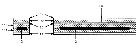
The present invention relates to an
immunochromatographic assay device (FIGS. 1, 2 and 3) for
detecting the presence of an analyte in a sample. The
assay device comprises means for conducting an
immunochromatographic assay 12 in contact with and sealed
within a substantially air-tight and substantially fluid-
tight manner within means for sealing (18a and 18b). The
invention further comprises a support means 20, which
supports the assay means. The sealing means contact and
support the assay means. The sealing means are adapted
to be opened by the user to provide access to the assay
means, i.e., to apply sample.
SUBSTITUTE SHEET (RULE 26)
WO 94/24563 ' . O PCT/US94l04072
8
The invention further provides a detection system
that gives a positive result in a competitive immunoassay
format when an analyte of interest is present in a
sample.
As used herein, the term "sample" refers to an
aqueous liquid sample suspected of containing an analyte
of interest. Such samples include but are not limited to
blood, plasma, serum, urine, saliva, sweat, effusions,
fluid, and materials reconstituted or dissolved in a
suitable aqueous solvent, e.g., a buffer solution.
As used herein, the term "analyte" refers to a
molecule of interest. Analytes may be any antigen, but
small analytes (MW of 100 to 1000 Daltons) are of primary
interest. Such analytes include therapeutic drugs and
metabolites thereof, illicit drugs and metabolites
thereof, steroids, and peptide hormones. Nevertheless,
assays may be for larger molecules such as protein
hormones, e.g., insulin, or viral antigens, bacterial
antigens, serum proteins, antibodies or any antigen of
interest where detection of the presence (or absence) of
the analyte in a rapid, specific, sensitive assay is
desirable.
SUBSTITUTE SHEET (RULE 26)
WO 94/24563 PCT/(JS94104072
9
5.1. IMMUNOCHROMATOGRAPHIC ASSAYS
The device of the invention comprises means for
conducting an immunochromatographic assay
("immunochromatographic assay means"). Many
immunochromatographic assay means and formats are known
in the art (see Section 2.1, supra), and can be used in
the practice of the present invention. Generally, an
immunochromatographic assay involves use of a solid phase
support for conducting a liquid. As used herein, the
term "solid phase means for conducting a liquid" refers
to a solid support that allows migration of a liquid
therethrough, e.g., via capillary action.
In the practice of the present invention, the solid
phase support should behave as a hydrogel. Although not
intending to be limited by any particular theory, it is
believed that hydrogel materials permit the migration of
a liquid sample into the substantially air-tight
environment of the device of the invention. A suitable
solid phase immunochromato-graphic assay means is a nylon
membrane, which is the preferred solid phase support for
this purpose, although any solid phase which permits
movement of the sample may be used. Other suitable solid
phase supports for include, but are not limited to,
coated plastic and coated glass, e.g., such as is used
for thin layer chromatography, filters, polymer beads,
silica gel, paper, membranes, agarose gel, polyacrylamide
gel, gelatin, etc. In a further embodiment, the solid
phase support may be impregnated with hydrogel materials
such as, but not limited to, proteins (e. g., collagen,
gelatin, albumin, etc.), polyethylene glycol, charged or
neutral polysaccharides (e. g., hyaluronic acid,
xanthates, alginates, guar gum, agarose, etc.) and
starches. In another embodiment, adhesive polymers used
to seal the immunochromato-graphic assay means can also
have hydrogel properties.
The immunochromatographic assay means of the
invention will preferably be a membrane strip, more
SUBSTITUTE SHEET (RULE 26~
WO 94/24563 . . ~ ~ 6 016 Q pCT~594/04072
preferably a nylon membrane strip. However, it is
contemplated that the immunochromatographic means may be
formatted for radial migration of liquid sample, e.g., as
on a disk.
5 The present invention can be used with any assay
format adaptable to an immunochromatographic assay.
Although not limited by any particular example,
Generally, depending on the assay format, the
immunochromatographic assay means will contain a
10 mobilizable labeled reagent and a detection zone for
detecting the labeled reagent. As use herein, the term
"labeled reagent" refers to labeled receptor specific for
the analyte of interest, or labeled analyte, and the term
"mobilizable" means that the reagent will move along the
solid phase support with the liquid sample. The
mobilizable labeled reagent is located on the solid phase
support so that it can be mobilized by the liquid sample
and moved to the detection zone. In a specific
embodiment, infra, the labeled reagent is labeled
analyte. In a competitive assay format, the detection
zone contains a specific binding partner of the labeled
reagent immobilized in the detection zone, i.e., analyte
if labeled receptor is used, or receptor if labeled
analyte is used (see Section 2.2, supra). As pointed out
in Section 2.2, supra, there is an inverse correlation
between detection of label in the detection zone and the
presence of analyte in the sample.
In a sandwich immunoassay format, one receptor is
labeled, and another receptor, which does not compete
with the first receptor for binding to analyte, is
immobilized in the detection zone. When analyte is
present, it will bind both labeled receptor and
immobilized receptor, thus localizing the label in the
detection zone. In this case, a signal directly
correlates with the presence of analyte.
Preferably, the immunochromatographic assay means
includes a control to indicate that the assay has
SUBSTITUTE SHEET (RULE 26~
~, WO 94/24563 ' PCTIUS94104072
11
proceeded correctly. The control can be a specific
binding spot more distal from the sample application
point on the solid phase support than the detection zone
that will bind to labeled reagent in the presence or
absence of analyte, thus indicating that the mobilizable
receptor has migrated a sufficient distance with the
liquid sample to give a meaningful result.
The term "receptor" refers to a molecule that can
specifically bind to analyte. Suitable receptors for use
in assays of the invention include antibodies, cell
surface receptors (or a fragment of a cell surface
receptor that contains the binding site of analyte and
ligand), enzymes (or the substrate binding site of an
enzyme), or any other molecule or macromolecule capable
of specifically binding to and forming a complex with a
ligand and complex with an analyte. Antibodies and cell
surface receptors are preferred, with antibodies more
preferred. In a preferred embodiment, receptor is
generated or selected to be specific for the most unique
epitope on the analyte.
Suitable labels include enzymes, fluorophores,
chromophores, radioisotopes, dyes, colloidal gold, latex
particles, and chemiluminescent agents. When a control
marker is employed, the same or different labels may be
used for the receptor and control marker. In a specific
embodiment, infra, the label is a colored latex particle.
5.2. SEALING AND SUPPORT OF THE DEVICE
The device also comprises a means for sealing the
immunochromatographic assay means ("sealing means"), also
referred to as a sealing member. As used herein, the
terms "sealing means" and "sealing member" refer to a
material that can be used to seal the
immunochromatographic assay means in a substantially air-
tight and a substantially liquid-tight manner. The
sealing means should also be substantially non-wettable,
i.e., it does not absorb significant amounts of water.
SUBSTITUTE SHEET (RULE 26)
WO 94/24563 ~ ~ PCT/US94/04072
12
According to the present invention, material useful as
sealing means includes, but is not limited to, adhesive
tape, plastic, mylar and the like. In pne embodiment,
the sealing means are sealed with an adhesive. In
another embodiment, the sealing means are sealed with a
sonic weld. Although not intending to be limited by a
particular theory, it is believed that sealing with an
adhesive confers non-rigidity to the device, allowing
migration of air into interstitial spaces of the adhesive
or slight separation of the sealing means to allow for
the movement of liquid in the solid phase support.
The device also comprises a means for supporting the
device t"support means"), also referred to as a support
member. As used herein, the terms "support means" and
"support member" refer to a material which can act to
reinforce the solid phase means for conducting a liquid,
i.e., to buttress or brace a membrane strip. The support
means is selected from a material that can be attached to
the sealing means, e.g., via an adhesive. In a specific
embodiment, the support means can also act as sealing
means. Materials for use as support means include, but
are not limited to, glass, plastic, mylar and the like.
In a preferred embodiment, the support means is
transparent plastic that is stiff enough to support a
nylon membrane. Alternatively, a relatively non-stiff
material can have suitable stiffeners attached thereto.
In the device, the solid phase means for conducting
a liquid is laminated between the sealing means and the
support means. Thus, the dimensions of both the support
means and the sealing means are larger that the
dimensions of the immunochromatographic assay means.
Preferably the device defines a transparent window
aligned with the test zone for viewing the assay results. '
The invention further contemplates sealing more than '
one immunochromatographic assay means, e.g., membrane
strips, in a single device, to allow for multiple assays.
SUBSTITUTE SHEET (RULE 26)
WO 94/24563 ~ ~ ~ PCTlUS94/04072
13
5.3. PREFERRED EMBODIMENTS OF THE INVENTION
Referring now to FIGS. 1, 2 and 3,,in a specific
embodiment, a membrane strip 12, onto which the assay
reagents have been previously impregnated and stabilized,
is attached directly to a sealing member 10 (plastic,
mylar or other suitable material) by means of any
lamination technique such as double sided tape or
adhesive film 18a, or sonic welding. A further
application of double sided tape or adhesive film 18b is
then applied to the remaining exposed membrane surface,
followed by a layer of stiff transparent plastic sheet 20
to totally enclose the membrane strip. Alternatively,
the stiff transparent plastic sheet 20 can be sealed to
the sealing member 10 by sonic welding.
In a further embodiment, the transparent plastic
sheet 20 can then be overlaid indirectly with double
sided tape or adhesive film 18c followed by white or
colored tape or plastic 22 (or alternatively directly
with white or colored adhesive backed tape or plastic 22)
leaving an appropriate window 14 (or windows) to allow
for viewing of the test result and control reaction.
Alternatively, the transparent plastic sheet 20 can be
painted, stained, or colored to define a window 14.
Fabricated as described above,~the membrane strip is
totally enclosed within non-wettable sheets in packaging
which serves the duplicate function of membrane holder
and moisture barrier pouch.
The assay is carried out simply by exposing the end
of the membrane strip, e.g., by cutting the laminate or
by pealing off a protective cover. Preferably, cut marks
16 are indicated on the device. This exposed membrane is
then dipped into the sample (which enters through the
exposed end) and migrates up the membrane. Alternatively
the device can just be immersed in the sample (for
example, a full urine collection vessel) and the sample
will enter through the exposed end of the membrane (the
SUBSTITUTE SHEET (RULE 26)
CA 02160160 1999-OS-04
14
only channel of access available) and migrate up. In
another embodiment, the sample can be applied to the
exposed membrane. The result is read as the presence or
absence of label in the detection zone. In a preferred
aspect, the label is observed through the viewing window
14. Moreover, because the device is an integral arrange-
ment of the immunochromatographic assay totally enclosed
in the packaging, it can be removed from the sample,
dried off, preferably resealed (e. g., with adhesive tape)
and stored conveniently as a permanent record.
Selection of membrane and lamination method can be
controlled so that such devices have controlled pore
sizes and effectively eliminate particles above a certain
size, such as red-blood cells, making such a device
useful for whole blood.
The device of the present invention provides for
enclosing any immunochromatographic assay means, e.g., a
membrane strip, such as used in immunochromatographic
assays in dipstick or flow-through format for pregnancy
(hCG), fertility (hLH), infectious mononucleosis
(IN),Strep A, chlamydia and drug abuse, using lamination
so that the membrane is contacted on all sides and
totally enclosed. The laminate can then be completed
with plastic, mylar or any other material as required for
rigidity and or cosmetic appearance. As pointed out
above, the membrane is thus held in a device that acts as
an integral holder and packaging pouch.
CA 02160160 1999-OS-04
14a
The device of the present invention provides good
control of liquid sample migration, since the volume of
sample entering the device is limited by the physical
dimensions of the enclosed test-strip membrane. The
migration of the sample is strictly controlled by the
a
PCTIUS 94/0407 2
_ d
r e~ Trr1
15 ~'~1 ~~~ ~ pC ~~~ °TC 2 6 JUN 1995
ph~rsical limitations imposed by the confinement of the
test-strip membrane in the laminate. The volume of
sample that enters is controlled by the area of membrane
used.
In one preferred embodiment the laminate is
assembled from test-strip membranes and sheets of
materials (adhesive films, tapes, transparent plastic
sheets, etc.) then the final laminate is die-cut at the
required dimensions. This eliminates the cost involved
in using precut or molded components and the accuracy
that would be involved in the assembly of individual
components into a laminate.
Referring now to FIG. 4, in another preferred
embodiment a colored indicator strip 20 that can indicate
the presence of moisture levels (blue for low humidity,
pink for high humidity), is incorporated into the device.
Suitable indicator strips are sold by Multiform
Desiccants. Alternatively, a suitable moisture indicator
can be impregnated in a part of the solid phase support
that is not intended to contact sample. The colored area
is given its own viewing window at the top or bottom of
the laminate. The indicator serves as a confirmation of
package integrity. When the colored area is pink --
indicating high moisture -- the laminate integrity has
been compromised and the test should not be used.
Satisfactory package integrity is indicated by the
colored area remaining blue.
Referring now to FIG. S, in yet another embodiment
the laminate is incorporated into a sample collection
vessel 30, such as a urine collection vessel, thus
allowing immediate specimen testing in the collection
container. The collection vessel should be transparent
to allow for viewing of the result. The device is
attached to the inside wall of the collection vessel by
adhesive film, double sided tape, or a sonic weld. In
this embodiment the laminate can be precut, with the
test-strip membrane exposed (i.e., as if the test is
AMENDED SHEET
o .
PCTIU~ 94/0407
~f 60l 6~ ,
51 ~~cd PCTt°; ~ 2 s JUN 19
16
ready to run). The collection vessel is then sealed (cap
32 screwed on, etc.) to provide the package integrity.
Alternatively, the device may include a removable
adhesive strip so that the membrane strip remains sealed
within the laminate until the test is ready to run. In
this case, the adhesive strip is removed prior to adding
the sample. The test runs automatically when the sample
is collected in the vessel. The amount of sample
collected need only be sufficient to be above the level
at which the test-strip membrane is exposed.
5.4. PREFERRED DETECTION ZONES OF THE INVENTION
In addition to the integral immunochromatographic
assay device, the present invention provides an
advantageous detection zone for a competitive assay
format. The detection zone of the invention gives a
positive signal when analyte is present in the sample,
and a negative signal when analyte is not present in the
sample, in contrast to standard competitive assay
techniques (see Section 2.2, supra). The
immunochromatographic assay includes a mobilizable
labeled reagent, such that in the presence of a liquid
sample the labeled reagent is transported with the sample
along the assay strip a detection zone. The label has
been immobilized in part, but not all, of the detection
zone such that there is a contrast between the label and
the membrane strip in the detection zone. The detection
zone also includes a specific binding partner of the
labeled reagent, as described above. However, when
labeled reagent binds to the specific binding partner in
the detection zone, i.e., in the absence of analyte in
the sample, the contrast between the label in the
detection zone and the membrane strip is lost. When the
labeled reagent does not bind to the detection zone,
i.e., in the presence of analyte, the contrasting signal
remains. Thus, a contrasting signal in the detection
zone indicates the presence of analyte in the sample and
AMENDED SHEET
_ ~ ~CTIUS 9 4 / 0 4 p 7 2
(~ b
51 Recd PCT;'~T~ 2 6 J U N ;5,- w~
a non-contrasting signal in the detection zone indicates
the absence of analyte in the sample. In this way, the
signal directly correlates with the presence of analyte
in the sample.
In another embodiment, the detection zone contains a
specific binding partner of the labeled reagent in one
section, and a non-specific, i.e., control, binding
partner of the labeled reagent (or alternatively a
binding partner of a control labeled reagent) in another
part. In the presence of analyte, the control reagent
will bind in the control area of the detection zone,
while the labeled reagent will not, thus producing a
contrast between labeled and unlabeled areas of the
detection zone, indicating (1) a "positive" result and
(2) that the assay ran correctly. In the absence of
analyte, the entire detection zone will be labeled,
indicating (1) a "negative" result and (2) that the assay
ran correctly. If the detection zone is not labeled at
all, the assay failed to run.
It can be readily appreciated that the contrasting
areas in the detection zone can be arranged in shapes,
such as "+" signs or letters.
As can be appreciated by one of ordinary skill in
the art, any label system commonly used for
immunochromatographic assays can be used according to the
present invention.
The present invention will be made more clear by the
following example, which is intended to be exemplary of
the invention and not limiting.
AMENDED SHEET
CA 02160160 1999-OS-04
18
6. EXAMPLE: DETECTION OF COTININE IN A SAMPLE
6.1. MATERIALS AND METHOD
Preparation of cotinine coated particles. Blue dyed
latex (0.318 ~m diameter, Seradyn, IN) was coated with a
solution of cotinine chemically linked to bovine gamma
globulin (cotinine-BGG) overnight at room temperature in
0.05 M phosphate buffer, pH 7.2-7.5. The final
concentration of blue latex was 0.25% and that of the
cotinine-BGG was 3 ~g/ml. The particles were washed
twice with 5 mg/ml BSA in phosphate buffer, pH 7.2-7.5,
and the coated latex solution was suspended in phosphate
buffer containing 5 mg/ml BSA.
Rabbit antibody to cotinine. Rabbit antibody raised
against a cotinine derivative were prepared against a
trans-4-hydroxycotinine-KLH conjugate using standard
procedures, as described in International Patent
Publication No. WO 93/03367, published February 18, 1993.
The antibody was affinity purified batchwise by affinity
chromatography on Sepharose-cotinine. (Sepharose is a
Trade-mark). The affinity purified material was diluted
in 0.5 M sodium carbonate, pH 9.5, at a concentration of
350 ~g/ml.
Control antibody. Rabbit anti-sheep antibody
(Jackson Immunoresearch Labs) was diluted to 1.2 mg/ml in
0.5 M sodium carbonate, pH 9.3.
CA 02160160 1999-OS-04
18a
Preparation of the test-strip membrane. Nylon
membranes (Biodyne A - Trade-mark, 5 ~m pore size, Pall
Biomembranes, NY) were cut into 7 x 10 cm sheets.
Affinity purified rabbit anticotinine antibody and the
control antibody were applied to the sheet of nylon as
lines (20 ~l per line, 8 cm long at the rate of 2.5
~,1/cm) . The line widths were 2 cm and 2.5 cm,
respectively, from the bottom end of the sheet, parallel
to the 10 cm side. The application was performed using a
mechanized air-brush applicator with 25-75 psi nitrogen.
The sheets were then allowed to air dry at room
temperature for 1 h followed by 30 minutes in an
incubator at 37°C. The sheets were then soaked, with
agitation, in an aqueous solution of 0.5% casein, 5%
sucrose, 0.1% TRITON X-100 (Trade-mark), 0.05 M Tris,
0.003 M MgCl2, 0.9% NaCl, 0.02% NaN3, pH 8.0, for 30
minutes . The sheets were air dried for at least 4 h at
room temperature.
Blue latex coated with cotinine-BGG diluted to 0.25%
in 20% sucrose was applied to the sheet of nylon as zones
adjacent and parallel to the affinity purified rabbit
anti-cotinine line. Each zone was approximately 0.3 x
0.5 cm with approximately a 1.5 mm gap between each zone.
The gaps were visible. The zone was applied about 1.5 cm
from the bottom end of the sheet. The application was
performed using a mechanized air-brush applicator with
25-75 psi nitrogen. The sheets were then allowed to air
dry at room temperature.
CA 02160160 1999-OS-04
19
The 7 x 10 cm sheet was cut into strips 0.5 x 7 cm
with each strip having a visible blue latex zone and an
affinity purified anti-cotinine line and a control
antibody zone.
Assembly of test-strip membrane in its integrated
holder/packaging pouch.
(a) Referring to FIGS. 6 and 7, a test-strip
membrane 12 was placed onto a piece (2 x 8 cm) of
Arclad 7148 (Trade-mark) double sided-tape 60
(Adhesives Research, PA). The uncoated surface of
the membrane (side not impregnated with the coated
blue latex and antibodies) was attached to the
exposed surface of the tape and the membrane was
located centrally within the piece of tape.
Membrane 12 was placed on tape 60 with the strip 62
of blue latex is positioned approximately as shown
in FIG. 6.
(b) Referring now to FIGS. 8 and 9, the
membrane strip 12 was totally enclosed by covering
the exposed surfaces of membrane and adhesive with a
piece (2 x 8 cm) of Arclad 7530 adhesive film 66
(Adhesives Research, PA). The covering over the
second adhesive surface of the 7530 was removed and
WO 94/24563 ~ PCT/US94/04072
the exposed surface (the top surface) was covered
with transparent plastic film 64, i.e., the support
member. The adhesive film and plastic film were
firmly pressed on all sides to allow complete
5 contact of all adhesive surfaces and sealing of the
membrane totally within the assembly. ,
(c) Referring now to FIGS. 10 and 11, the
transparent plastic top surface was then covered by
white or colored tape or plastic 70 (directly with
10 adhesive backed material, although the same effect
could be achieved indirectly with double sided tape
or adhesive film followed by white or colored tape
or plastic) leaving an appropriate window 14 (or
windows) above the affinity purified anti-cotinine
15 line and the control antibody line on the membrane
strip 12 to allow for viewing of the test result and
control reaction. Additionally, the bottom end of
the laminate was marked with 2 lines 16, just below
the strip 62 of coated blue latex. The space
20 between the two lines was cut in order to expose the
test-strip membrane 12 to sample for performing a
test. The whole assembly, test-strip membrane in
its integrated holder packaging pouch, is the device
for an assay for cotinine.
Assay for Cotinine. The test kit was prepared for
use by cutting the device at the bottom where indicated
by the two marker lines. This exposed the end of the
test-strip membrane.
The device was put into sample such that the cut end
of the device was in contact with the sample. The device
was left to stand in the sample for 10 minutes, after
which time the results were read. Samples were urine
samples containing known amounts of cotinine.
6.2. RESULTS
Sixty tests were performed with urine samples having
known cotinine concentrations received from the Centers
SUBSTITUTE SHEET (RULE 26~
WO 94/24563 PCT/US94104072
21
for Disease Control (CDC). The results of assays using
the device ("Detection with the Device") are shown below
in Table 1 and compared to the cotinine, concentrations of
the samples and the positive cut-off value of 100 ng/ml
proposed by the CDC.
TABLE 1. Results of Tests For Cotinine
in Urine Samples
Cotinine Detection based Detection
Sample Conc. ng./ml on the CDC with the
# in the sample cut-off 100ng/ml* Device
1, 3080 + +
2. 1360 + +
3, 2860 + +
4, 1250 + +
5, 2450 + +
6. 37 - _
7. 194 + _
g, 23 - -
9. 6.9 - -
10. 590 + -
11. 102 + -
12. 25.5 - -
13 . 540 + +
14. 6460 + +
15. 32.2 - _
16. 17.5 - _
17. 520 + +
18. 50.5 - _
19. 56.2 - -
20. 24.2 - _
21. 2310 + _
22. 1060 + _
23. 15.3 - _
SUBSTITUTE SHEET (RULE 26)
WO 94/24563 216 016 0 p~/ps94/04072
22
Cotinine Detection based Detection
Sample Conc. ng./ml on the CDC with the
# in the sample cut-off 100ng/ml* Device
24. 230 + +
25. 276 + +
26. 122 + +
27. 122 + _
28. 320 + +
2g. 510 + +
30. 9.5 - -
31. 590 + +
32. 960 + +
33. 1450 + +
34. 890 + +
35. 27.3 - _
36. 13.8 -
37. 232 +
38. 272 + +
39. 590 + +
40. 26.1 - '
41. 55.2 - '
42. 326 + +
43. 106 + +
44. 780 + +
45. 2070 + +
46. 510 + +
47. 670 + +
48. 54 - -
49. 67.8 - _
50. 570 + _
51. 1500 + +
52. 154 + +
53. 200 + _
SUBSTITUTE SHEET (RULE 26~
WO 94/24563 ~ . .:~. s PCTlUS94104072
23
Cotinine Detection based Detection
Sample # Conc. ng./ml on the CDC with the
in the sample cut-off 100ng/ml* Device
54. 164 + +
55. 176 + -
' S6. 1500 +
57. 7470 + +
58. 1860 + +
59. 5860 + +
60. 238 [ + -
* The samples were graded as positive or negative by
the CDC based on the detected concentration of
cotinine; negative <100ng/ml., positive >100ng/ml.
Table 2 summarized the results of Table 1. The
number of samples that registered as positive for
cotinine under the CDC criteria and using the device are
in the "+,+" square (32); the number of samples that
registered positive using the device of the invention,
but that were negative according to the CDC criteria are
shown in the "+,-" square (0); the number of samples that
registered as negative using the device, but that are
. positive according to the CDC criteria are in the "-,+"
square (11); and the number of samples that registered
negative using the device and that are negative according
to the CDC criteria are shown in the "-,-" square.
TABLE 2. SUMMARY OF COTININE ASSAY RESULTS
Result, cut-off 100ng/ml cotinine
+ -
Device + 32 0
- 11 17
The following observations about accuracy (the
percentage of correct assay results using the device),
SUBSTITUTE SHEET (RULE 26)
CA 02160160 1999-OS-04
24
sensitivity (the percentage of correct positive results
using the device) and specificity (the percentage of
correct negative results using the device) are available
from the data in Table 2. The overall accuracy of the
assay using the device of the invention to test samples
of known cotinine concentration, in which the cut-off for
a positive result is 100 ng/ml, is 81.5% (49 out of 60).
This value is calculated by adding all of the "+,+" and
"-,-" values and dividing by the total number of tests.
The sensitivity of the assay using the device, which is
calculated from the number of samples that registered
positive divided by the total number of positive samples
according to the CDC criteria, was 74.4% (32 out of 43) .
The specificity of the device, which is calculated from
the number of samples that registered negative divided by
the total number of negative samples according to the CDC
criteria, was 100% (17/17).
The results clearly show the present invention is
able to detect cotinine in the urine samples.
The present invention is not to be limited in scope
by the specific embodiments described herein. Indeed,
various modifications of the invention in addition to
those described herein will become apparent to those
skilled in the art from the foregoing description and
accompanying figures. Such modifications are intended to
fall within the scope of the appended claims.