Note: Descriptions are shown in the official language in which they were submitted.
o 94125597 PcT/Isg4/00205
~G14~5
Description
METHODS AND COMPOSITIONS FOR DETECTION OF SALMONELL~
5 Cross-Reference To Related Applications
This application is a cortinl~tion-in-part of United States patent
application Serial No. 08/054,452, filed April 26, 1993 and pl~senlly pending.
Technical Field
The present invention relates generally to diagnostic tests and, more
particularly, to nucleic acid based disgnostic tests, and antibody-based r~ nostic tests
directed to Salmonella.
Background of the Invention
In 1980, the World Health O~ .;,nl;on ei,l;.. A~ed that in developing
co~ ;es food poisoning from h~,Lioll with Salmonella b~ct~ (SAImnn~llrtsis)
c~.lLlil u~ed to more than 1 billion cases of acute di~lLea in children under the age of
five years (Kve.lbel~, and Archa, Food Technol. 40:77-98, 1987), and at least 5 million
deaths (this l~,f~.~nce, and all other l~,f;,.~ ce,s cited herein, is hereby CA~ S~
20 incGl~ la~ed herein by r~,fe.ence in its entirety). Since the mid-1980s, the worldwide
inri~nce of salmonellosis has increased steadily. S. enteritidis, in particular, has been
imrli~ted in the sharp increase in food borne ;..r~l;O~- since 1983. Indeed, the current
frequency of S. enteritidis h.r~cl;Qn~ con;,~ilule a worldwide pAn~lemir, (Rodrique et al.,
Epidemiol. Infect. 105:21-27, 1990).
The severity of the disease is greatest in infants, the elderly, the infirm
and in other p~.~ons with inadequate or hll~ d ;.. ~ e ~y~l~,lls, inrl~ltling the
malnourished. In third world countries where malnutrition is more co.~ o~-ly a
comp!ic~ting factor, mortality rates due to S. enteritidis illr~lion as high as 28% have
been reported. In both the clinical and industrial settir~c~ the situation is also
30 complicated by the fact that many people are &Sylllp~OllldliC carriers. Salmonella spp.,
incll~rling S. enteritidis, often possess several plasmid encoded antibiotic rçeictAnce genes
that comp!ic~te the lle~ .l of human infections.
In the industrialized world, it is the c~ -AI;nn of food products by
Salmonella bacteria that is most directly lluc ~c~ to human health. Hence, it is not
35 ~ul~lising that the inc,~se in salmonellosis in first world countries parallels tfie
centrAli7~tion of food production and processing despite contin~.ed improvements in
epi~içmiological and microbiological metho~s
WO 94/2s597 ~ ; 4 o~ PCT/IB94100205
The ci~nific~nce of the problem is rPflected in one aspect in the poultry-
related industries. For example, in the U.S. alone hatcheries produce app,o~ tPIy 100
million broiler chicks per week and chicken egg production in the U.S. has reached 5
billion annually. A large proportion of S. enferitidis infections have been associated with
S the co~ ;on of the eol~ of whole shell eggs r~clllting from vertical
l~ u...:csion of this pathogen from breeder stocks due to transovarian infection. This is
significant since common procedures deci~ned to decont~min~te the external shellsurface are not effective. The problem presented by S. enterifidis is exacerbated by the
fact that infection in the adult laying hens may be a~,y"")~o,l~alic. Typically, S. enteritidis
10 infection of laying birds does not have a ~ c~ adverse effect on fertility,
hatcl~abili~y- or egg prod~lstion Similarly, broiler ~ enc may be asymptomatic
throughout their lifpJtimp~ ~hhough losses of about 20% do occur in infected flocks due
to death in chicks, retardation of growth and rejectiQn of co..l~ d birds at
gl.~ Ccsl~ h~h~ed poultry feed may be a major source of infection, but stress
to poultry due to h~nrllin~ ,p~ al;on and ov~, .,ruwdillg add to the problem by
i".;~ , the shçdrling of Salmonella from infected ~ ls The end result is that the
majority of modern processing plants, which process about 10,000 birds per hour, are
co-ll~ d and S~ ' ~"ella are typically icolstçd from 40% to 70% of turkey or
chicken carcasses s---~le ~ in the U.S. and Canada.
The overall c,cononuc costs of the rising inr;d~nce of food borne
f~.ctiQnc have been ~;gn;~c~ ~I The U.S. General ~Gco~ , Office has r~cGnlly
~1;."al~ the cost of S. enteritid~s food pr~i~o~ g in the U.S. ~_I..~n 1985 to 1990 at
$118 million in lost productivity, me~ l and hospital costs rçslllting from about 9,500
illnPscPs The U.S. Center for Disease Control receives more than 40,000 case reports
25 annually but attributes greater than 2 m-illion cases and roughly 2,000 deaths per year in
the United States to ,c~lmQr Pllosic (Cohen and Tauxe, Science 234:964-969, 1986). The
econûlluc cost related to ~ lle ll of ~lmonpllosic in the U.S. was e~lhl~led to be $50
million in 1986. About 8 million cases involve physician conclllt~tion and an .,,~I;Ill~t~,d
250,000 cases require hospit~li7~tion Non-hosrit~li7r~ cases are thought to have30 accuul,~ed for about S680 million in medical costs and minim~lly $2 billion in lost
productivity. Others e~ le the total costs of salmonellosis in the U.S. arising from
mer3i~ rt.~ ul and lost productivity to be as high as $23 billion per year (Kvenberg
and Archer, supra).
The losses absû.l,ed by the food industry from liability and product loss
35 are l~ndo~lbteAly passed on to the col~sulller. Thus, there is a need for an effect*e nsk-
l~, .,.r..l prc~ Il to monitor the di~ phases of poultry prod~lction inclllding
bl~lulg, raising, cl~ lghtPring paçl~h~g and further proces~ distribution and
2161~as
0 94/2~97 PCT/IB94100205
pl~p~alion~ and con~ lmption. The development of strategies for creating Salmonella-
free feed, the control of Salmonella in breeder flocks, hatcheries, and product operations
will include development of more effective diagnostics. Accordingly, there is a general
need for a technology which could be applied to the in. ,~n~i~re, rapid ~etection of all
5 Salmonella food bome pathogens.
One type of assay for Salmonella comprises the standardized culture
tests for Salmonella in the food industry. These culture assays are recognized by
di~erenl names in di~.t;n~ countries but they share the same basic approach. In the
United States, the procedures are known as the "Bacteriological Analytical Manual"
10 (BAM), published in 1984 by the Association of Official Analytical ChPmictc (AOAC,
Arlington, Va.). In C~n~ the procedures are known as the "Official (:~n^ 1i~n Wet
Culture Method" (WCM), the protocol most often used to test food s~"plas is MFD-20.
Within the standardized tests, S~mple5 are inc~lb~ted at 30C to 37C for
18 to 24 hours in a rich, non sele~ re .. ~eAi.. to promote recover,v of the cells and
allow them to begin to replicate to the levels detect~hle by current technologies. There
will likely be an excess of other microG,~S~..;c...c in the sample, some of which may be
from the closely related family Enterobacteriaceae. Th_.erole, a selecti~e growth step
is con~iucted to enrich for Salmonella bacteria, for ~,Y~mrle, by inoc..l~ting a small
20 sarnple of the pre-enrir.hm~nt culture into a selenite-cysteine broth, tetrathio~ite broth,
or Rappapo,~-V~cci~ iic broth for 18 to 24 hours, typically at an elevated t.,..l~e.~ e
such as 43C. The cells are then plated on a select*e ..~ .., such as brilliant green
agar or xylose-lysine-de~ ycllolate agar, and i~.c~ e~ ov~.lliglll. P~ e colonies
are then ll~lsr~ d to various biochemical or metabolic test media for collLIllalion.
25 Pure cultures of S~ ' )nella are then grown ov~.luglll on agar slopes for 3e.cllylJing. In
total, three to four days are required to obtain pre;,~ e posili~c results, and a five to
seven day wait can be ~-ec.;s~.y before final col~tillllalion and id .~;r.-,A~;on of the
Salmonella.
An alternative test to assay for the presence of,S~ ~ ,.clla is based on
30 nucleic acid probes. One such test uses probes constructed from a part of the fimA gene
of Salmonella ~ ", and is pl~f~.~.llially based on two particular seq~l~ncec
(Madonna and Woods, EP Publication No. 383,509, Or~ho~i~ostic Systems, Raritan,
N.J.). Briefly, a nucleic acid molecule of a known seq.l~nce is introduced to a sarnple
under conditions suitable for hybridization of the nucleic acid molecule to its target
35 nucleic acid seq~lence in the DNA or RNA of ,S~ ' ,,.ella. Alternative hybridization-
based assays include the colGlull~ ic Gene-Trak~ Salmonella Assay (Gene-Trak
WO 94125597 ~6~4 4 PCT/rB94100205
Systems, Fr~mingh~m MA), Fitts et al., Appl. Environ. MicrobioL 46:1146-llSl,
1983
Another alternative test is a fluo,~scell~ antibody assay (FA test
Thomason, J. Food Protection 44:381-384, 1981), which inr~ f~s the Salmonella
5 Flouro-Kit (Incstar, Stillwater, Minn.). Such a test uses polyvalent antisera prepared
against Salmonella flagella (anti-H) and lipopolysaccharide (LPS) O-chain (anti-O).
The assay can also use purified polyclonal IgG antibodies. However, an FA test is
laborious, has a high level of false-positive results, yields only presumptive positive
samples, and requires visual dett;",-",&lions to be made by highly skilled pe, ~o-~lel using
10 c ,.~.,ns;~e equipment.
Another test is an enzyme imm~lnoaccay (EIA). In general, as with the
FA test described above, antibodies to flagella or lipopolysaccharide form the basis of
most EIAs. EIAs can use either polyclonal or morlocl~n~l antibodies. However, as with
the FA test, false-positive results are a si nifir~nt problem. Further, these assays can
15 take an extensive testing period, and some diagnostic tests using monnçlcn~l antibodies
to Salmonella flagtollin have r.,po.lcd si~ifir~nt pr~' nc with false-negative results.
F.~ 'es of such EIAs include the TECRA .~-' ,.clla immlln~capture ELISA
m~mlf~ctllred by BjGC,lL~ ,l;3~S Pty. Ltd., Roseville, NSW, Australia, and the Dynatech
Labo,a~D,;es, Inc. (Chantilly, Virginia) ,~ .ella MlCROELISA~-92 and
20 MlCROELISA~-32 D~ te~,l;on kit.
Still another type of test is an aggllltin~tiQn assay (Benge, Eur. J. Clin.
Microbiol. Infec~. Dis. 20 8:294-298, 1989), such as the W~ lf~Y-Colour ,c ~ ,.ella
assay (Wellcolne; Bouret and Jeal1je n, J. Clinical Microbiolo~ 30:2180-2186, 1992),
which is based on anti-,S~ 'r. i.ella ~ o~ s can, g5lt~d to latex beads. This form of
25 test is relatively simple, but l~ S at least two to three days to provide results from
food or ellvil O~ nl ~l s~ p'~ s, and has a relatively low level of sel,s l;vily.
A filrther type of test is the selective motility assay, in which a sample
potentially co..l~ g Salmonella is introduced into a pre-enr;~llmf~nt or selective
growth medillm in the first cl..u,~ ,r of a double-G~ bclcd device. (IIun~ et al.,
30 Letters in Applied Microbiology 10:245-249, 1990) The motile S~ ' ..clla thenfavorably grow and move across the pre-enrirlment growth --eJ;~ r.l~ , the
second ch~,-ber, which co~ a serni-solid meflillm having a sample of antisera at the
far end. As the motile Salmonella replicate and migrate into and across the second
c.halllb~,r, the antisera diffuses toward the ol~co~ g b~cten~, forming an
35 imml--~opre~ e line at the point where the bactf ri~ contact the antisera.
Yet another test is a bacteriophage assay, such as the Vitek System's
Salmonella test (McDonnell-Douglas Health Systems Co.). This assay uses
~o ~ 4 Q5 ' ' ' '
94125597 - PCT/IB94/00205
5 ~.' f ~ .
bacteriophage that specifically recognize receptors on Salmonella. An enzyme is
conjug~ted to the bacteriophage and is used for detection purposes. This test requires a
miniml~m of 48 hours and is subject to false positive and false negative results.
Yet another test is an enzyme-linked ampe~oll" ~liC immlm- sensor, a tvpe
of biosensor format (Brooks et al., Journal of Applied Bacteriology 73:189-196, 1992).
Accordingly, the present invention discloses compositions and methrJ~S
suitable for the diagnosis of Salmonella in a sample, inr.~ ing isolated nucleic acid
m :~lccl~lrs~ i~ol~ted proteins, probes and primers.
Summary ofthe Invention
In one aspect, the present invention provides an i.colnted nucleic acid
molecule CGIll~,l;S;llg a sefU2UlBCD gene cluster, which is inr,ll~ded in the
sefU2UlABCD gene cluster. In an alternative embo~im~nt, the icolsted nucleic acid
molecule comprises a sefABCD gene cluster or a sefU2UI gene cluster. In further
alternative embo~lim~nt~ the present invention provides i~o!~ted nucleic acid molecules
C~III~J1;~111g a sefA gene, a sefB gene, a sefC gene, a sefD gene, a sefUl gene, or a sefU2
gene.
The present invention also provides an i~ol~ted nudeic acid molecule
CGIll~ an agfA gene.
Still further, the present invention provides an isolaled nucleic acid
mol-cule cc,~ 2 a tctCBA gene cluster. In ~ltprn~tive embo~ c the present
invention provides i~o~ted nucleic acid mole~lles CG~ lllg a tctA gene, tctB gene or
tctC gene.
~lef~.ably, the i~olsted nucleic acid mrJIe ~lles are l~olllbin~ll, which
means that the mrl1ecllle has been constructed using recollll)inalll nucleic acid techniqlues
and inr,l~-des nucleic acid se~ -ces ~tt~rhed to the gene that are not naturally ~tt~r.hed
to, or in some cases plO~ e to, the gene.
In anolller aspect, the present invention provides probes based on one or
more of the sefA, sefB, sefC, sefD, sefUI, sefU2, agfA, tctA, tctB, or tctC genes. In
particular, these probes comprise at least a portion of the nucleotide seq~enre depicted
in Figures2A-2D from nucleotide No. 136 to nucleotide No. 633 (SEQ ID
No. ); Figures 2A-2D from nucleic acid No. 755, to nucleic acid No. 1495
(SEQ ID No. ); Figures 2A-2D from nucleic acid No. 1512 to nucleic acid
No. 3956 (SEQ ID No. ); Figures 2A-2D from nucleic acid No. 3953, to
nucleic acid No. 4402 (SEQ ID No. ); Figures 3A-3B from nucleic acid No. 2
to nucleic acid No. 1108; Figures 4A-4B from nucleic acid No. 3323 to nucleic acid
No. 4420 (SEQ ID No. ); Figure 5 from nucleic acid No. 2727, to nucleic acid
WO 94/25s97 . ~ ~ `PCT/IB94/00205 --
4~ 6
No. 3236 (SEQ ID No.); ~igures 6A-6B from nucleic acid No. 1293 to nucleic
acid No. 2270 (SEQ ID No. ); or, ~igure 7A from nucleic acid No. 1, to nucleic
acid No. 361 (SEQ ID No. ); Figure 7B from nucleic acid No. 1 to nucleic acid
No. 45 1 .
In one embodiment, the probes comprise one or more of the seJA gene,
the sefB gene, the sefC gene, the sefUl gene and the sefU2 gene, and the probes are
capable of sperific~lly hybridizing to S. enteritidis, S. berta, S. pullorum, S. dublin and
5. gallinarum under conditions of high ~ ;el.cy.
In another aspect, the present invention provides vector constructs
10 co~,.i:,illg a sefU2UlBCD gene cluster. In an alternative embodiment, the vector
construct comprises a sefABCD gene cluster. In still further alternative embo~iments
the vector construct cG...p,ises a sefA gene, a sefB gene, a sefC gene, a sefD gene, a
sefUl gene or a sefU2 gene. The present invention also provides a vector construct
CGnl~ illg an agfA gene. The present invention further provides a vector construct
15 CGIllplisil.g the tctCBA gene cluster. In alternative embo-limPnte, the vector construct
con-p.ises a tctA gene, a tctB gene or a tctC gene.
In pr~,f~.-.d embo~limpnte~ the vector construct of the invention
colllplises an eA~ules;~;on vector. Even further p.~f~.~Lbly, the eA~ ,s ,;on vector is able to
express the gene or gene cluster upon introductirJn of the e ,~re~,s;on vector into a cell of
20 a living o~al~is.." further ~ ft.~bly a plant or on which in some embo~liments is animal.
The host cell for the e.~ s;,;on vector construct is ~ ,f~,~ly E. coli, a Salmonella, a
Shigella spp., Citrobacter, Enterobacteria, Pse~/dA ~nas, Sl~lu~yces, R~lrll/~
Staphylococcus aureus, further pr~,f.,.~l~ an E. coli or a Salmonella.
In a further aspect, present invention provides a probe compricing at least
25 a portion of the nucleotide sequence shown in Figure 7A, from nucleic acid No. 1 to
nucleic acid No. 361, or Figure 7B from nucleic acid No. 1 to nucleic acid No. 451, the
probe capable of s}.~;r.c~lly h~ ;L;ng to the DNA of GWPQ-L..~.;&c (SEQ ID
No. ~ enro~ g e.l1e~0p9~l~0gPnic bacteria of the family Enterobacteriaceae underco~-1itions of moderate ~l~i..g~.~;y. Condilions of moderate stringency are such that a
30 ...;~ .h of a single base pair or similar small number of base pairs does not prevent
hybridi7~tion, yet only nucleic acids encoding a GVVPQ-type fimbrin amino acid
sequ~nr~ are able to hybridize to the probe.
In still a further aspect, the present invention provides probes capable of
sperifir~lly hybridi7ing to a nucleic acid mole :~le from greater than 99% of Salmonella
35 strains that are p~thr~nic to warm-blooded animals relative to nucleic wid molccllies
~om other, p~elably virtually all, microu~ s It is particularly ~ef.,.le~ that the
probes be able to tli~tin~ h such strains from all other microol~m~ In a pl~f~
~0 94/2SS97 21~Z PCT/IB94/0020S
embodimpnt~ the probes are able to specifically hybridize to greater than 99.5% of such
Salmonella strains.
In yet a further aspect, the present invention provides a primer suitable
for a nucleic acid amrlifi~-s-tion procedure wl,~ hl the primer is able to speçificslly
5 hybridize to a nucleic acid molecule from greater than 99% of Salmonella strains that
are pathogenic to warm-blooded animals relative to nucleic acid molecules from virtually
all other microbial o~n~ In a pl~re"~d embodiment, the primer is able to
~re~.ific-slly hybridize to greater than 99.5% of such Salmonella strains. In a plefe.lt;d
embo~imPnt the primer is one of a set of two primers that are able to hybridize to
10 opl)osil,g strands of a target seq~lPnce, such that the set is suitable for use in the PCR
reaction.
In yet a further aspect, the present invention provides a method for
detecting the presence of Salmonella in a sample Co~ illg ~ illg cells co..l~;..ed
within the sample to expose the cellular nucleic acids, then ;..c.ll,~ g the cellular nucleic
acids with one or more of the probes, pr~ft;lLbly labeled, des.,libed above under
co~-litionc suitable for desired hybridization, and then detecting the pl~s.,~ce of the
hybridi_ed labeled probe. In a p~.led emhotlim~nt the e-l.osed cellular nudeic acid
is sul~e te~ to an smplific~s,tion ploc~lule, such as PCR or LCR, prior to i .~ n
with the labeled probe to give a hybrirli7ed labeled probe or a product build up that is
detected spe~,lrophulu.-~- tlically.
In a further aspect, the present invention provides a method for ~etecting
the plesellce of antibodies to Salmonella that are in a sample. The s. mple is conts-cte~
with TctC protein that is bound to a solid phase, preferably composed of styrene, under
cQntlition~ suitable for the antibodies in the sample to bind to the protein and then the
antibodies are dPtecte~l In al~ ,&live embodimPntc, the protein is a SefA protein, a
SefC protein, or an AgfA protein~ a FimA protein or a SefD protein, or a fimhri91 or
aggr~liv-e ~I,u~lule il,collJu,alillg such p,ole.,.s, such as SEF14 (for SefA), SEF21
(for FimA), SEF17 (for AgfA) or SEF18 (for SefD). In a further alternative
embo~lim~nt the method is for det~;~ g the p,~ence of Salmonella in a sample, and
co-,-y-i3es conts~cting the sample with a labeled antibody to SefD protein under con-lition~ suitable for the antibody to bind to the SefD protein, and then ~t~l;i~g the
presence of the bound labeled antibody. In another alternative embo~im~-nt, the
- antibodies are to a SefA protein, a SefC protein, an AgfA protein, a FimA protein, or a
TctC protein.
In still arulL~. aspect, the present invention provides mçtho~l~ able to
,1;~;.~ ,.;~1. greater than 99% of the strains of ,Sar ,,ella that are pathogenic to warm-
blooded animals from virtually all other microbes in less than 24 hours. In these
WO 94125597 ` PCT/IB94/00205 --
2~14~ 8
methods, cells from a sample are treated to expose cellular nucleic acids, then the
cellular nucleic acids are incub~ted with one or more of the probes, p~ bly labeled,
des~ ed above under conditions suitable for desired hybridization, and then the
hybridized labeled probe is detecte~ In a p,~,~.-ed emborlimçnt, the exposed cellular
5 nucleic acids are amplified prior to the incubation with the labeled probe In a further
p,~r~.~ed embodimrnt, these methods are able to ~ietin~ich greater than 99 5% of the
strains of Salmonella. In an alternative embodiment, the methods of the present
invention provide for dete~il;i-g the p~esence of GWPQ (SEQ DD No ~ fimbria-
encoding, or SefA-type, SefD-type or FimA-type fimbriae encoding, enteropathogenic
10 bacteria of the family Enterobacteriaceae in a sample In this method, cells within the
sample are treated to expose cellular nucleic acids, the cellular nucleic acids are
inrllb~ted with the labeled probe (as desclil)ed above) under conditions suitable for
hybridization, and then the hybridized labeled probe is cletected In a ~ler~,ledemborlim~nt the exposed cellular nucleic acids are ~mplified prior to the incuh~tion
15 step
In still yet another aspect, the present invention provides a method of
~t~ g greater than 99.5% of Salmonella in a sample, prcr~ bly greater than 99 9%of Salmonella in a sample, and further preferably all of such .~ ' ,.ella. The method
con.~.ises a nucleic acid probe assay, an antibody assay and/or a protein assay, as
20 des~;.il,ed above and set forth more fully below, ~l,~..,i- the method targets a group of
the above genes, and/or utilizes a cor~il of the .~,sl,e~ , gene products, COlllyliSillg
the agfA gene, thefimA gene, and the tctC gene Further, the group and/or corl~ may
con,~,.se a sefA gene
These and other aspects of the present invention will become evident
25 upon re;relence to the following ~ iled drscrirtiQn and ~tt~che~ d-~w,~-gs In addition,
various ~eÇere.~ces are set forth below which des~,il,e in more detail certain procedures
or conll~Gs;lions (e.g, pl~cm;~lC, etc ); such er~ . ces are inco.l~olaled by ~;reler~ce in
their entirety
30 BriefDes~ ulion ofthe Dlawing,~
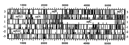
Figure 1 is a sr~ l;c illustration and open reading frame analysis of the
seJU2UlABCD gene cluster
Figures 2A-2D depicts the nucleotide se~lpnr~s of sefA, sefB, sefC and
sefD, and the co..~,~onding predicted amino acid sequenr,Pc
Figures 3A-3B depicts the nucleotide sequPnces of sef(JI and sefU2 and
the co......... esponding predicted amino acid seqllrnc~s
~0 94125597 ' ` ~f ~ 5 PCT/IB94/00205
~1 9
Figures 4A-4B depicts the nucleotide seq~l~nce of tctA and the
coll~s~onding predicted amino acid sequence.
Figure 5 depicts the nucleotide seq~lense of tctB and the corresponding
predicted amino acid sequence.
Figures 6A-6B depicts the nucleotide sequence of tctC and the
coll~ onding predicted amino acid sequPnee.
Figure 7A depicts the nucleotide sequence of an agfA gene fragment
~mplified from S. enteritidis 27655-3b TnphoA mutant strain and cloned into pUCl9,
and the co,-~sl,onding predicted amino acid sequence. The solid arrows inriie?te PCR
primer pairs TA~3 and TAF4; the dashed arrows in~ie~te TAF5 and TAF6.
Figure 7B depicts the seq~lon~e of the full agfA gene of
S. enteritidis 27655-3b and the corresponding predicted amino acid sequence.
Figure 8 depicts a Western blot id~ntific~tion of ploleuls from
Salmonella strains and other ~ ..I.e.~ of the family Enterobacteriaceae that are15 immllnologically cross-reactive with AgfA of S. enteritidis27655-3b. Lanes: 1,
5. enteritidis 27655-3b; 2, S. hadar F9-1; 3, S. hamburg, 4, S. heidelberg, 5, S. infantis
S41-16; 6, S. paratyphi B; 7, S. javiana; 8, S. mbu,~, 9, S. mikawasima; 10,
S. typhi; 11, S. w~"h."~1071, 18, S. entefftidis 27655-3b. The m~le~ r weights (in
th~l.~nrl~) of collu~LIllg, pre~ f,d protein sl~ld&ds (Ret~les~ Research
20 Labolalol;es) are noted.
Figures 9A and 9B depict agarose gel ele~,llophores;s of the results of
PCR ~mplific~tion of DNA L,t~ from various Enterobacteriaceae using
oli~om~leQtide primers decigred from the agfA gene of S. enteritidis. Lanes: 1, no
DNA control; 2, S. enteritidis 27655-3b; 3, S. ~, .u". SU453; 4, S. infantis S41-
16; 5, S. hadar F9-1; 6, S. agona; 7, S. ~ .Jr~, 8, E. coli C600; 9, E. coli HB101; 10,
E. coli NG7c; 11, E. coli Gambia 3; 12, E. coli Vietnam I/l; 13, Enterobacter
a~ ,.e:" 14, Citrobacter ~eu~ulii 8090; 15, Shigella sonnei; 16, S. choleraesuis; 17,
5. typhi; 18, S. pafratyphi A subsp. du~azzo ATCC 11511; 19, DNA ~agment size
lll&ll~l~ p.epaled byMspI ligestion of pBR322.
Figure lOA depicts an autoradiograph of the hybri~ii7~tion Of [32p]
labeled a~A gene probes derived by PCR from S. enteritidis 27655-3b TnphoA mutant
2-7f to l. ~ ,sen~ re panels of HGMF colony blots. Panel positions: A1-F20, E. coli
strains; G1-G12, Citrobacter spp.; I1-I16, Enterobacter spp.; J1-J5, Hafnia spp.; K1-
K15, ~rot~us spp.; L1-L7, Klebsiella spp.; M1-N3, Shigella spp.; N9-P20, Yersinia
spp.; Q1-Q4, Ae,. ,~s spp.; Q5, Boriella; Q6-Q7, Erwinia spp.; Q8-Q9, Providencia
spp.; Q10-Q13, Serratia spp.; Q14-Q15, Acinetobnrter spp.; Q16-Q17, Ach,o). ~bacter
spp.; Q18-Ql9, Alcaligenes spp.; Q20, Serratia ,i.~ces~ , Rl-R12, Pse1~0monas
WO 94125597 . 3 ' `' PCT/IB94/00205--
spp; R3 and Sl-T20, Salmonella spp.; G13-H20, I17-I20, J6-J20, K16-K20, L8-L20,
N4-N8, R13-R20, no bacteria applied.
Figure lOB depicts an autoradiograph of the hybridization of [32p]
labeled agfA gene probes derived by PCR from S. enterifidis 27655-3b TnphoA mutant
5 2-7f to ~ r~selllali~e panels of DNA dot blots. Panel positions: Al-A7 and B2-G7,
Salmonella spp.; Al-A7, B2-C3, S. enteritidis i~ol~te~; C4-C6, S. berta i~ol~tes; C7-D4,
S. pullorum isolates; D5, S. gallir~u~ "~, D6-E4, S. dublin isolates; E5, S. drypool; E6,
S. eastbourne; E7, S. albany; Fl, S. u~ "l, F2, S. arizonae; F3, S. cerro; F4, S.
choleraesuis; F5, S. dahomey; F6, S. florida; F7, S. gu,,.i,u.,u, Gl, S. havana; G2, S.
10 ",i",.csola; G3, S. ~,~,I~u,l, G4, S. neinstedten; G5, S. tennessee; G6, S. typhi,,..~ri~.l,.,
G7, S. wo, Ihi~ on; Hl, Citrobacter fre~lulii 8090; H2, E. coli; H3, Serratia
marcescens; H4, Shigella sonnei; H5, Klebsiella ~ ",ae 13883; H6, Enterobacter
ae,~,.cs; H7, Hafnia alvei; I1, Erwinia caratovora; I2, Proteus vulgaris; I3,
Providencia sp.; I4, Pseudomonas a~,~i"osu, 15, Aeromonas hydrophila; I6,
15 Aeromonas salmonicida: I7, Ra( i~ subtilus; Jl, herring sperm DNA; J2, Salmonella
paratyphi A; B 1 and J3-J7, blank.
Figure 1 lA depicts an wtoradiograph of the results of eAyl~ci.;,ion of the
sefA, sefB ând sefC genes in an E. coli in vitro ll~ulSG~ iOIl-~ ;ol~ system. Lane 1,
pTZl9; Lane 2, pKXl; Lane 3, pSCl; Lane 4, delB15; Lane 5, delB23; Lane 6, delD10;
20 Lane 7, Western blot of the in vi~ro llall~ClilJLiOn-Ll~SIaliOl~ of pKXl de~lopcd using
antisera ~,ne.~LfA against denalured SEF14 fimbrin. The size of the m~e~ r weight
.h.Lt;l~ is inrlic~tf d on the left (103 Mr)
Figure llB is a s~h~ ic Icpres ~;on ofthe sefgene cluster showing
the inserts of various dPle~ion s~lbclorle.s used in the in vi~ro ~ s.;l;~1iol~-ll;..-L~ ;nn
25 ~A~ ds.
Figure 12 depicts a rf sfriction map of the region of the S. t~htm~rt~
chromosome f~Od;~ tctDCBA. The poeition and olie~1~l;on of the open reading
frames of tctD, tc~C and ctB and the tctA open reading frame are in~lir~ted by the
underlying boxes and arrows. ~l~fe.l~d regions of tctC and tctB useful as Salmonella
30 DNA probes are in~iir~ted by the cross-h~trhsd boxes. Abbreviations: A, ApaI; Av,
Ava~; B, Bgm; Bg, Bgll; Bs, BsfE~; D, DraI; E, EcoRV; K, KpnI; N, NcoI; S, SalI;Sr4 SmaI; Sn, SnaBI; Ss, SspI; X XmnI
Figure 13 depicts a PCR-...F ~ d DNA ~rnrlifir.~fion of 308-base tctC
fr~ nte using primers TPP1 and TPP2, from repr~3e,ll~ /e Salmonella serovars and35 other Enterobacteriaceae. Lanes: 1, S. enteritidis 27655-3b; 2, S. tJv~hi~u~ .u". F18-1;
3, S. infantis S41-16; 4, S. choleraesuis; 5, E. coli C600; 6, C freundii 8090; 7,
Shigella sonnei; 8, no DNA (control); 9, S. typhi; 10 and 17, DNA fragment size
~0 94/2SS97 21 6~ PCT/IBg4/00205
ma,kel~ of 217, 238, 242, 309, 404 and 527 bp ple~aled byMspI digestion of pBR322;
11, 5. parafyphi A subsp. durazzo ATCC 11511; 12, S. enteritidis HWC989; 13,
Shigella dysenteriae ATCC 29027; 14, Shigella boydii A~C:~ 870n; 15, Shigella
flemeri ATCC 12022; 16, S. berta ATCC 8392; 18, S. p2.l~rum ATCC 9120; 19,
5 Enterobactera~g~c"cs; 20, Klebsiel~npneumoniae 13883; 21, Serratial,.J,cesc~,.s.
Figure 14 depicts an analysis of the serological cross-reactivity between
the four fimbriae of S. enteritidis 3b and their antisera. Panel A depicts a Western blot
analysis of Factor Xa cut MalE'-SefD fusion (lane 1), purified SEF14 (lane 2), SEF17
(lane 3) and SEF21 (lane 4) with anti-SefD antiserum. Panel B depicts a western blot
10 analysis of SefD with SEF14 (lanes 1 and 2), SEF17 (lanes 3 and 4) or SEF21 (lanes 5
and 6) antisera. SefA, AgfA and FimA are the s~.~ s of SEF14, SEF17 and SEF21
fimbriae, respectively. In both panels A and B, the size (kDA) of the molecular weight
~kw~ is inrlir~ted on the left.
Figure 15 depicts the nucleotide sequ~nr,e offimA of S enterifidis~ and
15 the co.l.,.,~,onding predicted amino acid sequPnr~.
Detailed Dw~ )tion of the Invention
The present ul~h~liOn provides mPthotls and co",pos;l;Qns for the
detection of Salmonella. These methods and compositions include ~u~ ,r~ls ;CQI9ted
20 genes specific to Salmonella, vector constructs, r,u",~,.o~s ;.~O A~Cd plol~llS specific to
S~ ' ,,.ella and r1i~gnostic tcsts. These methnd~ and COIlll~GS;liOl-S are dcs_,il,ed further
below.
I. Genes Specific to S~-~ ,,.clla
A. Genes Generatly
The present i"~e.,lio" provides i~ol~ed DNA msle~ulss ~"""is"~g the
sefU2UlABCD gene cluster, the sefABC gene cluster, the sefU2UI gene cluster, thesefA gene, sefB gene, sefC gene, the sefD gene, the sefUI gene, the sefU2 gene, the
30 a~A gene, the fin~4 gene, the tctCBA gene cluster, the tctA gene, the tctB gene, and/or
the tctC gene. ~hho~l~h one embodiment of each of these m~!ecll1ss is shown in Figures
2 to 7, it should be understood that within the context of the present invention, reference
- to one or more of these genes inrludes derivatives of the genes that are s~lbslAl~l;Ally
similar to the genes (and, where apl)~op-idle, the protein (inr.lutlin~ peptides and
3~ polypeptides) that are cnroded by the genes and their dc.iva~ es). As used herein, a
nucleotide seq~lpnce is deP.mP,d to be "s~ iatly simitar" if: (a) the DNA seq~lPnce is
derived from the coding region of a native gene of any Salmonella serovar and l~ A;~lA;"~
WO 94ns597 ~16 1~ 0 5 PCT/IB94/00205
s-lbst~nti~lly the same biological activity (in~ ing for CA~lllple, portions of the
seql~nre or allelic variations of the sequ~nr,es ~liccl~esed above); (b) the DNA sequ~nce
is capable of hybridization to DNA seq~lences of the present invention under moderate,
high or very high ~ gel-cy (see Sarnbrook et al., Molecular Cloning A Laboratory5 Manual, 2nd ed., Cold Spring Harbor Laboratory Press, NY, 1989); or (c) the DNA
seqU~nres are d~el~e.ale as a result of the genetic code to the DNA sequPnr,~e defined
in (a) or (b). Within the context of the present invention, moderate ~L,i,~gency means
conditions such that an a~l)-o~-iate nucleotide seq-nPnce is able to selectively hybridize
to nucleotide sequence~e from Salmonella and to conserved nucleotide se(~ nre-e in
10 other bacteria such as E. coli. High SLIU~gC,.I~ means the nucleotide sequpnre is able to
selectively hybridize to nucleotide se~u-~ces from Salmonella. Very high stringency
means the nucleotide sequence is able to selectively hybridize to a single Salmonella
species, such as S. enterifidis, S. dublin or S. gallinarum.
B. sef(Jl. sefU2. sefB. sefC. sefD genes and the sefU2UlBCD gene cluster
In one aspect, the present invention provides the nl~leo~ide se~lences of
the sefUI, sefU2, sefA, sefB, sefC, and sefD genes, which are located in a single cluster,
the sefU2UlABCD gene cluster. As can be seen by ,~,.ence to ~igure 1, which is asC~ ;c illustration and open reading frarne analysis of the sefU2UlABCD gene
20 duster, the sefB and sefC genes co,n~,ise an appro~ A~Ply 3.4 kil~b~ee pair region of
Salmonella DNA. In vifro e,.~,ie~s;on di,~,lcd by a 5.3 kilobase pair DNA fragment
Pl~r~ c.;.~g sefA, sefB and sefC in~ ted that the SefB and SefC p.ole~s have an
appro,~ ely 14 K, 28 K and 90 KMr molec~ r weight, r~ecl;~ely. See Figure 1 lA.
The present u,~.,l;on pr~ bly involves one or more of a sefA, sefB, sefC, sefD,
25 sefU~, or sefU2 gene. Further inrullualion with respect to the sefA gene and its
products may be found in U.S. Applic~tion Ser. No. 08/054,542. See also Applic~tiQn
Ser. No. (all.""~'s docket no. 920043.403Cl), and Application Ser. No.
(allU111~S docket no. 920043.403C3). As noted above, these applic~ti~ns and all other
references cited herein are c~.lJleS5l~ incorporated by r~rencc herein in their entirety.)
~ c;nlal results with S. enteritidis i~d;~nle that sefB and sefC are
not e,~ ,ssed in the ~hsence of sefA. Primer ~ ;on analysis of sefABC gene clusters
revealed two major l,~nscli~.lion start sites located ul.sl,~" of sefA (~igure 12).
Tl~sc.ipl;on of sefB and sefC is also ;~.;l; ~ed from the sefA p.omoler region.
Seconr1~ry structure analysis ûf the mRNA Ir~1s.;.il)l frûm sefABC predicted theform~tio~ of two stable stem-loop structures in the uderc;~lronic regiûn ~etween sefA
and sefB, which is indicative of d;~renl;al re~ tion of sefA as opposed to sefB and
10 94l2~597 I~OS PCT/IB94/00205
sefC translation. The nucleotide sequence~, and corresponding arnino acid sequences, of
sefB, sefU~ and sefU2 are depicted in Figures 2A-2D and 3A-3B.
sefUI and sefU2 are ove,lal)ping open reading frames oriented in the
opposite directions (i.e., çneo(ied on opposite strands). sefD abuts sefC and a concP.nc.lc
S Shine-Dalgarno ribosomal binding site seq-lPnee is just inside the sefCOrf. No promoter
is recognized ;.. P~ PIY upsL-e~,. of sefD; cAIJiesa;on may be directed by the sefA
promoter.
C. a~fA gene
The agfA gene codes for a structural fimbrin protein composing very thin
fimbriae, apl)lo~.lalely 3-4 nano,nete.a in r~i~mP~ter~ that are highly agglega~ e and
stable. The ag~;~,aLh~e plopelly of the fimbriae is believed to be due to its
hydrophobicity. The gene product of the agfA gene, AgfA, is found in SEF17 fimbriae,
and comprises an app~ ely 14-15 KMr m~!~cnl~r weight protein. AgfA
15 contributes to heavy pellicle form~tiQr. in static cultures, colony l.~d.ophobicity, and
~t~egalion of cells in culture. SEF17 rl..ll";ac are immlmolo~yc~lly distinct from
SEF14 and SEF21, as ;..~ieAtcd by the lack of cross-reactivity with polyclonal antisera
raised in rabbits against SEF14 or SEF21 (Collinson et al., Purification and
characL~.~alion of thin, aggregali~e fimbriae from S. enteriti~s," .J. Bacteriol.
20 173:4773-4781, 1991). Figure 7A depicts the nucleotide sequPnre of an a~fA gene
L~lc.l~ that was ~mplifie~ from the S. enteritidis 27655-3b TnphoA mutant strain and
then cloned into pUCl9. The bases underlined in the agfA sequpnce of Figure 7A are
GOIll~llOn to pG.lions of the PCR primers TAFl and TAF2, which were used in
~mrlificA~tirJn of this fr~ nt Below the agfA Sequpnre in Figure 7A is the ll~ rlsled
25 amino acid se4u~ .ce As J;c~ cced further below, the nucleotide se~uenres targeted by
the ~ ~ostic PCR primer pairs TAF3 and TAF4 (solid arrows) and TAF5 and TAF6
(dashed arrows) are also il~dirAte~ in Figures 3A-3B.
The sequPnr,e of the full agfA gene of S. enteritidis 3b is shown in
Figure7B. The se~uçnce ~- ed in Figure7B refiects two nucleolide ~I~AI~gS (at
30 pos;l;o~-c 69 and 75 of Figure 7B) from the sequpnre of Figure 7A, but the chA~ges do
not alter the encoded amino acid sequçnr,e
D. tctA. tctB, and tctC ~enes. and the tc~I, tctII and tctIn ope. ons
The tctA, tctB, tctC and tctD genes are located in the tct~ operon, which
3~ is one of three op~,-uns for tricarboxylic acid ~ ulL within ,~ .clla spp. The
other Salmonella tricall,uAylic acid ll~.s~ y~ .ls are termed tctII and tctIII. It is
believed that the trica,l.u~ylic acid ll~.s~,o.l sy-stem of Salmonella is lacking from
WO 94/25597 2 i ~ 1 ~ O S PCT/IB94/00205--
14
certain other related genera. tctI and tctIII both encode protei,.s responsible for citrate
transport and are in~llced by growth of Salmonella typhimurium on minim~l media
supplemented with citrate as a carbon source. tctII is normally not ~ sed. Each
system demonstrates p,ererelllial ~lar,~po" of various tricarboxylic acids and di~.~
5 concentrations of the monovalent cations Na+ and K+. The tl~U~SIJOl~ of llichll~uAylic
acids, particularly citrate, are important to the intr~cel~ul~r survival of Salmonella, as
such transport provides a means of scavenging IlllLliC.-ls from a host cell. Acc~rdil,~ly,
; in these various tct systems, plerelably in S. ty~h."...,i. ". or S. enteritidis,
create ~tt~n~ted strains of Salmonella capable of being taken into a host cell, and
capable of pc.:,;;,lu.g for a limited time, yet not able to proliferate within such a host cell.
In a p~ef~ ,d embodimpnt~ one or more ofthese tct ~ are coupled with a mutant
unable to utilize s~lcçin~te to provide a "back-up" system to assure attPm~tion
The nucleotide seq~P,nces for tcfA, tctB and tCfC, along with their
co"~pond,llg amino acids, are depicled in ~igures 4A-4B, S and 6A-6B, rc~cclively.
E. fimA gene
fimA Pnr.odec the Salmonella typel fimbriae, which is also known in
Salmonella enteritidis as SEF21 L"~li&c. Prior to the licclQsllre of the subject
spplication, Type 1 fimbriae were believed to be the only Salmonella fimbrial type
imrlic~ted in p~th-)g~ y. polydonal antisera studies have int1ic~ted that Type 1fimbrial Pntig~nc are among the majority of ,Sa' ,.ella S_.ulylJeS. The sequp-nre of the
fimA gene of S. enteritidis 3b is shown in Figure 15.
II. Vector Cûnstructs Cûnlyl is;"g the Gene Seq~lences of the Present Invention
A. Vectûr Constructs Generally
The present invention provides for the m~niplul~tion and c,~ ,sDion of
the above des~; ;l,ed genes by culturing host cells co~ ing a construct capable of
c,.~.ressi"~, the above-described genes, incl~ldirlg s~ s~ l;ally similar derivatives thereo
Nu~ ,.ous vector constructs, inrl.~-li~ all or part of the nucleotide
seqnenr~çs of a native or d~,ivalive sefUl, sefU2, sefA, sefB, sefC, sefD, agfA, tctA,
tc~B, and/or tc~C genes, as ~es il,ed above, can be p,.,~ar~d as a matter of con~,e.~icnce.
Within the context of the present invention, a DNA construct is understood to refer to a
DNA molee~ " or a clone of such a molecl-le (either single-stranded or double-
stranded), that has been modified through human intervention to contain s~ of
DNA co",billed and ju~posed in a manner that as a whole would not otherwise exist in
nature. Vector constructs of the present invention COnllJl;SC a first DNA seen.P.-I
o 94/2sss7 21 6i l 05 pcTlIs94loo2os
~nr.odin~ one or more of the sefUI, sefU2, sefA, se~B, sef~, sep~, a~A, tctA, tctB,
and/or tc~C genes operably linked to additional DNA se~g,..f~ required for the
c AI,le~s;on of the first DNA se~S...P .I Within the context of the present invention,
additional DNA se~,,..r~nl~; will include a promoter and will generally include
5 t,ai~s~ ,Lion tG~una~ol ~ and may further include P nh~nr,fj. ~ and other P,l~ ..f ..l ~
Mutations in nucleotide sequences constructed for eA~"~ssion of variant
proteins preferably preserve the reading frame of the coding sequences. Furthermore,
the mllt~tionc will p,GL.~bly not create compl-~..F.~ regions that could hybridize to
produce secondary mRNA structures, such as loops or hairpins, that would adversely
10 affect llnr~ l;on of the mRNA. ~hhollgh a ,..,~l~lion site may be predete.,."ned, it is
not l-ecess,. y that the nature of the mllt~tior~ per se be predet~,.",llled. For ~ ,le, in
order to select for o~ "~", characteristics of m~lt~ntc at a given site, random
ml-t"gf.-es:c may be cond~lcted at the target codon and the e,~,iessed ~ hl~l~ sc,~ned
for indicat*e biological activity.
M.. ~;.l;nnc may be introduced at particular loci by syrthe~i7ing
oligom~rleotides ~,,I~;..;.~g a mutant se~upnce~ fianked by restriction sites enabling
ligation to fr~n~ntc of the native sequçnre- Following ligation, the res..lting
rfcon~l,u~,led se~lence rncode5 a de.i~ali~le having the desired amino acid in3~.liol1,
~b~l;l.... .....l;ol-, or dPle.tion
A1lG111~ Y~ oligo"u~leolide-dilccl~d site-specific mllt~genPcic
procedul es may be c."~lo~ cd to provide an altered gene having particular codons altered
acco,di"g to the s lbtitution~ df~k,~irJ~ or insertion r~ uil~d. FY~ ~IP1LIY mPthorl~ of
making the alterations set forth above are ~ rlosed by Walder et al. (Gene 42:133,
1986); Bauer et al. (Gene 37:73, 1985); Craik (BioTechniques, January 1985, 12-19);
25 Smith et al. (Genetic E,.~i,.c~",,~. Pr",i~les and Methods, Plenum Press, 1981); and
Sambrook et al. (s~pra).
The pli~ll~y amino acid structure of the above des.,lil,ed ploteills may
also be morlified by rO~"Iing covalent or ag~t,gali~e conj~ tes with other chemical
moieties, such as glycosyl groups, lipids, phG5ph~ acetyl groups, or with other
plole.ns or polypeptides.
Within a further embotlimrnt~ the above desc-il,cd proteins may be fused
with other peptides that f~rilit~te purifir~tion or ir~Pnfifir.~tion of these proteins. For
- ~Y~mrhP~ â protein can be p~ ed as a fusion protein with the FLAG polypeptide
se~1Pnre (see U.S. Patent No. 4,851,341; see also Hopp et al., Bio/Technology 6:1204,
35 1988). The FLAG polypeptide sequPnce is highly ~nfigPnir, and provides an epitope for
binding by a specific mQn~rl~n~l antibody, Pn~hling rapid puriication of the c,~ ed
WO 94/25597 ~ 16 PCT/IB94/00205 --
recol~lbina..~ protein. This sequence is also specifically cleaved by bovine mucosal
enterokinase at the residue imm~t~iAt~ly following the Asp-Lys pairing.
B. Expression Vectors
S One type of vector construct, known as an e~yles~;on vector, can contain DNA
se~ n~ rceccz~ to direct the secretion of a polypeptide of interest. Such DNA
sP~ C can include at least one secretory signal seq-~çnce. Plef~ ,d secretory signals
include the yeast alpha factor signal sequPnce (pre-pro sequPnre; Kurjan and
Herskowit, Cell 30:933-943, 1982; Ku~an et al., U.S. Patent No. 4,546,082; Brake,
EP 116,201), the PhoS signal sequence (Beck et al., WO 86/00637), the Suc2 signal
seq-lPnce (Carlson et al., Mol. Cell. Biol. 3:439~47, 1983), the a-2 plasrnin inhil~itor
signal sequPnce (Tone etal., J. Biochem. (Tok~yo) 102:1033-1042, 1987), the tissue
plas,l""ogen activator signal sequence (Pennica et al., Nature 301:214-221, 1983), the
E. coli PhoA signal sequPnre (Yuan et al., J. Biol. Chem. 265:13528-13552, 1990), or
any of the other bacterial signal seqUpnc~pc known in the art, such as those reviewed by
Oliver (Ann. Rev. Microbiol. 39:615-649, 1985). Alternatively, a se_~tul~ signalse~ n~e can be synthP~i7Pd accord",g to the rules e~lubli~l,ed, for ~ _ rle~ by von
Heinje (Eur. J. Biochem. 133:17-21, 1983; J. Mol. Biol. 184:99-105, 1985; Nuc. Acids
Res. 14:4683-469û, 1986). Se~_ ~,to,y signal se~u ~ces can be used singly or in
c~n,l,u,aliol,.
For ~Ay~ ;on~ a DNA mole~l-le as ~Ic,~il,ed above is ins_.lcd into a
suitable vector construct, which in turn is used to lr~n~rol". or ~ r~, ayployliale host
cells for CAyi~Sa;Oll. Host cells suitable for use in practiring the present invention
include ~ ""Ali~An, avian, plant, insect, bacterial and fungal cells. ~,fe.,~,d t,uk~u~ulic
25 cells include ~ullur~,d ,~ n~ n cell lines (e.g., rodent or human cell lines) and fungal
cells, in~ ing species of yeast (e.g., Sacck~u"..yces spp., particularly S. cerevisiae,
Schi o.~ncch~v".yces spp., or Kl?yveromyces spp.) or filz~ lous fungi (e.g,
Aspergillus spp., Neu,v~or~ spp.). Strains of the yeast Sacc~h.,o".yces cerevisiae are
particularly p,.,~ ,d. ~etho~l~ for producing r~con~ z~l proleins in a variety of
30 prokaryotic and eukaryotic host cells are generally known in the art (see, "Gene
Expression Technology," Methods in E;n~ymology, Vol. 185, Goeddel (ed.), ~dçmic
Press, San Diego, Calif., 1990; see also, "Guide to Yeast Genetics and M- lec~ rBiology," Methods in E;nzymology, Guthrie and Fink (eds.), .A~d~mic Press, San Diego,
Calif., 1991). In general, a host cell will be sP~e~,l~ on the basis of its ability to produce
35 the protein of interest at a high level or its ability to carry out at least some of the
procP~ing steps necess~y for the biologi~l activity of the protein. In this way, the
~O 94125597 17 ~l~4o PCT/IB94/00205
number of cloned DNA sequences that must be introd lc into the host cell can be
. . .;. .;. . .; ~ed and overall yield of biologically active protein can be lll~;.~ fd
Suitable yeast vectors for use in the present invention include YRp7
(Struhl et al., Proc. Natl. Aca~ Sci. USA 76:1035-1039, 1978), YEpl3 (Broach et al.,
Gene 8:121-133, 1979), pJDB249 and pJDB219 (Beggs, Nature 275:104-108, 1978)
and derivatives thereo Such vectors will generally include a s~leel~,lc marker, which
may be one of any number of genes that exhibit a doll"ll~l phenotype for which aphe.~ y~ic assay exists to enable ll~lsr~".,l~ts to be selected Pl~Eelled select~ble
lll~el~ are those that complement host cell auxotrophy, provide antibiotic reciet~nce or
enable a cell to utilize specific carbon sources, and include leu2 (Broach et al."bi~),
ura3 (Botstein et al., Gene 8:17, 1979), or his3 (Struhl et al., ibi~). Another suitable
s~lect~hle marker is the cat gene, which confers chlo,~llphel~icol le~;cl~nce on yeast
cells.
P~.,fe.,ed promoters for use in yeast include pro"lote.~ from yeast
glycolytic genes (TT;~ - etal., J. Biol. Chem. 255:12073-12080, 1980; Alber and
Kawasaki, J. Mol. Appl. Genet. 1:419-434, 1982) or alcohol dehydrog~n~ce genes
(Young et al., in Genetic Er~ .7~ of Microo~u"i~".s for Ch~?micn~ ro!l~n-l~r
et al. (eds.), p. 355, Plenum, New York, 1982; A,llll,~ ;l, Meth Enzyrnol. 101:192-201,
1983). The eAI ,~,s~ion units may also include a l.~sc,;plional t~."li"alor.
In nd~litisn to yeast, prole.ns of the present invention can be c~ ssed in
fil~ C fungi, for e~L __ptt-, strains of the fungi Aspergillus (~Knight et al., U.S.
Patent No. 4,935,349, which is incG~Glaled herein by r.,re,~nce). F~ s of usefulpromoters include those derived from Aspergillus nidulans glycolytic genes, such as the
adh3 prol"otw ~cRni~ht et al., EA~O J. 4:2093-2099, 198S). An ~Y~mple of a
suitable t~ or is the adh3 te.",inalor (~rXnight et al., ibid., 1985). The
c.~ s .;on units utili7ing such components are cloned into vectors that are capable of
ills~ ;on into the chrnmnsom~l DNA of Aspergillus.
TCC~ 4~'eS for h~ru~ ing fungi are well known in the Lle.alul~, and
have been desc,il,ed, for i..~ cr" by Beggs (ibid.), Hinnen et al. (Proc. Natl. Aca~ Sci.
30 USA 75:1929-1933, 1978), Yelton etal. (Proc. Natl. Acad. Sci. USA 81:1740-1747,
1984), and Russell (Nature 301:167-169, 1983). The geno~y~c of the host cell will
generally contain a genetic defect that is co rl-~nted by the s~lect~hle marker present
on the e,~ie~;,;on vector. Choice of a particular host and sFIG~ hle marker is well
within the level of oldill~ ~ skill in the art.
3~ In ~d~lition to fungal cells, ~iulluled ~ n cells may be used as host
cells within the present il,~,nlioll. ~r~ d cultured ~ Alistn cells for use in the
present ill~,.,.llioll include the COS-1 (ATCC No. CRL 1650), COS-7 (ATCC No. CRL
W O 94/25597 ' 2 1 B 1 4 Q5 18 PCT/nB94/00205
1651), BHE~ (ATCC No. CRL 1632), and 293 (.~TCC No. CRL 1573; Graham et al., J.
Gen. Virol. 36:59-72, 1977) cell lines. A l),e~"ed BHK cell line is the BHK 570 cell
line (deposited with the American Type Culture Collection under accession number CRL
10314). In ~ tion~ a number of other ,..F..,~ n cell lines may be used within the
S present invention, inelu-ling Rat Hep I (ATCC No. CRL 1600), Rat Hep II (ATCC No.
CRL 1548), TCMK (ATCC No. CCL 139), Human lung (ATCC No. CCL 75.1),
Human k-,p~lo~ (ATCC No. Hl P~-52), Hep G2 (ATCC No. HB 8065), Mouse liver
(ATCC No. CCL 29.1), NCTC 1469 (ATCC No. CCL 9.1), SP2/0-Agl4 (ATCC No.
1581), HIT-T15 (ATCC No. CRL 1777), and RINm SAHT2B (Orskov and Nielson,
F~BS 229(1):175-178,1988).
~ mm~ n CAyle;~i~;on vectors for use in c6~ out the present
invention will include a plU-I-Ot~. capable of .lir~ ~ li,.g the ~ s~.-;ylion of a cloned gene
or cDNA. ~ d plc"l.ol~.., include viral p,c""ole.., and cellular p-o",ùle, ,. Viral
plul"olc~., include the ;"".~eA;~e early ~to".egalovirus plon,oter (Boshart et al., Cell
41:521-530, 1985) and the SV40 plolllûtcr (SUblCUI~U~ et al., Mol. Cen BioL 1:854-
864,1981). Cellular plc,l"olel:, include the mouse metallothionein-l plolllùllr (Palmiter
et al., U.S. Patent No. 4,S79,821), a mouse Vj prolllote~ (~c.{~ l et al., Proc. Na~l.
Acacl Sci. USA 81:7041-7045,1983; Grant et al., Nuc. Acid~Res 15:5496,1987) and a
mouse VH promoter (Loh et al., Cell 33:85-93, 1983). Such eA~ ;OIl vectors may
20 also contain a set of RNA splice sites located duv~-~lwlll from the plulllole. and
Up~ l from the DNA sequ~onre enco~ing the peptide or protein of interest. ~ ~,f~ d
RNA splice sites may be obt~ ed from adello~ s and/or ;.. ~.oglobulin genes. Also
Co~ d in the ~Aylei~ion vectors is a poly..d~,nyl~lion signal located dowl-sl~ of
the coding sequence of interest. S~ polyadenylation signals include the early or25 late pol~denylalion signals from SV40 (R~llfm~n and Sharp, ibi~), the polyadenylation
signal from the Adenovirus 5 ElB region and the human growth holll,one gene
t.,.ll~il,alor (l)eNoto et al., Nuc. Acid~Res. 9:3719-3730, 1981). The cAI.l.,.a~;on vectors
may include a noncoding viral leader sequence~ such as the Adenovirus 2 ~ ile
leader, located be~..cen the promoter and the RNA splice sites. ~,f~ d vectors may
30 also include çnh~nr,~r seqllçncPs such as the SV40 f~.nh~nr,er and the mouse I ~nh~nrPr
(Gillies, Cell 33:717-728, 1983). Expression vectors may also include seq~lenr,ç~
Pnr,orling the adenovirus VA RNAs. Sl-it~ble cA~"es~ion vectors can be obtained from
cc .. e~cial sources (e.g, Str~t~g~n~ La Jolla, Calif.).
Vector constructs co",~ i"~, cloned DNA sçq~Pnr,~s can be introduced
35 into cultured m~mm~ n cells by, for example, calcium phosph~te-mçdi~ted tr~n~fection
(Wigler et al., Cen 14:725, 1978; Corsaro and P~u~on, .S~ n~ Cell Genetics 7:603,
1981; Graham and Van der Eb, Virology 52:456, 1973), elc~llopolalion (Ne~m~nn
0 94/25597 21 61 ~ Q5 PCT/IB94/00205
etal., EA~BO J. 1:841-845, 1982), or DEAE-dextran metli~ted l,~nsÇG.,Iion (Ausubel
et al. (eds.), Curren~ Protocols in Molecular Biolo~y, John Wiley and Sons, Inc., NY,
1987), which are incolporated herein by l~,f~.e.-ce. To identify cells that have stably
integrated the cloned DNA, a selçctakle marker is generally introduced into the cells
along with the gene or cDNA of interest. Pl~f~.lt;d sele~ble lll~.~ for use in
cultured "~ n cells include genes that confer r~ ce to drugs, such as
neomycin, hyglolllycin, and methol~ e. The s~le~ ble marker may be an amplifiable
s~l&.,l~ble marker. Pl~f~ ,d ~mplifi~hle s~le~hle m~k~.~ are the DHFR gene and the
neomycin le~ nee gene. Selectable ",&rl~ are reviewed by Thilly (Mf7~nmn~ CeU
Technology, ~ull.,.wcl~lh Publishers, Stoneh~m~ MA, which is incc)l~,ulaled herein by
l.,f~,.e.,ce). The chûice of select~ble markers is well within the level of ordii al~ skill in
the art.
As d;c~ ssed further below, naked vector constructs can also be taken up
by m-lcc~ r cells ~ se.lLIent to injection intû the muscle of a ".~.".n~l (or other
1 5 animals).
~ele~l7.b'e nl~l~.~ may be introduced into the cell on a separate vector
at the same time as the sefUl, sefU2, sefA, sefB, sefC, sefD, a~ffA, tctA, tctB, and/or
tc~C genes sequences, or they may be introduced on the same vector. If on the same
vector, the sFle~ble marker and the seflJl, seJU2, sefA, sefl3, se~, sefD, agfA, tctA,
tctB, and/or tcfC genes seq~l~nrPs may be under the control of dilr~,rl,nl p~ lùle~a or the
same promoter, the latter ~UI' ~gf-~- nl pro~uçing a dicialr~nic mP~Q~P Constructs of
this type are known in the art (for example, Levinson and Su,lonsen, U.S. Patent No.
4,713,339). It can also be adv~ geo~c to add ~ tion~l DNA, known as "carrier
DNA," to the lIUAIUI~ that is introduced into the cells.
M~mm~ cells c~ g a suitable vector are allowed to grow for a
period of time, typically 1-2 days, to begin eAl~le~s;ng the DNA se~v nee(S) of interest.
Drug sPlection is then applied to select for growth of cells that are eA~ u~g the
sPlect~hle marker in a stable f~hion For cells that have been tl;~n~r~led with an
~mplifi~ble, se~ ble marker the drug cç~nc~ alion may be incleased in a stepwisemanner to select for i.~cleased copy number of the cloned se~up~nre-s~ thereby incleaalng
cAIJiesa;on levels. Cells cA~lesalll~, the introduced se~uPnr,es are sPlecte~ and scl~ned
for pro~uction of the protein of interest in the desired forrn or at the desired level. Cells
that satisfy these criteria can then be cloned and scaled up for productiQn
Nu~"c.uus plant host cells known in the art can also be useful within the
present invention, in light ofthe subject specifir~tiQn
The pl~fe.lèd prokaryotic host cell for use in eA~ sing the gene
sequenr,es of the present invention is Salmonella. Other pl. felled host cells include
WO 94/25597 ?,~ 6~ 4 20 PCT/IB94100205
strains of the bacteria E. coli ~lthough R~ h/~, Shigella, Ps~t -J~ r~s, Sl,~on~yces
and other genera are also useful. Techniquec for transrol.,ung these hosts and
C~,IJlt;SSil-g foreign DNA sequences cloned therein are well known in the art (see, e.g,
Maniatis et al., Mol~ nr Cloning A Laboratory Manual, Cold Spring Harbor
S Laboratory, 1982, which is incGI~laled herein by lerertnce; or Sambrook et al., supra).
Vectors used for ~ t;S~ g cloned DNA sequences in bacterial hosts will generallycontain a sele~ LAble marker, such as a gene for antibiotic reC;~t~n~ç~ and a promoter that
functions in the host cell. Appr~. ;a~e promoters include the trp (Nichols and yAllor~ky~
Me~h EnymoL 101: lS5-164, 1983), lac (C~s~ b~n et al., J. Bacteriol. 143:971-980,
10 1980), and phage ~ (Queen, J. Mol. AppL Genet. 2:1-10, 1983) p,omol~. systems.
Plasmids useful for llAll;~rollllillg bacteria include the pUC pl~cn~?~c (Mes;.~lg, Meth.
EnymoL 101:20-78, 1983; Vieira and M_s~i~, Gene 19:259-268, 1982), pBR322
(Bolivar et al., Gene 2:95-113, 1977), pCQV2 (Queen, ibid.), and derivatives thereof.
Plasmids may contain both viral and bacterial e
Given the teachingc provided herein, ~IOlllO~C~ te-llu IAlOl:i and metho
for introducing c~iei,;,;on vectors encoding sefUI, sefU2 sefA, sefB, sefC, sefD, agfA,
tc~A, tctB, and/or tc~C genes of the present invention into plant, avian, fish, and insect
cells would be evident to those of skill in the art. The use of baculoviruses, for ~ r~ ' e,
as vectors for e.~ ,ss...g heterologous DNA sequ~- ~ces in insect cells has been reviewed
20 by Atl~nson et al. (Pesffc. Sci. 28:215-224,1990). In ad~litiQn, the use ofAgrobacterium rh.~,.es as vectors for e,.pi. i~ genes in plant cells has been
reviewed by Sinkar et al., (J. Biosci. fBangaloreJ 11:47-58, 1987).
Host cells CG..~ i~ vector constructs of the present invention are then
~,ullul~,d to express a DNA mole;llle as des~,lil,ed above. The cells are cullured
25 acc~lding to ~lAndard methods in a culture ~P.I;~ 8 nu~ hls l~ui~d forgrowth of the chosen host cells. A variety of suitable media are known in the art and
generally include a carbon source, a nitrogen source, esC .,1;~1 aunino acids, Vil~llills and
minerals, as well as other co.,l~,on~ , e.g., growth factors or serum, that may be
lequihGd by the particular host cells. The growth ...Pfl; ~.. will generally select for cells
30 cG~ ;nE the DNA construct(s) by, for Py~mrle~ drug selection or deficipnry in an
eecrnti~l nutrient which is complemPnted by the s~k~ i ~kle marker on the DNA construct
or co-tr~n.cfected with the DNA construct.
Suitable growth colltlitiQnc for yeast cells, for e-i~n~ include culturing
in a rh~omir~liy defined ...e-~i~..n co...l,.;sing a nitrogen source, which may be a non-
35 amino acid nitrogen source or a yeast extract, inorganic salts, vi~..,..s and esc~n'ti~lamino acid su~ s at a te.ll,.)eralllre between 4C and 37C, with 30C being
particularly l)rere.led The pH of the .~e~ n is plc;rwcbly ...~ .PA at a pH greater
lO 94/25597 21 2~ PCT/IB94/00205
than 2 and less than 8, more preferably pH 5-6. Methods for I~lA;ll~ ;llg a stable pH
include buffering and CQ~ pH control. ~,fGII~d agents for pH control include
sodium hydroxide. P~.,f~,.led bu~.ing agents include s~ccinic acid and Bis-Tris (Sigma
Chemical Co., St. Louis, Mo.). Due to the te~ r of yeast host cells to
5 hy~ lycosylate heterologous proteins, it may be plere.~ble to express the sefUl,
sefU2, sefA, sefB, sefC, sefD, agfA, tctA, tctB, and/or tctC genes of the present
invention in yeast cells having a defect in a gene required for asparagine-linked
glycosylation. Such cells are pl~felably grown in a II.eAi~ CGllI;~ an osmotic
stabilizer. A pr~,f~;l,ed osmotic stabilizer is sorbitol supple~ ed into the me~ m at a
10 conc~ lion ~ ,en 0.1 M and 1.5 M, prefe,.~bly at 0.5 M or 1.0 M.
Cultured ,.. ~ cells are generally cultured in col.. ~ ,;dlly
available serum-cG.. ~ g or serum-free media. Selection of a .. e~l;.. and growth
contlitiQnc applu~,lia~e for the particular cell line used is well within the level of ordina,y
skill in the art.
m. ~l~t~-S
A. ~ .l.s Generally
As noted above, the present invention also provides icol~ted prl)le,l~S.
20 Within the context ofthe present in~e.llion, such proteh~s are und~ lood to include the
whole, or portions, of a gene product derived from one or more of the sefUl, sefU2,
sefA, sefB, sefC, sefD, agfA, fimA, tctA, tctB, and/or tctC genes, or d~,.i./ali.~es thereof
as .l;c~iucsed above. Where the protein is a portion of a native gene or is ~ncoded by
derivative of a native gene, the protein ..~ c ~b~ 11y the same ~ ~r~l
25 activity of the native protein. The structure of the plOle~l~s CGIl~,SyOn~ 8, to the sefUl,
sefU2, sefA, sefB, sefC, sefD, agfA, fimA, tctA, tctB, and/or tctC genes can be pl.,di.;led
from the plilll~y translation products using the Lyd~ophobr-;ly plot filn~tion of, for
example, P/C Gene or TntP~ ;CS Suite (TntPllig~nPtirC Mountain View, Calif.), oraccG,di,-g to the methods desc,il,ed by Kyte and Doolittle (J. Mol. Biol. 157:105-132,
30 1982).
B. Purification of Proteins
Plotc;ns can be iColntpd by, among other methn~l,s, culturing suitable host
and vector systems to produce the lecolll~ l tr~nQlntior- products of the present
35 invention. Sllpc,l,aL~ILs from such cell lines, or protein ;--r~ c or whole cells where
the protein is not eAcreted into the ~lpC,.Ilal~ll, can then be treated by a variety of
purifir~tion procedures in order to isolate the desired ~,lote;ns. For ~Anlll~)le~ the
WO 94125597 2 ~ ~ ~ 4~5 22 PCTIIB94100205 ~
s~lpt;l,la~lL may be first col~æ ~ ed using collllllereially available protein
conr,çntration filters, such as an Amicon or Millipore Pellicon ultrafiltration unit.
Following conc~lllr~l;on, the conct~ a~e may be applied to a suitable purification matrix
such as, for eY~mple an anti-protein antibody bound to a suitable support.
5 Alternatively, anion or cation ~-~.I-A~ge resins may be employed in order to purify the
protein. As a further alternative, one or more reverse-phase high ~.,.rull~ ce liquid
chrc.lllalography (RP-HPLC) steps may be employed to further purify the protein.Other methods of i~ol~ting the prote~lls of the present invention are well known in the
skill of the art.
A protein is deemed to be "isolated" within the context of the present
invention if no other (undesired) protein is detected pursuant to SDS-PAGE analysis
followed by Coom~ blue ~tAinin~ Within other embo~ the desired protein
can be icolsted such that no other (unde;,;r~d) protein is ~etected pul~&ll to SDS-
PAGE analysis followed by silver sl~ g
C. Pl~y~alion of A-llibod;cs
In another aspect, the prote.lls of the present invention are utilized to
prepare s~e iL~lly binding An~;lJo~ s Accol.li l~ly, the present i.l~ Lioll also provides
such &~libod;es. Within the context of the present u~ nlion, the term "~aLil,odies"
20 inrl~ s polyclonal antibodies, ..-o~nclo~~AI &-~il.od;~s, anti-idiotypic antibodies,
L~ r.l~ thereof such as F(ab')2 and Fab L~ , and r~l..l.il.al.lly produced
binding pa,l.l~.~. Such binding p&lll~,.S h cc,l~olale the variable regions that permit a
monoclonal antibody to sperifir~ y bind, which means an antibody able to selectively
bind to a peptide produced from one of the sefUI, sefU2, sefA, sefB, sefC, sefD, a~A,
25 firnA, tctA, tctB, and/or tctC genes of the invention. The affinity of a rnono.~,lon~l
antibody or binding partner can be readily dct~ ed by one of oldillaly skill in the art
(see Scal~,ll~d, Ann N Y. Aca~ Sci. 51:660-672, 1949).
A variety of assays can be utilized in order to detect antibodies that
specific~lly bind to the desired protein or peptide. r~ ?~ assays are des.,lil.ed in
30 detail in Antibodies: A Laboratory Manual, Harlow and Lane (eds.), Cold Spring
Harbor Labolaloly Press, 1988. Rep.eie.llali~le examples of such assays include:CO~ ,;ull~ immllno el~Ll~ph~ll,S;S (CIEP), radi~;n....~nn~s~ys,
radioimm~nop.cc;~ ;ons, ~nL~Illc-linked imm--no-sorbent assays (ELISA), dot blotassays, inhibition or col~ ion assays, and sandwich assays, ;~ .os~iC~ (dipsti~
35 assays, s;~ n~us immllno~cs~ys, immlmocl~olllalo~a~hic assays, illl~ lnofil~rallon
assays, latex bead ~,I.J~ l;c~n assays, imm..nnfl~-Qr~scenl assays, b nS~n~or assays, and
~0 94/25597 f ~ o s 23 ,, PCT/IB94100205
low-light detection assays (see U.S. Patent Nos.4,376,110 and 4,486,530; see also
Antibodies: A Laboratory Manual, supra).
Polyclonal antibodies can be readily generated by one of cldin&ly skill in
the art from a variety of warm-blooded animals such as horses, cows, goats, sheep,
5 dogs, Cl~iC~.lS, turkeys, rabbits, mice, or rats. Briefly, the desired protein or peptide is
utilized to ;.. ~.; e the animal through i~L~)e.iloneal, inll~.. cc~ r, intraocular, or
subcut~neous il,je~,Lions. The imm~-nogenicity of the protein or peptide of interest may
be in~ ased through the use of an adjuvant such as Freund's CCIll,.~ or inc~ rl~ e
adjuvant. Following several booster ;.. ~ ;ons, small sa~ )les of serum are
10 CQllP,GtPd and tested for reactivity to the desired protein or peptide.
Particularly pl~ d polyclonal antisera give a signal that is at least
three times greater than background. Once the titer of the animal has reached a plateau
in terms of its reactivity to the protein, larger qu~ntities of polyclonal antisera may be
readily oblaincd either by weekly ble~~ g~ or by eYc~ngllin~ting the animal.
Monorlo~l antibodies can also be readily gcnc.utcd using well-known
ter.hn~ es (see U.S. Patent Nos. RE 32,011, 4,902,614, 4,543,439, and 4,411,993; see
also Monoclonal Antibod es, Hybridomas: A New Dimension in Biological Analyses,
Plenum Press, ~ennett lU~Xe~n, and Re~htol (eds.), 1980, and Antibûd~es: A
Lubu,.~ Manual, Harlow and Lane (eds.), Cold Spring Harbor Laburalo-y Press,
20 1988). Briefly, in one c.lll~o~ a subject animal such as a rat or mouse is u~;ected
with a desired protein or peptide. If desired, various techniques may be utilized in order
to i.~c~se the reslllt~n~ ;""".i~e .~*,ollse ~s~ne~aLed by the protein, in order to develop
greater ~ ody reactivity. For c ~ . .?1&, the desired protein or peptide may be coL ~ 1~ d
to another protein such as ov~lb~ in or l~hole limpet hemocyanin (~CLH), or ll~o~gh
25 the use of adjuvants such as Freund's complete or c ~1 e adjuvant. The initial
e~ t~tior~ of an ;~ r l~,sl.o~ may ~n,~,.ubly be through ill~ c.ilol cal,
c~ r, intraocular, or s ~bcu~ lc routes.
BeL~ n one and three weeks after the initial ;n.. ~ ;r)n the animal
may be l~:--------l-;,ed with another booster immllni7~tior~ The animal may then be test
30 bled and the serum tested for binding to the desired antigen using assays as desc.il,ed
above. ,~rlition~l immllni7~tion.C may also be ~ccQmrliched until the animal has reached
a plateau in its reactivity to the desired protein or peptide. The animal may then be
given a final boost of the desired protein or peptide, and three to four days later
sacrificed. At this time, the spleen and Iymph nodes may be harvested and disrupted into
35 a single cell ~v~lJr l.~;on by passing the organs through a mesh screen or by rupturing the
spleen or Iymph node lll~ es which enr~r~s~ t~ the cells. Within one embo~lim~nt
WO 94/25597 ~ 6~4~ 24 PCT/IB94/00205 /d~
the red cells are subseq~entIy lysed by the addition of a hypotonic solution, followed by
i....~.e~ e. return to isotonicity.
Within another embodiment, suitable cells for prcp~ing monoclon~l
antibodies are obt~ined through the use of in vitro immllni7~tion terl-n~ es Briefly, an
5 animal is sacrificed, and the spleen and Iymph node cells are removed as described
above. A single cell ~ yv~sion is p,~ed, and the cells are placed into a cultureco~ ini~ a form of the protein or peptide of interest that is suitable for genc.~ g an
immlln~ ,~sponsv as desv-il,cd above. Subseq~lçntly~ the Iymphocytes are harvested and
fused as described below.
Cells that are obtained through the use of in vifro ;.. ~ 1 ;on or from
an ;.. ~ d animal as desvlilJed above may be immortalized by ~ r~ ;or with a
virus such as the Epstein-Barr Virus (EBV). (See Glasky and ~ ing Hybridoma
8(4):377-389, 1989.) Alternatively, within a pr~,f~"~d embo~im~nt the harvested
spleen and/or Iymph node cell suspensions are fused with a suitable ",~.lo",a cell in
1~ order to create a "hybridoma" which se~"~tes monncIon~l ~ntihotlies SnitP~e myeloma
lines are preferably defective in the construction or e.~"ei.sion of antibodies, and are
~lAiti~n~lly :,y,,g~ne;c with the cells from the ;n.. l.;,ed animal. Many such myeloma
cell lines are well known in the art and may be ob1~ ed from sources such as theAmerican Type Culture CoII~ction (ATCC), Rockville, Maryland (see Catalogue of Cell
20 Lines&Hybrid 7C, 6thed.,ATCC, 1988). R,cl)r~ er.,~elonalinesinclude: for
hllm~-ls, UC 729-6 (ATCC No. CRL 8061), MC/CAR-Z2 (ATCC No. CRI. 8147), and
SKO-007 (ATCC No. CRL 8033); for mice, SP2/0-Agl4 (ATCC No. CRL 1~81), and
P3X63Ag8 (ATCC No. TIB 9); and for rats, Y3-Agl.2.3 (ATCC No. CRL 1631), and
YB2/0 (ATCC No. CRL 1662). Particularly ~)rcfe.,~ fusion lines include NS-1 (ATCC
25 No. TIB 18) and P3X63 - Ag 8.653 (ATCC No. CRL 1580), which may be utilized for
fusions with either mouse, rat, or human cell lines. Fusion ~ n the myeloma cellline and the cells from the ;.,...~n;~d animal can be ~ li l rA by a variety of
methr ds, incluAir~ the use of polyethylene glycol (PEG) (see Antibod~ies: A Laboratory
Manual, Harlow and Lane, supra) or electrofusion. (See Zi"..,le...,an and Vienken, J.
30 MembraneBiol. 67:165-182, 1982.)
Following the fusion, the cells are placed into culture plates co~ g a
suitable meAil~m such as RPMI 1640 or DMEM (Dulbecco's Modified Eagles M~i~....~RHB;OS~, P~.ÇeC, T ~PX~, Kall.). The meAium may also contain additional --~d;enls,
such as Fetal Bovine Serum ~BS, e.g., from II~.,lone, Logan, Utah, or lRH
35 B;OSG; ~eeS), thymocytes that were h~ ~ ,led from a baby animal of the same species as
was used for ;.. ~ ;on, or agar to solidify the l.. Pdii~ AAitinn~lly, the mPAiIlm
should contain a reagent which selectively allows for the growth of fused spleen and
--O 94l25597 `.;~ PcTlIs94loo2o5
myeloma cells. Particularly ple~,-ed is the use of HAT me(iil~m (hy~u~ e,
~",nol,Lerin, and thymidine) (Sigma Chemical Co., St. Louis, Mo.). A~er about seven
days, the res--ltin~ fused cells or hybridomas may be sclcened in order to de~ ",nc the
ples~,nce of antibodies which recognize the desired qntig~n Following several clonal
5 ~lil-ltion~ and reassays, hybridoma producing antibodies that bind to the protein of
interest can be i~olsted
Other techniques can also be utilized to construct monoclonal antibodies.
(See Huse et al., '~Gen~.aLion of a Large Col.,b;.~l;on~l Library of the Tmmllnoglobulin
Rel)c;. Loile in Phage T .qmb~ " Science 246:1275-1281, 1989; see also Sastry et al.,
10 "C.loni~g of the Tmmllnolcgical Rc~c.Loi e in Escherichia coli for Generation of
Mrnorlon~l Catalytic Antibodies: Construction of a Heavy Chain Variable Region-
Specific cDNA Library," Proc. Natl. Aca~ Sci. USA 86:5728-5732, 1989; see also
Alting Mees et al., "l~onoclor-ql Antibody E,.~ ss;on Libraries: A Rapid Alle~,.&L;~e to
Hybridornq-~," Strategies in Molec~ r Biology 3:1-9, 1990; these r~,fel~,nces describe a
15 cQ..~ c-~i;al system available from Sl,aL~.;yle, La Jolla, California, which enables the
pro~llçti~n of qntihorliPs through ~ tP~hn;ques ) Briefly, mRNA is j~lqtp~
from a B cell population and utilized to create heavy and light chain i.. ~nogloblllin
cDNA c,~le~a;on libraries in the ~IMM[JNOZAP~H) and ~MMUNOZAP(L) vectors.
These vectors may be s.,r~ned individually or co c,.~ sed to form Fab frq-gm~-ntc or
20 h"l;l,od;es (see Huse et al., supra; see also Sastry et al., supra). Positive plaques can
l;seq.lently be co.,~.lcd to a non-lytic plasmid which allows high level e,~ ;On of
norlol~_l antibody L-.~-.- .~1~ from E. coli.
Similarly, binding p&.l,.c.:, can also be constructed lltili7:ing r~mh;~
DNA techniques to il~co~GIale the v~ -''e regions of a gene that e ncodp e a spe~ ;r.r-~lly
25 binding a~ ody. The construction of these binding p~ ll,~ can be readily
acco---l liched by one of ~dil~y skill in the art given the rlict;lo~c~re provided herein.
(See Larrick etal., "Poly...~ sc Chain Reaction Ucing Mixed Primers: Cloning of
Human Monoclonal Antibody Variable Region Genes From Single Hybridoma Cells,"
Biofechnolo~y 7:934-938, 1989; ~ h.. A ~1~ et al., "I?ÇC1~ g Human Antibodies for
30 Therapy," Nafure 332:323-327, 1988; Roberts et al., "Generation of an Antibody with
F.nh~nce~ Affinity and Spc~ cily for its Antigen by Protein F~-e~,-ing," Na~re
328:731-734, 1987; Vc.l.o~ ,n etal., llp~P.~hA~ Human ~ntibot1ie~s: Grafting an
Antily~...c Activity," Science 239:1534-1536, 1988; ChA~ hAry et al., "A
p~r~..,~.;..A.~I Tmmlll.oluAill Coll~:c~ of Two Antibody Variable Do...~ s Fused to
35 ~ -~ ~nas ~olu~l"" Nafure 339:394-397, 1989; see also U.S. Patent No.
5,132,405 entitled '~Bio~rllth~lic Antibody Binding Sites".) Briefly, in one e."bo~1;... -.I,
DNA SF,~y~ encoding the desired protein or peptide interest-specific antigen binding
WO 94/25597 ~ 614~ 26 PCT/IB94/00205
dom~ine are ~mplified from hybridomas that produce a speçifit ~lly binding monoclonal
antibody, and are inserted directly into the genome of a cell that produces human
antibodies. (See Ve~l,oeyell et al., supra; see also PcPir.hm~nn et al., supra.) This
ter.hni~ e allows the antigen-binding site of a specific~lly binding mouse or rat
5 monn~nn~l antibody to be ~ sre.,ed into a human antibody. Such antibodies are
preferable for ll,cl~t;.ltic use in humans because they are not as ~nti~enic as rat or
mouse antibodies.
In an alternative embotlimt~nt~ genes that encode the variable region from
a hybridoma producing a monoclonal antibody of interest are ~mrlifie~l using
10 oligornlcleQtide primers for the variable region. These primers may be synthpci~pd by
one of o,dil,a.y skill in the art, or may be pu,chased from cQ.. r.Gially available
sources. For ;~ re, primers for mouse and human variable regions irlcl~ling among
others, primers for VHa, VHb, VHC, VHd, CH1~ VL and CL regions, are available from
Stratacyte (La Jolla, Carif.). These primers may be utilized to amplify heavy or light
chain variable regions, which may then be il,je.led into vectors such ss
IMMUNOZAP~M~H) or IMMUNOZAP~) (Strstacyte), ~ ,ecli~ly. These vectors
may then be introduced into E. coli for e.~ s~;on. Utilizing these teçhninuPe, lsrge
~mo~lnte of a single-chain protein c~nl~ , a fusion of the VH and VL ~orn~ine may be
produced (see Bird et al., Science 242:423-426, 1988).
Mono.,lon~ ;l,o~l;es and binding ~ "e.:i can be produced in a number
of host systems, incl~di~ tissue cultures, b~Pr-~ euksryotic cells, p1ants and other
host systems known in the art.
Once suitable ~ntibot~ or binding ~ .,e.~ have been ob~;ned, they
may be icol~ted or purified by many te ' ; e9 well known to those of o,.l"la,y skill in
the art (see Antibod~es: A Laboratory Manual, Harlow and Lane, supra). Sllit~le
ter~ es include peptide or protein afflnity col~ , HPLC or RP-~'LC, purifir~tio~on protein A or protein ~ col -mn~, or any co~h~ n of these t~ ec Within the
context of the present invention, the terrn "icolDted" as used to def~ne antibodies or
binding ~al l"e- ~ means "s~,bs~ lly free of other blood COn-l)One.~
The antibodies and binding p& Ill~ i of the present invention have many
uses. For ~ , antibodies may be utilized in flow ~lo",~ y to sort cells bearing
such a protein. Briefly, in order to detect the protein or peptide of interest on cells, the
cells are in~lbated with a labeled monoclonal antibody which syer;r~r~lly binds to the
protein of interest, followed by detection of the p,~s.,nce of bound antibody. These
steps may also be accG"lplished with ~dditiol ~l steps such as washings to remove
mhollnd antibody. Labels suitable for use within the present invention are well known
in the art inct~ding among others, fl~u~sc~ isothio~;y~e (FITC), phy~c.ylhlin
- ~\
~O 94/25597 27 2~6~ PCT/IBg4/00205
~E), horse radish peroxidase (HRP), and colloidal gold. Particularly p.ere"ed for use
in fiow ~;ylo"~cL-y is FITC, which may be conjl-g~ted to purified alltibody accordil~g to
the method of Kçllk~ in "Conjug~tion of Fluo~escein Isothiocyanate to Antibodies.
I. Ex~,c.il..t..ls on the Con-litiorlc of Co~ g~tion," Immunology 18:865-873, 1970. (See
5 also Kelllc;....l-, "Conjugation of Fluorescein Isothiocyanate to Antibodies. II. A
Reproducible Method," Immunology 8:875-881, 1970; Goding, "Conj~g~tion of
Antibodies with Fluorochromes: Modification to the Standard Methods," J. Immunol.
Mefhods 13:215-226, 1970.) The antibodies can also be used in bloçl~i~ assays or for
identific~tiQn of recel)lo. ~ for Salmonella fimbrin or eukaryotic cells.
IV. Di~gnostic Tests
A. Nucleic Acid Based Di~gnosl;c TeR~ts
Another aspect of the present invention provides probes and primers for
15 dct~l;..g S~ 1 . ,.clla.
In one embodiment of this aspect of the invention, probes are provided
that are capable of i,l,ee;l~c~lly hybridizing to sefUI, sef U2, sefA, sefB, sefC, sefD, agfA,
tctA, tctB, and/or tcfC genes DNA or RNA. For purposes of the present invention,probes are "capable of hybn~ ing" to se~J~, sef U2, sefA, sefB, sefC, sefD, agfA, tctA,
20 tctB, and/or tcfC genes DNA or RNA if they l~,;d;~e under cQn~ R of either high or
Illode.~Le ~ gen~ (see Sambrook et al., supra). P~l~f~,~ably~ the probe may be utilized
to hybridize to i,~.i~b'~ n~lc~ lc se~uçnces under highly ;~ 5e.ll co~ ;l;ol..~, such as
6x SSC, 1x Denh&ld~'s sc' ~I;ol- (Sambrook et al., supra), 0.1% SDS at 65C and a
least one wash to remove excess probe in the ples_nce of 0.2x SSC, 1x Denh~dl's
25 sol ltion, 0.1% SDS at 65C. Except as otherwise provided herein, probe se~ 5 are
de~ie~d to allow hybri~li7~fion to S~ ' ".ella DNA or RNA sequrnces, but not to DNA
or RNA SG~ n~,s from other ol~ c"J~~ l~ly other br.vl~ ;~, inrlur~ine other
genera of the family E,.t~,~ba~-t~c"aceae sequenr~s The probes are used, for example,
to hybridize to nudeic acid that has been e.~l,osed from a cell in a sample. The30 hybridized probe is then de.le~;led, thereby inrlir~ting the pl~sence of the desired cellular
nucleic acid. P-~;r~.~bly, the cellular nucleic acid is sul)je ~e~ to an ~mplifir~tiQn
procedure, such as PCR, prior to hybril1i7~tion.
Probes of the present invention may be colll~)osed of either
d~ylibo..~rleic acids ~DNA) or ribo~.lrl~ic acids (RNA), and may be as few as about
35 12 nucleotides in length, usually about 14 to 18 nucl~lides in length, and possibly as
large as the entire seqU~nce of the sefUI, sefU2, sefA, sefB, sefC, sefD, agfA, tctA, tctB,
WO 94125597 ~ PCT/IB94100205 ~
?,~ 6~ 4~S 28
and/or tc~C genes. Selection of probe size is s(s~ .lla~ depel~drnt upon the use of the
probe, and is within the skill of the art.
Suitable probes can be constructed and labeled using teçlln;quç.s that are
well known in the art. Shorter probes of, for example, 12 bases can be gen~a~ed
synthetirAlly. Longer probes of about 75 bases to less than 1.5 kb are pl~f~.~bly
~e"e~a~ed by, for ~ , PCR Amrlific~tiQn in the presence of labeled precursors such
as [a-32P]dCTP, tli~oxi~nin-dU-FP, or biotin-dATP. Probes of more than 1.5 kb are
generally most easily Amplified by transfecting a cell with a plasmid COl~ g therelevant probe, growing the l~ r~cted cell into large q-lAntiti~c~ and purifying the
10 relevant sequenr,e from the ~ r~ cd cells. (See Sambrook et al., supra.)
Probes can be labeled by a variety of l"~k~-." inr~ lin~ for ~ox~mrle
radioactive ",~1~.~" fluo,~,scel~ ",~,." en~ymatic l"~h,.~" and chromogenic Illa~ 7.
The use of 32p is particularly pl~f~ d for Illalk;ll~, or lAh.o,ling a particular probe.
It is a feature of this aspect of the invention that the probes can be
15 utilized to detect the plese.lce of Salmonella rnRNA or DNA within a sample.
However, if the bacteria is present in only a limited number, then it may be brn~firiq1 to
arnplify the l~lc~all~ sequPnre such that it may be more readily dete~l~ or oblained.
A variety of mPthl)d~ may be utilized in order to amplify a s~ 1c;l
se~u~nr,e, inr.lll~ing~ for; ~ '~ RNA AmrlificAtion (see Lizardi et al., Bio/Technology
20 6:1197-1202, 1988; Kramer et ~., Nature 339:401-402, 1989; Lomeli etal., Clinical
Chem. 35(9):1826-1831, 1989; U.S. Patent No. 4,786,600), and DNA Amplifir.~tion
8 LCR or Pol~l,lc.~se Chain Reaction ("PCR") (see, U.S. Patent Nos. 4,683,195,
4,683,202, and 4,800,159) (see also U.S. Patent Nos. 4,876,187 and 5,011,769, which
describe an alternative d~ te~l;nn/Amr1ifir~tion system CO~ g the use of scissile
25 lin~-Ages), or other nucleic acid ~.,,I.l;l;r.~l;Qn procedures that are well within the level of
o~ ~y skill in the art. ~lth respect to PC~ for example, the method may be mntlified
_s known in the art. Tl~scl;~tiol~al f.-h~ .n~ of PCR may be ~c~o~ 1.ed by
incorporation of bacteriophage T7 RNA polyl"~-~se promoter seq~t~nGes in one of the
p~ l~y oligonurleotides, and imm~m-)el.,yl~Lic detection of the products from the
30 enhAncec~ emitter may be ~rr~;~ed using anti-RNA:DNA antibodies (Blais, Appl.Environ. Microbiol~ 60:348-352, 1994). PCR may also be used in co...~inalion with
reverse dot-blot hybn~i7~tion (Iidaetal., FEMSMicrobiol. Lett. 114:167-172, 1993).
PCR products may be 4u~ ely analyzed by i..cG,~o.alion of dUlP ~Duplàa et al.,
AnaL Biochem~ 212:229-236, 1993), and .C- ..I-1Gs may be filter s,- ..ple~ for PCR-gene
35 probe detection (Be; et al., AppL Errviron~ MicrobioL 57:3529-3534, 1991).
Within a particularly pr~fe.~d e...bo~ ..e.~, PCR &...pl;r,.~ ~;on is utilized
to detect ~n~ ,-clla DNA. Briefly, as desc.il.ed in ~eater detail below, a DNA sample
94/2~s97 2161~ PCT/Is94/00205
is dcl,aLu~ed at 95C in order to generate single-stranded DNA. Specific primers are
then ~nn~led to the single-stranded DNA at 37C to 70C, depending on the p~ulJollion
of AT/GC in the primers. The primers are eYt~n~ecl at 72C with Taq DNA polymerase
in order to gen_.ale the opposite strand to the t~mrl~te These steps constitute one
5 cycle, which may be repeated in order to amplify the selected sequence.
Within an alternative p~vr~ed embo~im~nt LCR ~mrlific~tio~ is utilized
for amplification. LCR primers are synth~ei7ed such that the S' base of the upsl, val,.
pnmer is capable of hybridizing to a unique base pair in a desired gene to speçific~lly
detect a strain of Salmonella harboring the desired gene.
Within another p,vfv.. vd embo~imP!nt the probes are used in an
Z ~lo...~G~ non-isolG~ ~ strategy wherein target nucleic acid seq~n~çs are amplified by
PCR, and then desired products are detv.~ ,cd by a COIOIIJIIeLI;C oligo~cleQtideligation assay (OLA) (Nickerson et al., Proc. Natl. Aca~ Sci. USA 81:8923-8927,
1990).
Primers for the ~ !;r.r~;ol~ of a scle~,lcd sequçnee should be s~Dl~
from sequçnces that are highly specific and form stable duplexes with the targetsequence The primers should also be non-comple...v~ , esper;slly at the 3' end,
should not form dimers with tl.v.,lsvhres or other primers, and should not form
seconda,~ structures or duplexes with other regions of DNA. In general, primers of
20 about 18 to 20 nucleotides are plefv.,vd, and can be easily ~ c;~cd using terhn:ques
well known in the art. PCR products, and other nucleic acid ~ ;r~ ;on products,
may be .~ cd using lc-~h..:q~les known in the art (Duplàa et al., Anal. Biochem.
212:229-236, 1993; ~i~l-`.hi et al., Bio/Technology 11:1026-1030).
B. Antibody-based Diagnostic Tests
Still another aspect of the present invention provides antibodies, as
dic~ sed above, for d~te~ g Salmonella in '1;A~O~I;C tests.
Such antibodies are useful in a wide variety of antibody-based assays. As
dicc..ssed above, ~ y assays are desv~ ed in detail in An~ibodies: A Laboratory
30 Manual, (supra); U.S. Patent No. 4,736,110; and U.S. Patent No. 4,486,530.
These antibody-based ~1;A~.OSI;C tests include the following tests.
A fiuoresvc.,l ~l~ibo.ly test (FA-test) uses a fiuore3vrv.,~ly-labeled
antibody able to bind to one of the plole.l,s of the invention. For cletection~ visual
detv.,,,,,,&lions are made by a te~hn;~i~n using fluolvsc~ iCloSCol~y~ yielding a
35 qualitative result. In a p~vfv~led embo-lim~nt this assay is used for the t~ ;.. ";on of
tissue ~mrlçs and hictologic~l sectionc.
WO 94/25597 2 1 6 ~ 4 0 S 30 PCT/IB94/00205 ~
In latex bead ~ggl~-tin~tiQn assays, ~.~;I,odies to one or more of the
plul~;ll5 of the present invention are conjugated to latex beads. The antibodiesconjl~g~ted to the latex beads are then contacted with a sample under conditionsp~lllillu-g antibodies to bind to desired proteins in the sample, if any. The results are
S then read visually, yielding a qualitative result. In a prcfe.,~d embo-liment this format
can be used in the field for on-site testing.
Enzyme immlmo~cs~ys (EIA) include a number of lirrerelll assays able to
utilize the antibodies provided by the present invention. For example, a heterog~neollc
indirect EIA uses a solid phase coupled with an antibody of the invention and an affinity
purified, anti-IgG ;.. n-oglob~lin pl~palaLion. Pler~"~bly, the solid phase is apoly~y~ c microtiter plate. The antibodies and imml~noglobulin p,~ lion are thencontacted with the sample under co~litions pc~"uLlu,g antibody binding, which
con.litionc are well known in the art. The results of such an assay can be read visually,
but are ~I.,relably read using a s~.ecLIophotometer, such as an ELISA plate reader, to
15 yield a ~lv-n~ re result.
An ~ , solid phase EIA format in~ des a plastic-coated ferrous
metal beads able to be moved during the procedures of the assay by means of a magl ~t
Yet another all~.~ ali~C is a low-light d~e~tion imml~noacQ~y format. In this highly
sensitive format, the light Pmicr;on produced by app-op,;alely labeled bound antibodies
20 are q~n~ ed a~ ;r~lly. ~l~fGl~bly, the reaction is p~ru~l~led using l.ucroliler
plates.
In a capture-anlil,ody sandwich enzyme assay, the desired protein is
bound bct~een an antibody ~tt~rheA to a solid phase, pl~rG,~ly a poly ,ly.~,nc microtiter
plate, and a labeled ullil,ody. Preferably, the results are l..eas~lcd using a
25 ~,e ,l-opholo-.. ,ler, such as an ELISA plate reader.
In an ~krrn~tive emboAiment, a r~io~ctive tracer is .ub~ d for the
enzyme l~.rA;~P,d dele~l;on in an EIA to produce a r~ioin~ n~Q~ (RIA).
In a seq~lpnti~l assay format, reagents are allowed to incub~te with the
capture antibody in a step wise f~hion The test sample is first incllbated with the
30 capture antibody. Following a wash step, an inrl~b~tion with the labeled antibody
occurs. In a sim~llt~nP,ous assay, the two ;. cub~l;Qn periods dcsclil,ed in the s~uçnti~l
assay are combined This el;...;~-r"es one incubatiQn period plus a wash step.
A dipstir~fimm--r~ostir~ fo~nat is essrnti~lly an immllnQasSay except that
the solid phase, instead of being a poly~ c ,.uc..,til~. plate, is a poly~lyl."le paddle or
35 ~lipstirk Re~.ont~ are the same and the format can either be simlllt~neous or sequenti~l
In a Clu.,...aLo~al,h;c strip test format, a capture antibody and a labeled
antibody are dried onto a ClllOI~ O~a~hiC strip, which is typically nitrocellulose or
~0 94ass97 3 ~ ?oS PCTm394/ûO205
nylon of high porosity bonded to cellulose acetate. The capture antibody is usually
spray dried as a line at one end of the strip. At this end there is an absoll,c.,l material
that is in contact with the strip. At the other end of the strip the labeled antibody is
deposited in a manner that prevents it from being absorbed into the m~."b,~ne. Usually,
5 the label ~tt~rh~d to the antibody is a latex bead or colloidal gold. The assay may be
ted by applying the sample imm~ t~oly in front of the labeled antibody.
Tmmnnofiltration/immllnoconcellLIa~ion ro""a~, combine a large solid
phase surface with direction~l flow of sample/re~g~nt~ which conce"t~ales and
accelerates the binding of antigen to antibody. In a p-~;rt "ed format, the test sample is
10 pr~inc~lb~ted with a labeled antibody then applied to a solid phase such as fiber filters or
nitrou~lllllose ",.,."1.,2~es or the like. The solid phase can also be preco~t~ d with latex
or glass beads coated with capture antibody. Detection of analyte is the same as~dald imm~nrJ~cs~y. The flow of sample/reagents can be mo.l~ ted by either
vacuum or the wicking action of an underlying zbsoll,~.,l m~tr~ri~l
A threshold biosencor assay is a sensitive, il"L~u.~-~n~ed assay ~ Lle
to s~ .ni~3 Iarge number of s~ ~l~s at low cost. In one l ~-hor~ such an assay
cb...l.,;c~Lc the use of light add,~,s~a~le potr~ntiometric sensors ~l.er~ the reaction
involves the dcte~l;Qn of a pH change due to binding of the desired protein by capture
antibodies, bridging antibodies and ul~sc conjugated ~ntiborlirs Upon binding, a pH
20 change is e~cted that is ",e8~, b'e by l~ ;on into electrical pote.,~ volts). The
assay typically occurs in a very small reaction volume, and is very sensitive. Moreover,
the reported rd~ t~l;QI- limit of the a~say is 1,000 mcle ~ s of urease per minute.
EXAMPLES
EXAMPLE 1
Cloning of the a~fA gene of Salmonella enteritfdis
An AgfA-negative Tn~hoA insertion mutant of S. enteritidis 27655-3b,
30 named strain 2-7f, was constructed (Collinson et al., "Thin, a~,al;~e L"~,;..c ...~;~e
binding of 5~7~ ,,.ella enteritidis to Lbrone~;l;..," J. Bacteriol. 175:12-18, 1993). The
strain Coll~ s an a~A-TphoA gene fusion. Stock cultures of strain 2-7f were
- pr~paled using cells from mid eAI~o~ l phase cultures that were mixed with 15%
glycerol and then stored at -80C in Luria-Bertani (LB) broth. Isolated colonies of
strain 2-7f were ~cd by inoculation of solid LB l.. e~;.. followed by in~b~tion at
37C for 24 hours. An individual colony was inoc~ ted in 2 ml of LB broth co,~ ed
in a sterile 18 mm x 150 rnm test tube and the inoclll~tion was ;...,~ led for 48 hours at
WO 94/25597 ` PCT/IB94/00205 ~
21~1 405 32
37C under static conditions. For the purpose of isolation of DNA for ~mrlifie~tion of
agfA, cells may be equally effectively pre~ ed by growth in LB broth, Coloni7~tiQn
Factor Antigen (CFA) broth, T (tryptone)-me~ m other suitable proteolytic digest-
based m~ m or other me~jllm suitable to support the growth of Salmonella. Cells
5 may also be grown under aeration, such as by growth of the culture in an Erlenmeyer or
other f~ask positioned on a rotary or ~,ylalOIy shaking device. Pltifelably~ the culture is
grown at tc~ al~res between 20C and 37C.
Cells of strain 2-7f colll~)osing the pellicle at the surface of a static culture
and cells in suspension were mixed by volL~ ing for 1 minute. Cells were harvested
10 from 1 ml of this cell sl~prn~ion (appl~ ely 20 mg wet weight of cells) Llar.sr~ d
to a 1.5 rnl polypropylene microfuge tube and centrifuged (16,000 x g for 5 min. at 21
C). The pelleted cells were saved and subsequently resuspended in 1 ml of distilled
water. The cells were Iysed to release cellular DNA by boiling the 1 ml sample in a
sealed microfuge tube for 5 minlltes The cell Iysate was partially clarified by
15 centrifilg~tion (16,000 x g for 10 min. at 4C) in a microfuge to pellet cell debris. The
crude DNA prepal~lion (~ .na~ fraction) was used as a ~ul~ ale for ~mplifirDtionof a 394 bp DNA fragment enroding the n,ajo-;ly of the SEF17 _mbrin s~ mit, agfA.
Standard mo1es~ r cloning techniques were p~,r~-...ed accorL~g to
protocols desc.il,ed by Sambrook et al. (Mol~ nr Cloning A Laboratory Mamlal,
20 CoJd Spring Harbor Press, Cold Spring Harbor, New York, 1989), or by illcol~OIaL;l~g
rninor mr rlific~tiorlc thereto that are well known in the art. To fi~rilit~te cloning of an
~mrlified agfA fr~gmrnt PCR primers were constructed to include restriction
en~omlrle~e clea~ sites. The 5' PCR primer, called TAFl, was a 34 bp biased,
de~,enc,ale oligl)nllcleotide dGGCGGAAGCTTGAATTCGT[A/C/T]GT~A/C/T]C
25 C[A/G/TJCA[A/G]TGGGG) (SEQ Il) No. ~, of which the 17 bases at the 3' end
co~ )olld to amino acid residues 2 to 7 of the AgfA N-t~ us (the final nucleotide of
amino acid 7 is dege,le.ate, and the,~fo,~; was not made a part of the primer). The
amino acid sequ~nr,e of the N-terminus had been previously dete,.. td by Collinson
et al. ~s~ pra). The underlined sequçnr~s were required to create Hintlm and EcoRI
30 cleavage sites in the product of DNA ~rnrlific~tiQn. The 3' PCR primer, called TAF2,
(dGGGAAAGGTTGAATTCAGGACGCTACTTGTG) (SEQ ID No. ~, into which
three nucleotide Gh '~gCS (u~ldc,LIcd) were introduced to create an EcoRI site in the
PCR product, was co.n~ to the IS50L sequenre residing at the junction of
Tr.phoA generated ~Ik~lin~ phoS~JhAl~ce gene fusions. The &,ll~l;Lcd 394 bp agfA35 fragment was i~ol~ted after a~&use (1.5%) gel ele~,l.ophol~;s using a 40 mM Tris-
acetate, 1 mM EDTA buffer system and then purified using Gene Clean II ~l~c~milk
~0 94125597 , . . S
following the 'double Gene Clean' protocol ~eco.,....~-Aed by the m~nnf~ rer (Bio 101
Inc., La Jolla, CA).
App~ù~ ely 0.5 ~g of the ~mrlified a~fA fragment was cleaved with
the restriction endQn-~r~ e EcoRI at a conce~lLlalion of 5 units per ~g of DNA. In
5 ~dAifion 0.5 ~g of the plasmid vector pUC l9 (Yannisch-Perron et al., "Improved M13
phage cloning vectors and host strains: nucleotide sequences of the M13mpl8 and
pUC19 vectors," Gene 33:103-119, 1985) was similarly ~igested with EcoRI. A~er
AigestiQn the DNA s~"~les were extracted once with 0.75 volume of buffered-phenol
and then with 0.75 volume of chlo,ofo,-" to separate enzyme from DNA. The tli~ested
10 DNA fr~gm~n~c were p,~c;~ ed in the presence of 2.5 volumes of 95% ethanol and
0.3 M sodium acetate, pH 5.4. Residual ethanol was removed from the DNA pellets
under vacuum.
The DNA was dissolved in 20 ~l of 10 mM TrisHCl - l mM EDTA, pH
8Ø T ig~tion of a8~A DNA fr~gmentc into pUCl9 was cond~cted in 50 Ill ligation15 buffer using 7 units of T4 DNA ligase. Clones CQn~ .g r~,cGl~ pl~cmiAc were
ob~led by ~ sru~ll'alion of co...~ P..l cells of E. coli strain DH5a ob~ ed fromGIBCO BRL Life Technûlogies Inc. (BUIlill~ loll, Ontario, Canada) with a sample of
ligated DNA. T,~r,sru""cd cells were s~ cd on solid LB l~eA;~ co.~ g (200 1
g/ml) ~mpicillin 50 IlM IPTG (isoprop~ -D-thiog~l~r,tQpyranoside) and û.005% X-
20 GAL (5-bromo~-chloro-3-indoyl-~-D-g~l~ctos;de). Ampicillin ~ CO1O~ S that
co..lS~;..ed the r~_co"~l,in~,~ pl~cm; ic were id~ ;ed by their white color. ~Pc~...h;~
plasmid DNA was purified from ~ ru,l"ed cells using a morlifir.~ti~n of the ~ d&d,
small scale, ~lk~line Iysis technique desc,;l,ed by Sambrook et al. (supra). Reco",bi~
p1~cmi~ic were purified from three individually ico1?ted colc~ s. These three, app&enlly
25 id~ntic~l pl~emir~c were Aeci~ted pAGFl, pAGF3 and pAGF4.
In order to obtain a clone ~ln;~ g the full agfA gene, ch-romoso~
DNA of S. enteri~i~is strain 27655-3b was pu,il,ed by CsCl ~nd;e~lL c. .~1.;r.l~l;On and
Aigested separately with Hindm or DraI and analyzed by So~th~rn hybriAi~tiQn at
65C using a random-primer, [a-32P]dATP labeled agfA PCR fragment accord..,g to
30 m~thoAs described by Sambrook et al. (supra). Following sl.;.~g~ ~ filter ~l.."g at 60
- C to 62C as described above, a ge ~-o. .~ic fragment of app,oY ;. .. J.I ely 3 kb was id~ntifieA
HindIII or DraI Aigt~!cted strain 3b DNA was fraction~ted accor~ g to size by sucrose
gradient centrifi~tion (Sambrook et al., supra). DNA fi~gm~ntc CG~ ed in the
fraction found to hybridize to the [32P]-labeled a~A PCR L~ were ligated into
35 M13mpl8 (Yannisch-Perron et al., supra) at the Hindm site or SmaI site within the
multiple cloning site at 12C using 12 units of ligase and a total of 1 ~lg of DNA at a
final co~ alion of 50 ~g/lll. Recol"l~in~,l plaques p.epued on a lawn of E. coli
WO 94/25597 PCTIIB94100205 ¦
~6~4~ 34
JM109 (Yannisch-Perron et al., supra), co~ ;..;n~ af~A were identified by dot blot
hybridization (Sambrook et al., supra) using the [32P]-labeled agfA PCR fr~ mPnt The
l~,ccmbina.ll M13mpl8 bacteriophage cc...~ g a~A were plaque purified in the
replicative form, double-stranded DNA was purified and the insert fr~gmPnts encoding
5 agfA were cloned into pUC18 (Yannisch-Perron et al., supra), and ll~nsrull..ed into
E. coli strain DHSa as dcs.-ibed by Sambrook et al. (supra). The l~colll~in~ll plasmid
composed of pUC18 and the app-t., ;...~lely 3kb DraI ~agment of strain 3b DNA was
named pDAG6. The r~cctmlJi~ -l plasmid formed from pUC18 and the app.u,.;...~l~.ly
3kb Hindm fragment of strain 3b genOIlllC DNA was named pHAG10.
The following F.- - p'~ s are offered by way of illustration, and not by
way of limitption
EXA~PLE 2
Dete. ~ n~lion of the DNA sequence of aPfA
The DNA sequ~pnrps of both strands of the agfA PCR fragment of
,le 1 were d t~ ed by a .,.ot~;r.cA~ of the el~ll~lic, dideoxy-t~ ;n~
20 s~lnPnri~ method (Sanger et al., DNA seq~,e .t; ~ with chain-te;l...in~l;s~ inh~ lc.l~,
Proc. Natl. Acad Sci. USA 74:5463-5467, 1977) using the universal rc.,~ud and
reverse primers and the lea~,~,.ltS and protocols i,,pplipd in the ~e~ nz~e Version 2.0
kit (United States B;OCht n~:~A1~ Cl .~ nd, Ohio). The sequPnre was co.~...cd byd~t~...;..il~g the DNA seq~lPnre of agfA cO.n~ :ned in two of the indtpen-ltn~ plasmid
25 clones. For this d-tw...;n-~iQn an Applied BiG~l~."s model 373A ~ ted DNA
seq~l~nrPr and ~c~oc:-~ed le~e 1~, pr~,tocols and son~ ue (version 1.10) for thennal
cycle se~ pnring (Applied Bio~l~."s Canada Inc., Mississauga, ON) was used. The
Ul~ . ;,al fo, ~d and reverse seq~lpnring primers (Yannisch-Perron et al., st~pra) were
used. The DNA seqllence of a~A P.nr,o~led on the o~,e.lap~ g HinalII and DraI
30 fir~m~Pntc doned in pHAG10 and pDAG6 was d~t~."".,cd on both strands using
clollble-~trand DNA st.~e .c; ~g lec~ ;qu~Ps
Comrl~tP-~ analysis of the DNA seqllPnce for open reading frames and
gene llnr.~ ;on was pc.~u---,cd using DNA Strider, version 1.1. The start of the open
reading fTarne of the ag~A gene enco~ing the mature AgfA fimbrin was recogni7P,cl by
3~ cc,..-p--;col- of the tr~ncl~ted sequçnre to the N-tc."~ al amino acid sequçnce of AgfA.
The a~A PCR fragment DNA seq~lence is pl~s~.~lcd in Figure 7A.
YO 94/25597 PCT/IB94100205
~ 0~
The full a~A gene seq~çnce cloned into pHAG10 (and the a~fA gene
sequçnce from a separate clone, pDAG6) was dete""incd on both strands using double-
stranded seq~.çnr.ing techniques. The sequence is provided in Figure 7B. The ~ ted
DNA seq~l~nre of the single open reading frame co"~sl,onded p,. c;sely to amino acid
5 residues 2 to 31 det~."--"ed by N-terminal seq~lenring of AgfA (Figures 7A and 7B).
The amino acid co",~osilion of the tr~nrl~ted seqllçnce had a similar high glycine
content (16%), high co",bined alanine, serine plus glycine content (37%), low basic
amino acid content (4.5%), and nearly 30% asparagine plus aspartic acid content
col,si~l~.,l with the total amino acid analysis of native AgfA fimbrin (8). Based on the
10 çstim~ted molecular weight of AgfA of 17 K Mr~ and the expected moler~ r weight of
the protein ~nr~ocled in the 333 bp region of a~A, about 12 K - 13 K Mr~ it appears that
about three quarters of SEF17 fimbrin is re",~3en~ed in the fr~gJn~-nt
EXAMPLE 3
II~,id;,~;on ofthe a~fA probe to dot blots and
colony blots of e. b&clc. ;al DNA
Thc strains of.~ r~ ..ella, other E,-~,oba-t~c,.aceae, and c~bz~l..;n
used for hybri.l; .I;ol~ studies and PCR assays, and the sources of the b~ are
20 provided in Table 1. Panels of Ent. ,oba. t. ,.aceae in arrays on l,~d,opho~ c grid
~u~ e filters (HGMF; Gelman Sc~^nce~ Montreal, PQ) were l~plic~Gd (HGMF
R~.p!icAtor, Richard ~.~c~r Research, Ottawa, ON) and grown as des_.;l,cd by
Sharpe et al. ("T~ ' q~Je for ~ 8 and SCl~.l~ many microbial ~iullules~ Food
Microbiol. 6:261-265, 1989). All bz_l~.;a were grown on LB ~ A;...~ or Nutrient broth
25 ...e~ -.- at 37C except isolates of Serratia mL~, cesc.;,.s and Erwinia caratowra which
were grown at 25C and Aeromonas salmonicida and A~,. .~s hydrophila which
were grown at 20C.
The af~A gene fragment r~lesenled in Figure 1 was hybridized to DNA
from a total of 896 bzcl~.;al strains ~p-~s~..led as colony blots on HGMF or as DNA
30 dot blots on nylon .lc.--~ es (Table 1). DNA dot-blot hybridization was used to
screen 58 Salmonella icol~tç~, 27 other Enterobacteriaceae spp. and 4 other e.~b~ ;A
Chromosom~i DNAs were y~ icd from plolein&se K-treated cell lysates by repç~ted
phenol-elllGrorc,lll. extraction followed by ethanol ~,re~ on and l~ Jf ~ on
(Sambrook et al., supra) The DNA s~ 1 '~s were q~ ;r.ed by sl,ecl.oscol,y and
35 0 5 ~lg samples were applied to HyBond-N+ nylon lllhl~ f s (A~ h~.. Canada Inc.,
Oakville ON) using a dot-blot m~nift ltJ The re-~ g 546 Salmonella s- ~U~ , 239
other Enterobacteriaceae strains, and 22 isolates of other eob~cteria were scrcu~_d for
W094/25597 2 1 6 1 4 ~ 5 PCT~94/00205 -
~ 36
a~A related genes as colony blots plepaled on HGMF as described by Peterkin et al.
("Scr~e~ gDNA probes using the hydrophobic grid-membrane filter," FoodMicrobioL6:281-284,1989).
The p~esence of Salmonella DNA was detected by hybridization with a
5 mixture of total DNAplep~d from S. enteritidis, S. t.~h.~,.u,.~,. and S. ber~a and
labeled with [a-32p]dATp by random-primer-directed DNAsynthesis(Sambrook et al.,supra) An agfA gene probe was similarly p,epared by radio!~helil~g the 394 bp agfA
PCR~mplifiedfragnf~nt DNA blots were p-eh~,idi_ed at 65C in buffer (Sambrook etal., supra) COII~A;ni~ 20 ~g/ml herring sperm DNA. Following hybri-1i7~tion at 65C,
10 the nle.,ll)r~es were washed at a high ;,~lge.,~,y(0.2XSSPE buffer (Sambrook et al,
supra), 0.1% SDS, 63C) to .,.-~;..;,e the signal di~,rellce b~ ..e.,n :"lon~ and
weakly hybridized bacterial gf-nomic DNA ~--ple,s Thehybridi_ationresultswere
recorded by autoradiography on X-OMATAR5 film (Kodak, Rocl,~ , N.Y.).
Virtually all (603 out of 604,or99.8%) ~ ,.ella isolates tested
h~ id~id to the agfA gene probe. These 603 Salmonella strains l~r~se~lled 90
serovars from 23 serogroups. The serovars surveyed inr1~lded those most frequently
r~ ,onsil,le for ga~huf ~ ;s worldwide, S. enteritidis and S. t.~h;~u~iu~ other
plo""n~ causes of ~ )n~ollos;s in North America and Europe, S. heidelberg,
5. infantis, S. hadar, S. newport and S. agona, and for enteric fever and septicemia,
S. typhi, S. paratyphi A, S. paratyphi B and S. cholG~e~uis. A single isolate of S. saint-
paul did not L~l;di~,c to the agfA probe, ~Ithough six other S. saint-paul isolates did
hybridize DNA prep~al,ûns from 235 of 266 strains of other genera of
Enterobacteriaceae and DNA ~ ple s from 26 ~ ated e~,la.~ did not hybridize to
the agfA gene probe. The agfA gene probe hybridized very wealdy to heterologo~
DNA on HGMF derived from 1 1 of 120 E~. coli i~:olste~:, 2 of 12 Ci~robacter spp. and 1
of 16 Enterobacter spp. The relatively nr~ ble levels of hybridization to these isolates
ng~ hçd them from ~Llollgly~ di~ing Salmonella DNA. No hybridization was
detected to species of ~;rwinia, Hafnia, Rlebsie~ Proteus, Providencia, Serratia,
Shigella or Yersinia or several other eob~ctPrial species.
Th~ fore, under highly ~lingellL con~itinns the agfA nucleotide probe
in col.lbina~ioll with HGMF technology delllûll~ ed e~rc~llPnt inclusivity and
exclusivity prop.,.lies (Pigure lOA). Alternatively, DNA dot blots pl~ d from 0.5 llg
~---ov~ of purified g~ntlmic DNA provided a more sensitive, but less practical, means
of scrce.~-g using the a~A DNA probe (Figure lOB). Accordingly, wealc hybridi7~tion
was detecte~ to DNA from 15 of 17 E coli i~ol~tes, 10 of which produced GVVPQ
fimbrin d~te~;l~l~ by Western blotting, and to a single strain Shigella sonnei but not to
other Enterobacteriaceae DNA s~mrl~ ^
o 94/25s97 ~ PcT/Isg4/00205
37
-~S
TABLE 1
Hybridization of agfA DNA probe to Salmonella and
other Enterobacteriaceae
Bacterial Species Hybri~li7~tinn
or Serovar Strain Sourcea Serogroup toagfA probeb
10 Salmonella
S. agona HPB B 13113
5. alachua HPB O 1/1
5. albany TJT C3 +
HPB C3 6/6
15 S. anatum UVJ,C E1 +
HPB El 14/14
S. arizonae Trr +
HPB 4l4
5. ,~,~ HPB E3 4l4
20 S. bardo HPB C3 1l1
S. barielly HPB Cl 12112
S. berta 8392 ATCC Dl +
89-4065 PVL Dl +
90-1271 PVL Dl +
HPB Dl 4l4
5. binza HPB E2 6/6
5. blocldey HPB C2 5l5
S. b,~ p HPB Cl 5/5
5. b,u,~,.bu,,~ HPB B 3l3
S. bredeney HPB B 8l8
S. brunei HPB C3 1ll
5. ccili.ru~;a ~B B 212
S. cerro Trr K +
HPB K 7l7
S. chester HPB B 1ll
S. chol~,~;esuis BBF Cl +
HPB Cl 1/1
S. colindale HPB Cl 1ll
S. cubana HPB G2 2l2
S. dahomey Trr X +
S. derby HPB B 3l3
S. d~ool UVIC E2 +
~B E2 1ll
5. dublin 15480 ATCC Dl +
89-3349 PVL Dl +
894189 PVL Dl +
W O 94/25597 pcTlrB94loo2o5
. '~ 38
2161~`05
90-243 PVL Dl +
90-1176 P~L Dl +
89-3320 P V L D 1 +
HPB Dl 1/1
S. ealing HPB O 1/1
5. eastbourne uvr,c D 1 +
HPB Dl 2/2
S. ei,,,~bll~itel HPB C4 2/2
5. elisabethville HPB El 1/1
S. enteritidis 27655-3b TW Dl +
27036 2I TW Dl +
27036 2II TW Dl +
13076 ATCC Dl +
4931 ATCC Dl +
31194 ATCC Dl +
809 LCDC Dl +
813 LCDC Dl +
907 LCDC Dl +
913 LCDC Dl +
914 - LCDC Dl +
930 LCDC Dl +
939 LCDC Dl +
955 L C D C Dl +
972 LCDC Dl +
JTSel JT D 1 +
27655-3a TW Dl +
EPB Dl 7/7
S. flint HPB Z 1/1
5florida TJT H +
30 S. galli,~". 9184 ATCC Dl +
HPB Dl 1/1
5. ~".;,~ TJT I +
5. give ~B El 1/1
5 god~sb~ B N 1/1
35 S.)uaardt FIPB C3 9/9
5. h~dor EIPB C2 56/56
5. hn~m~t~ ~IPB E2 1/1
5 hc~nburg T ~ B +
5. havana TJT G2 +
EDPB G2 5/5
S. heidelberg HE'B B 26/26
5 indiana EIPB B 10/10
S. infantis JTSil JT Cl +
~DPB Cl 25/25
S. isangi FfPB Cl 1/1
5 j~iana FfPB Dl 1/1
5 jok~.,.esburg E~B R 22/22
O9412S597 ~ PCTI~9410020~
~s~ros
S.~,~ HPB C3 11/11
S. Iandau HPB N 1/1
S. Iexin~on HPB El 1/1
S. Iille HPB Cl 2/2
S S. Iitchfield HPB C2 1/1
S.li~ "c HPB Cl 7/7
S. Iondon HPB El 4/4
S.m~,.hullu~ HPB C2 1/1
S. mbandaka HPB Cl 27/27
10 S. meleagridis HPB El 2/2
S.. i,.,,csota TJT L +
HPB L 2/2
S. montevideo HPB Cl 14/14
S.~ ch~n HPB C2 1/1
15 S. muenster HPB El 6/6
S."~v~ .vick HPB E2 2/2
S. newington HPB E2 13/13
5. newport TJT C2 +
HPB C2 5/5
20 S. nienstedten TJT C4 +
HPB C4 1/1
S. ohio HPB Cl 11/11
S. oranienberg HPB Cl 6/6
S. orion HPB El 2/2
25 S. panama HPB Dl 2/2
5. para~phi A.subsp.,~,~
11511 ATCC A +
S. paratyphi B HPB B 3/3
S. poona HPB Gl 2/2
30 S. pullorum 9120 ATCC Dl +
10398 ATCC Dl +
19945 ATCC Dl +
89-2331 PVL Dl +
90-1175 PVL Dl +
HPB Dl 2/2
S.reading HPB B 2/2
S. saint-paul HPB B 6/7
S. san~ego HPB B 1/1
S.schwu,G,,~,~nd HPB B 18/18
40 S.~L~J~ r~ HPB E4 28/28
S. stanley Trr B +
S. taksony HPB E4 1/1
S. t~,.,.e~ee TJT Cl +
HPB Cl 14/14
45 S. Ihv.. ~ille HPB E3 3/3
5.Ih~".~s~n HPB Cl 18/18
5. typhi HPB Dl 2/2
W O 94/25S97 ` PCT/nB94/00205 -
2~4~ 40
5. typhimurium F18-1 TJT B +
F112-2 TJT B +
S736 TJT B +
96Z TJT B +
rrst2 JT B +
JTst3 JT B +
HPB B 34/34
S. urbana HPB N 1/1
S. wus~ HPB Z 1/1
S. weltsevreden HPB El 1/1
S. westh.. ",~t~" HPB El 1/1
5. wu,lh;r,~lu,, TJT G2 +
HPB G2 6/6
1~ OtherEnterobacteriaceae
Citrobacter freundii 8090 UVIC
HPB -/+2C/
Citrobacter sp. GS /+c
HPB 0/1
20 EnterobacterG~,o~,.es UVIC
HPB 0/2
Enterobacter aggl .. ~ ~,s HPB 0/6
Enterobacter clc?nrno F~PB /+l C18
Erwinia c,lu~u~a UVIC
HPB 0/2
Escherichia coli 11775 ATCC
C600 ATCC /+c
HB101 ATCC /+c
E1049a-13 TJT /+c
B41M LE
HM1475 LE /+c
438 H~ TJT /+c
B4 GS /+c
-- Vietnam I/l LE /+c
3~ VietG LE /+c
Gambia G3 LE /+c
NG7c LE /+c
NG7cl LE /+c
135+ GS /+c
314-H GS /+c
654-H GS /+c
H2 TlT /+c
HPB -/+Cl l/120
Hafnia alvei UVIC
HPB 0/5
Klobsie/la o~ytoca HPB 0/1
Rlebs~elln~, ~: ~nia 13883 ATCCjp36
~0 94125597 ~ pcTlIB94loo2os
41 ~O~
HPB 0/6
Proteus mirabilis ~B 0/4
Proteus morganii HPB 0/1
S Proteus rettgeri HPB 0/4
Proteus vulgaris UVIC
HPB 0/5
Proteus sp. HPB 0/1
Providencia rettgeri UVIC
Providencia alcalifaciens ~B 0/1
Providencia sp. HPB 0/2
Serratia fonticola HPB 0/1
Serratiamarcescens UVIC
~B 0/4
Shigellaboydii HPB 0/2
Shigella dysenteriae HPB 0/3
Shigella flemeri HPB 0/14
Shigella sonnei UVIC l+c
HPB 0/4
Yersinia enterocoli~7 ~B 0/27
Yersinia frederiksenii HPB 0/1
Yersinia i,.t~",.clia HPB 0/3
Yersinia pseu~sfuberculosis HPB 0/1
Other ~ P.~l~. ;a
Ac*romobnrter spp. HPB 0/2
Acinetobacter ~n~c~ret~ ~B 0/2
Aeromonas hydrophila TJT
HPB 0/4
30 A~ mv,~s salmonicida WWK
Alcaligenesfnec71i~ HPB 0/2
Rnri/~ ~s subtilis UVIC
Bordetella bronchisep~ica HPB 0/1
Pseu~ ,~s a~,~si,.osc~ UVIC
HPB 0/5
Pseudomonasflu-J,es~,.s HPB 0/2
Pseudomonas put da HPB 0/2
Pseudomonas stu~zeri HPB 0/1
Pseno'or. ~nas sp. HPB 0/1
a AlCC, ~menC~n ~lype Culhlre Co~ n BE~, B.B. ~Inlay, n ~ clo"y
Labo.~-y and D~,l,~l.l..,.lt~ of R~ .y and Mic.~biclcLy, U~ ily of British
Columbia, Vallcou~_., Canada; GS, G. Sarlos, U~...,;ly Medical School, Institute of
Mi~ !.c~,~, Pécs, II~,aly; HPB, Health Protection Branch of health and Welfare Canada,
4~ Ottawa; JT, J. Tomas, D~al~.ll~,.ll Microbicl~,,ie, Uui~ il~ de 13~1w~a, Spain; LCDC,
H. Lior, T ~bu. ~ y Centre for Disease Control, Ottawa, Canada; LE, L. Emody, Ulf~ ;ly
Medical School, Institute of Mi~ !c~,y, Pecs, II~ ;aly, PVL, G. Thiele, Plu.il~,;al
Vet~.i~y T~o.<,lu-y of British Cs' ' a, ~hbo~ru~ l, Canada; TlT, T.J~ Trust,
WO 94/25597 ~ 21~14 ~ 42 PCTIIB94/00205
D~ Lnle~lL of R~ .y and Microbiology, U~ aiLy of Victoria, Canada; TW, T.
Wadstrom, University of Lund, Sweden, UVIC, Culture CollPrtif~n of the D~ ln~.lt of
ni~~ y and Mi..-obi,c'~,y, Ul.~ y of Victoria, Canada; WWK, W.W. Kay,
D~pall,.l.,.lt of P;~l~ r and Mi~,lu~i21~,y, Universit~ of Victoria, Canada. The strains
5 .1~ as ~.. ;gi.. ,~ g from source ~IPB' (Health PIU~;LjUII Branch of Health and Welfare
Canada, Ottawa) were screened as panels of colony blots on HGMF .~ es (32, 40) for
n to the agfA DNA probe only.
b II~ ' - of samples of .ll~ b.~-e bound b~^tPn~l DNA to the 394 bp agfA gene
probe. The results of hyl" ;~ to dot blots of purified bacterial DNA are reported as
10 positive '+', negative '!, or +/- for very weak L~ n Il~~ l;nn of the a&fA probe
to DNA samples l~ el~t~d as colony blots on HGMF (HPB) panels are reported as the
nurnber of pOa;~ ly h~. ;.li,:.~g strains per total number of st~ains tested.
c DNA from these strains h~il..i~l~ very wealcly to the 394 bp a~fA probe.
EXAMPLE 4
aPfA-based PCR assays l~ , Salmonella
A set of oligonu~4oti~e primers was de~;~..ed for ~mplifie~tion of a~A
from Salmonella seq~lences Accordingly, primers TAF3 and TAF4 allowed Salmonella20 specific PCR ~mrlific~tion of a 261 bp a~A DNA L,.B...e..l Spe~ifir~lly~ PCR primers
TAF3 (dTCCGGCCCGGACTCAACG) (SEQ ID No. ~, and TAF4
(dCAGCGCGGCGTTATTACCG) (SEQ ID No. _) targeted complc~ ,..l~y strands
of agfA in re~ons ~I~ onding to AgfA amino acid re,ri~les 19 to 24 and 100 to 105,
ely (Figure 1). As de3v.il,ed above, bz_l.,.ial DNA s~nples for PCR analyses
25 were p~ed from cells (20 mg wet weight) resl~sp~n~ed in 1 ml of ~listille~l~ deio~ ed
H20 and Iysed by boiling for 5 minllteC. Cell Iysates co-~ g DNA were d~iLed by
centrifugation (16,000 x g, 5 min., 25C). If not used ;~ ly, the samples were
stored frozen at -20C.
~ mrlific~tir rl was carried out in a 10 ~I reaction volume co.~ g 1 ~l
30 of heat-dcnalu.ed bact~ri~l DNA sol~tion~ 5 pmol of each primer, the four
deoxyn~leoti~e trirho~h~.s at 0.5 mM each, and 0.4 units of Taq DNA polyl..e.ase(S~ (Ag~-~e, La Jolla Calif.) in reaction buffer conc~ g of 50 mM Tris-HCl, pH 8.5, 20
mM KCI, 2.5 rnM MgCl2 and 0.5 mg/ml BSA. Thermocycling was l)elrul,lled on
s~mpl~s cGnl~ e(l in sealed glass capillary tubes inserted in an Idaho Te~hnslrgy air
35 driven thermocycler for 30 cycles of denaturation (95C, S s), ~nne~1ing (68C, 1 s) and
çlc!ngp~;on (74C, 30 s). ~nnP~ling telllpel~L.lres of 68C to 70C were used to provide
Salmonella specific PCR ~mrlific~tion of an internal a~fA f~gmf~nt Products of DNA
~mrlific~tion were separated by el~l-opho,.,i,;s in 15% polyacrylamide gels and
v~ f>A by W ill~..n;.~l;on following ethidium brornide ~
~o 94/25597 ~6~ PcT/Isg4/00205
43 ~ ~ ~S
Under these conditions, in a survey of Salmonella, inrluriing strains of
S. agona, S. choleraesuis, S. enteritidfs 27665-3b, S. hadar F9-1, S. heidelberg ATCC
8326, S. infantis S41-16, S. newport, S. paratyphi A ATCC 11511, S. t~vphi and
S. typhimurium SU453, TAF3 and TAF4 primed Salmonella specific ~mrlific~tiQns ofS agfA DNA yielding fra~nPnts of app,ox;~ çly 260 bp (Figure 8), cQ-leictçnt with the
~l~di.,~ed size of 261 bp. Using TAF3 and TAF4 there was no ~mrlifir~tinn from DNA
isolated from strains of E. coli, Shigella sonnei, C. freundii, Enterobacter a~ ,.es,
Erwinia caratovora, H. alvei, K pneumoniae, Proteus vulgaris, Serratia ml ces~r~ or
Providencia at ~nn~ling t~ ,.al.lres above 55C.
EXAMPLE S
a~ffA-based PCR assays targeting Salmonella
and other e.ll~. opalllogenic bacteria
A set of oligon~lcleotide primers was de-c;~-ed to amplify agfA-related
genes present in other enterop~thogPnic b~ct~n~ Accol.lin&ly, primers TAF5 and
TAF6 allow ~mrlifir~fion of appr~ lrly 92 bp DNA L, ~.~ from both agfA and
related genes. Speçifi~slly~ the 5' PCR primer was TAFS (5'-
dGGCG&CGGCAATA[G/A]TTCCGGCCCG-3') (SEQ ID No. ~ ~--~ olll-.l8 to
20 amino acid residues 14 to 21 of AgfA and the 3' PCR primer was TAF6 (5'-
dCGGGCATCGCTTTGCAGAGGAAGCGC-3') (SEQ ID No. ) co,,~ ol~ g
to amino acid residues 36 to 44 (Figure 7A).
Bacterial DNA samples for PCR analyses were p.~alcd from cells (20
llg wet weight) ~ )cn~1ed in l ml of tlictillP,rl dP o~ ed H20 and Iysed by boiling for
25 5 ...;..~les Cell Iysates co..~ g DNA were clarified by c~Pntrifilg~tion (16,000 x g, 5
min, 25C) and stored at -20C. ~mplifi~tio~ was carried out in a 10 ~I reactionvolume corl;~ P. 1 ~11 of heat-denaLL~red bacterial DNA sol~tio~ 5 pmol of each
primer, the four deoxymlcleotide triphosph~tes at 0.5 mM each, and 0.4 units of Taq
DNA polymerase (Str~t~g~ne, La Jolla, Calif.) in reaction buffer con-:~lh~g, of 50 mM
30 Tris-HCl, pH 8.5, 20 mM KCI, 2.5 mM MgC12 and 0.5 llg/ml BSA. Thermocycling
was pe rol.llcd on samples CG~ ;.. Pd in sealed glass capillary tubes ins-~led in an Idaho
Terhnology air driven thermocycler for 30 cycles of denaturation (95C, 5 s), ~nnP~ling
(55C, 62C, or 68C, 1 s) and elor~tion (74C, 30 s). ~nnP~ling tc.llyc.~ res of
68C to 70C were used to provide ,S~ ' ,.clla specific PCR ~mplifir.~tion of an internal
35 agfA L~..c-.l The products of DNA ~mp!ifir~tirJn were separated by cle~,llophoresis
in 15% polyacrylamide gels and vic~ 7Pd by W illllmin~tion following eth~ llm
bromide st~ining
WO 94/25597 . PCTIIB94/00205
~ Z1~1405 44
In a survey of Salmonella, inrlurlin~ strains of S. agona, S. choleraesuis,
5. enteritidis 27665-3b, S. ha~r F9-1, S. heidelberg ATCC 8326, S. infantis S41-16,
5. ,.~ , S. paratyphi A ATCC 11511, S. typhi and S. t~/~h.~,.u,i"l,. SU453, at an
AnnP~Alin~ te..lpe~ ure of 68C, TAF5 and TAF6 allowed amplific~tion of a a~fA
5 fragment of ~pplO~ P~ly 90 bp from all Salmonella 3~ )Val~ listed i~ ..eA;~ely above,
co~istPnt with the PYpected La~ll~.lL size of 92 bp (Figure 9). An app~ ; ..A~ely 90 bp
DNA fragment was ~Amplifiçd efficiently from E. coli NG7c, and was inPffici~Pntly
AmrlifiP,d from E. coli strains HB101, C600, Gambia 3 and Viet G as well as from a
strain of Shigella sonnei Under the same con-litions, no amplification products were
10 generated from F~. coli clinical isolates 438Hf, B41-M, or Vietnam I/l or from
Citrobacter J',e.l"lii 8090, Enterobacter a~,o~,.es, Erwinia cu,lo.~o,a, H. alvei,
K ~, ~niae 13883, Proteus vulgaris, Serratia ~ ,ces~e,~ or a Providencia sp At
an ~nnç~ling tc...p~r~ re of 55C, this set of primers allowed h~ l-l;rr-liQn of a
fragment of a~ u~ ely 90 bp from Salmonella spp., ~ coli strains HB101, C600,
15 438Hf, B41-M, NG7c, Gambia 3, Vietnam I/l and Viet G and Shigella sonnei. Nû
PCR fr~gmPnt~ were ~_n~,.alcd from C~,el~,ulii, Enterobacter a~ ,.es, ~rwinia
caratovora, H. alvei, K pneumoniae 13883, Proteus vulgaris, a Providencia sp., or
Serrafia .,.~, ces~ at the lower ~nnP~ling tc...pc.al~lre
EXAMPLE 6
Se~uencing of the tctCBA gene cluster
of Salmonella ty~hi,,..l,.u~.
The tric~o,~lic acid ll~ls~ol l (tcfI) operon of Salmonella f.~h.".ul .u".
2~ LT2 was i~ol~tPd on an 8 kb Eco~-BamF~ L~,.c.,l cloned into the vector pBR322 to
create the ~cco..~ plasmid pKW101 which co,~ n.,d a citrate-~ti1i7irlg (cit+)
I)hc~,oly~ on E. coli MC4100 (Widenhorn etal., "Cloning and PnjlllOIe.~ of the
Salmonella ~hi,,.~ u,,. tricarboxylate II~IS~)OI L operon in Escherichia coli,"
J. Bacteriol. 170:883-888, 1988). pKW101 was ~lige~ted with the restriction e,~y",es
30 XimI and PstI to yield a fragment of 4.5 kb, c~ g the entire tctI operon, which was
inserted into the M13 cloning vector M13mpl8 (Yannisch-Perron etal., supra) to
produce the ,eco",l--lal,l clone KSl016. KS1016 was ~ig~o~ted with the restriction
c~",es ~coRI and Hindm (which cut only in the vector DNA) to release the tcfI
L~ l for subcloning into the M13 cloning vector MWB2349 to produce MKS3.
35 (~3arnes et al., "Kilo-Sequen~ing Creation of an Ordered Nest of AsyTnrnetric Deletio,~s
Across a Large Target Seq~ence Carried on Phage M13," Meth. ~;nz. 101:98-122,
1983 )
I~O 94/25597 4S gOS PCT/IB94/00205
To f~r.ilit~te detern~ining the DNA sequPnce of tcfCBA, the 4.S kb
EcoRI-HindIII ~ctI fragment was subcloned from KS1016 into the Ml3 cloning vector
MWB2341, whose multiple cloning site is oriented in the opposite direction relative to
the binding site of the universal DNA seqUpnring primer. This resulted in the creation of
5 MKSll.
The rcconlbina"L clones MKS3 and MKS 11 were subjected to a
procedure ~,ne,~ling nested deletions (Dale et al., "A rapid single-stranded cloning
sLlaLe~y for producing a sequential series of ov~,lapl)h~g clones for use in DNAseq~lPnr.in~ ApplirAAfion to sequpncing the corn mitor.11onrlrial 18S rDNA," Plasmid
10, 13:3140, 1985). Briefly, single-stranded rccG,..l~ An~ M13 DNA was icol~tPd by
standard procedures (Sarnbrook et al., supra). DNA was Ih~e&i~d by Hindm ~ esfion
following hybridi~Lion of the oligonllcleotide WK-10, 5'-TGAATTAATTCCA
CAAG~1 1 1 1 1 1 1 1 1 1 1-3'; to MKS3 (SEQ ID No. ~ or WK-9, 5'-
CGACGGCCAGTGCCAAG~; 1 1 1 1 1 1 1 1 1 1 -3' to MKS l l (SEQ ID No. ~ to
15 create a double stranded restriction ~n-ionur~ e cleavage site in an otherw-ise single-
stranded mole~ p
The lil-P .; ed DNA was subsequently degraded by the 3' to 5'
eYon~cl~PA~e activity of T4 DNA polymerase. Samples were withdrawn at 15, 30, 45,
60, 7S, 90 and 105 mimltPs The poly",~a~e was inactivated by treating the sample at
20 65C for 2 .~ e5 and the samples stored on ice. By w;lLdlawing s- . 1P9 at dirr~r~
times, poplll~tionx of DNA LA~ nIX with .l;~ g degrees of deletion were created.All time-point s~ rl P 9 were pooled and the DNA was treated with terminal ~ sr~ se
in the prescnce of ATP to create a polyA tail. The deleted DNA mole;llles were
hybridized with oli~o2~ leQtide WK-10 by heating to 65C for S minl~te~ followed by
25 slow cooling for 30 mimltç~, then ligated by T4 DNA ligase for 1 hour at 21C and
overnight at 4C. The ligated frAgm~nt~ were L~sro~lllcd into E. coli WB373 madeco---p~,t~ by the procedure of Mandel (l~fandel and Higa, IIC'AIri~lm de~p..~
bacteriophage DNA infection," J. Mol. Biol. 53: 159, 1970). Well-i~olqted M13 ~'-ques
were picked, and grown up o~e,ll.~lll (Sambrook et al., supra) into 2X-YT broth. The
30 size of each r~conll)inanl DNA molecule was el,l;.--A~ed by &~aruse gel ele~,llo~,hul~,~ls.
From over 100 plaques analyzed, 35 leCOIll~lnall~ phage repl~ n~;..g deletions of 0.3 -
4.8 kb were s~lected for DNA sequçnce analysis.
DNA sequ~ncçs were dct~,.".llled using standard modifi~Ations of the
tl~ylllalic dideoxy t~ -aliOl- method of Sanger et al. (supra). To resolve the
35 sequence of regions that proved ~iffic~lt to d~,te.lllille by ~ dard protocols, the
se~ n~ing reaçtion~ were pe.Çulllled using Seq~nA~e (a G~ lY morlified form of
T7 DNA poly",c. aSe; Us Biochemicals) and either deuAyil~ûs,.le or 7-
WO 94125597 . ~ PCT/IP94100205 ~
21~1 4~ 46
clç~7~deo~yy,,~ osine in place of deoxy~nosine, or sL~ndald protocol sequencingre~ctiQI~ were analyzed by wedge-gel ele.,~lo~hore~.;s. Whereas, most seql~Pnringreactions utilized the universal forward seq~lenr.ing primer, specific internal primers were
also used. The names and seqlle~ ces of the internal primers used are: WWK-19 5'-
5 GGGCGACTATCGCGTTA-3', WWK-20 5'-AGCCACTTGTAGCGGCC-3', WWK-21
5'-GGAAGTGCATTTTACGT-3', WWK-22 5'-CATGCTGCCAAGACAGG-3', WWK-
23 S'-C TTTGGATCTGCCAGGC-3', WWK-24 5'-GCGCCGTCATGATCGCC-3'
(SEQ ID Nos. , and , l~syc~ rely). The sequences for fctA,
tctB and tcfC are shown in Figures 4AAB, S and 6A-6B, I ~spe~,L~ely.
The se~ es of tctA, tctB, and tctC were collrlll.led by automated
DNA seq~lçnr.ing using an Applied Biosystems, Inc. Model 373A automated DNA
segurnrer and the reagents and protocols provided by the m~mlf~r.~lrer for cycle-
sequenrir~ (Applied Biosystems, Inc., Foster City, CA). DNA o!i~on~lrleotide
sequenring primers used for this purpose inr1llded
5 TCGGGATGCTGTTCGGCG3 (SEQ. ~. No.
S CTGCCTGCGGAGTCGGC3 (SEQ.ID. No.
5 GTCGCAAGGCCAAGACCG3 (SEQ.nD.No.
5 GTGTATCGGCACCACCCTG3 (SEQ.ID. No.
5CCCGGCGATGTTCACCG3 (SEQ.rD.No.
5 CCAATACCGCGCCGGAG3 (SEQ.ID. No.
5 GCGGAGGCAATGATGAGCG3 (SEQ.ID. No.
5 TGCCGCCATACTCACAGCC3 (SEQ.ID. No.
5 TCTTGGCAGCATGATGGCG3 (SEQ.DD.No.
5CTGGCAATGGTCGCCCG3 (SEQ.DD.No.
5 GCAATCAGCAGCGCAGC3 (SEQ. ID. No.
A restriction map plcl)ared from the DNA s~u~ ~ appears in Figure 12.
The Figure shows the positions of Pnr~om~cle~ce cleavage sites for ApaI, Ava~, Bgm,
30 BglI, BstIL DraL EcoRV, K~7nI, NcoL SalI, SmaI, SnaBI, S~pI and XmnI relative to the
open reading frames of tctC, tcfB and tc~C.
DNA sequences encoding the TctC protein were leco~ed by
ccl-l?a.;son to the complete sequ~nre of TctC protein; the predicted amino acid
sequer~ce agreed with the amino acid s~q~enr,e de~c,mil~ed by peptide seq~l~nring studies
35 on the purified protein. The predicted size of the TctB protein based on the nl~c~otide
se~uen~e agreed with the size of an ~A~.essed protein as detelll.-ned by SDS-PAGE.
~0 9412s597 ~ pcTlIs94loo2o5
EXAMPLE 7
tctC- and tctB-based diagnostic probes for Salmonella spp.
Two fr~m~ntc of the tctI operon were applied as ~ gnnstir, probes for
5 Salmonella. One was composed of a fragment encoding the entire tctB gene. Thisprobe was ~enelaled by PCR amp!ific~tion using primers (TTB-l, 5'-
ATGGATACCTGGATATAl ~ CTCAGGG'-3' (SEQ ID No. ~; TTB-2, 5'-
TATTTATTTAAGCCGGGTTT-3' (SEQ ID No. ~) that are le~l.ec~ ely
comple.l.c~lL~y to the 5' and 3' termini of the tctB gene, as depi~ted in Figure 5. The
10 second is 591 base pair fragment of tctC ge.lc"aled by the digestion of the ~ecG,.~
plasmid pKW132 with the restriction ~ .nes SmaI and DraI. This ~igestion produces
a fragrnent from nucleotides 1553 to 2144 ofthe tctC gene (see Fy~mrle 6).
These fragn ~ntc co"~spondl.~g to tctB and tctC were used to screen the
dot-blots and HGMF panels of the bacterial strains tested in Exarnple m following
15 random primer l~bF~ g with [a~32P]dATP and inc~lb~ti~ at room t~llpGla~llre for 3h.
The DNA blots were pl~,h~lidized in buffer (Sambrook etal. supra) co..~ g
20 ~g / ml herring sperm DNA at 65C, and then hybridi_ed to one or the other of the
rliagrostir, probes at 65C. The blots were then washed under high s~ genl ~ using 0.2
x SSPE - 0.1% SDS at 65C prior to det~l;ol- of hybri~ii7~tiQl by au~ul ~ o~ly on
20 X-OMAT ARS film.
The tctB probe det~led 609 out of 612 .~ ' "ella isolates (99.5%). As
shown in Table 2, of the 252 other Enterobacteriaceae tested with tctB, the pos;~ ely
hybridizing strains in~ ded 10 of 13 Citrobacter 8pp. 11 of 17 1~,oba~-t~, spp. and 2
of 6 Serratia spp. The tctC probe detected 609 out of 612 (99.5%). The tctC probe did
25 not detect any of the 250 non-Salmonella Enterobacteriaceae tested. Neither the tctB
nor tctC probes detected any of the non-Enterobacteriaceae et~bact~ria that weretested.
2~6~4~ 48 PCT/IB94/00205--
TABLE 2
Hybridization of tctB and tc~C probes to Salmonella and
related bacteria
s
Bacteria Source(s)a O-Serogroup Hybridization of bacterial DNA tob
tctB PCR tctC restr.
fragment fragment
10 ~ t. 7, ~nserovars
S. agona AGG MRD TJT B 13/13 13/13
5. alachua MRD O 1/1 1/1
S. albany MRDTJT C3 7/7 717
5. anatum AGGMRDUVJ,C El 15/15 15/15
S. Uli~Uld~e MRDTJT 5/5 515
5. u,~ AGGMRD El 4/4 414
S. bardo MRD C3 1/1 1/1
5. barielly MRD Cl 12/12 12/12
5. berta ATCCPVLHPB Dl 7/7 717
S. binza AGGHPB E2 6/6 616
S. blûckley HPBMRD C2 5/5 5/5
5. brae,~,ul, HPB Cl 5/5 5/5
5. b,~,.b~ AGGMRD B 3/3 313
5. bredeney AGGMRD B 8/8 818
S. brunei MRD C3 1/1 1/1
5. calilu,~.. a MRD B 2/2 212
5. cerro AGG HPB MRD TlT K 8/8 718
5. chester MRD B 1/1 1/1
5. choleraes~is AGGBBF Cl 2/2 212
S. colindale MRD Cl 1/1 1/1
S. cubana MRDTJT G2 212 212
S. dahomey TJT X 1/1 1/1
5. derby AGGMRD B 3/3 313
5. drypool MRDUVIC E2 2/2 212
S. dublin AGG ATCC PVL Dl 1/1 1/1
S. ealing HPB O 1/1 1/1
S. eastbourne MRD UVIC Dl 3l3 313
5. eimsbuettel MRDHPB C4 2/2 212
5. eli~n~lh~ille FfPB El 1/1 1/1
S. enterit disAGG ATCC HPB Dl 24/24 24124
lT MRD PVL TW
S.flint MRD Z 1/1 1/1
S.florida TJT H 1/1 1/1
S. gallir,~,ul,,ATCC AGG Dl 1/2 1/2
S. 25~.".i,~ T~T I 1/1 1/1
S. give MRD El 1/1 1/1
0 94/25597 ~~S`PCT/IB94/00205
5. godesbergNRC N 1/1 1/1
5. good MRD L 1/1 1/1
5. haardt HPB MRD C3 9/9 919
5. hadar HPB TJT C2 56/56 56/56
S. h~m,sf~ MRD E2 1/1 1/1
5. hamburg TJT B 1/1 1/1
5. havanaAGG HPB MRD TJT G2 516 616
5. heidelbergAGGATCCHPB MRD B 26/26 26126
S. indianaAG&HPB MRD B 10/10 10/10
5. infantisAGGHPB MRD TJT Cl 26/26 26/26
5. isangi MRD Cl 1/1 1/1
5. javiana ATCC MRD Dl 1/1 1/1
5. j ohL.,.,.esburg AGG ~B MRD R 22/22 22122
5. kentucky HPBMRD Cl 11/11 11/11
S. Iandau NRC N 1/1 1/1
5. I~i"~l~", ~B El 1/1 1/1
5. Iille HPB Cl 2/2 212
5. Iitchfield MRD C2 1/1 1/1
5. Ii~ ".e HPB MRD Cl 7/7 717
S. Iondon AGG HPB MRD El 4/4 4/4
5. mL.,~lt~,.HPB TJT C2 1/1 1/1
5. mbu,~a HPB MRD TJT Cl 27/27 27/27
5. meleagridis~B MRD El 2/2 2/2
5. ".i,."esDlaHPBNRC TJT L 3/3 3/3
S. ~ nt.,~ideoHPB MRD Cl 14/14 14/14
5. m- ,.ch_,.HPB TlT C2 1/1 1/1
5m ,~tG, AGG MRD El 6/6 616
S.~ b~u~lick HPB E2 2/2 2/2
S.~ AGG HPB MRD E2 12/13 13/13
S.,._.l~,lHPB MRD TJT C2 616 616
5. nienstedtenHPB TJT C4 2/2 2/2
5. ohio HPB Cl 11/11 11/11
5. oranienbergATCC HPB MRD Cl 616 6/6
5. orion HPB El 2/2 2/2
S.~u,~.. a HPB MRDDl 2/2 2/2
5. paratyphi A ATCC A 1/1 1/1
5. paratyphi B MRD TJT B 3/3 3/3
5. poona AGGHPB Gl 2/2 2/2
S. p~Jllorum ATCC PVL AGG Dl 7/7 717
S. reading AGGMRD B 2/2 2/2
5. saint~ l AGGHPB MRD TJT B 717 7/7
5. sandiego MRD TJT B 1/1 1/1
S.sc-h,~ Gr~r~dAGGHPB MRD TJT B 18/18 18/18
5. ~G~ tGI~ AGG HPB MRD E4 28/28 28/28
S. stanley Trr B 1/1 1/1
5. taksony MRD E4 1/1 1/1
5.tG,.,.e~see~DAHPB MRD TJT Cl 15/15 15/15
wo 94125~97 2 i 6 1 ~ Q ~ PCT/IBg4/00205
S. thomasvilleAGG E3 3/3 3/3
S. thompson HPB MRD Cl 18/18 18/18
S. typhi MRD Dl 212 2/2
5. t~h.".u/.. ~",AGGATCC HPB B 40/40 40/40
s rr MSRD Trr
S. urbana NP~C N . 1/1 1/1
S. W~GrI~UI MRD Z 1/1 1/1
S. weltevreden MRD El 1/1 1/1
5. ~ve;,lh,J".~to,. HPB El 1/1 1/1
S.wo,lh."glon AGG~B TJT G2 7/7 5/7
~). .clla spp. HPB 7/7 7/7
Other F~ . ~b~ter aceae
Citrobacterfreundii AGG ATCC MRD UVIC 9/12 0/12
Citrobacter sp NRC 1/1 0/1
Enterobactera~,~G-,.csATCCNRCUVIC 3/3 0/3
Enterobacter agglo".~,,~,s AGG MRD 1/6 0/6
Enterobacter cloacae AGG ATCC MRD NRC 7/8 0/8
Erwinia caratovoraNRC UVJC 0/3 0/3
Escherich~ia coliATCC CDC FOD LCDC LE 0/121 0/121
MRD NRC TJT UG UT UVIC
Hafnia alvei MRD NRC UVIC 0/6 0/6
lebsiel~n oxytoca NRC 0/1 0/1
Klebsfel/a~ ,.. ae AGG ATCC MRD NRC 0/7 0/7
Proteus mirabilis MRD 0/4
Proteus1 !~,gu,-.i AGG 0/1 0/1
Proteus rettgeri MRD UVIC 0/4 0/4
Proteus vulgaris AGG MRD NRC UVIC 0/6 0/6
Proteus sp. MRD 0/1 0/1
Providencia n~cn~ifn~iens F~!B 0/1 0/1
Providencia rettgeri UVIC 0/1 0/1
Providencia spp MSRD UVJC 0/2 0/2
Serratiafonticola MRD 0/1 0/1
Serratia ~,.. u eesc~,-s MRD UVIC 2/S 0/5
Shigella boydii ~D 0/2 0/2
Shigella dysenteriae MRD 0/3 0/3
Shigellaflexneri MRD 0/14 0/14
Shigella sonnei MRD UVJC 0/5 0/5
Yersinia ent~,ocoliticaAGG LCDC MRD 0/27 0/27
Yersiniafrederiksenii MRD 0/1 0/1
Yersinia ir~lel~cdia MRD 0/3 0/3
Yersinia pseudotuberculosis MRD 0/1 0/1
Other ~ bn~ t~ria
45 Achromobacter xyloso~i~s NRC 0/1 0/1
Achromobacter sp MRD 0/1 0/1-
O 94/25597 , 61f~0$ PCTIIB94/00205
Acinetobacter calcoaceticus ATCC MRD 0/2 0/2
Aeromonas hydrophila ~D NRC TJT 0/5 0/5
Aeromonas salmonicida WWK 0/1 0/1
Alcaligenesf~o~ic NRC 0/2 0/2
Bacillus sub~ilis UVIC 0/1 0/1
Bordetella bronchisepticaMRD 0/1 0/1
Pseudomonas a~, ~;".osa ATCC MRD NRC UVIC 0/1 0/1
Pse~ dom~nas dimuta MRD 0/1 0/1
Pseudomonasfluol esc~"s NRC 0/2 0/2
Pse1 ~omonasputida NRC 0/2 0/2
PSnl~J~ stutzeri MRD 0/1 0/1
rs~ nas sp. AGG 0/1 0/1
aAbh.~ of source names: AGG, Agric~ e Can-ddd Research Station, Guelph, Ontario;
15 ATCC, American Type Culture Co~ n; BBF, B.B. Finlay, Rirt--'~ y Ld~uldk~-y and
D~u~ t~ of r ~ y and Mi~ rly, U~ y of British Col~ml~i~ V~uv~il,
Canada; CDC, Centers for Disease Control, Atlanta, Georgia; FDA, U.S. Food and Drug
n, W~ u~ D.C.; GS, G. Sarlos, Ul~h._.~lly Medical School, Institute of
Mi~.,ot s'r~,y, Pecs, II~ul~y, HPB, Health Protection Branch of Health and Wel~dre Canada,
Ottawa; lT, J. Tomas, D~m~t Mi~l.'--1-6 ~, Unl._. .;~t de 13~1~,1.a, Spain; LCDC, H.
Lior, National Enteric Reference Center of the T-A1J~ JI Y Centre for Disease Control, Ottawa,
Canada; LE, L. Emody, Ulfi._.~ Medical School, Institute of Mic,~ ~Ic~,y, Pécs, II~y,
MRD, l~ficl~t ~lo" 1 Rcsearch Division of Health and Wel~dre Canada, Ottawa; NRC,
National Rese-arch Council, Ottawa, Ontario; PVL, G. Thiele, ~o.hl~,;al Veterinary ~ o.~
25 of British Cr' ' Ab~brold~ Canada; T~T, T.J. Trust, D~,~LIll~.lt of ~ h~ and
Mi.,l.~ , UL.~_. ,;ly of Victoria, ~qn~A~; TW, T. Wadstrom, U~.,_.~ity of Lund, Sweden;
UG, U~_~a;ly of Guelph, Guelph, Ontario; Unl-_.~;ly of Toronto, Toronto, Ontario; UVIC,
Culture ~11~ 1;---- of the D~all.ll.,.lt of rt;~~ y and M",l~ U~-_.~ily of Victoria,
Canada; WWEC, W.W. Kay, D~ali.ll~,.lt of r ~-1-- -.-:-1-~ and M;.,1~-AIC,,~, Ulll-_.~;ly of
30 Victoria, Canada.
bII~ Of 509 bp PCR r.~ c~;.,8 tctB or a 591 bpSnzal-DraI ,c~ l;OI~r."~,... 1 derived from tctC to dot blots of purified DNA or colony blots on HGMF panels are
reported as the number of ~ .iti~_ly L~ strains per total number of strains tested.
Strains OL~ P~ from BBF, PVL, TJT, UVIC and WWK were s."~ed by dot blot
35 L~ ... using 0.5 ~g DNA samplcs bound to Hybond-N+ ' ~~s (~ -.. Canada
Ltd., Oakville, ON) as AP~-~;bC~d by Doran etal., ~ Clin. Microbiol. 31:2263-2273, 1993).
Other strains were r~ L~ on HGMF .-~ and s.l~ed by colony blotting as
.,;ousl~ ~esc-;l-PA (Peterkin ctal., Food Microbiol. 6:281-284, 1989). II~ of
samples of l--~ --P ~UI d bacterial DNA to the probe. Thc rcsults of L~ to dot
40 blots of purified DNA are reported as positive '+', nc2~i~_ '!. Hyl";~ --- of the probe to
DNA samples ,~ ,.l~d as colony blots on HGMF (HPB) panels arc rcported as the number of
~nJsi~ strains per total number of strains tested or `ndl~ not fi~
WO 94/25597 ~ 52 PCTIIB94/00205
EXAMPLE 8
tcfC-based PCR assays l~ge~ g Salmonella
Oligonucleotides TTCI, 5-GGGACTGTGGTCGCCTTTTCCG G3
5 (SEQ ID No ~ and TTC2, 5-CCGCCCTCAAAGGCA ACGTAGCGC3 (SEQ ID
No ~, were ~ç~i~ed to allow an amplification of an appro,.; ~ y 180 bp DNA
fragment spe~ifir~ily from Salmonella bacteria Bacterial DNA s~,.~'o~ for PCR
reactions were pl~,p&red from cells (20 ~g wet weight) resuspended in 1 ml of ~ tille~l,
d~ioni7ed H20 and Iysed by boiling for S min Cell Iysates cGI~lAi~ g DNA were
10 clarified by centrifugation (16,000 xg, 5 min, 25C and stored at -20C)
~ ,~pl;r.,,~;on was carried out in a 10 ~l reaction volume co,.~ ;. g 1 ~11
of heat-denalulcd bacterial DNA solution, 5 pmol of each primer, the four
d~y~ 'e lide triphospkAIes at 0 5 mM each, and 0 4 units of Taq DNA pol~",e~_se
(SllA~ e, La Jolla, CA) in reaction buffer cs~ ;l.g of 50 mM Tris-HCl, pH 8 5, 20
15 mM KCI, 2 5 mM MgC12 and 0 5 mg/ml BSA Th~,.."o~cling was p~,.~l..~ed on
~ 'os c~n~ ed in sealed glass capillary tubes i~ ed in an Idaho Technology airdriven thc....o~,le, for 30 cycles of dellalu.alion (95C, 1 s), ~ A~ (63C-70C,
1 s) and elongation (74C, 30 s) The products of DNA amplification were ~e~ cd by
ele~ ophor~i~;s in 15% polyacrylamide gels and vis..~ d by W ill.. ;.~,.lioll following
ethidium bromide ~tpinir~g
A 308 bp fragment of the tctC gene was ~ ;r~p~d from S. u~i~u~e~ S.
berta, S. choleraesuis, 5. enteritidis, 5. galli,.,u"" 5. hei~lherg, 5. infantis, 5.
paratyphi A, S. paratyphi B, S. pullorum, and S. t~,~h,~,,u,.u", but not from S. typhi.
No PCR fi~grnPnts were generated from ~. coli, Profeus vulgaris, and Shigella spp
that do not utilize citrate or from R pneumoniae, Providencia rettgeri and Serratia
m~cesc~,.s that do grow on citrate Wealc ~mpl;r.~l;Qn of a 308 bp fragment was
observed for citrate-uti~ ng species C. J;eun.lii and Ent,,obaeler a~ ,.es when PCR
was contlucted at a m~gnç~i~lm conc~;nllalion from 15 to 15 mM This band was
el;. ;n~ed when conc~ ~ alions were greater than 18 mM
No tctC ~ia~nelll was ~mp!ified from S. typhi, or~y the Salmonella
serovar tested that does not utilize citrate, ~lthough sequences h()mologous to tcfC and
tcfB were detected by L~id~aliOn
~IO 94125597 53 161~o~ PCT/IB94100205
EXAMPLE 9
Sequencing and chara~ ion of seJU2UIABCD
from the sef ~ene cluster of Salmonella enteritidis
A 5.3 kb Hindm fragment ca~Tying the SEF14 fimbrin structural gene
and part of the sef gene cluster was subcloned from cos48, a lecol,lbi~ cosmid
carrying 44 kb of S. enteritidis chromosomal DNA, into pTZ19R to ~,n~ e pKXl
(Feutrier et al., "Cloning and cA~.~ss;on of a Salmonella enteritidis fimbrin gene in
10 Escherichia coli," J. BacterioL 170:4216-4222, 1988; Muller et al., "Fimbriation genes
of Salmonella enteri~idis," J. Bacteriol. 171:4648-4654, 1989). A series of ov~,.lapping
deletion subclones of pKXl (DpKXl) were gene~aled using pKXl l;~ d with
EcoRl and varying degrees of DNAse I digestion acco.dillg to the method of Lin et al.
("An improved DNA sequenring strategy," Anal. Biochem. 147:114-119, 1985), to
15 create a collection of 50 nested cleletionc The re,s ~lting linear DpKXl subclones were
treated with the Klenow fragment of DNA poly.ncil~se I and then blunt end ligated with
T4 DNA ligase to re~ l~e the plasmids. The various DpKXl were ~ sru""ed
into E. coli ~-1 Blue (str~t~g~ne~ La Jolla, CA) using ~t~dard ploc~lures (Sambrûok
et al., supra). The DpKXl were ~Ju~iLed by sland~ line lysis procedures
20 (Sambrook et al., supra) and run on a 1% agarose gel. A series of DpKXl ;~ClOlleS
S_~J~a~ed by about 200 to 400 bp in size were chosen and named delA10, delBlS,
delB23, delCl, delD5, delD8, delD9, delD16, delDl9, delE1, and deE21. Large
., ,n~ c of these ~l~cm;~c were pllrified by ~lk~line Iysis for DNA sequ~n~ing
The ~pKXl s~bclon~s were se~ nced by the dideuAy.. ~cleotide chain
25 le.ll~ aliûn method (Sanger et al., "DNA s~u~nril~g with chain te--~ g inhibitors,"
Proc. Natl. Acad Sci. USA 74:5463-5467, 1977) using T7 DNA polyl~ se (T7 DNA
Polyl-lt;l~se Sequen~ing Core System, Deaza kit, ~lu-llega, Madison, WI) and
deoxyad~noinP 5'-[a35S] L~;l)hos~.k~le (New Fngl~nrl Nuclear, Markham, ON)
accor~ g to the m~nllfi~lrersl speçific~ti~n~
The result of DNA seql~en~i~ reactinn.e were ele~l.ui~holescd through a
6% polyacrylamide gel (45 W, 55C-60C) using a riiec~..l;..~)o~c buffer system
(Carninci et al., "A simple ~ieCQntin.-Q..e buffer system for increased reso!lltion and
speed in gel ele-;LIophoretic analysis of DNA sequ~nc~- " Nucleic Acid Res. 18:204,
1989). Following ele~,llopholesis, the gels were fixed in a solutiQn of 12% .~ QI
35 and 10% acetic acid for 15 min, and dried onto 3MM paper (Whatman Intl. Ltd.,~irleton~ F.ngl~nrl) under vacuum at 80C for 2 hours on a Savant gel drying
app~al.ls (Savant Ir~ll.llll~,.lls Inc., Farrning~le, NY). Dried gels were exposed to X-
wo 94125~97 .~ PcTlIs94loo2
7.i614~5 ~4
Omat K XK-l film (Kodak, Rochester, NY) and the seq~l~onr.e read directly from the
developed films. Both DNA strands were fully sequenced, using the 17 bp reverse
primer for the coding strand (GTCATAGCTGTTTCCCG) (SEQ ID No. ~ and 12
custom made internal oligom~cleotide prirners (ULTRA Di~enostics Co~ ion,
5 Seattle, WA) for the opposite strand. The sefA, sefB, seJ~ and sefD seq~lenr~s are
depicted in Figures 2A-2D.
The genes for sefUI and sefU2 were similarly seqll~nred on o.~llapp,..g
subclones and these sequences are depicted in Figures 3A-3B.
The programs conlained in MacVector (Tnt~lligPnP,tir.e, Mountain View,
CA) were used to determine the order of the o~,~.lap~ g DNA seq~enrec
DNA StriderlM version 1.1 wa~e used to identify the open reading frames for sefA, sefB
and sefC, which were predicted to encode polypeptides of 14,436Mr, 28,012Mr and
90,268 Mr"e~e~ ely. The open reading frames for sefUl and sefU2 and sefD were
similarly recogJ-i7~1 The predicted amino acid sequpnces of the SefA, SefB and SefC
plol~".s were co.,l~,ared to pr~,teil~s listed in the t~TenR~nL- (release #66.0), SWISS-
PROT (release #16.0), and GENPEPT (release #64.3) data bases and the MACAW
pro{~ (NCBI, Reth~ MD) to align regions of local similarity among proteins
e ~ ;ly.
Co.~.p~ r analysis showed that the gene sefA enr~des a novel protein
whose predicted Mr, and amino acid composition match those repo,led previously by us
for the SEF14 fimbrin (~eutrier et al. 1986, supra). Mor~ , the first 60 p,~l;~ed
amino acids are i~lrntic~l to the N-t~ ,l,lal amino acid sequ~nr,e lepGIlcd for the SEF14
firnbrin (Fe.~ll;e. et al. 1986, supra). These results ~ ns~ale that sefA Pncosles the
structural subunit of SEF14 fimbriae, Se~.
The AdjPr,ent do~"~ gene, sefB, ~nr~eS a f~mbrial pe~ c
ch_pe.~ e protein.
sefC, the gene ;~ eAi~e1y dow~LIw-" of sefB, ~ncodes a f~mbrial outer
,,nbl~ e protein that col~ .c r~ine putativc l"~,n,~ e-~A~ , do. ~il;5 Up;,l.",open reading frames, sefUl and sefU2, sre of u,~nu~,. function.
The nucleotide s~.~ .ce of DNA ;.. P.l~ .ly do~ LI~l of sefABC
revealed a fourth open reading frame (ORF) d~ led sefD. This gene had the same
ll~slaLional polarity as sefABC (Figure 1). In fact, the AUG start codon for sefD
o~,.lapped the UGA stop codon of sefC. The gene Ol~ .;".~ inn of the gene cluster, has
been col~llled on the chromr~some by Southem blot analysis of KpnI rligejlcd
35 S. enteritidis 3b cL.o",oso",al DNA hybridized with sefA and sefD specific probes.
~l~dillg the sefD 0~ by 8 bp was a co~s~ C Shine-Dalgarno seq~l~nce for
~0 94125597 ~6~S PCT/IBg4/00205
L,~lslalion initiation (GGAG). The sefD 0~ was 447 bp and the predicted molecular
weight ofthe en~oded protein, de~ ted SefD, was 16,722 Daltons.
The predicted amino acid sequence of SefD had a putative signal
peptidase cleavage site ~vL~.~een Ser-24 and Ser-25 as dctel,.uned by the method of von
5 Heijne (1984). The presence of a putative leader sequence s~lgge.,led that the protein
was c,~JO~ led from the cytoplasm to either the pc.iplas"~ic space or the outer ",~""l~ e.
To confirm that sefABC encoded proteins of the predicted sizes, proteins
were t,~n~ ed in vitro from pKXl. The plasmid-encoded plOIe;nS were labeled with[35S]-...I Ih;ol.inç using a cell-free coupled ~ svli~Jlion-ll~rlslalion system (Prokaryotic
10 DNA-Directed T~ sc,i~ion-Translation System Kit, A",er~l~", Oakville, ON)
accoldi,lg to the m~m~f~lrers' instructions.
Plasmids carrying either the 5.3 kb fragment of the sef operon or
dPIetinnc thereof (deB 15, de?B23, delD10) were used as DNA t~ lstes (Figure 1 lB).
Plasmids purified by alkaline Iysis (Sambrook et al., supra) were in~lb~ted with the
15 other reaction ~I"pol~enls in a final reaction volume of 30 ~11 and in~1b~ted at 37C for
30 min. Unl~bpled methionine (5 ~1) was added, the ll~ was ine~b~ted a further 5min and then the reaction was t~ ed by placing the ,~zcl ;onc at 0C. Ten 111 of the
reaction "~lu,e was added to 2x T ~çmmli sarnple buffer (I.~mmli, "Cleavage of
structural prote.l~s during the assembly of the head of ba~ .iophage T4," Na~ure20 227:680-685, 1970) and then s '~,ected to microdialysis (Marusyk and Sergent, "A
simple method of dialysis of small volume samples," Anal. Biochem. 105:403-404,
1989) on Millipore filters ~Millipore Corp., I~edrold, MA) for 10 to 15 min prior to
SDS-PAGE analysis on 12% gels poured with a 5% st~ç~i~ gel (I ~mmli, supra). Theac,yl~ll.de gel was fixed for 30 min in 7% acetic acid, dried onto 3MM paper for 1 hour
25 at 80C and then ~ osed to X-Omat-AR5 film ~Kodak, ~nçhester, NY) to visualize the
labeled prole,ns.
Several l~n~ ;Qn products were i~entifipd ~Figure 11A, lane 2). The
14KMr protein was i~1Pnfified on Western blots as SefA (Figure 11A, lane 7). The90KMr protein was id~PntifiPA as SefC. The 27KMr protein was itlP.ntifiP~d as SefB.
30 The 70K, 44K and 40KMr bands were likely minor degradation products of SefC
b~,~. .ce these bands were absent when pSC1, which co~ n~d a deletion in sefC, was
used as the temrl~te (Figure 11A, lanes 2 and 3). The 16KMr band seemed to be a
- minor degradation product of SefB as this band ~ inP~d when pSCl was used as a
t....r1~le ~Figure 11A, lanes 2, 3). When the three DNasel d~PlP~tiQn s~.bc1~nPe delB15,
35 delB23 and delD10, were each used as temrl~tes, the bands for SefB, SefC and their
minor degradation products were absent (~;igure 11A, lanes 4-6) in~ic~ting sefA and/or
W O 94/25597 ` '` ~ 56 PCT/nB94/00205
its Llp~Lrea.l, region is l-eces~,r for the e~ ssion of sefB and sefC, as was predicted
from the DNA sequ~nre analysis (Figures 1, 2)
To confirm that translation of SefB and SefC was depen~nt on the
presence of sefA and/or the region UpS~I~ll of sefA, the ~ scliplion start sites for
5 sefA, sefB and sefC were de~e.l.lined Primer ~Yt~ncion studies concictently revealed
l,~sc.i~ion start sites ;"",.e.li~tf.ly ups~.eat.~ of sefA. These incl-lrled two major
extension products as well as several rninor ones (Figure 11-3) When the primer
eYt~ns;on reaction was pe-ru-l~led at 50C, a t.,m~ela~.lre expected to ~ctQhili7e
secondQ~ structures, reverse ~ s.ilipLase still stopped at all the sites with the same
10 frequency suggesting that stem-loop structures were not blocl~in~ the migration of
reverse ~ s.;li~ lase. No ~ sc.iplion start sites could be found ;....~e~ rly U~LI~Il
of sefB or sef~. These results inr~irQted that the 5' end of the rnRNA tl~nscl;~Jt of
sefABC was initiQted ul,s~le~ll of sefA.
EXAMPLE 10
SefD-based diQ.~ostic probes for Salmonella spp.
SefD was e,~,e~ied as a MalE'-SefD fusion protein (New F~glQnd
Biolabs, Beverly, MA) Purified SefD, ~ ed by Cl~Villg the fusion protein and
20 eluting SefD from SDS polyacrylamide gels, was used to raise polyclonal antisera
Western blot an~ysis of SefD prodl~ction in S. enferitidis reve~ed that the protein
migrated at apl,ro~ y 18,000 Mr and that the protein was ~"cssed in large
4~ s This result su~e~ed that Sefl) was a structural protein. Since S. enteritidis
produces three other known fimbriae, SEF14, SEF17 and SEF21, filrther Western blot
25 studies were pel~ll.led to d~te,.. u~c if SefD was serologically distinct from the ~lblms
of these other fimbriae. Western blot analysis revealed that the purified fimbriae SEF14,
SEF17, and SEF 21 did not react with polydonal antisera raised to SefD ~igure 14).
Similarly, purified SefD did not react with immllne sera raised against purified SEF14
(SefA), SEF17 (Ag~), or SEF21 (FimA). In lane 1 of Figure 14, two high MW bands30 were detected in additiQn to the ~ ed 18 kDa band. The anti-SefD antisera wasg~ al~d using the purified 18 kDa, Factor Xa cleavage product which c~ te~ of
SefD plus 4 amino acid residues from MalE' (44 l~a) and the uncleaved MalE'-SefDfusion (70 kDa). In Figure 14, the lanes labeled SefD co~ 1 Factor Xa cleaved
MalE'-SefD. In lane 5, two bands were present due to cross-r~_livi~y of anti-SEF2!
35 with MalE' (44 Id)a) ~l~sed by the Factor Xa cleavage and with the MalE' in the fusion
protein (70 kDa) that was not completely cleaved with Factor Xa. Anti-SEF21 did not
cross react with SefD since an 18 l~Da band was not detected
~0 941t5597 ~6~o PcT/Iss4/0020s
SefD was localized to the outer cell surface by immlmogold elecL.o
microscopy using polyclonal immllne sera raised to affinity purified, lec~s,~in&~" SefD.
These studies revealed fil~m~ntous, immllnogold-labeled structures le3f ..1~ g L,lbl;ac
on the surface of S. enteritidis 3b cells. Thus, SefD was con~luded to be the major
S subunit of these fimbriae-like structures design~ted SEF18 (S. enteritidis Lml)l;&c-like
proteins cG",posed of 18,000 Mr subunits). Interestingly, SEF18 was often
conc~ ed at the junction of two a~ çent cells or found b~l~!een cells. Thus, SefD is
serologically distinct from SefA, AgfA, and FimA and morphologically distinct from
SEF14, SEF17, and SEF21. Fwlll~.lllole, the SEF14 gene cluster çnc~ode~s two unique
10 fimbrin-like ~rolc;ns which are ~ss~mbled into two distinct cell surface structures,
SEF14 and SEF18.
PCR amplified sefD gene was hybridized to DNA from a total of 73
bacl~.,al strains l~,pl~s~.lled as DNA dot blots on nylon ~ es under high
sllillge.l~ condi~ions (Table 3). The sefD gene probe hybridized to all .~ he-~, ofthe
15 family Enterobacteriaceae tested with the eYceptiQn of Serratia marcescens. Strong
hybri~li7~tiQrl signals were ob~ r~i with DNA isolated from S. enteritidis, S. dublin,
5. pullorum, S. galli,~ul,., and S. berta, as well as with DNA from
Klebsiella pneumoniae and Shigella sonnei. DNA i~!l9t~ from various E. coli strains
and the other Salmonella species hybridized to the sefD probe with moderate ill~e.ls:;y
20 ~he.eas DNA j~ol~ted from Enterobacter, Citrobacter, Erwinia, Hafnia, Providencia,
Proteus and Shigella sonnei hybridized weakly to the sefD probe. DNA ~le~alions
from strains of other e~lb~ ia did not hybridize to the sefD probe (Table 3).
Proteus vulgaris and Providencia rettgeri produced an ;.. I,~nlogit~lly cross-reactive
band that migrated with an app~e.ll MW of 17,000. Those strains negali~e for sefD by
25 DNA dot blot analysis were also negative for SefD production as cl~t~ ...;-lP~ by
Western blot analysis (Table 3). Slll,s~ immllnogold EM studies; ~ that
SEF18 Llll.lizc-lilce structures were also produced on the surface of E. coli. The
ap~ ,..l morphology and distribution of the ;....~ nogold labeled structures on these
strains varied from filA~ o.ls structures of variable length M~ fing from the cell to
30 amorphous structures. These structures lacked the un;r~JIlll length and rigid, ~ .Pled
morphology of Type 1 fimbriae.
wo 94125597 ~40~ 58 PcT/Isg4/00205--
TABLE 3
The distribution of sef~ and SefD among Salmonella isolates and other eub~cteria
Bacterial Strains Sourcea Serogroup SefD cross- Hybrid-
Species reactive izationC to
proteinb sefD probe
,~ ~ o~ PP
5. albany TJT C3 +
S. anatum UVic El nt
5. u/,~v,ul~ TJT +
S. berta 8392 ATCC Dl +
89-4065 PVL Dl nt
90-1271 PVL Dl nt 111
5. bovis- T~T C2 +
morbificans
S. cerro TJT K +
S. choleraesuis BBF Cl +
S. da*omey Trr X +
S. drypool Wic E2 +
S. dublin 15480 ATCC Dl +
89-3320 PVL Dl nt
89-3349 PVL Dl nt
89-4189 PVL Dl nt
90-243 PVL Dl nt
90-1176 PVL Dl nt
S. eastbourne Wic Dl +
5. enteritidis 27655-3b TW Dl +
27655-3a I~W Dl nt
27036-2II TW Dl +
809 LCDC Dl nt 111
813 LCDC Dl nt
907 LCDC Dl nt 111
913 LCDC Dl nt
914 LCDC Dl nt 111
930 LCDC Dl nt 111
939 LCDC Dl nt
955 LCDC Dl nt
972 LCDC Dl nt 111
4931 ATCC Dl +
13076 ATCC Dl nt
31194 ATCC Dl nt
89-2749 PVL Dl nt 111
JTSel JT Dl nt
S.florida TJT H +
5. gal~inarum 9184 ATCC D1 +
~0 94/25597 ~6 PCTIIB94/OOZo5
59 ~S
S.8~.".~ a TJT G2 + ++
5. hwana TJT G2 + ++
- S. infantis JTSi1 JT C1 + ++
S. javiana 10721 ATCC Dl + nt
S. m~tl~,. TJT C2 + nt
S.,,.i,.,.eso~a TJT L + ++
5. newport TJT C2 + ++
5. nienstedten Trr C2 + ++
S. oranienburg 9230 ATCC Cl + nt
S. pullorum 9120 ATCC Dl + +++
10398 ATCC Dl nt
1994S ATCC Dl nt
89-2331 PVL Dl nt
90-1175 PVL Dl nt
S. t~,.,.e~ze TJT C1 + ++
S. ~7hi Wic Dl + nt
5. t~,~h.~ ,.u". 962 TJT B + ++
Bowmerl 1 TJT B + ++
F18-1 TJT B nt ++
F112-2 TJT B + nt
JTSt2 JT B nt ++
JTSt3 ]T B nt ++
S736 TJT B + ++
5.1v~J~lh.~ lc". TJT G2 + ++
Other F- r ..7b~t~ DP
Citrobacter
ii 8090 Wic + +
Enterobacter a~, ~. ,.es W 1C + +
Erwinia caratowra Wic + +
Escherichiacoli 11775 ATCC + ++
C600 ATCC + nt
HM101 ATCC nt ++
E1049a-13 TJT + ++
438Hf TlT + ++
B41M LE + ++
HM1475 LE + ++
Vietnam I/l LE + ++
Gambia G3 LE + ++
NG7c LE + ++
NG7cl LE + ++
VietG LE + ++
Hafnia alvei W~c + +
RleWel~
~,.~u.. ~,.. ae 13883 ATCC + ++
Prot~us vulgaris Wic +d +
WO 94/25~97 ~ PCTm~94/00205--
21~ 5 60
Providencia rettgeri Wic +d +
Serratia".~cescL,.s Wic - -
Shigella boydii Wic + nt
Shigella dysenteriae Wic + nt
Shigellaflemeri Wic + nt
Shigella sonnei Wic + ++
Other eubacfena
A~ ~ hy~ophila TJT
A~ ,~ sh.. ".. cida WWK
R~ril~ ~ subfilis Wlc
Ps~u ' ~. ,,~s a~,~i,.~sa Wlc
StaphylQcocc1~ WIC - nt
aATCC~ ~m~nr~n Type Culture Co~ BBF, B.B. Finlay, r ' -lc~y T ~,~"y and
D.,~ ,.,b of r;~ ~ y and Mi~ ~Ic~, Ull.~ ity of British Cc' ' , V~uuu~
Canada; JT, J. Tomas, De~ ~.,.~,~t M;cl~*'~ie, U~._,~it~ de B~c~l.,na, Spain; LCDC, H.
Lior, Na~ional Enteric Reference Ccnter of the ~ho. ..~ Centre for Disease ContTol, Ottawa,
Canada; LE, L. Emody, U~ y Medical School, Institute of Mi~,lo~ic'~"y, P~cs, II~y;
PVL, G. Thicle, P~u.i~ Vc~,~y 1 of British Co1 lmh~ b~r~d, Canada;
TlT, T.J. Trust, De~ of r-- ~ y and ~ y~ U~-_~a;~y of Victoru, Canada;
TW, T. Wadstrom7 U~ y of Lund, Sweden; Wic, Culture Coll~nn of the D~l~h~ t of
r- - -k.. ~.y and Mic,~ ,y, Un--_. ,ity of Victoriq7 Canada; WWK7 W.w. Kay, DL~ul-".,.lt
of 1~ y and Mi~ BY, U~_. ,iiy of Victoria, Canada;
bs~ains grown ze.~ 11y o~ ..gl t in LB were s~.,~i for; ~ _ SefD proteins
25 was .~u.J~d as positive (+), negative (-) or not tested (nt).
ClIyl,; ~ ;.... of ~..F....l ~ e-~iO~d b~ .nq1 DNA to the 442-bp sefD probe. The results of the
h~b; 1i :;~... are reported as wealcly (+), r-~l~ (++)~ or strongly ( I I I ) positive or ne~li~,
~e
SefD ~ 11y cross-reactlve band mlgrated wlth an ~ p, ~ .. 1 MW of 17,000
~her than with an a~ MW of 18,000 as ûbse~,_d with S. enterfffdfs 27655-3b SefD
fimbrin.
EXAMPLE 1 1
sefB or se~ based PCR assays
for de~ection of S. enferifidis~ S. berfa. S. dublin.
5. ~alli,."~r~ and S. pl~llomm
The procedure descl;bed in F~n.pl~ 4 of .,.,.1;~l ~ndi..g cells in di~tilled
40 water and briefly boiling them was found to be an effective method of cell lysis that
p~ led access to the chromosomal DNA for ~mplific~tion. The prirners for the PCRwere a 24-mer and a 21-mer as follows: 5'-GATACTGCTGAACGTAGAAGG-3'
~0 94/25s97 61 ~S PCT/IB94/00205
(21 mer; SEQ ~ No. __~; S'-GCGTAAATCAGCATCTGCAGTAGC-3' (24mer;
SEQ ID No. ~.
~ Amrlifir~tion was carried out in a 10 ~ll reaction volume co.~ g 1 ,ul
of heat-den~lul~,d bacterial DNA solution, 5 pmol of each primer, the four
S deoxynucleotide Llil)hosl,hales at 0.5 mM each, and 0.4 units of Taq DNA polymerase
(Str~t~genç, La Jolla, CA) in reaction buffer consialillg of 50 mM Tris-HCl, pH 8.5, 20
mM KCl, 2.5 mM MgCl2 and 0.5 mg/ml BSA. Thermocycling was p~;lrolllled on
S rle~ co..l~ d in sealed glass capillary tubes inserted in an Idaho Technology air
driven thermocycler for 30 cycles of denaturation (95C, 1 s), ~nnP~ling (55C, 1 s) and
10 elong~tion (74C, 21 s). The products of DNA ~mplific~tisn were separated by
electrophoresis in 15% polyaelyl&~lide gels and vic~ ed by W illl~min~tisn following
eth~ -m bromide st~ining
The PCR assay det~Pcted less than 1 pg of target chromosom~l DNA in
the presenee of a 106 fold excess of DNA from a wide variety of other
15 Enterobacteriaceae in~ tlin~ strains of E. coli, Citrobacter, Enterobacter, Shigella and
Serratia spp. This r_plese..La a ~lete~tion limit of 1 to 10 cells using as an assay the
vi~ li7~tion of PCR products by ethidium bromide s~ g of s~mples of ~mp!ified
DNAclcel~opho~s~ in agarose gels (Figure 14). The signal a~ ~lb of the assay wasincreascd by many orders of ...a~ le using the specific DNA probes desclil,ed in20 FY~ . Ie 14 to detect the p-~,s_,lce of ~. ..pl;l;~ sefA gene L, ~.-- ~-1 s
idi~lion of PCR ~ d S. enteri~dis sefB and sefC genes to
DNA dot blot panels l~pi~F~ g app~ ;...n~ y 600 isolates of Salmonella and
ap~ ely 300 isolates of other E,.t~, oba~,;aceae (for a general list of the i~o1ste
see Table 1 ) under ,l 3t . ~1 cor~liti~nc showed that the distribution of the sefB and se~
25 genes were limited to isolates of S. enterifidis~ S. berta, S. dublin, S. galli,~, . .,. and S.
pullorum. PCR assays reliant upon DNA primers de~ rd from the terminal sequencesof sefB (~a~ge~ lACTTTCCGAAGA' (SEQ ID No. ) and
'CTAATAATC;l~;l lATAATT' (SEQ ID No. )) and sefC (~.,hl~g
'AGTCGAAATTATATTGTCT' (SEQ ID No. ) and
30 'TCATTTGCACACTCCATTT' (SEQ ID No. )) of S. enterifidis were
co~ cted as oulLlled above. The sefB and sefC genes ~ntrlified well from S. berta,
S. dublin, 5. enteritidis, 5. galli,~ ~.". or 5. pullonJm.
EXAMPLE 12
Antibody-based assay for detection of SEF17 or SEF18
~ .
wo 94125597 ~ . pcTlIs94loo2o5--
2i6~4QS 62
Crude IgG was first pl el~ ed by caprylic acid ,~, e-,iy;L~Lion of extraneous
serum plU~C;~.s as outlined in Harlow and Lane, supra (Antibodies; ~1 I,aboratory
Marnal, Cold Spring Harbor, 1988). Pure IgG is ~,ei)ared by ProteinA affinity
eluu~ ography accord;llg to established methods for adsorption and elution of rabbit
5 IgG. Purity is cl-~r~ed by sodium dodecyl sulfate-polyacrylamide gel elc~,Ll~pho,~i,;s
(SDS-PAGE). The purified IgG was passively cross adsorbed against other (non-
Salmonella) Enterobacteriaceae. The cross adsorbed IgG is r.l,..."i~.~lly coupled to the
enzyme horseradish peroxidase (HRP) by the method of Nakane and Kawaoi (J.
Histochem. Cytochem. 22:1084, 1974).
Twelve well l.ucl~ er strips are coated with 100 ~Vwell of cross-
adsorbed rabbit anti-SEF17 or anti-SEF18 at a col-c~ ion of 10 ~glml in 50 mM
carbonate/l,;ca,l,onale buffer pH 9.6. The strips are inr3)h~t~d overnight at 4C or 3
hours at 37C.
The strips are washed 5 times with 300 ~l of phosphate buffered saline
pH 7/O.OS% (Tween 20), then bloc~ed for 1 hour with 300 ~Vwell of PBS pH 7/0.05%Tween 2015% glycine. The strips are washed as before, then washed three more times
with PBS pH 7. The strips are post-coated for 10 minlltes at room ~c.ll?~.a~llre with 5%
g~ncQs~ r in H20. The strips are lyophilized overnight then sealed with cellnlose
acetate and stored at 4C in a foil pack ~.~ g a sachet of desic-c~nt
Test ~--ples are enriched by in~clll~ting growth media with the sample.
Growth media is prerelably either Luria broth or Modified Davis Minirnal Media.
Growth con(lition~ for Luria broth (10 g/L ~ly~,~one, 5 g/L yeast extract, 10 g/L NaCl)
are 18 hours at 37C for st~tictic~lly~ Growth con~1itic)n~ for Modified Davis Minimal
Media (5.25 g/L K2HPO4, 4.28 g/L Na2HPO4, 2.25 g/L KH2P04, 2.29 gtL NaH2P04,
0.05 g/L MgSo4, 1.0 g/L (NH4)2SO4, 4gtL protease pe~one #3, 20mM sodium
citrate) are 37C for 18 hours with vigorous sh~kir~ The test ~ S are then
dec~nted and conre .~ d.
The test samples are first diluted in extraction buffer then boiled for 15
mimltPs and finally cooled on ice. 50 ~1 of sarnple is added to a well of a l~rep~ed strip.
50 111 of pre~ -ted rabbit anti~SEF17 HRP or anti-SEF18 HRP conjl~gPte, in PBS pH
7/0.05% Tween 20/5% glycine is added to the well that COI~ the sample and gentlyshaken. Wells are inr,~b~ted at room telll~Jc.~u-t; for 30 mimltes, then washed 7 times
with PBS pH 7/0.05% Tween 20. 100111 of tc~l~"cll~l brn7~line (TMB) is added to the
wells and color development is allowed to proceed for five minutes at room
twl,l)~a~u, e. Color development is stopped with the ~d~ition of 25 111 of lM H3PO4.
Results can be read either visually or with a micl~Jpla~e reader at an
absoll,a Ice of 405 nm.
~0 94125597 l~os PCT/IB,9~/00205
In a p,t;~elled embodiment, the anti-SEF17 HRP or anti-SEF18 HRP
conjugate are combined in a cocktail, and are further preferably co~ ,ed in a cocktail
with anti-SEF14 HRP conj~g~te, anti-SEF21 HRP conjugate, and/or anti-TctC HRP
conj.lg~te. Labels other than HRP can also be used acco~di"g to the needs of the user.
EXAMPLE 13
FimA-based tli~gnostic probes for Salmonella spp.
Purification of SEF21 fi"lb-;zc
SEF21 fimbriae were isolated from sn LPS O-polysaccharide defiti~nt
SEF17 fimbriae non-prod~ring~ Tn~hoA derivative of S. enteritidis 27655-3b, strain 3b-
TnphoA-7. Serial culturing was con~lucted to o~L;.~e SEF21 production through
selection of cells glOWillg in the surface pellicle. Subsequently, cells were grown
statically to sn optical density (OD60onm) of 10 by inc~lb~tion for 90 h in 20 L of LB
15 ...r-li...,l st 28C; a t~ pf-lalLlre restrictive for the productiQI of SEF14 fimbriae.
Appr~ f Iy 95 to 100 g wet weight of cells were harvested by centrifi~gPtion (12,000
x g, 15 min, 4C), washed with 1.2 L of 0.15 M eth~nslsmine buffer, pH 10.5, andres~lcp~ndçd in 1.2 L of eth~nol~mine buffer. Fillll.l;c were sheared from the cells using
a Waring blender and unbroken cells and cell ..I_.ll~l~les were removed by
20 centrifugation. Trace ~ll~ulll~ of SEF14 fillll,l;ae were selectively ~le~;~;~led by 18 h
dialysis against two ch~rlges of 6 L of 10 mM Tris-HCI buffer, pH 7.5, cc~ h;~ e 0.2%
SDS (13RL, Gailhc.~ulg, MD), and hhl~ ed by c~nt~ifi~gPtiQn (15,000 x g, 15 min,4C). ~Q;~ l SEF14 fimbriae were similarly pelleted following concentration of the
supelllalanL to 30 ml by dialysis against polyethylene glycol 20,000. SEF21 fimbriae
25 present in the ~.lp_lllal~ll were collected by centrifugation (250,000 x g, 2h, 4C) and
l~,;..l;,l,~,nded in 2 ml of eth~nolsmine buffer. Final purification was achieved by rate
zonal centrifi~gPtiQn (60,000 x g, 2.5 h, 4C) in a sucrose gl~d;_.lL (15% (w/v) to 75%
(w/v) sucrose in 0.15 M eth~n. l~mine~ pH 10.5, co..~;; .;.~g 0.5% deo"y.,holate). Peak
fractions were identified by SDS-PAGE, pooled and dialyzed against 8 L of riictilled
30 water. SEF21 fimbriae were r~s..~p~-dcd in di~tilled water and passed through a
Detoxi-Gel (Pierce Ch~mic~l Co., Rockford, IL) column to remove trace levels of LPS.
Samples of SEF21 ~illlbl;zc were analyzed by SDS-PAGE to confirm the absence of
- other ploleills or LPS ~l~tect3hle by silver ~ g The purity of the SEF21 fimbrial
pl~ lion was collfilllled by Western blotting and immunoelccllon mi~ scopy.
Tmmunoassays
.
WO 94/25597 ~ ~ PCT/IB94100205--
To prepare antisera to S. enteritidis 27655-3b SEF21, purified SEF21
fimbriae, or n~ e-bound FimA structural fimbrin protein p,~&,ed by Western
blotting onto Trans-Blot~ nitroc~ llose "I~;lllIl~le (Bio-Rad Laboratories, Hercules,
CA), were used to imml-ni7e New 7.~o~1Qn~ white rabbits. Polyclonal antisera to native
5 SEF21 (titre>108) was used provided more sensitive detec,tion of Salmonella Type 1
fimbriae and fimbrin protein than antisera to the SEF21 structural fimbrin protein, FimA
(titre~104). Antibodies that were weakly reactive with C fre~ ndii or C. amalanaticus
were removed by repeated adsorption against cells of Citrobacter spp. without
co"l~ro~ c;,-g immllnodetection of Salmonella Type 1 and Type 2 L,~b,;Lc. Anti-
10 SEF21 IgG was obln~ned by HPLC using a mono-Q column (P}l~lll&cia, Uppsala,
Sweden). Anti-SEF21 antibodies were purified by affinity cl~ullldlography using SEF21
fimbriae coupled to .AminoT.inkTM Gel (Pierce Chemical Company, Rockford, IL).
Cross-reactive, Salmonella Type 1 fimbrin ~l~t~;ns were detected by
Western blotting as previously de~ ed. Type 1 or Type 2 L,~,.ac were detected onwhole cells by c~ "e-linked ;.. -~ oso,l~ assays (ELISA) p~,.roll,.ed by standard
rnethotls Microtiter plates (Costar EL~/RIA places, Costar Corp., Cambridge, MA)were coated by il.c~ ;Qn ov~.lugll~ at 37C with washed cells ~ ,t~ ed in PBS to an
OD620nm of 0.1. Goat, anti-rabbit, IgG-allcaline pho~h~ e col~ g,~te (Ced~la.,c
Laboratories Ltd., Hornby, ON) se~ved as the second~ry antibody. The results of the
20 assays are set forth in Table 4.
TABLE 4
Detection of Type 1 L~,~,;ac ~ntig~n;c~lly related to S. enteritidis SEF21.
Bacterial Strain Sourcea Serogroup Western blot detectionb ELISAC
Species
LBd NB PW CFA CFAP
spp.
S. agona Rosy-l TJT B ++ + + + + 16
BTR-II-1 TlT B + + + + + nd
S. albany TJT C3 + + + ++ ++ nd
S. anatum uvr.c El + + + + + nd
S. arizonae Trr + + + + + 5 t
S. berta 8392 ATCC Dl ++ + + + + 43
89-4065 PVL D1 + + + + + nd
90-1271 PVL Dl ++ + + + ++ nd
S. bovis-
morbificans FW22-1 TlT C2 + + + + + nd
S. bu~ a UVIC + + - + + nd
5. cerro TJT K ++ + + + ++ 52
0 94n5597 65 ~ S PCT/rs94loo2o5
S. choleraesuis BBF Cl ++ - - + + S9
S. cubana W~nl-2 TJT G2+ + - + + nd
S. dahomey TJT X+ + - + ++ nd
5.o~ypool IrVlC E2++ + + + ++ nd
S. dublin 15480 ATCC Dl + + + + ++ 39
89-3320 PVL Dl++ + + + + nd
89-3349 PVL Dl + + + + + nd
89-4189 PVL Dl + + + + + nd
90-243 PVL Dl + + + + + nd
90-1176 PVL Dl + + + + + nd
S. eastbourne UVIC Dl + + + + + nd
S. enteritidis 27655-3b TWDl +++ ++ ++11 1 1 11 100
27036-2I TW Dl++ + + ++ + nd
27036-2II TW Dl + + - + + 19
4931 ATCC DlI I I ++ ++ ++ ++ 108
13076 ATCC Dl++ - - + + 67
31194 ATCC Dl + + + + +
89-2749 PVL Dl++ ++ + ++ + 14
801 LCDC Dl++ ++ ++ ++ ++ 17
809 LCDC Dl++ + ++ + ++ nd
813 LCDC Dl ++ ++ ++ ++ ++ nd
907 LCDC Dl+ + + ++ ++ nd
913 LCDC Dl+ + + + ++ nd
914 LCDC Dl++ ++ + ++ + nd
930 LCDC Dl + + + + + nd
939 LCDC Dl + + + + + nd
955 LCDC Dl + + + + - nd
972 LCDC Dl + + + + ++ nd
989 LCDC Dl - - - - - O
sel JT Dl + + + + + nd
se2 JT Dl + + + + + 19
se3 JT Dl + + + + + nd
se4 JT Dl + + + + + nd
S. florida TJT H + + + + + nd
S. galli~u.,.~,,. 9184 ATCC Dl ++ - - + + 41
5. ~U~ a TJT I + - - + + nd
S. hadar F9-1 TJT C2++ + + + + 38
S. hamburg RTl-l TJT B++ ++ + ++ ++ nd
S. havana TJT G2 + + + + + nd
S. heidelberg 8326 ATCC B ++ + - + - 43
S. infantis S41-16 JT C2++ + - + + 44
S. infantis sil JT C2++ + + + + nd
S. infantis si2 JT C2 + + + + + nd
S. infantis si3 JT C2++ + + + + nd
S. infantis si4 JT C2++ + +++ + nd
S. javiana 10721 ATCC Dl + + + ++ 22
5. manhatten Bowmer-6 TJT C2++ + + + ++ 55
WO 94/25597 ~,~6'~ PCT/IB94/00205--
~ . 66
`.
S. mbandaka S108-8 TrT Cl ++ + + ++ ++ nd
5. mikwasima IrVIC Cl + + + + + nd
S. ".i,.nesot TrT L + + + + + 12
S. muenchen Rosy-3 TrT C2 + + + + + 10
5 S. muenchen Bo~ner-9 Trl C2 + + ++ + + nd
S. negev HV~-6 Trr S + + + + - nd
S. newport TJT C2 + + + + ++ 13
S. nienstedten 5FC3 Trr C4 ++ + + ++ ++ nd
S. oranienburg 9239 ATCC Cl ++ + + + ~+ nd
10 S paratyphi A 11511 ATCC A + + + + + 2
S.paratyphiB S10-2 ATCC B++ + + ++ + 30
S. pullorum 9120 ATCC Dl+ + + + + 20
10398 ATCC Dl+ + + + + nd
19945 ATCC Dl+ + - + - nd
15 89-2331 P~L Dl + + - - + nd
90-1175 PVL Dl + - - - - nd
5. saint-paul S43-7 Trl B ++ + + + + nd
S. sandiego M BL-l TJT B + + + + + nd
S. schw,~,.g,und S108-6 TJT B ++ + + ++ + nd
20 S. stanley M TV-I TJT B + + + + + nd
S. te,.,.e~æe TJT Cl + + + + + nd
S. typhi TJT Dl + + + - - nd
S. t~h.~,.ur~u". F18-1 TJT B + + + + + 17
SU453 TJT B + + - + + 7
~1 JT B + + + + + nd
S. wide".~,~h Monm-6 TJT O ++ + + + + nd
S.lv~,l~.i,.~lon Trl G2 ++ + + ++ ++ 17
Other E).t~ t~, nce 7
Citrobacter
freundii 8090 IrVlC - - - - - O
Citrobacter
amalanaficus GG - - - - - O
Citrobacter
35 diversus G G - - - - - 0
Enterobacter aerogenes UVJ.C ~ - O
Erwinia caratovora UVIC - - - - - O
Escherichia coli H~ TJT - - - - - O
Hafnia alvei UVJ,C - - - - - O
Klebsiella ~ u~Jr~iae UVIC - - - - - O
Kluyvera cryo~-, esc~,~ G G - - - - 0
Proteus vulgaris UVIC - - - - - O
Providencia rettgeri UVJ.C - - - - o
Serratiamu~cescer~s UVlC - - - - o
Shigella sonnei UVIC - - - - o
~0 94/25597 ~6~ PCT/IB94/00205
67 ~S
Other: b~ct~ria
Aeromonas hydrophila TJT - - - - - O
Aeromonas salmonicida WWK - - - - - O
S R~rri~tl~ subtilis UVIC - - - - - O
Ps~2 ~smQnas a~,~i"osa UVIC - - - - - O
aAbb~ ons of sources: ATCC, American Type Culture C.olhfcti~-n; BBF, B.B. Finlay,
~;ulf ~ -lc~,y T~ho~ y andD~al~ of R;o~ yandM;~ b~!~,y,U~U~ ;LYof
British Colllmhi~ Vancouver, Canada; GG, G. Gol~ f.-~i, Plu~ ,ga Corporation, M~rlicnn,
10 WI; JT, J. Tomas, D.,~ Microbiolovie~ U~ l de 13~1u..a, Spain; LCDC, H.
Lior, National Enteric Reference Center of the T Ahn~t~ ry Centre for Disease Control, Ottawa,
Canada; PVL, G. Thiele, Pluvul~,;al Veterinary ~ ~or~tory of British Col~mhi~, Abb~bro~
Canada; TJT, T.J. Trust, D~al I~ ,.ll of r ~~ y and Microbiology, U~.i._.~;ly of Victoria,
Canada; TW, T. Wadstrorn, Uriversity of Lund, Sweden; UVIC, Culture Cclllfctil~n of the
1~ D~ of Ri~.l~ y and Mi."ub ~!c~,y, University of Victoria, Canada; WWK, W.W.
Kay, Dc"~ L.ll~ of Pio l-f ~ y and Mi~,lub i-~ ~,y, U~u~e~ ~;ly of Victoria, Canada;
bThe resu1ts of Westem blotting d- ~ - l;.... of Type 1 firnbrin protein c . ~ a~ldi.lg to
Muller et al. using polyclonal antisera to native SEF21 L..l, - were quaL~~ ly cu...~,~ to
FimA p.u~ by S. enteriffdts strain 27655-3b grown in LB broth as follows: I I I equally
20 high level ~r~1.~ ..; ++, ...o.l. 1~ r~l--- 1;4-~ +, low level p~ , no dstectable fimbrin
band.
CELISA ~ ~l;-.-- of Type 1 r...~ on whole cells was c~ - ~i by .t~d~l methods as~lfS~ ;1~ in M~t~n~l~ and M~.ll.~Yk using po~ al antisera to native SEF21 r....k.;~. The
results are ~I~_.-t~,d as a ~r~t~,~ of the results o~ i using cells of S. enterit~dis 27655-3b
25 o~ ;---~ from LB broth cultures. "O" ' an ELISA reaction equivalent to ba~rùu,ld
l~liu~, ~. The values ~ ~ an average of three C~ ,.illl~ltS. nd, not done.
dAb~ . LB, Lu- ;~ D~,. l ul. Broth; NB, Nutrient Broth; PW, r~ tu.l~-Water broth; CFA,
Ca' 7~tir,n Factor Antigen Broth; CFAP, solid CFA plate medium.
EXA~LE 14
A~fA-based ~ nostic probes for Salmonella spp.
A~regali~e L"~l;ac from colo~ics grown on T ..-e~ . for 5 days were
solubilized and su~;P ~d to Western blot analysis as desc,il)ed by Collincon et al. (8).
35 Anti-AgfA immllne serum served as the p,i-"~y antibody. Vis~ i7~ti~n of pro~eins that
were immlm(logically cross-reactive with AgfA was ~-comrlich~d by using goat,
anti-rabbit, immlmQglobulin G-alkaline phosph~t~ce conj~lg~tec (Caltag Laboratories,
San Fr~n~icco, Calif.), the substrate 5-bromo-4-chloro-3-indolyl phosph~fe~ and the
Pnh~ne~r Nltro Blue Tetra_olium (Sigma Ch~mLiG~l Co., St. Louis, Mo.). The results of
40 the analysis, along with moleclll~r weights (in thousands) of comigrating, p,e~ ed
protein standards (Rethesd~ Research Laboratories) are shown in Figure 8.
Wo 94/25s97~ PcT/Iss4/00205
68
EXAMPLE lS
Subcloning and Sequencing of thefimA Gene
To isolate the S enteritidis fmA gene, a ~enomic DNA library was
S prep~ed in a BamHI-digested~ dephosphorlyated cosmid cloning vector, pHC79 (Hohn
and Collins, Gene II: 291-198, 1980), using 35 to 40 kb frAgln~nt~ of strain 27655-3b
chromosomal DNA generated by partial Sau3AI digestion (Sambrook et al., supra).
Cone~A..lP-ic DNA was p~r~ ed in bacteriophage ~ using an in vitro ~i ~p~c~
pn^1~Aging extract (Str~t~gçne, La Jolla, CA) and ~ r~led into E. coli DHSa.
10 Following chlo~ ph~"ficol Amplifiç~tion (Sambrook et al., supra), a~-u,.;~ ely 600
colonies were scr~ ed by hybridization to a [y-32P]-end labeled, 44-mer
oligonucleotide fimA probe, fAII
(CAGCTTTACGGCGATTGGTAATACGACTGCGCAGGTGCC (SEQ. ID No.
at 63C (Doran et al., J. Clin. Microbiol. 31: 2263-2273, 1993). A posit*ely -
lS hybridizing rccv-.lbin~l~ cosmid, pPB523 was purified and analyzed by Southern
hybri(1i7Ation A 4.4 kb EcoRI fragment enr~ofling fimA was isolated by agarose gel
cle~llophol~sls and subcloned into pUC18 to create plasmid p~D12, which was
h;~r.d in E. coli DHSa.
To d~ t~ e the seq~l~once of both strands of S enteritidis fimA, a series
of o~e.lappl,~ subr~ones were pl~ared by ~hOL~;UII s~cloning or by cloning gel-
purified
(Sambrook et al., supra) HinPI, Hpa~, Sau3AI, or TaqI L~Q~e-~ into M13mpl9
(Yannisch-Perron et al., supra) and prop~fir~ the ~ bacteriophage in E.
coli JM109. Al)propl;ale r_coln~il~ll plaques were ide-~l;r.ed by L~ ;On ofthe [y-
25 32P]-end labeled probe fAII to dot blots of r~,co,.~hi~ l M13mpl9. E~ lllalic DNA
seq~lenring re~ctionQ were con~lnctçcl using the T7 DNA polymerase and Taq-Track~
sequencing Systems ~ulllega Corp., ~Atlieon WI) and by cycle s~u~ r;,~g con~ucted
for analysis using an ABI model 373A a.~lo,nal~d DNA seqmPncpr (Applied Biosystems
Inc., ~i~cieQ~llg~ ON). DNA sequencing polyacrylamide gels cG..IA;~ .g 40%
30 r~ e and sequencing reactions lltili7ing 7-deaza-~ oc;..r, were used to resolve
sequPnces that were fimA genes using the programs available on DNASIS (Hitachi
Software Fl-~ P~ ,l-g Co., San Bruno, CA) and GeneWorlce (TntPlligt~nPtir,e Mol-ntAin
View, CA).
From the ~or~o,h~g, it will be appl~iâ~ed that, although specific
35 ~mbo-limP,ntQ of the invention have been described herein for purposes of illustration,
various mn-lifir~tiQns may be made without deviating from the spirit and scope of the
invention. Accordil- Iy, the invention is not limited except as by the appended claims.