Note: Descriptions are shown in the official language in which they were submitted.
W094/25872 216 2 0 4 ~ PCT~S94/04754
CELL TEST FOR AT-~TM~ 'S DISEASE
FIELD OF THE lNv~NllON
The present invention relates to methods for
diagnosing Alzheimer's disease. The technique utilizes
newly discovered differences between cells ~rom healthy
donors and those with Alzheimer's disease. In one method,
differences in the existence of functional potassium
ch~nnelg are agsessed. In another method, differences in
intracellular calcium levels in response to
depolarization by a potassium ch~nnel blocker are
assessed. In yet another method, differences in
intracellular calcium levels in response to a chemical
known to increase intracellular calcium levels by
releasing calcium from intracellular stores are assessed.
~ACKGROUND OF THE lN V~N'l'lON
Alzheimer's disease is associated with extensive
loss of specific neuronal subpopulations in the brain
(Sims, N.R., et al. (1987) Annals of Neuroloqy 21:451),
with memory loss being the most universal symptom.
(Katzman, R. (1986) New England Journal of Medicine
314:964). Alzheimer's disease has been linked to a
genetic origin. (Schellenberg, G.D., et al. (1992)
Science 258:668; ~i, G., et al. (1991) Psychiatric Clinics
of North America 14:267; St. George-Hyslop, P.H., et al.
(1989) Neurobioloqy of Aginq 10:417; St. George-Hyslop,
P.H., et al. (1987) Science 235:885). Early-onset
familial forms of the disease exhibit a genetic defect on
chromosome 21. (St. George-Hyslop, P.H., et al. (1987)).
Cellular changes, leading to neuronal loss and
the underlying etiology of the disease, remain unknown.
Proposed causes include environmental factors, (Perl, D.P.
(1985) Environmental Health Perspective 63:149; Katzman,
W094/25872 PCT~S94/04754
4~
0 - 2 -
R. (1986)), including metal toxicity, (Perl, D.P., et al.
(1980) Science 208:297), defects in ~-amyloid protein
metabolism, (Shoji, M., et al. (1992) Science 258:126;
Joachim, C.L. and Selkoe, D.J. (1992) Alzheimer Disease
Assoc. Disord. 6:7; Kosik, K.S. (1992) Science 256:780;
Selkoe, D.J. (1991) Neuron 6:487; Hardy, H. and Allsop, D.
(1991) Trends in Pharmacoloqical Science 12:383), and
abnormal calcium homeostasis and/or calcium activated
kinases. (Mattson, M.P., et al. (1992) Journal of
Neuroscience 12:376; Borden, L.A., et al. (1991)
Neurobioloqy of Aqinq 13:33; Peterson, E., et al. (1989)
Annals of New York Academy of Science 568:262; Peterson,
C., et al. (1988) Neurobioloqy of Aging 9:261; Peterson,
C., et al. (1986) Proceedinqs of the National Academy of
Science 83:7999)
Al~he;m~r's disease is well characterized with
regard to neuropathological changes. However,
abnormalities have been reported in peripheral tissue
supporting the possibility that Alzheimer's disease is a
systemic disorder with pathology of the central nervous
system being the most pr~m;n~nt. (Rizopoulos, E., et al.
(1989) Neurobioloqy of Aqing 10:717; Peterson (1986)).
Potas ium rh~nnels have been found to change
during memory storage. (Etcheberrigaray, R., et al.
(1992) Proceedinq of the National Academy of Science
89:7184; Sanchez-Andrés, J.V. and Alkon, D.L. (1991)
Journal of Neurobiology 65:796; Collin, C., et al. (1988)
Biophysics Journal 55:955; Alkon, D.L., et al. (1985)
Behavioral and Neural Biology 44:278; Alkon, D.L. (1984)
Science 226:1037). This observation, coupled with the
almost universal symptom of memory 105S in Alzheimer's
patients, led to the investigation of potassium ch~nn~l
function as a possible site of Alzheimer's disease
pathology and to the current invention.
W094/25872 ~16 2 ~ ~ 8 PCT~S94/04754
The so-called patch clamp technique and
improvements thereof, have been developed to study
electrical currents in cells. The method is used to study
ion transfer through channels. To measure these currents,
the membrane of the cell is closely attached to the
opening of the patch micropipette so that a very tight
seal is achieved. This seal prevents current from leaking
outside of the patch micropipette. The resulting high
electrical resistance across the seal can be exploited to
perform high resolution current measurements and apply
voltages across the membrane. Different configurations of
the patch clamp technique can be used. (Sakmann, B. and
Neker, E. (1984) ~nn~l~l Review of Physioloqy 46:455).
Currently, there is no laboratory diagnostic
test for Al~he;m~r's disease. Therefore, there is a great
need for a method to rapidly and clearly distinguish
between Alzheimer's patients, normal aged people, and
people suffering from other neurodegenerative diseases,
such as Parkinson's, Huntington's chorea, Wernicke-
Korsakoff or schizophrenia. Although some investigatorshave suggested that calcium imaging measurements in
fibroblasts were of potential clinical use in diagnosing
Alzheimer's disease (Peterson et al. 1986, 1988, supra),
other researchers using similar cell lines and techniques,
have shown no difference in calcium levels in Alzheimer's
and normal control fibroblasts. (Borden et al. 1991,
supra). Thus, the latter work refutes the findings of the
former work.
The methods for diagnosing Alzheimer's disease
of the present invention using cells isolated from
patients are needed and will greatly improve the now very
complicated clinical diagnostic process for Alzheimer's
disease. These methods are especially important because
they are able to distinguish patients with Alzheimer's
W094/25872 PCT~S94/04754
-- 4
o
disease from patients with other neurodegenerative
diseases.
SUMMARY OF THE INVENTION
The invention provides a method for assaying ~or
Alzheimer's disease using cells isolated from patients.
In one embodiment of the invention, the presence or
absence of a particular potassium rhAnnel is measured. In
a cell from a healthy control, potassium channels with
slope conductances of 113 pS (picosiemens) and 166 pS are
present and functional. In Alzheimer' 8 cells, the 113 pS
potassium rh~nnel is missing or nonfunctional.
In a second embodiment of the present invention,
the effect of potassium ch~Annel blockers specific for the
113 pS potassium chAnnel on intracellular calcium levels
is assessed. In this method, intracellular calcium levels
are found to be elevated in response to potassium chAnn
blockers in normal cells, but not in cells from donors
with Alzheimer's disease. The preferred potassium chAnne
blocker is tetraethylAmm~;um ("TEA") at a final
extracellular concentration of 100 mM. However, other
potassium chAnnel blockers which specifically block the
113 pS potassium chAnnPl may also be used. Furthermore,
when TEA is used, other final concentrations of TEA may be
used as long as the level of TEA causes intracellular
calcium levels to be elevated in normal cells, but not in
cells from donors with Al~he;mPr's disease.
In a third embodiment of the invention, sample
cells from a patient are contacted with an activator of
intracellular calcium release, in an amount sufficient to
release calcium from intracellular storage sites, and the
resulting increase in intracellular calcium levels is
measured. In this embodiment, both normal cells and cells
from Alzheimer's patients exhibit an increase in
intracellular calcium; however, the increase in
~ W094/25872 PCT~S94tO4754
21620~8
Alzheimer's patients is much greater. When an inositol-
1,4,5,-trisphosphate (IP3) activator is used to increase
intracellular calcium levels, the preferred embodiment
utilizes bombesin added to a final extracellular
S concentration of 1 ~m. However, other final
concentrations can be used.
As shown in the examples, the combination of the
second and third embodiments of the invention can be used
in series to provide a very accurate method of diagnosing
AD, with no false positives or false negatives.
Furth~rmore, these methods are able to distinguish
patients with Al~hP;mPr's disease from patients with other
neurodegenerative diseases. Cells from patients with
Parkinson's disease, schizophrenia, Huntington's chorea,
and Wernicke-Korsakoff exhibit responses of ~Qrm~ 1 cells
when treated with either TEA or bombesin.
It is not known at the present time if the
defects detected by the methods of this invention appear
prior to or concurrently with the clinical onset of
Al~h~;m~r~s disease. However, if the former is true, it
is anticipated that the methods of this invention will
have predictive as well as diagnostic utility in the
detection of Al7hP;m~r's disease.
BRIEF DE5CRIPTION OF THE FIGURBS
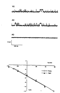
Figures. lA-lB. 113pS ch~nnel. (lA). Cell
attached recordings from Alzheimer and control
fibroblasts. A potassium ch~nnel of ~4.5 pA unitary
current size (O mV pipette potential), with identical
kinetics appeared in age-matched control (AC) and young
controls (YC) fibroblasts, but was entirely absent in the
recording of AD fibroblasts (lA, bottom) Do.~lwdrd
deflections represent the open state. (lB). I/V
relationships and slope conductances. I/V relationships
and slope conductances (detPrm;neA by linear regression)
SUBSTITUTE St IEET (RULE 26~)
W094/25872 PCT~S94/04754
~6~ 18
-- 6
o
were almost identical within the voltage range explored,
113.2+0.9 pS (mean+S.D., n=8) for YC and 112.9+3.2 pS
(n=7) for AC fibroblasts.
Figures 2A-2B. 166pS ch~nnel~ (2A). Cell
attached recordings from Alzheimer and control
fibroblasts. A second rh~nnel (166 pS) was recorded under
the same conditions from fibroblasts of all three groups
(AD, YC and AC). (2B). I/V relations and slope
conductances. I/V relations as well as slope conductances
[YC= 174+5.7 pS, n-4; AC= 169.2+2.8 pS, n-4; AD= 157.6+4.7
pS, n~6 (Mean+S.D.)] were approximately the same across
groups. Membrane potential was g;m;l~r in control (-
42.6+5.4, Mean+S.D., ne7) and in AD (-45.4+6.9, n-3)
fibroblasts.
Figures 3A-3C. (3A) and (3B). Percent of cells
responding to the addition of 50 mM potassium chloride and
average [Ca2+]i (nM) of responding cells. High potassium-
induced depolarization caused [Ca2+]i elevation (at least
100~ increase) in all three groups (AD N= 13 cell lines;
AC N=10, YC N=6). The proportion of responding cells and
the [Ca2+]i peak values were significantly higher in YC
(n= 183 cells) fibroblasts (x2- 14.22, p ~ 0.001), as
compared to AC (n-299) and AD (n=268) fibroblasts (3A and
3B). (3C). Sample traces of time courses of the Ca2+
response in cells after the addition of 50 mM RCl. The
[Ca2+]i peak occurs 10 to 15 seconds after stimulation,
returning to basal levels after 100 seconds. No responses
were observed if external [Ca2+] was lowered ~"n~m;n~lly
Ca2+ free" solution, 5 mM EGTA was added (estimated free
Ca2+ = 0.04 ~M)], or Ca2+ ch~nnel blockers (0.1 mM LaCl3,
10 mM CoCl2, 10 mM NiCl2, 10 mM CdCl2 or 10 ~M nifedipine)
were added before stimulation (no Ca2+").
Figures 4A-4C. [Ca2+]i elevation in response to
TEA. (4A) Percentage of cells responding to the addition
of TEA and (4B) Average [Ca2+]i response in the cells after
SU8STITUTE S~EET (RULE 26)
W094/25872 216 2 0 4 8 PCT~S94/04754
TEA treatment. 1 mM TEA application elevated [Ca2+]; in YC
fibroblasts (n= 130 cells) but not in AC (n= 184) or AD
fibroblasts (n= 195). 10 mM TEA elevated [Ca2+]j in YC (n=
176 cells), AC (n= 231), but not in AD (n= 204)
S fibroblasts (X 134.00, p c O.001). Similarly, 100 mM TEA
elevated [Ca2+]; in YC (n= 532 cells), AC (n= 417), but not
in AD (n= 738) fibroblasts, x2 231.44, p ~ 0.001 (also see
Table 2). Basal [Ca2+]; levels were virtually the same
(S.E. c 2 nM), therefore, st~n~rd error bars are not
disting~l;shAhle from the bar representing the arithmetic
mean for those groups. (4C). Time course of Ca2+
responses. The [Ca2+]i peak occurs 20 to 30 seconds after
100 mM TEA addition in YC and AC fibroblasts, returning to
basal levels after 100 seconds. Note that no response
meeting criterion (10~ of cells in a line with ~ 100
elevation) was observed in AD cells. Similarly, the
response was absent in control cells when external [Ca2+]
was lowered.
Figures 5A-5B. (5A). Ca2+ mobilization induced
by 1 ~m bombesin in the absence of extracellular calcium.
(5B). Ca2+ responses at 42 sec after 1 ~M bombesin
application. The [Ca2+]j levels in AD cells are much
larger than in AC and YC cells. The numbers of cell lines
(N) are 9, 8 and 6 for AD, AC and YC, respectively. The
values are means + S.E.M.
Figures 6A-6B. (6A). Ca2+ responses induced by
1 ~m bombesin in the presence of extracellular calcium.
~m bombesin elicited a fast peak of [Ca2+]j, followed by a
sust~;ne~ phase for YC and AC cells, but not for AD cells,
in the presence of extracellular 2.5 mM CaCl2. The arrow
indicates drug application. (6B). Bar graph illustrating
differences evident 90 seconds after bombesin application.
In the presence of normal extracellular calcium (2.5 mM),
a sust~;n~ calcium entry follows the initial bombesin
response in control cells but is completely absent in AD
SUBSTITU~ S~EET (RULE 26~
W094/~872 PCT~S94/04754
~2~
-- 8
O
fibroblasts. The difference evident 90 seconds after
bombesin application is shown and has a significance level
of p ~ 0.001.
DETAI~ED DESCRIPTION OF THE lNV~NllON
The invention concerns methods of diagnosing
Alzheimer's disease (AD). These methods are based upon
detecting the absence of a particular potassium ion
rh~nn~l in the cells of an AD patient; upon differences in
intracellular calcium ion concentration in AD and non-AD
cells in response to potassium rh~nnel blockers specific
for the potassium ion rh~nnel that is absent in the cells
of an AD patient; and differences between AD and non-AD
cells in response to activators of intracellular calcium
release such as activators of inositol-1,4,5-trisphosphate
(IP3).
The fir t embodiment of the invention i8 based
upon the discovery by the inventors that cells from people
not suffering from AD have (at least) two types of
functional potassium rh~nn~ls, with conductances of 113 pS
(picosiemens) and 166 pS, as measured by the patch clamp
technique (see Example 1). The 113 pS rh~nn~l is either
missing or not functioning in people with AD. The first
embo~;m~nt of the invention involves diagnosing AD by
determ;n;ng whether cells of the patient have a
functioning 113 pS potassium rh~nnel. The presence of a
functioning 113 pS potassium rh~nnel indicates that the
patient does not have AD. However, the absence of a
functioning 113 pS potassium ch~nnel indicates that the
patient does have AD.
In this embodiment of the invention, a suitable
method of recording electrical conductances in the cells
must be used to detect functional potassium ch~nnels in
cells. Any technique which can measure electrical
conduct~nc~s in a cell can be used. Examples include
intracellular microelectrode recording (indirect
SUBSTITUTE SHEET (RULE 26~
W094/25872 PCT~S94/04754
'~162~
g
o
measurement), two microelectrode voltage clamp, and single
microelectrode voltage clamp. The patch clamp techni~ue,
as described herein, is a preferred method for measuring
electrical conductance in small structures. In an
embodiment of the invention, the cell attached mode of the
patch clamp technique is used to record the existence of
potassium ch~nnel S and the inside-out and outside-out
patch configurations are used to record the sensitivity of
potassium channels to various chemicals.
The second embodiment of the invention concerns
another method for diagnosing AD. In this second
embodiment, the cells are contacted with a potassium
ch~nn~l blocker that blocks the 113 pS channel but not the
166 pS ch~nnel. This blocker may substantially block the
113 pS channel but not substantially block the 166 pS
ch~nn~l. An example of such a blocker is TEA, or
tetraethyl~mm~n;um. The blocker has the effect in non-AD
cells of transiently increasing intracellular Ca2+
concentrations. In AD cells, the blocker has
substantially no ef~ect, allowing for variation within
observational or technical error. In contrast, the
intracellular calcium ion concentration increases several
fold in non-AD cells after being exposed to 100 mM TEA
(see Fig. 4B). The intracellùlar Ca2+ concentration can
be measured in various ways, such as by adding fluorescent
indicators or absorbance indicators or by using a Ca2+
electrode. Preferably, because of ease of operation,
fluorescent indicators are used.
In this embodiment of the invention, the cells
are first cultured with a Ca2+ indicator, such as quin or
- fura-2, that fluoresces with an intensity proportional to
the calcium concentration. The cells are then contacted
- with a select potassium channel blocker that has the
ability to block the 113 pS ~h~nn~l but not the 166 pS
~h~nnel. The fluorescence intensity of the cells before
W094l25872 PCT~S94/04754 ~
. ~ .
o ~162~ o
and after the addition of the potassium channel blocker is
measured. In cells from people not suffering ~rom AD the
fluorescence intensity increases rapidly, peaks and then
drops back down (Fig. 4C). This shows that the blocker
S has the effect of increasing, transiently, the calcium ion
concentration. In cells from AD patients, the
fluorescence intensity is substantially the same before
and after the blocker is added. This is a reflection of
the fact that the 113 pS channel is missing or non-
functional in AD patients and thus potassiu-m-~ ion ch~nnel
blockers that block the 113 pS chAnnel, but not the 166 pS
ch~nnel, do not have any effect on AD cells.
As mentioned above, the select potassium ch~nnPl
blocker used in this second embodiment of the invention is
one that has the ability to block the 113 pS potassium
~h~nnel but that has little or no effect on the 166
potassium ch~nnel. One example of such a blocker is TEA,
with any biologically compatible counter anion.
Preferably, the counterion is chloride. Other suitable
potassium rh~nnel blockers can be easily found using the
following method. Using the patch clamp technique
described in Example 1, the 113 pS and 166 pS ch~nn~ls are
detected in a viable human cell. The candidate potassium
~h~nnel blocker is added to the culture cont~;n;ng the
cells, and the patch clamp technique is used again. If
the 166 pS ch~nn~l is still functional, but the 113 pS
channel is not, then the candidate blocker is suitable for
use in this invention. Candidate potassium rh~nnel
blockers include the known potassium ch~nnel blockers
charybdotoxin, ~p~m~n, dendrotoxin, kalidotoxin, MCD-
peptide, scyllatoxin, barium, cesium, leiurotoxin I and
~oxiustoxin. As shown in Bxample 2, TEA concentrations
between 10 mM and 100 mM worked well. It is easy to
extend this range of workable concentrations by using AD
and non-AD control cells.
W094/25872 PCT~S94/04754
~162018
Example 2 exemplifies the second embodiment of
the invention for diagnosing AD using a select potassium
channel blocker, TEA, and measuring the effect on
intracellular calcium ion. This method is so simple, with
a yes or no answer, that the exemplified sophisticated
apparatus is not required to make the diagnosis. Any
method which will tell one if the intracellular calcium
ion concentrations has increased or not as a result of
contact with the select potassium ion ch~nnel blocker will
suffice to give a diagnosis. In the preferred method,
fluorescent calcium ion indicators are used. In this
case, any method which will tell one if the fluorescence
of the indicator has increased or not as a result of
contact of the cells with the select potassum ChAnn~l
blockers will suffice. Any method used must be able to
make the measurements in the short time available. The
calcium ion influx peaks a short time after contact with
the blocker, and then decreases to the baseline value. In
Example 2, the time it takes to peak i8 less than one
minute.
A simpler method for detecting a fluorescent
calcium ion indicator would involve using a fluorimeter, a
device with a light source for exciting the calcium ion
indicator and a light meter for measuring the intensity of
a the fluorescence. Fluorimeters are well known and
commercially available. At the simplest level, the
calcium ion indicator is added to the cells taken from the
patient (either fresh or ~p~nA~d in culture). After an
hour or so of being in contact with the indicator (at
about 2 micromolar concentration) the cells in suspension
are placed in the fluorimeter and the fluorescence
intensity from the indicator is measured. Then the select
- potassium channel blocker is added; if TEA is used, it is
added to a concentration of about lO0 mM. The
fluorescence is measured again. If the intensity, within
W094/25872 PCT~S94/047~4
- 12 -
a time period between 20 seconds and 40 seconds, is
substantially the same as before the TEA was added (taking
account of changes in volume due to the addition of the
TEA), then a positive diagnosis of AD is made. If the
S intensity increases within 30 seconds and subsides after
another 30 seconds, then the patient does not have AD.
It is within the skill of the art to improve the
simple scheme outlined above. For example, one could use
a fluorimeter with dual sample holders, in which the
difference in fluorescence from two samples is measured.
Starting with identical samples of patient's cells (after
incubation with the indicator) in each sample holder, the
select potassium ch~nnpl blocker is added to only one of
the samples. If there is no change in the difference
signal (that is, it rPm~;n~ as essentially zero), a
diagnosis of AD is made. If the difference signal changes
significantly, then the patient does not have AD. The
advantage of the differences method is that it has a built
in control which increases the accuracy of the
measurement. It is still within the skill of the art to
add the select potassium ch~nn~l blocker automatically and
to make more than one measurement at a time; i.e., to
automate the method for a commercial medical laboratory.
Before making any diagnoses ùsing the methods taught here,
the methods should be optimized for the particular
apparatus and conditions in the laboratory by using non-AD
and AD control cells, which are commercially available.
The third e-mbodiment of the invention is yet
another method of diagnosing AD. This method concerns the
effect of agents that activate inositol-1,4,5,-
trisphosphate (IP3) or otherwise induce the release of
calcium from intracellular storage sites. Such storage
sites include the endoplasmic reticulum and other
organelles that have receptors for IP3. The preferred IP3
activator is bombesin. Other agents that activate the
W094/25872 PCT~S94/04754
2162048
release of calcium from intracellular stores which are
useful in the invention include thrombin, bradykinin,
prostaglandin F2a and vasopressin. See, e.g., Berridge,
M.J. and Irvine, R.F. ~1984) Nature 312:135).
S It has been discovered that cells from people
not suffering from AD and cells from people suffering from
AD both transiently release calcium ion in response to
bombesin, but the resulting intracellular calcium
concentration is much larger in AD cells than in non-AD
cells. The determ;n~tion is easily made using any method
of measuring intracellular calcium ion concentration, as
discussed above with respect to the second embodiment of
the invention. Again, the use of flourescent calcium
indicators is the preferred method. The same experimental
setup as described above for measuring fluorescence
intensity can be used, i.e., a fluorimeter. In this
method, it is also possible to st~n~rdize the
fluorescence apparatus using non-AD and AD cells as
controls. In this way, later measurements of just the
patient's cells can provide a diagnosis. Alternatively,
the patient's cells can be compared with non-AD cells as a
control.
Example 3 exemplifies the third embodiment of
the invention concerning the diagnosis of AD using
activators of IP3 and measuring their effect on calcium
ion release into the cytosol from intracellular storage
sites after contact with said activators. The amount of
released calcium is larger in AD cells compared to non-AD
cells. The increase in intracellular calcium
concentration is transient: the concentration peaks soon
- after contact with the activator and is back to baseline
value with 90 seconds. This effect is enhanced when the
- extracellular calcium ion concentration is zero or near
zero (which is generally accomplished by washing the cells
with BSS nom~ n~l ly free of calcium, however, other methods
W094l25872 PCT~Sg4/04754
~ 0~ - 14 -
of tying up or negating the effect of the extracellular
calcium ions can be used, such as adding EDTA, or adding a
calcium channel blocker such as nifedipine, respectively).
After contact with an IP3 activator, such as bombesin, the
intracellular calcium ion concentration in AD cells
reaches a higher peak value and takes longer to return to
the baseline value than either young or aged control cells
(Fig. 5A). In the experimental setup described in Bxample
3, it was found that 42 seconds after the bombesin was
added to the cells that the difference between the
intracellular calcium ion concentrations in AD cells and
in control cells was at a m~X; mllm, and that at that time
period, i.e., at 42 seconds after bombesin was applied,
the concentration of calcium ions was always greater than
300 nM in AD cells and was always less than 300 nM in
control non-AD cells (Fig. 5B). Basal levels of both AD
and non-AD fibroblasts were at 80 nM + O.5 nM. However,
it should be noted that control values might differ from
80 nM, necessitating a criterion level of calcium signal
greater or less than 300 nM. Furthermore, differences in
measuring conditions might req~ire a time longer or
briefer than 42 seconds to show m~x;m~l differences
between the calcium signals of AD and non-AD fibroblasts.
Again, it is not nècessary to use the
sophisticated methods and apparatus exemplified herein.
This method of diagnosing AD can be performed more simply.
One need not measure the absolute concentration of
intracellular calcium; a measurement of its relative value
will also work. In Example 3, the basal level of
intracellular calcium ion concentrations in resting (i.e.,
nonactivated) cells was the same for both AD and control
non-AD cells, 80 nM + O.5 nM. Thus, at the time where the
concentration differences between AD and non-AD cells was
m~;mllm (i.e., at 42 seconds using bombesin and the
inventors' apparatus, but the time would need to be worked
~ W094/25872 21~ 2 0 4 8 PCT~S94/04754
out empirically for different activators and different
setups) the intracellular calcium concentration in non-AD
cells would be less than (300/80 =) 3.75 times the basal
level whereas the intracellular calcium concentration in
AD cells would be greater than (300/80 =) 3.75 times the
basal level. Using commercially available AD and non-AD
cells, one can easily determine the time at which the
calcium concentrations are maximally different between AD
and non-AD cells. This involves measuring relative
intracellular calcium concentrations for resting cells,
adding bombesin or another IP3 activator, following the
relative calcium ion concentrations for a minute or SQ,
and finding the time (after the activator is added) at
which the difference in relative calcium ion
concentrations is at its m~x;mllm. Then, for any real
sample from a patient, one simply needs to measure the
relative basal intracellular calcium concentration by any
means known in the art, add the activator to its
prescribed concentration (about l micromolar for
bombesin), wait the predet~rm;ne~ time and again measure
the relative intracellular calcium concentration. If the
ratio of the intracellular calcium concentration "after"
the addition of the activator to the intracellular calcium
concentration "before" the addition of the activator is
greater than 3.75, the patient has AD; if it is less than
3.75, the patient does not have AD. It is not necessary
to determine the time of m~x;m~l difference in calcium
concentrations -- any time where there is a reproducible
difference between these ratios can be used. It is only
necessary to work out the particular ratios for the time
chosen from known AD and non-AD control cells.
The calcium ion indicators used in the second
- and third embodiments include any compounds which can
enter the cell, are biocompatible, and which can bind to
calcium ions to produce a species whose concentration is
W094/25872 PCT~S94/04754
2162~ 16
O
easily measured using any physico-chemical means and is
proportional to the calcium ion concentration. Preferably
the means is fluorescence or absorbance. Preferable
fluorescent indicators are the commercially available
indicators fura-2 AM, fura-2 pentapotassium salt, quin-2,
and indo-l from Molecular Probes (Eugene, OR). The
Chemical Abstracts name for fura-2, AM is 5-
oxazolecarboxylic acid, 2-(6-(bis(2-((acetyloxy)methoxy)-
2-oxoethyl)amino)-5-~2-(2-(bis(2-((acetyloxy)methoxy)-2-
oxoethyl)amino)-5-methylphenoxy)ethoxy)-2-benzofuranyl)-,
(acetyloxyl)methyl ester. The Chemical Abstracts name for
fura-2, pentapotassium salt is 5-oxazolecarboxylic acid,
2-(6-(bis(carboxymethyl)amino)-5-(2-(2-
(bis(carboxymethyl)amino)-5-methylpheno~y)ethoxy)-2-
benzofuranyl)-. Other fluorescent calcium indicators
include Fluo-3, Rhod-2, Calcium GreenTM, Calcium OrangeTM,
Calcium CrimsonTM Fura RedTM and Calcium Green DextranTM
(Molecular Probes (Eugene, OR)). Generally, the cells are
incubated with the indicators at a concentration of about
2 micromolar for about 60 minutes. An absorbance
indicator which may be used is arsenazo. Finally, calcium
levels could also be measured for this invention with
calcium electrodes inserted into the cells.
In the exemplified e-mbodiment of the invention,
fluorescence was measured using an imaging system under
the control of a personal computer. For excitation, 340
nm and 380 nm band path filters with a neutral-density
filter were used. Images of fluorescence were obtained
using a dichroic mirror, barrier filter and objective
lens. The whole image can be recorded or portions
thereof. A ~m~m~tSU Photonics Argus 50 Calcium Imaging
system imaging 60 cells in a microscopic field at l0 x
magnification was used. Fluorescence from the cells was
quantified in 1~ of the field at l0x magnification. Such
an imaging system (and other similar currently available
~ W094/25872 PCT~S94/04754
2162048
systems) with its microscope could be custom designed for
everyday clinical laboratory analysis of cells' calcium
signals. Other instrumentation and/or measurements would
have to be adapted for the use of other calcium
indicators.
In the methods of the invention, the cells that
are taken from the patient can be any viable cells.
Preferably they are fibroblasts; buccal mucosal cells;
blood cells such as erythrocytes, lymphocytes, and
lymphoblastoid cells; or nerve cells such as olfactory
neurons. The cells may be fresh or may be cultured (as
described in the examples). The fibroblast potassium
channel dysfunction and resulting absence of TEA-induced
calcium signals described herein suggest that AD, which
primarily affects brain cells, is likely to alter
potassium rh~nnel function in many different types of
cells in the body. Similarly, AD is likely to alter
calcium released by bombesin and related agents in many
different types of cells in the body. The methods
described herein to measure potassium ch~nnel function and
calcium release, therefore, should be applicable for AD
diagnosis using other cell types.
A punch skin biopsy could be used to obtain skin
fibroblasts from a patient. These fibroblasts might be
analyzed directly with the techniques described herein or
be introduced into cell culture conditions. The resulting
cultured fibroblasts would then be analyzed as described
for the cultured fibroblasts obta;nP~ from the Coriell
Cell Repositories described below. Other steps would be
required to prepare other types of cells which might be
used for analysis such as buccal mucosal cells, nerve
cells such as olfactory cells, blood cells such as
erythrocytes and lymphocytes, etc. For example, blood
cells can be easily obtained by drawing blood from
peripheral veins. Cells can then be separated by stan~rd
W094/25872 PCT~S94/047~4
2~
- 18 -
procedures (e.g., by using a cell sorter, centrifugation,
etc.) and later analyzed in suspension or on a solid
support (e.g., in petri dishes).
The present invention will now be described by
way of examples, which are meant to illustrate, but not
limit, the scope of the invention.
Example 1
Patch-clamp Diagnostic Test
Cultured skin fibroblasts (described in Table 3)
from the Coriell Cell Repositories (C~m~n, NJ) were grown
under highly st~n~rdized conditions. Cristafallo, V.J.
and Chapentier, R.J. (1980) Tissue Culture Methods 6:117.
The following cell lines were used for the experiments:
Young Control Fibroblasts ("YC") 3652, 3651, 2987, 4390,
3377, 8399 (21.5+2.8 years, Mean +S.D); Age-matched
Control Fibroblasts ("AC") 3524, 6010, 6842, 7603, 9878
(65.2+6.0 years); and Alzheimer's Disease Fibroblasts
("AD") 6848, 7637, 5809, 8170, 6840, 8243, 6263 (60.6+6.8
years). Five AD lines were from familial patients. Some
of the lines (2 AC and 4 AD) were from C~n~ n kindred.
In agreement with the literature, the data
indicate the time to phase out does not vary between the
AD and control lines (YC and AC). Cells were seeded
(approximately 5 cells per mm2) in 35 mm Nunc petri dishes
in Dulbecco's Modified Eagle Medium (DMEM, Gibco),
supplemented with 10~ fetal calf serum and used when cell
density was equivalent for all cell lines, between days 2
and 4 after plating. On average, fibroblasts from AD
patients and controls took the same time to reach erosion
density (50 cells/mm2).
Patch-clamp experiments were performed at room
temperature (21-23C), following st~n~rd procedures set
forth in Sakmann, B. and Neher, E. (1983) Sinqle Channels
Recordinqs (Plenum New York) and Kukuljan, M., et al.
~ W094/25872 ~16 2 0 4 8 PCT~S94/04754
- 19
(1991) J. Membrane Biol. 119:187. Before recordings,
culture medium was replaced with the following solution:
150 mM NaCl, 5 mM KCl, 2 mM CaCl2, 1 mM MgCl2, 10 mM HEPES
(NaCl) pH=7.4. Pipettes were made from Blue Tip capillary
tubes (I.D. 1.1-1.2 mm) using a BB-CH Mecanex puller, and
then filled with a high potassium solution of 140 mM KCl,
2 mM CaCl2, 1 mM MgCl2, 10 mM HEPES (NaOH), pH=7.4.
Pipette resistances were approximately 6 MQ. Records were
obtained using an Axopatch-lC amplifier (dc-10 kHz),
stored on tape (Toshiba PCM-video recorder), and later
transferred to a personal computer using an Axolab
interface. Only recordings lasting for at least 3 minutes
were considered for final analysis. The pClamp suite of
programs was used for single-~hAnnel data acquisition and
analysis. Amplifier, interface and software were obt~;ne~
from Axon Instruments (Foster City, CA).
In the cell-attached mode, two types of
potassium ch~nnels were recorded from human skin
fibroblasts. Since pipettes were filled with a high
potassium solution, potassium currents were inward as
expected, and their reversal potential approximately
corresponded to the cell resting potential. A potassium
channel (113 pS) of approximately 4.5 pA unitary current
size (O mV pipette potential), with identical kinetics
appeared in YC and AC fibroblasts, but was entirely absent
in the recording of AD fibroblasts (Fig. lA). Downward
deflections represent the open state. I/V relationships
of the same channels in Fig. lA (Fig. lB) and slope
conductances (detPrm;ned by linear regression) were almost
identical within the voltage range explored, 113.2+0.9 pS
(Mean+S.D., n=8)) for YC and 112.9+3.2 pS (n=7) for AC
fibroblasts.
A second ch~nnPl (166 pS) was recorded under the
same conditions from fibroblasts of all three groups (Fig.
2A). I/V relations (Fig. 2B) as well as conductance
W094/25872 PCT~S94/04754
C~1~20 ~8
- 20 -
o
(YC=173.4+5.7 pS, n-4; AC= 169.2+2.8 pS, n=4; AD=157.6+4.7
pS, n=6 (Mean+S.D.)) were approximately the same across
groups. Membrane potential was similar in control (-
42.6+5.4, Mean+S.D., n=7) and in AD (-45.4+6.9, n=3)
S fibroblasts.
Both ~h~Annels had linear voltage-current
relationships, with slope conductances of 113 pS and 166
pS respectively (Figs. lA-lB and 2A-2B). At 0 mV pipette
potential, the ch~nnels could easily be identified by
their unitary current size (Figs. lA and 2A) and by their
percentages of open time, approximately 60~ for the 113 pS
K+ rh~nnel and approximately 10~ for the 166 pS R+
~hAnnPl. For both ch~nnpls~ the percentages of open time
showed no significant voltage-depPn~Pnce (+60 to -40 mV
pipette potential). The 113 pS K+ rh~nnPl was found in
47~ of YC cells (n~30) and 94~ of the AC cells (n=17),
while it was never found in AD fibroblasts (n=24) (X2 =
18.96, p c 0.001 (Table 1)). There were no AD cell lines
(N=6) that had fibroblasts with an observable 113 pS
chAnnel. By contrast, all AC cell lines (N=5) and three
of six YC cell lines had fibroblasts with observable 113
pS ch~nnels (X2-11.93, pcO.005 (Table 2)). The 166 pS
chAnnPl found was s;m; 1 ~r fre~uency in all three groups
(X2=0.89, N.S. (Tables 1 and i)).
The 113 pS ch~nnpl found to be "absent" in the
AD fibrobla~ts, could be present but not functional. Such
dysfunction could involve structural changes in the
ch~nnel and/or alteration in processes involved in ch~nnPl
activity regulation.
Using cell-free patches, following the method
described above, it was observed that both chAnnpls were
sensitive to 50 mM Ba2+ (inside-out, n=4 for each
~h~nnPl), but only the 113 pS ch~nnel was sensitive
(outside-out, n=4 YC, n~3 AC) to the K+ ~hAnn~l blocker
tetraethyl~mm~n;um (TEA). The TEA-blockade of the 113 pS
SUBSTITUTE St~EET (RULE 2&)
W094/25872 PCT~S94/04754
~620~
- 21 -
o
channels (possibly together with other channels)
significantly affects membrane potential since control
cells (n=4) depolarized 13-20 mV after 100 mM TEA
addition.
s
Table 1
Number of Cells
113 pS K+ 166 pS K+
Condition Total ~hAnn~l rh~nn~
Young Controls 30 14(47~) 6(20~)
Aged Controls 17 16(94~) 6(35~)
Alzheimer Patients 24 0(0~) 8(33~)
Table 2
Number of Cell Lines
113 pS K+ 166 pS K+
Condition Total Ch~nnel Ch~nnPl
Young Controls 6 3 4
Aged Controls 5 5 3
Alzheimer Patients 7 0 4
When using control cells, it is best to use age-
matched control cells.
Example 2
TEA-Ca2+ Diagnostic Test
Cultured skin fibroblasts (described in Table 3)
from the Coriell Cell Repositories (C~m~n, NJ) were grown
as described in Example 1.
r Thirteen AD, ten AC, and six YC were used for
the calcium-imaging experiments. Culture medium was
replaced and washed three times with basal salt solution
("BSS") consisting of 140 m~M NaCl, 5 mM KCl, 2. 5 mM CaCl2,
3~ 1.5 mM MgCl2, 5 mM glucose, 10 mM HEPES (NaOH), pH 7.4.
W094/25872 PCT~S94/04754
21620~
- 22 -
Nsm;n~1ly Ca2+ free BSS was prepared as BSS without adding
CaCl2 .
Fura-2 (acetyloxymethyl ester) (Fura-2AM) was
purchased from Molecular Probes (Eugene, OR) and stored as
a 1 mM solution in dimethylsul~oxide. Fura-2AM was added
to a final concentration of 2 ~M and cells were incubated
at room temperature (21-23C) for 60 minutes. After
incubation, cells were washed at least three times with
BSS at room temperature before tCa2+]i det~rm~n~tions.
Fluorescence was measured with a u~m~m~tsu ARGUS 50
imaging system (~m~m~tSU Photonics, Japan) under the
control of a personal computer (~m~m~tSU imaging software
package). Excitation at 340 nm and 380 nm was attenuated
with neutral density filters. Fluorescent images were
obt~; ne~ with a 400 nm dichroic mirror and a 510 nm long-
pass barrier filter. The objective lens was an X10 Nikon
W fluor. Fluorescence was measured within a uniformly
illuminated fraction (~) of the whole image.
The averaged Ca2+ responses within 15 x 15
pixels in cytosolic and in nuclear cellular compartments
obt~; ne~ were quantified with ratios between emitted 510
nm fluorescence activated at 340 nm and fluorescence
emitted at 510 nm with activation at 380 nm. These ratios
were transformed to absolute values of [Ca2+], after
calibration based on the following equation:
R - R~ + (R~ - R~)/(1 + ([Ca2+]j/Kd) b),
Here R denotes fluorescence intensity
illuminated by 340 nm divided by fluorescence intensity
illuminated by 380 nm (F340/F380), and R~ and ~ are the
values of R when the concentration of calcium is at a
m~x;mllm and a m;n;mllm (i.e., the m~x;mllm and m;n;mllm value
measurable by the machine under the measuring conditions),
respectively. Kd is a dissociation constant of fura-2 for
Ca2+ and was determined as 240 nM. The value of b, which
determined the degree of asymmetry, was 1.2. TEA
W094/25872 PCT~S94/04754
2~62V~8
- 23 -
o
application caused a m;n;mllm of 100~ [Ca+2]j elevation in
at least 18~ of cells in every control cell line except
one young control. A response of 100~ [Ca+2]i elevation in
at least 10~ of cells in a line was, therefore, considered
to be a conservative criterion for a positive response.
Only one AD cell line had cells with any response (100
[Ca+2]j elevation in 4~ of cells), well below the
criterion).
Depolarization of the fibroblasts by perfusion
in elevated external potassium caused greater elevation of
intracellular Ca2+ ([Ca2+]j) in YC as compared to AC and AD
cells (Fig. 3A-3C). This depolarization-induced [Ca2+]j
elevation was eliminated by lowering external calcium or
by adding calcium chAnn~l blockers (Fig. 3C). High K+-
induced depolarization caused a marked [Ca2+i] elevation
(at least 100~ increase) in all three groups (AD, nc 13
cell lines; AC, n= 10; YC, n~ 6). The proportion of
responding cells and the [Ca2+]; peak values were
significantly higher in YC (n= 183 cells) fibroblasts
(X2=14.22, p c 0.001), as compared to AC (n= 299) and AD
(n= 268) fibroblasts. The [Ca2+]i peak occurs 10 to 15
seconds after stimulation, returning to basal levels after
100 seconds. No responses were observed if external
calcium was lowered by addition of "n~m;nAlly Ca2+ free"
2S solution or 5 mM EGTA (estimated free Ca2+=0.04 ~M) or Ca2+
chAnnel blockers (0.1 mM ~aCl3, 10 mM CoCl2, 10 mM NiC12,
10 mM CdCl2 or 10 ~M nifedipine) before stimulation.
Depolarization of control fibroblasts by TEA
also caused [Ca2+]; elevation, that was eliminated by
lowering external calcium or by adding calcium ~h~Annel
blockers. AD fibroblasts, however, only showed [Ca2+];
elevation in elevated external potassium and had no [Ca2+];
response with addition of even 100 mM TEA. Every AC cell
line (N=10) and all but one YC cell line (N=6) had cells
responding to TEA, while none of the thirteen AD cell
SUBSTITUT~ S~lEET (RULE 2~)
W094/25872 PCT~S94/04754
~16,~
- 24 -
o
lines ~m; ned had cells responding to 100 mM TEA
(X2=25.66, p<0.001) (Tables 3 and 5).
Table 3
Number of Cell Lines
Increase in [Ca+2];
Condition Total with 100 mM TEA
Young Controls 6 5
Aged Controls 10 10
Alzheimer's Patients 13 0
1 mM TEA application elevated tCa2+]i in YC
fibroblasts (n=130 cells) but not in AC (n=184) or AD
(n=195) fibroblasts. 10 mM TEA elevated [Ca2+]j in YC
(n=176) and AC (n=231) but not in AD fibroblasts(n-204).
Similarly 100 mM TEA elevated [Ca2+]; in YC (n=532) and AC
(n=417), but not in AD fibroblasts (n=738) (X2=231.44, P~
0.001). At least 417 cells were explored in each
experimental group (Table 4). The [Ca2+]j values of the
responding cell were s;m; 1 ~r in YC and AC cells after 10
and 100 mM TEA addition. Basal [Ca2+]j levels were
virtually the same (S.E. c 0.5 nM), therefore st~n~rd
error bars are not distingll;Rh~hle from the bar
representing the arithmetic mean for those groups (Fig.
4B). Time courses of Ca+2 response shows that the [Ca2+];
peak occurs 20 to 30 seconds, after 100 mM TEA addition in
YC and AC fibroblastæ, returning to basal levels after 100
seconds. No response was observed in AD cells (10~ of
cells in a line with 2 100~ elevation). Similarly, the
response was absent in control cells when external [Ca2+]
was lowered (Fig. 4C).
wog4l258n PCT~S94/04754
~16~048
Table 4
Number o~ Cells
Increase in [Ca+2];
Condition Total with 100 mM TEA
Young Controls 532 145 (27~)
Aged Controls 417 119 (29~)
Alzheimer's Patients 738 4 (0.5~)
TEA-induced [Ca2+]; elevations were repeated
using a coded subsample that included Alzheimer's and
control fibroblasts. Experiments and analyses were
conducted without the experimenter's knowledge of the cell
lines identity. The results were in complete agreement
with the non-blind sample. None of the blindly e~m;ned
AD cell lines (N=ll) showed [Ca2+]; elevation in response
to TEA and all but one of the control cell lines (4 AC and
6 YC) had TEA responses (X2=17.33, p c 0.001 (Table 5)).
Since [Ca2+]j elevation in response to high
potassium was virtually the same for AC and AD cells, the
lack of AD cells response to TEA is almost certainly due
to dysfunction of K+ channels and not to Ca2+ ch~nnel
dysfunction.
The [Ca2+]; measurements are in agreement with
the patch-clamp measurements insofar as they both indicate
potassium rh~nn~l dysfunction in the AD fibroblasts. See
Table 5.
WO 94/25872 PCT/US94/04754
~ ~ 6 ~ 26 -
o
Table 5
Line # Age Gender Race Diag. Criteria 113 K+ TEA Response
Channel N Blind
Blind
,AI7h,~im~r's Disease Fil"ubla~l~
AG06840+~ 56 M W Clinical - Fam. H.
AG06848+2 55 F W Clinical - Fam. H.~ - - N.T.
AG07637+ 55 F W Clinical - Fam. H.
AG08170+ 56 M W Clinical - Fam. H.
AG06844+ 59 M W Clinical - Fam. H.~ N.T. N.T.
AG04400~: 61 F W Clinical - Fam. H. N.T. N.T.
AG04401~ 53 F W Clinical - Fam. H.~ N.T.
AG05809 63 F W Clinical - Fam. H. - - N.T.
AG08243 72 M W Clinical - No Fam. H. - - -
AG07375 71 M W Clinical - No Fam. H. N.T.
AG07376 59 M W Clinical - No Fam. H. N.T.
AG06263 67 F W Clinical - No Fam. H.
AG07377 59 M W Clinical - No Farn. H. N.T. N.T.
Age-Matched Control Fil,-ul l&.it~
GM03524 67 F B Normal + + N.T.
AG06010 62 F W Norm~l + + +
AG06842+ 75 M W Normal-Fam. H. + N.T. N.T.
AG07603+ 61 F W Normal-Fam. H. + + N.T.
AG09878 61 F B Normal + + +
AG08044 58 F B Normal N.T. + N.T.
AG6241 61 M W Normal N.T. + N.T.
AG4560 59 M W Normal N.T. + N.T.
GM04260 60 M W Normal - N.T. + N.T.
AG07141 66 F W Norrnal N.T. N.T. +
AG11363 74 F W No~mal N.T. N.T. +
Young Control Fil~-ubl~ ~
GM03652 24 M W Normal + + +
GM03651 25 F W Normal + + +
W094/2~872 ~16 2 0 4 ~ PCT~S94/04754
- 27 -
o
GM02987 19 M W No~
Line # Age Gender Race Diag. Criteria 113 K+ TEA Response
Channel N Blind
Blind
GM04390 23 F W Normal + + +
GM03377 19 M W Normal - + +
GM08399 19 F ? Norm~l - + +
Alzheimer's fibroblasts were from familial (N=8)
and non-familial cases (N=5). Five (t) are members of the
Canadian family 964, only 1 and 2 are immediate relatives
(sibs). "~" are members (sibs) of family 747. Autopsy
confirmed Alzheimer's Disease in three cases (*). Two of
the age-matched control (N=11) cell lines are unaffected
members of the Canadian family (964). All young control
lines (N=6) are from normal and without AD family history
individuals. Criterion tca2+]; responses (to 100 mM TEA),
indicates as +, were observed in all AC lines used and in
all but one of the YC lines. The presence of the 113 pS
K+ channel is indicated by the "+" sign. None of the AD
lines exhibited "positive" response. A blind protocol was
conducted to measure TEA responses in Alzheimer's (N=11)
and control (YC=6, AC=4) fibroblasts. The results exactly
reproduced those of the non-b~ind sample: no AD cells
line exhibited TEA responses and 9 out 10 control cells
showed TEA responses, x2=17.33, p < 0.001. The notation
"N.T." indicates cell line/conditions that were not
tested.
30Example 3
Bombesin - Ca2+ Diaqnostic Test
Human skin fibroblasts listed in Table 3 were
used. The average age for the AD cell lines used is 60.5
+ 5.9 years; for the AC cell lines is 62.3 + 9.6 years;
35and for the YC cell lines is 21.5 + 2.2 years. The method
W094/25872 PCT~S94/047~4
21~2Q~
- 28 -
o
of maintenance for the cells was described in Example 1,
i.e., maintained 3-5 days at 37C in C02/air (5~/95~) to
reach a density of 50 cells/mm2 before calcium
measurements. The number of culture passages were less
than 19.
Bombesin was purchased from Calbiochem (San
Diego, CA). Bombesin was stored as a 1mM solution in
distilled water. Fura-2 (acetyloxymethyl ester), fura-2
(pentapotassium salt) and omega-conotoxin (~-CgTX) GVIA
were from Molecular Probes (Eugene, OR). Fura-2 AM was
stored as a lmM solution in dimethylsulfoxide; fura-2
pentapotassium salt was stored as a 6mM solution in
potassium acetate, and ~-CgTX was stored as a lOO~M
solution in distilled water. All of the chemicals except
for phenytoin were maint~;ne~ at -20C and protected from
light.
The cells were incubated with 2~M fura-2 AM in
BSS (described in Example 1) at room temperature (21 -
23C) for 60 min. After being washed at least three times
with BSS, the cells were used for measurement of [Ca2+],
at room temperature. Cell fluorescence was measured as
described in Example 2. Absolute calcium values were
calculated as shown in Example 2.
Bombesin was added to the cells at a final
concentration of l~M. Calcium mobilization levels were
measured from -30 seconds to 150 seconds after bombesin
treatment. (Fig. 5A) The particular experimental set up
resulted in a m~;mllm difference in [Ca2+]; between AD
cells and control cells at a time of 42 seconds after
bom.besin was added.
Forty two (42) seconds after bombesin treatment,
ïn the absence of extracellular Ca2+, the [Ca2+]; levels in
Alzheimer's disease cells are much larger (pcO.OOO1) than
in age-matched and young controls. The numbers of cell
lines (N) are 10, 8, and 6 for Alzheimer's disease, age-
W094/2~872 2 l ~ ~ ~ 4 ~ PCT~S94/04754
- 29 -
O
matched and young cells, respectively. The values are
means ~ S.E.M. (Fig. 5B)
Bombesin stimulated IP3-induced Ca2+ release from
intracellular storage sites in fibroblasts from all
S groups, but it caused a larger and more prolonged response
in AD fibroblasts. This larger and prolonged response in
AD cells was independent of extracellular Ca2+. On the
other hand, the IP3-mediated Ca2+ responses in AC and YC
cells were followed by Ca2+ entry. When this Ca~+ entry
was ~;m~ n; shed by removal of extracellular Ca2+, or
blocking with inorganic Ca2+ blockers, the bombesin-
elicited Ca2+ responses in control cells were found to
return to the basal level faster than in AD cells (Fig.
5A). The results shown in Fig. 5A are for cells washed
with BSS no~;nAlly free of Ca2+.
Since Ca2+ influx induced by bombesin was not
observed in AD cells, this pathway of Ca2+ entry following
the decrease of stored calcium seems to be altered. This
test independently confirmed the diagnoses made by the
previously described test based on potassium ~h~nnel
dysfunction. In particular, the Ca2+ responses at 42 sec
after 1 ~M bombesin stimulation in AD fibroblasts in the
absence of extracellular Ca2+ were always higher than 300
nM. In contrast, the [Ca2+], in AC and YC were less than
300 nM and 200 nM, respectively (Fig. 5B).
In a variation on the above experiment, Ca2+
responses were induced by l ~m bombesin in the presence of
extracellular calcium. In the presence of 2.5 mM
extracellular CaCl2, l ~m bombesin elicited a fast peak of
[Ca2+];, followed by a sustained phase for YC and AC cells,
but not for AD cells. (Fig. 6A). This difference was
evident 90 seconds after bombesin application and with a
significance level of p c 0.00l. (Fig. 6B). This
difference in response of AD and non-AD cells to bombesin
in the presence of extracellular calcium can be used to
W094/25872 PCT~S94/04754
~62~ 30
provide a "yes or no" diagnosis of AD. Detection methods
similar to those described above with respect to the
second embodiment of the invention involving the diagnosis
of AD by detecting differences between non-AD and AD cells
in response to select potassium channel blockers (e.g.,
TEA) may be used. Furth~rmore, the combination of this
diagnostic test with any one of the above diagnostic tests
further increases the confidence level of a correct
diagnosis as AD or non-AD.
Example 4
Responses In Neuropathological Non-AD Fibroblasts
Using the techniques described in Examples 2 and
3, cells from donors with other diseases were measured for
intracellular calcium levels in response to either TEA or
bombesin.
Fibroblasts from a Parkinson's disease donor had
normal TEA (indicated as +) and bombesin responses ("N"),
and did not significantly differ from responses observed
in the age-matched control group. Fibroblasts from two
schizophrenic patients also had normal TEA and bombesin
responses. In addition, normal TEA responses were
observed in five out of seven cases of Huntington's
disease, and the bombesin response was normal in all
Huntington's cases. Furth~r~nre, normal TEA and bombesin
responses were observed in four out of four cases of
Wernicke-Korsakoff disease (Table 6). These responses are
significantly different from those of AD fibroblasts to
the level of p ~ 0.0001 (Fisher's exact test). "*"
indicates autopsy confirmation.
W094/25872 PCT~S94/04754
~2Q'~ -
- 31 -
o
Table 6
Line ~ Age Gender Race Condition TEA Bombesin
AG08395 85 F W Parkinson's* + N
5 GM01835 27 F W Schizophrenia + N
GM02038 22 M W Schizophrenia + N
GM06274 56 F W Huntington's + N
GM02165 55 M W Huntington's + N
GM00305 56 F W Huntington's - N
10GM01085 44 M W Huntington's + N
GM01061 51 M W Huntington's + N
GM05030 56 M W Huntington's - N
GM04777 53 M W Huntington's + N
15 7504 50 M W Wernicke-Kors. + N
7505 52 F W Wernicke-Kors. + N
7507 63 M W Wernicke-Kors. + N
7508 64 M W Wernicke-Kors. + N
Every reference cited hereinbefore is hereby
incorporated by reference in its entirety.
The invention has been described in detail with
particular reference to the preferred embodiments thereof,
but it will be understood that the invention is capable of
other and different embodiments. As is readily apparent
to those skilled in the art, variations and modifications
can be affected within the spirit and scope of the
invention. Accordingly, the foregoing disclosure and
description are for illustrative purposes only, and do not
in any way limit the invention, which is defined only by
the claims.