Note: Descriptions are shown in the official language in which they were submitted.
WO 94/27137 21629 96 PCTIUS94/05567
APPARATUS AND METHODS FOR MULTIANALYTE HOMOGENEOUS
FLUOROI]VIMLTNOASSAYS
Background of the Invention
Technical Field: This application relates to the art of analyzing samples for
particular substances by means of fluorescent binding assays, and more
particularly to
apparatus, compositions and methods for such assays employing evanescent
light.
State of the Art: Biosensor apparatus based on optical detection of analytes
by
fluorescence of tracer molecules, have attracted increasing attention in
recent years.
Such apparatus are useful for both diagnostic and research purposes. In
particular,
biosensors for a solid-phase fluoroimmunoassay, in which an antibody or
antibody
fragment specific to the desired analyte is immobilized on a substrate, and
binding of
the analyte to the antibody results either directly or indirectly (for
example, by means
of a labelled tracer) in a fluorescence signal, are becoming an important
class of
optical biosensor.
In most solid-phase fluoroimmunoassays, to achieve adequate sensitivity a
"wash" step is required to remove unbound tracer before measuring the
fluorescence.
This problem is particularly true for detection of analytes present at
concentrations
below nanomolar, as is the case for many analytes of interest in body fluids
including
blood, serum and urine. However, the wash step is tedious, and care on the
part of
the technician is required to produce repeatable and accurate results.
Accordingly, it
is highly desirable to provide a fluoroimmunoassay system in which sensitivity
to
analyte concentrations of 10'10 to 10'13 molar or below is achieved without a
wash step.
An optical technique known as total internal reflection (abbreviated TIR)
provides one approach to such a system. Evanescent light is light produced
when a
light beam traveling in a waveguide is totally internally reflected at the
interface
between the waveguide and a surrounding medium having a lower refractive
index. A
portion of the electromagnetic field of the internally reflected light
penetrates into the
surrounding medium and constitutes the evanescent light field. The intensity
of
evanescent light drops off exponentially with distance from the waveguide
surface. In
a fluoroimmunoassay, evanescent light can be used to selectively excite tracer
molecules directly or indirectly bound to an immobilized binding agent, while
tracer
molecules free in solution beyond the evanescent penetration distance are not
excited
and thus do not contribute "background" fluorescence. The use of evanescent
field
properties for fluorescence measurements is sometimes referred to as
evanescent
sensing. For a glass or a similar silica-based material, or an optical plastic
such as
VVO 94/27137 2 1b 2 f.~ Q L PCT/US94/05567
-2- / U
polystyrene, with the surrounding medium being an aqueous solution; the region
of
effective excitation by evanescent light generally extends about 1000 to 2000
A
(angstroms) from the waveguide surface. This depth is sufficient to excite
most of the
tracer molecules bound to the capture molecules (antibodies, receptor
molecules, and
the like, or fragments thereof) on the waveguide surface, without exciting the
bulk of
the tracer molecules that remain free in solution. The fluorescence thus
resulting
reflects the amount of tracer bound to the immobilized capture molecules, and
in turn
the amount of analyte present.
The tracer fluorescent light will conversely also evanescently penetrate back
into the waveguide and be propagated therein. The maximum solution depth for
efficient evanescent collection by the waveguide approximates the depth of the
region
of evanescent penetration into the solution, and thus the waveguide-
penetrating portion
of the tracer fluorescence can also be used to selectively measure
fluorescence from
tracer bound to the waveguide surface.
U.S. Patents Nos. RE 33,064 to Carter, 5,081,012 to Flanagan et al,
4,880,752 to Keck, 5,166,515 to Attridge, and 5,156,976 to Slovacek and Love,
and
EP publications Nos. 0 517 516 and 0 519 623, both by Slovacek et al, all
disclose
apparatus for fluoroimmunoassays utilizing evanescent sensing principles.
Desirably, an immunofluorescent biosensor should be capable of detecting
analyte molecules at concentrations of 10-'Z M (molar) or below. To date, most
reports of evanescent-type biosensors indicate that at best, concentrations of
10-11 M
could be detected.
It is further desirable for speed and convenience in "routine" testing, for
example testing of blood bank samples for viral antibodies, to have an
evanescent
immunofluroescent biosensor which is disposable and which provides multi-
sample
measurement capability. Multi-sample capability would allow a test sample and
a
control sample (such as a blank, a positive control, or for a competition-type
assay, a
sample preloaded with tracer molecules) to be simultaneously illuminated and
measured. Simultaneous multi-sample capability would also speed up the process
of
analyzing multiple samples and would reduce the effects of variation in the
level of
exciting light which are known to occur with typical light sources. However,
in a
typical prior art evanescent light device such as that of Block et al, U.S.
Patent No.
4,909,990 issued March 20, 1990, the waveguide is a fiber optic rod whose
shape
makes it difficult to build a multi-well biosensor.
WO 94/27137 PCT/US94/05567
~162996
Another factor which affects the attainable sensitivity relates to the
intensity of
excitation light emitted from the waveguide. The intensity of fluorescence
emitted by
tracer molecules is in part dependent on the intensity of exciting light
(which is the
evanescent field). Therefore, increased evanescent light intensity should
provide
increased fluorescence which in turn would improve the detection sensitivity.
The
level of evanescent light is in turn dependent on the intensity of the light
beam
propagating in the waveguide, and this can be increased by decreasing the
cross-
sectional area of the waveguide.
Previous methods of immobilizing antibodies to optical substrates in
evanescent
biosensors also present some problems causing reduction in sensitivity. Many
such
methods utilize the e-amino groups of lysine residues in the protein. This
approach
has at least two significant disadvantages due to the fact that most proteins
have
multiple lysine residues. First, the presence of multiple potential coupling
sites
(multiple lysine residues) results in multiple random orientations of
antibodies on the
substrate surface. If the substrate-coupled lysine residue is near the N-
terminal of the
antibody molecule, the antibody's antigen binding site (which is near the N-
terminal)
may be effectively unavailable for binding of the analyte.
Second, if multiple lysines on the same antibody molecule are coupled to the
substrate, the molecule may be subjected to conformational strains which
distort the
antigen binding site and alter its binding efficiency. For capture molecules
immobilized by typical prior methods, generally only 20 b or less of the
binding sites
are functional for analyte binding. Thus, it is desirable to have a site-
specific method
for coupling of the antibodies or other proteins, so that the capture
molecules will be
uniformly oriented and available for analyte binding.
Another problem relates to the levels of non-specific binding to the antibody-
coated surface of the optical substrate. These levels are often sufficiently
high to
make detection of analyte at concentrations below about 10-10 M very
difficult.
Nonspecific binding can be reduced by including a wash step after the sample
is
incubated with the coated substrate, to remove unbound tracer molecules.
However,
as discussed above, a wash step is undesirable. Second, non-specific binding
can be a
serious problem unless the surface is "passivated" with a masking agent such
as
bovine serum albumin or with a thin coating of hydrophilic polymer such as
poly(ethylene glycol) or poly(methacrylate). Without such passivation (which
introduces yet another step into the procedure), non-specific binding can be
50 % or
WO 94127137 2162996 PCT/US94/05567
-4-
more of the specific binding. Even with passivated surfaces, non-specific
binding can
be sufficient to reduce detection sensitivity and reproducibility.
Thus, a need remains for an evanescent biosensor system which provides the
desired sensitivity in a homogeneous assay (homogeneous being defined for
purposes
of this application as meaning an assay that does not require a wash step). A
need
further remains for such an apparatus with improved sensitivity for detection
of
analytes at picomolar concentrations and below. A need also remains for an
immunofluorescent assay and biosensor with properties of low non-specif'ic
binding
and having uniformly oriented capture molecules. A need also remains for such
a
biosensor and assay system which are inexpensive and readily used by non-
skilled
persons.
Summary of the Invention
The invention comprises a system including both apparatus and methods for a
homogeneous immunofluorescence assay based on evanescent light principles,
capable
of detecting one or more analytes at concentrations less than pico-molar. The
overall
configuration of the apparatus is such that fluorescence-emitting tracer
molecules
bound to a waveguide surface are excited by an evanescent field penetrating
into the
adjacent solution from a light beam propagated within the waveguide, the
propagated
beam being introduced at an end or edge of the waveguide. The emitted
fluorescence
is then directly collected from the zone of evanescent penetration, e.g. not
from an
edge or end of the waveguide.
The apparatus includes a biosensor comprising a planar waveguide having first
and second parallel plane surfaces and an edge extending between them, the
edge
having a receiving region for receiving light to be inteinally propagated. A
semi-
cylindrical lens is integrally adapted to the waveguide adjacent the receiving
region, at
least one of the waveguide surfaces has a plurality of capture molecules
immobilized
thereon. The capture molecules are configured to specifically bind a chosen
analyte;
the capture molecules may be antibodies, antibody fragments, haptens, membrane
receptors, or any useful specific binding agent. The capture molecules may
include a
plurality of species each specific for a different analyte, and different
species may be
localized in different and mutually exclusive regions on the waveguide
surface.
The apparatus further includes a light source configured and disposed to
deliver a sheet beam of light into the waveguide through a receiving region on
the
WO 94/27137 216 2 9 9 6 PCT/US94/05567
-5-
edge, and detection means disposed for direct collection of the fluorescence
from the
evanescent zone, direct collection being defined as not requiring penetration
of the
fluorescence into the waveguide. The detection means is desirably an imaging
detector configured to simultaneously separately collect a plurality of
fluorescence
signals each originating from a different region on the waveguide surface. The
imaging detector includes a plurality of photodetection elements spaced from
each
other in the displaced parallel plane, and lens means positioned to focus each
of said
fluorescence signals onto a respective one of the photodetection elements.
In a highly preferred embodiment, the semi-cylindrical lens and the waveguide
are integrally molded of an optical plastic, and the lens is oriented to aim
the beam at
a selected angle to the plane of the waveguide, the selected angle being less
than the
critical angle of reflection at the waveguide-liquid interface. The waveguide
also may
have a serrated portion forming the portion of the edge opposite the receiving
region.
In other preferred embodiments of the biosensor, the waveguide surface is an
optical substrate coated with a passivating coupling coating which reduces non-
specific
binding. The capture molecules may be linked to the coupling coating via a
photo-
affuiity crosslinking agent. The capture molecules may also be coupled to the
coating
in a site-specific manner.
The invention further encompasses methods for immobilizing the capture
molecules to the waveguide surface, of producing the optical substrates with
patches
of different capture species, and methods for performing an evanescent
fluorescent
immunoassay without a wash step. A method for producing the waveguide with
patches of different capturP molecules involves the use of photo-affuzity
crosslinking
agents in combination with a masking system to selectively crosslink capture
molecules to selected regions of the waveguide surface. In a highly preferred
method,
the waveguide surface is coated with a coating agent which inhibits non-
specific
protein binding, and the photo-activated crosslinking agent couples the
capture
molecules to the coating via a thiol site on the capture molecules. The latter
coupling
provides site-specific immobilization of the capture molecules, so that 50% to
70% of
the capture molecules have the analyte binding sites readily available for
binding. The
method also provides for non-specific binding levels of less than 5 % to 10 %
and as
low as 1-2 %.
CA 02162996 2005-02-22
-6-
Brief Description of the Drawinjzs
FIG. 1 is a schematic diagram of a fluorescent immunoassay apparatus of the
invention;
FIG. 2 is a side view of a portion of the waveguide and the biochemical
components
of a competition immunofluorescent assay according to the invention;
FIG. 3A is a top view of a flow biosensor of the apparatus of FIG. 1;
FIG. 3B is a side cross-section view of the flow biosensor taken along line B-
B in
FIG. 3A;
FIG. 3C shows the waveguide in isolation as it could be arranged with respect
to a
cylindrical lens and incoming and reflected light waves;
FIG. 4A is an elevatiorial view of a two-channel flow biosensor of FIGS. 3A-3B
with
respect to exciting light beams and the collection of fluorescence in an
immunoassay;
FIG. 4B is a schematic diagram of the two-channel biosensor indicating the
arrangement of two components of the detection device, a CCD detector and the
entrance
spectrometer slit, with respect to the waveguide regions;
FIG. 4C is a diagram of fluorescence intensities as they might be detected
from the
two channels of a biosensor arranged according to FIGS. 4A and 4B;
FIG. 5A is an elevational view of an alternate embodiment of a multi-channel
biosensor;
FIG. 5B is a side view of the biosensor of FIG. 5A;
FIG. 5C is a side view of the biosensor of FIG. 5A in a vertical orientation
with a
sample solution therein;
FIG. 5D is a cross-sectional view of the biosensor shown in FIG. 5C;
FIG. 6 is an elevational view of an alternate embodiment of a multiwell
biosensor;
FIG. 7A is a chart depicting fluorescence intensity data from a sandwich
fluoroimmunoassay for detecting an antibody, and performed with the apparatus
of FIG. 1
according to a first assay format;
FIG. 7B is a chart depicting data from a sandwich fluoroimmunoassay performed
with
the apparatus of FIG. 1 according to an alternate assay format;
FIG. 8 is a chart comparing the fluorescence enhancement observed with the
assay
formats of FIGS. 7A and 7B;
. ,. . _
2162996 67
WO 94/27137 PCT/US94/055
-7-
FIGS. 9A-F are charts depicting data from an alternate scheme for a sandwich
fluoroimmunoassay for detecting an analyte using a corresponding antibody;
FIGS. l0A-D are charts depicting data from a displacement
fluoroimmunoassay performed with the apparatus.
FIG. 11A is an elevational view of another embodiment of a biosensor;
FIG. 11B is a side cross-section view of the biosensor embodiment of FIG.
11 A;
FIG. 11C is an end view of the biosensor embodiment of FIG. 11A;
FIGS. 12A and 12B are a top view and an elevation view, respectively, of an
alternate embodiment of an integrally molded biosensor;
FIG. 13 is a side view of a molded biosensor with an alternate embodiment of
the integral lens;
FIGS. 14A and 14B are a side view diagram of an improved imaging photo-
detection system and a top view of a photodiode array for use in the system;
FIG. 15 is a perspective diagram of a photo-masking set-up for producing a
waveguide with patches of different Fab' species;
FIG. 16 is a side view of a section of the waveguide surface showing in
schematic form, steps in a process of patterning a waveguide surface with
different
Fab' species;
FIG. 17 is a side view of a waveguide surface showing an alternate
embodiment of the patterning process;
FIGS. 18A, 18B, 18C and 18D show chemical formulas of photo-affuiity
crosslinkers useful in the invention, and partial chemical reactions for photo-
coupling
the crosslinkers to a base coating.
Detailed Description of the Illustrated Embodiments
A light source 100 provides a light beam 102 which is directed by means of
mirrors 104, 106, 108 to an optical biosensor indicated generally at 120 (FIG.
1). In
the working embodiment, light source 100 is an argon laser capable of emitting
light
at wavelengths of between about 488 and 514.5 nanometers (abbreviated nm). In
an
alternate embodiment, a laser diode emitting at wavelengths of 600 to about
700 nm
can be used as light source 100. Depending on the requirements of the
fluorescent
tracer, light source 100 may also be embodied as any other laser or other high-
WO 94/27137 21629 9 6 PCT/US94105567
-8- _.
intensity light source emitting a sufficient amount of light at an appropriate
wavelength to excite the selected tracer.
The embodiment of FIG. 1 further includes a 45 angle mirror 110 which is
positioned for making beam 102 a vertical beam prior to focussing the beam
onto the
biosensor. It will be understood by those skilled that the number and
arrangement of
mirrors 104, 106, 108, 110 may be varied as necessary to accommodate various
space
limitations, with the sole requirement being that a sufficient amount of light
be
directed to biosensor 120.
Biosensor 120 has an optical substrate 122 with one end 124 positioned to
receive light from beam 102. A focussing lens 126 is positioned between angle
mirror 110 and end 124 of waveguide 122, for focussing light from beam 102
onto
end 124. Focussing lens 126 is here shown mounted on an X-Y translation unit
so
that its position may be adjusted for best focussing. In contrast to the rod-
shaped
fiber optic waveguides typically found in immunofluorescent assay devices, in
the
present invention optical substrate 122 is of generally planar shape having
two planar
surfaces spaced by a width, as shown in FIG. 2. Optical substrate 122 may for
example be a square or rectangular glass microscope slide or coverslip, or the
like.
Materials for optical substrate 122 include glass, high-lead glass, quartz,
optical
plastic, and the like as are well-known in the art.
Light detection means indicated generally at 150 are positioned to detect
fluorescent light emitted from biosensor 120. The emitted light is reflective
of the
concentration of a selected analyte in a sample, as is better described
subsequently in
reference to FIGS. 2 and 7-10. Light detection means 150 includes a collection
lens
152 positioned to collect the emitted fluorescence from a direction
substantially
orthogonal to the direct of propagation of light beam 102 through optical
substrate
122.
The distance 154 between collection lens 152 and optical substrate 122 is
selected as known to those skilled to maximize the collection of light emitted
from the
region of evanescent light penetration. The light collected by collection lens
152 is
then sent to detection means 150, which responds by outputting signals
reflective of
the level of collected fluorescence light.
Detection means 150 may be any type of photodetector useful to detect light in
the wavelength region spanning the wavelength range of the emitted
fluorescence, as
known in the art. However, in a preferred embodiment for simultaneous multi-
analyte
WO 94/27137 21 6 2 9 9 6 PCT/US94/05567
-9-
assays, detection means 150 is an imaging-type detector providing direct
imaging of
each of the fluorescent signal(s) originating in the evanescent zone 240. In
the
apparatus of FIG. 1, detection means 150 is a CCD (charge-coupled device)
detector
which produces a signal like that depicted in FIG. 4C. Such imaging signal
collection
provides simultaneous measurement of multiple samples in a much simpler way
than a
system in which a separate optical element is needed to read each well or
patch. The
present imaging detection system also provides for collection of emitted
fluorescence
directly from the evanescent zone 240 (FIG. 2), rather than via evanescent
penetration
of the fluorescence into the waveguide.
Alternatively, detection means 150 may be a photomultiplier, a semiconductor
photodiode, or an array of such detectors. ln embodiments other than a CCD, an
array is generally preferable to a single detector for some purposes. With an
array of
small detectors, the user can determine that the peak fluorescence is being
detected
and is not inadvertently missed due to misalignment of the collection and
detection
optics. Optionally, a grating spectrograph is coupled to the CCD or other
detection
means, to provide spectral analysis of the detected light. In that case, means
are also
provided to integrate the signal function around each peak to determine the
total
collected fluorescence from a sample. Alternatively, in an embodiment for use
in a
setting such as in a testing laboratory, and for which all the parameters of
the assay
have been standardized, the spectrograph may be replaced by a filter which
passes
only wavelengths in the region of tracer fluorescence.
FIGS. 14A and 14B depict an alternate and presently preferred embodiment of
an imaging detection system, which may be substituted for the CCD imaging
detector
of FIG. 1. In this embodiment, an array of photodiodes 680 is arranged with
respect
to a detection lens means such that light from a given patch is focussed onto
a
corresponding photodiode (FIG. 14A). In this embodiment, the detection lens
means
comprises a pair of opposingly oriented lenses 682, 684. The apparatus having
the
imaging detection system of FIGS. 14A-B is substantially less expensive to
make than
one having a CCD detector. The patches 662, 664, 666 should be spaced
appropriately to correspond to the size and spacing of the photodiodes, the
focal
length and magnification of the lens, and the distance between the waveguide
surface,
the detection lens or lenses, and the photodiode array, as generally
understood in the
art of optics.
CA 02162996 2005-02-22
-10-
Desirably, one or more filters 688 are positioned adjacent the detection lens,
and
preferably between two detection lenses as shown in FIG. 14B. This arrangement
provides
for effective filtering of scattered excitation light and other stray light
prior to impingement of
the signal light on the photodiodes. The filter(s) can be of either bandpass
or long-pass type,
and the wavelength region to be passed will depend upon the wavelengths of the
excitation
light and the fluorescent light of the tracer molecule. For example, with
laser diode excitation
at 635 nm of the cyanine dye CY5 which has peak fluorescence at 670 nm,
filters passing
light longer than about 650 nni in wavelength are useful. In a presently
preferred
embodiment, a bandpass filter centered at 670 nm with a 40 nm line-width is
used.
For focussing light beam 102 onto the end of the planar substrate waveguide,
it is
preferred to replace the typical spherical lens with a lens of semi-
cylindrical shape, as better
seen in FIGS. 3C, 5A, and I lA. For purposes of this application, "semi-
cylindrical" is
defined to include both a transection of a right circular cylinder along a
plane parallel to the
vertical axis of the cylinder, and a transection of a right cylinder having
bases of an elliptical
shape. Thus, the lens shape may be similar to the type known as aspherical. A
hyperboloid
cross-section may also be suitable. The shape and dimensions of the curved
surface of the
lens should however be longitudinally uniform along the region used for
focussing of the
light beam.
As is better seen in FICi. 2, optical substrate 122 is embodied as a planar
waveguide
having at least one planar surface 200 spaced from a second surface 201 by a
width 202. At
least surface 201 is disposed iri contact with a sample solution 203. A
plurality of capture
molecules 204 are immobilized on surface 201. The sample solution contains a
plurality of
analyte molecules 210 of a selected analyte, and a plurality of tracer
molecules 220. The
capture molecules are chosen or constructed to bind to a binding moiety
present on each of
the analyte molecules 210. The tracer molecule 220 is chosen or constructed to
emit
fluorescent light in response to stimulation by light of the appropriate
wavelength. The level
of fluorescence emitted by the tracer molecules 220 is a measure of the amount
of analyte
bound to the capture molecule and is thereby reflective of the concentration
of analyte
molecules 210 in the solution.
When light is being propagated in the waveguide 122 and internally reflected
at the
surfaces 200, 201, an evanescent light field is produced having an intensity
curve 230 which
drops off with distance from the surface 200, as diagrammed relative to a
CA 02162996 2005-02-22
-11-
distance axis 232 and an intensity axis 234 (not to scale). An excitation zone
240 is the only
region of the solution in which the evanescent light intensity is sufficient
to excite a
significant or detectable fraction of tracer molecules 220 (not to scale).
Tracer molecules 220
outside zone 240 will contribute little or no induced fluorescence. Excitation
zone 240 is
typically between about 1000 and 2000 A in depth.
Capture molecules 204 may be whole antibodies, antibody fragments such as
Fab' fragments, whole antigenic molecules (haptens) or antigenic fragments,
and
oligopeptides which are antigenic and/or similar in 3-dimensional conformation
to an
antibody-binding epitope. Capture molecules 204 may also be a receptor
molecule of the
kind usually found on a cell or organelle membrane and which has specificity
for a desired
analyte, or a portion thereof carrying the analyte-specific-binding property
of the receptor.
In FIG. 2, a competition assay scheme is depicted (also termed a displacement
assay).
However, as will be apparent to the skilled person, alternate assay schemes
such as sandwich
assays may be performed with the present apparatus.
The capture molecules 204 may be immobilized on the surface 200 by any method
known in the art. However, in the preferred embodiment the capture molecules
are
immobilized in a site-specific manner. As used in this application, the term
"site-specific"
means that specific sites on the capture molecules are involved in the
coupling to the
waveguide, rather than random. sites as with typical prior art methods.
Examples I-III detail
methods for site-specific immobilization of capture molecules to the surface
of the optical
substrate by means of a protein-resistant coating on the substrate.
As previously stated, the intensity of evanescent light drops off rapidly with
distance
from the waveguide surface. Thus, only tracer molecules which are within an
effective
excitation range 240 (not necessarily to scale) from the waveguide surface,
will be excited by
the evanescent light to emit fluorescence. The range 240 is generally about
1000 to 2000 A.
This range is sufficient to ensure that essentially all tracer molecules 220
which are bound
(directly or indirectly) to capture molecules 204, will be detected, while the
bulk of the tracer
molecules which remain free in solution are outside the effective excitation
range.
In a working embodiment of the apparatus of FIG. 1, measurements of
fluorescence
are made by spectroscopy. For the examples involving rhodamine-
W 94/27137 21629 9 6 PCT/US94/05567
-12-
tagged molecules, light source 100 is an argon ion laser (a I.EXEL Mode195-2)
at an
emission wavelength of 514 nm. Fluorescence detection was done with a
monochromator (SPEX Industries, Inc., Model 1680C) and a charge-coupled device
(abbreviated CCD) (Photometrics Ltd. Series 200, or CH-250). Alternatively,
light
source 100 can be any light source emitting at the wavelength desired for
excitation of
selected fluorescent dyes. Also, once an assay procedure has been validated
and
standardized, it may not be necessary to measure the fluorescence spectrum or
spatial
distribution of fluorescence. The detection means may be simplified in
accordance
with the minimum requirements of the assay.
In another alternate embodiment, light source 100 is a laser diode emitting in
the red wavelength region of 600-700 nm, available from Toshiba (model No.
TOLD
9211) This laser diode provides about 5 milliwatts of power with a peak
emission
wavelength of about 670 nm. Laser diodes emitting at 630 nm are also available
and
can be used. For an embodiment using a wavelengths in this region, it is
necessary to
use dyes such as cyanine dyes, whose fluorescence can be stimulated by
excitation
with wavelengths in the red spectral region. An example of such a dye is CY5,
available from Biological Detection Systems, Inc., Pittsburgh PA (catalog no.
A25000). The CY5 dye can be conjugated to the desired tracer molecule by the
manufacturer's instructions and/or with a kit available from BDS. A second
dye,
CY7, which is available from the same source may also be suitable. The dyes
and
methods for conjugating are also characterized in the paper by Southwick,
P.L., et
al., titled "Cyanine Dye Labelling Reagents - Carboxymethylindo-cyanine
Succinimidyl Esters", Cytometry 11:418-430 (1990). The use of laser diodes as
a
light source permits the biosensor and waveguide to be formed of plastic,
which
considerably reduces the expense of manufacture and facilitates the integral
molding
of the semi-cylindrical lens with the waveguide and reservoirs.
In the embodiment of FIG. 2, the immunoassay is a competition assay in
which the tracer molecules 220 are constructed such that capture molecules 204
will
bind tracer molecules 220 in place of analyte molecules 210. Higher
concentrations
of analyte molecules 210 will cause most of the tracer molecules 220 to be
displaced
into the surrounding solution from capture molecules 204, thus reducing the
number
of tracer molecules within excitation range 240 of the substrate 122. This
reduced
binding of tracer molecules in turn reduces the amount of fluorescence. In
contrast,
CA 02162996 2005-02-22
-13-
lower concentrations of analyte molecules 210 will allow tracer molecules 220
to bind to
capture molecules 204, and thus to be held within the excitation range 240.
In the embodiment of FIG. 1, biosensor 120 is shown as a flow-through cell,
shown in
greater detail in FIGS. 3A-B. A planar waveguide 302 which may be for example
a
microscope slide or coverslip, is sandwiched between two plates 304, 306 which
are held
together by screw fittings 308A, 308B. A gasket 320 is seated between
waveguide 302 and
plate 306. Gasket 320 is configured with two internal openings which, when
gasket 320 is
securely sandwiched between plate 306 and waveguide 302, form reservoirs 322,
324. In
reservoirs 322, 324, waveguide 302 constitutes one wall, plate 306 constitutes
a second wall,
and the inner edges 322A, 324A of the gasket form the remaining walls.
Although the
reservoirs 322, 324 are here shown to be rectangular in shape, other shapes
could be used.
Also, instead of two reservoirs as depicted in FIG. 3A, the gasket could have
either just one
opening or more than two, creating corresponding numbers of individual
reservoirs.
Gasket 320 is preferably made of a semi-rigid material having an index of
refraction
less than that of the waveguide material in the wavelength range of the
exciting light. For
best results, it is believed that the index of refraction of the gasket
material should be as low
as possible compared to that of the waveguide. For a waveguide made of quartz
or glass the
index of refraction would typically be from about 1.46 to 1.52, higher for
high-lead glass. A
transparent (non-pigmented) silicon rubber (siloxane polymer) with an index of
refraction of
1.35-1.43 is a presently preferred material for gasket 320. TEFLON -type
materials such as
PTFE (polytetrafluoroethylene) or FEP (fluorinated ethylene propylene) have
indices of
refraction of around 1.34-1.35, and may also be suitable. However, because
TEFLON
surfaces tend to adsorb protein in a non-specific manner, silicon rubber is
generally preferred.
The lower plate 306 in FIG. 3B, has a pair of inlets 330, 332 and a pair of
outlets 340,
342. These inlets and outlets are arranged so as to permit solutions to flow
separately through
the respective reservoirs 322, 324. Desirably, the lower plate 306 may be made
from
aluminum alloy.
FIG. 3C shows the waveguide 302 in isolation from the remaining parts of the
biosensor. Lens 126 is shown receiving and focussing light beam 102 onto the
waveguide.
Desirably, the outer, surrounding edge 350 is coated with a reflective
material, except for an
uncoated region 352 at which the focussed light from lens 126
WO 94/27137 2 1U 2 9 9 6 PCTIUS94/05567
-14-
enters the waveguide (FIG. 3C). Arrows 354 indicate reflection from the coated
edges. In FIG. 3C, only one lens and one uncoated region are shown, however,
for
two or more channels, more portions of edge 350 may be left uncoated to allow
light
to enter the waveguide (see for example FIG. 4A).
The reflective coating reflects back into the waveguide, light that would
otherwise escape through the edge 350. The intensity of the evanescent light
wave is
thereby enhanced. Suitable reflective coating materials include aluminum,
silver, or
the like, as known in the art. Alternatively, in place of a coating,
reflectors could be
positioned about the edges to reflect escaping light back into the waveguide.
The design with at least two individual reservoirs has significant advantages
over a single reservoir embodiment for instances in which it is desirable to
measure
the test sample fluorescence simultaneously with fluorescence from a control
region on
the same waveguide. For example, the level of non-specific binding to the
waveguide
can be subtracted from the test sample fluorescence. Also, measurement changes
due
to fluctuations in intensity of the exciting light can be corrected for. In a
displacement assay, the "control" region could be the pre-loaded waveguide
with no
analyte present in the sample, or with a known amount of analyte. With three
or
more wells, fluorescence can be measured for both a no-analyte control and at
least
one known, calibration analyte sample in addition to the "unknown" or test
sample.
Fig. 4A depicts the flow-through cell of FIGS. 3A-3B as it would be used for
a waveguide excitation protocol. Here, the light beam is split into two equal
components 400, 402 passing through respective focussing lenses 404, 406 to
illuminate "channel 1" (CH. 1) and "channel 2" (CH.2) in the waveguide 302.
Solid
arrows 410 indicate the direction from which fluorescence in CH. 1 is
collected, while
dashed arrows 412 indicate the direction from which fluorescence in CH.2 is
collected.
When the focussing lenses 404, 406 are properly aligned with respect to
waveguide 302 and the light source, two illumin3ted strips 430, 432 (FIG. 4B)
are
visible which extend down the waveguide in the direction in which beam
components
400, 402 are aligned. Box 440 represents an approximate outline of the
detection
region of the CCD array while box 442 represents an approximate outline of the
spectrograph entrance slit. As previously described, one embodiment of a
detection
system comprises a spectrograph in combination with a CCD array. FIG. 4C
depicts
hypothetical expected results from such a detection system for simultaneous
CA 02162996 2005-02-22
-15-
measurement of a "blank" or no-analyte sample in CH. 1 and a test sample or
"unknown" in
CH.2. Curves 450, 452 respectively represent the fluorescence from CH.1 and
CH.2. The
fluorescent intensities of the blank and the sample would be compared by means
of the values
of the corresponding integrals of curves 450, 452 over the respective regions
460, 462. A
calibration curve for a series of calibration samples of known analyte
concentrations would
typically be made and used to determine the concentration of an unknown
sample, as known
in the art.
Of further interest in FIG. 4A is the orientation of lenses 404, 406 with
respect to
waveguide 302. It will be seen that the curved edges 404A, 406A face towards
waveguide
302, which is 180 from the orientation depicted in FIG. 3C. While the
focussing lens can be
oriented in either way, the arrangement of FIG. 4A is presently preferred for
illumination of
the waveguide with the flow-through cell.
FIGS. 5A-5D depict an alternate embodiment of a biosensor useful with the
apparatus
of FIG. 1. The biosensor indicated generally at 500 has an integrally mounted
or formed
focussing lens 502 and waveguide 504 arranged such that lens 502 focusses
light onto the
forward end 506 of the waveguide. Focussing lens 502 is configured and
positioned to focus
a light beam onto the receiving end 506 of the waveguide 504 (FIGS. 5A, 5C).
Side walls
511, 512, top and bottom walls 516, 517, and a removably sealing rear wall 518
enclose the
space about the waveguide 504 to create reservoirs 520, 522.
The integral focussing lens 502 replaces focussing lens 126 in the apparatus
of
FIG. 1. In the working embodiment of FIGS. 5A-5D, the focussing lens is molded
as part of
the waveguide holder 500 of an optical plastic such as polystyrene,
polycarbonate or the like.
Biosensor 500 also includes reservoirs 520, 522 best seen in FIGS. 513, 5C and
5D in which sample solutions can be disposed. Optionally, for some
applications it may be
desirable to provide lengthwise ribs 530 (FIG. 5D) along slot 505 which can
define separate
regions of the waveguide surface.
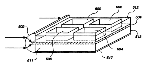
FIG. 6 depicts an alternate multiwell biosensor similar to that of FIGS. 5A-
5C, except
that a series of discrete wells 600, 602, 604, 606 are formed on the waveguide
504. The
embodiment of FIG. 6 would be used in a horizontal position, so that the wells
600, 602, 604,
606 need not be covered.
The biosensor including the lens may be formed by molding of a suitable
optical
plastic. A holder comprising the reservoir walls, the lens, and frame elements
WO 94/27137 216 2 9 9 6 PCT/US94/05567
-16-
as needed, may be pre-molded. A silica-surface waveguide is inserted
subsequently
with a refractive-index-matched adhesive to secure it in place and seal it as
needed to
create separate channels. Alternatively, the holder may be molded with a
silica-
surface waveguide in place, thereby eliminating the need for the adhesive.
In a presently preferred further embodiment, the waveguide is also formed of
the optical plastic and is molded simultaneously with the lens and/or the
reservoirs.
The latter type construction is not suitable for use with excitation
wavelengths of 488
to 515 nm, because known optical plastics tend to emit fluorescence when
excited in
this (the blue and green) wavelength region. This fluorescence would appear as
background fluorescence. However, an alternate embodiment of the apparatus
using a
light source emitting at wavelengths of 600 nm and above, would accommodate a
plastic waveguide. Molding the lens/wave-guide, or
lens/waveguide/reservoir(s), as a
single unit of plastic substantially reduces the cost of manufacturing and
makes a
disposable biosensor more feasible.
FIGS. 11A-C shows another embodiment of a biosensor similar to that
depicted in FIGS. 3A-C. In FIG. 11A, waveguide 10 is a glass coverslip
inserted in a
sawcut groove 12 (FIG. 11B) in a solid, colorless plastic lens 14. Transparent
walls
16, 18, 20, 22 are sealingly attached with index-matched adhesive to the lens
14 and
about the edges of the waveguide 10 to form a pair of separate reservoirs 30,
32
(FTG.11B).
In the embodiment of FIGS. 11A-C, the curved forward edge 34 of the lens 14
is spaced at a distance 40 fmm the forward end of the waveguide 10. Distance
40 is
selected so as to match the focal length of the lens 14. A mask 42 made of a
material
that is opaque to visible light, covers the rear edge 44 of lens 14. The
embodiment of
FIGS. 11A-C can be used in a vertical orientation as shown in FIG. 5C.
Alternatively, the biosensor may be oriented with the waveguide 10 in a
substantially
horizontal position, so that only one side of the waveguide 10 is used. In
such case, a
cap which can sealingly close the open ends of the reservoirs must also be
provided.
An advantage of the horizontal orientation scheme is that only a thin layer 50
of
sample solution is required (FIG. 11C). However, unless ribs along the
waveguide 10
are provided, like ribs 530 in FIG. 5D, the biosensor of FIG. 11C in the
horizontal
orientation has only one effective sample channel.
CA 02162996 2005-02-22
-17-
While the curved edge 34 of lens 14 is shown as being substantially a semi
right-
cylinder in shape, other lens shapes are possible as described previously
herein with respect to
FIGS. 3A and 5C.
In a further and highly preferred embodiment of the biosensor depicted in
FIGS. 12A and 12B, the end of the planar waveguide which is distal to the
receiving edge,
has a portion 650 of serrated or toothed shape. The angles 652A, 652B must be
less than the
critical angle for total internal reflection at the waveguide-air interface
(the critical angle for
polystyrene/air is about 51 ). Preferably, the sum of angles 652A, 652B
should be 90 , so
that the light is retro-reflected directly back along the longitudinal axis of
the waveguide; still
more preferably, angle 652A =: 652B = 45 . An advantage of this shape is that
it increases the
level of internal reflection without a reflective coating on the edges, thus
reducing
manufacturing complexity and costs. The increased TIR enhances the evanescent
field
intensity and thus improves sensitivity of the assay. Also, the serrated end
edge helps to
equalize (make more uniform across the whole waveguide) the intensity of light
within the
waveguide. The entire waveguide portion of the biosensor, should have an
optical-quality
surface, including the serrated end and the integral lens.
In a working embodiment, the waveguide is about 0.05 centimeters (cm) in
thickness
and the wells are about 0.08 to 0.1 cm in depth. The biosensor including the
waveguide is
about 2.5 cm wide and about 4.3 cm long.
The embodiment of FIGS. 12A and 12B also has a plurality of parallel wells 660
each
extending along the longitudinal direction from the light receiving end 624 of
the waveguide
622. Notably, each well contains a plurality of patches 662, 664, 666 each
comprising a
different immobilized Fab' species. The elimination of separation walls
between such
different species, which would extend crosswise to the direction of light
propagation in the
waveguide, further increases the sensitivity of the assay. The increased
sensitivity results
from 1) avoiding leakage of waveguide light through the walls, and 2) avoiding
scattering of
the excitation light which may excite unbound tracer molecules outside the
region of
evanescent penetration, undesirably increasing background fluorescence.
In another improvement, the sheet excitation beam is arranged to enter the
receiving
edge of the waveguide at an angle to the plane of the waveguide. FIG. 13 shows
an angled
integral lens 670 configured to accept such angled beam entry. For this
purpose, the beam
originating from the laser should be shaped to a sheet of width approximating
the width of the
CA 02162996 2005-02-22
-18-
receiving region of the waveguide 622, and of relatively narrow thickness
(preferably no more
than tenfold, and preferably one-to four-fold the waveguide thickness), using
cylindrical
and/or spherical lenses as known in the art.
The effect of so angling the beam entry is to increase the proportion of light
entering
the waveguide which excites the higher order modes that are propagated in the
multimode
waveguide, thereby increasing the intensity of the evanescent field.
Desirably, the mean
beam entry angle is selected tci be less than, but near the critical angle of
the
waveguide/solution interface. The closer the beam entry angle is to this
critical angle, the
greater the increase in evanescent intensity. However, a beam entry angle too
near the critical
angle could result in some of the entering light escaping at the
waveguide/solution interface,
thus decreasing the waveguiding efficiency and evanescent intensity. Thus,
there is an
optimum angle that can be determined experimentally. Generally, a beam entry
angle of a
few degrees less than the critical angle (from about 5 to about 15 degrees
less), will be useful.
For a polystyrene waveguide, the critical angle is about 33 , and the
presently
preferred beam entry angle is 25 ; with these values, an increase in
evanescent light intensity
of at least about a factor of two is achieved over using a 0 beam entry
angle.
The use of an angled beam entry necessitates adjustment of the orientation of
the
center of radius of curvature ol.'the semi-cylindrical lens with respect to
the waveguide
receiving end, as will be evident to a skilled person. In a molded integral
biosensor, the
integral lens/waveguide 622 may be formed as shown in FIG. 13.
The following examples detail several methods for attaching the capture
molecules to
the waveguide surface in a site-specific manner. The general scheme for
reducing the level of
non-specific binding is to coat the waveguide with a protein-resistant
material, and then
immobilize the antibody to the coating. The scheme further includes
derivatizing of the
protein-resistant coating combined with site-specific modification of the
antibody or other
capture molecule to be immobilized, so as to provide site-specific attachment
of the capture
molecule to the coating.
Of the examples presented, the procedures of Examples I and II gave generally
better
results. At present, the avidin-biotin coupling method (Example II) is the
most preferred.
Using either coupling scheme, at least about 75% of the immobilized Fab'
fragments were
active, and the levels of non-specific binding were typically no more
2162996
WO 94/27137 . PCTIUS94/05567
-19-
than 1-2 % of the specific binding. The modified PEG coating gave slightly
higher
levels of non-specific binding, in the range of 5 % to about 25 %.
EXAMPLE I
PREPARATION OF WAVEGUIDE SURFACE - HYDROGEL
A silica surface was prepared with a hydrogel coating comprised of
polymethacryloyl hydrazide (abbreviated PMahy). Fused silica slides of CO
grade
and thickness about 1 mm, available from ESCO, Inc., were suitable as
waveguides
(optical substrates).
To graft the PMahy to the silica, the surface was derivatized with aldehyde
groups. The derivatization was accomplished by silanization with 3-
aminopropyltriethoxy silane (abbreviated APS) to add an amino functional
group,
followed by reaction with glutaraldehyde to produce free aldehyde groups. The
PMahy was then be reacted with these aldehyde groups to form the hydrogel
coating.
Antibodies could be coupled to this hydrogel in at least two ways. In one
method, the carbohydrate groups in the Fc antibody region are oxidized to
aldehydes
by treatment with sodium metaperiodate. However, few antigen-binding fragments
contain carbohydrate moieties useful for this purpose. Thus, a preferred
method
comprised modifying the pendant hydrazido groups of the hydrogel to a
maleimido
group by treatment with succinimidyl4-(N-maleimido-methyl)cyclo-hexane-l-
carboxylate (abbreviated SMCC; Pierce Chemicals). These maleimido groups can
be
reacted with the free thiol groups typi.,ally found in the C-terminal region
of Fab'
fragments, thereby coupling the Fab' fragments to the hydrogel.
Polymethacryloylchloride (abbreviated PMaC1) was prepared by radical
polymerization of methacryloyl chloride (abbreviated MaCI) in dioxane under an
inert
atmosphere, as described in Jantas et al., J. Polym. Sci.. Part A: Polym.
Chem.
27:475-485 (1989).
A reaction mixture containing 21.1. mole% of MaCl, 78.1 mole% dioxane,
and 0.8 mole% AIBN (azobisisobutyro-nitrile), was allowed to react for 24
hours at
60 C with agitation. The PMaC1 so produced remained in solution during the
course
of the reaction. The mixture was then diluted with twice the amount of dioxane
used
in the reaction and slowly added to an excess of hydrazine hydrate, to achieve
a
volumetric ratio of 2:5 for diluted PMaCI. The latter addition was carried out
for
about 30 minutes in an ice bath under a nitrogen atmosphere. The resulting
mixture
WO 94/27137 -20- 21629 9 6 PCT/US94/05567
was then stirred for about an hour at room temperature. The product PMahy was
purified by evaporation of dioxane and the remaining unreacted hydrazine
hydrate,
followed by washing in distilled water. The washed product was then dialyzed
in a
SpectraPor dialysis membrane having a molecular weight cut-off of 3,500
daltons, to
remove unreacted monomer.
The polymer so prepared was shown to have a molecular weight of about
26,000 as measured by gel permeation chromatography for the hydrochloride
form.
The concentration of polymer in solution in the hydrochloride form was
estimated to
vary between about 5 % and 8 % (w/v). It has been found that the polymer can
be
stored in aqueous solution at 4 C under a nitrogen atmosphere, for at least 5
months
without undergoing a detrimental amount of spontaneous cross-linking.
Silica chips or glass or quartz microscope slides were cleaned with chromic
acid, then treated with 5 % APS/95 % deionized water (v/v) for about fifteen
minutes
at room temperature. The APS-treated surfaces were rinsed wi.th deoionized
water
and absolute ethanol, and incubated in a vacuum oven which had been flushed at
least
three times with nitrogen, at 120 C for 1 hour. The resulting silanized
surfaces were
then soaked in 2.5 % glutaraldehyde (E.M. grade from Polysciences) in 0. 1M
carbonate-bicarbonate buffer, pH 9.2, for two hours at room temperature.
Next, linear PMahy was reacted with the aldehyde groups on the treated chips
to create a cross-linked polymer film with many unreacted hydrazido groups in
the
chains. This was done by dipping the treated chips in solutions of PMahy of
between
about 5% and 8%(w/v), pH 5.2, at a temperature between about room temperature
and about 60 C, for a time sufficient to form apolymer film of thickness about
100 A
or less. The thickness of the hydrogel layer increases with time and
temperature of
incubation in the solution. It was found that optimal conditions for
preparation of the
film of 100 A thickness or less, comprised incubatir.g in 5_% (w/v) PMahy for
2 hours
at room temperature (about 25 C).
Next, the free hydrazido groups of the polymer film were modified by
treatment with SMCC to provide reactive maleimido groups on the ends of the
polymer side chains. This was done by immersing the PMahy-coated substrates in
a
solution of 0.19 % (w/v) SMCC in dimethylformamide for about 1 hour at 25 C.
Following derivatization with SMCC, the hydrogel-coated surfaces were
treated with a 1 mg/mi solution of Fab' fragments in phosphate buffer, ph 6.0,
with 5
mM EDTA. The waveguide surface so prepared was shown to immobilize Fab'
CA 02162996 2005-02-22
-21-
molecules at a surface density of about 1.4 x10'12 moles/cm2. Also the surface
was able to
immobilize Fab' fragments at their C-terminal thiol groups in a site-specific
way. The
thickness of the resulting polymer film was determined by ellipsometry to be
about 100 A, as
was desired. This film thickness is much less than typical previous polymeric
films, which
have thicknesses of 0.35 to 25 m (microns). The above-described method of
preparing the
PMahy polymers is superior to that described by von Kern et al. using
polymethacryloylacid
esters. Such esters suitable for reaction with hydrazine hydrate often have a
molecular weight
of 80,000 daltons or more, from which it is difficult to obtain a desirably
thin film on the
waveguide.
Finally, the Fab' fragments were coupled to the free maleimido groups pendant
from
the polymer-coated surface as follows. The prepared waveguide surface was
incubated for 24
hours at 4 C in a solution containing the Fab' fragments at a concentration of
1.5 x 10' molar,
in a phosphate buffer with 5 mM EDTA (pH 6.0).
EXAMPLE II
PREPARATION OF WAVEGUIDE SURFACE - AVIDIN-BIOTIN
This strategy was designed to exploit the very strong binding affinity of
biotin for
avidin (binding constant of around10'15). An avidin coating was readily made
by physical
adsorption on a silica surface. The Fab' fragments were then conjugated with
biotin to form
biotin-Fab' conjugates, also referred to as biotinylated Fab' fragments or b-
Fab' fragments.
The biotin is coupled at specific location(s) on the Fab' fragments. The
avidin coated surface
is then treated with the b-Fab' fragments, so that the biotin binds to the
avidin thereby
immobilizing the Fab' fragment to the surface in a site-specific manner.
In actual experiments, the procedure was as follows. Chromic acid-cleaned
silica
surfaces were immersed in a solution of 3 x 10⁻⁶ M (molar) avidin for about 3
hours at room
temperature. The surfaces were then washed several times in PBS to remove
unadsorbed
avidin.
Biotinylated Fab' conjugates were prepared from a solution of Fab' fragments
in PBS
(0.5-1 mg/ml), by addition of a sufficient amount of 4 mM biotin-HPDP in
dimethyl-
formamide to provide a 20-fold molar excess of biotin-HPDP. This mixture was
incubated
for 90 minutes at room temperature, and biotinylated Fab' fragments
(abbreviated b-Fab')
were purified by gel permeatiori chromatography with Sephadex G25
equilibrated in PBS.
WO 94/27137 2162" " " PCT/US94/05567
-22-
An alternate method was used for biotinylating whole antibodies, in which
biotin-LC-hydrazide was coupled to oxidized carbohydrate groups in the Fc
region of
the antibody. Mab designated 9-40 (a murine monoclonal IgG, antibody that
binds
fluorescein), was oxidized by incubation at a concentration of 1-2 mg/ml
protein in 10
mM sodium periodate, 0.1 M sodium acetate pH 5.5 for 20 minutes at about 0 C.
Glycerol was then added to a fmal concentration of 15 mM to quench the
reaction,
and the mixture incubated a further 5 minutes at 0 C. The oxidized Mab 9-40
was
purified by gel filtration chromatography on Sephadex G25 equilibrated with 0.
1M
sodium acetate buffer pH 5.5, aiid then reacted with 5 mM biotin-LC-hydrazide
for 2
hours at room temperature with agitation. Unreacted biotin-LC-hydrazide was
removed using a Sephadex G25 column equilibrated in PBS.
Avidin-coated surfaces were immersed in a 1.5 x 10' M solution of b-Fab'
fragments for about an hour at room temperature, followed by washing with PBS
to
remove unbound b-Fah' fragments. Optionally, polyethylene glycol (abbreviated
PEG) was coupled to surfaces that were previously coated with the b-Fab'
fragments,
by immersion of the b-Fab'-coated surfaces in a solution of between about 5
x10-$ and
1 x 10' M PEG. Unbound PEG was removed by washing in PBS.
The density of immobilized Fab' fragments obtained using the avidin-biotin
coupling chemistry was about 1.4x10⁻¹² moles per cm2 (square centimeter).
EXAMPLE III
PREPARATION OF WAVEGUIDE SURFACE - PEG-T:'PE
In this method, the terminal hydroxyl groups of polyethylene glycol
(abbreviated PEG) were converted to primary amine or hydrazide groups by
reaction
with ethylenediamine (abbreviated ED) or hydrazine (abbreviated HZ),
respectively,
to produce PEG-ED2 or PEG-HZ2. The PEG molecules so modified were then
coupled to APS-glutaraldehyde activated silica surfaces. One ED moiety on each
PEG-EDZ molecule couples to a free aldehyde group on the silanized-
glutaraldehyde-
treated waveguide surface. The other ED (or HZ, if PEG-HZ2 is used) is then
available to bind to ar. aldehyde moiety in a capture molecule (binding
protein) such
as an oxidized antibody or antibody fragment.
Monofunctional (PEG M2000, M5000) or difunctional (PEG 3400, PEG 8000,
PEG 18,500) of the indicated molecular weights in daltons, were reacted with p-
nitrophenyl chloroformate (abbreviated p-NPC; obtained from Aldrich Chemicals)
in
PCT/US94/05567
WO 94/27137 -23- 2162996
solution in benzene. The mixture was agitated at room temperature for= about
24
hours. Dry ethyl ether (less than 0.01 % water, purchased from J.T. Baker
Chemicals) was used to precipitate PEG-(o-NP)2 from solution. The precipitate
was
vacuum-dried overnight. Between about 50% and about 100% of PEG molecules
were converted by this treatment to PEG-Onp, as determined by hydrolysis with
0.1 N
sodium hydroxide to release the p-nitrophenol groups. The absorbance at 402 nm
was
determined spectrophotometrically and a mol.ar extinction coefficient of 18400
M-'
cm 1 used to determine the amount of conversion. The level of conversion
depended
somewhat on the molecular weight of the PEG of MPEG.
PEG-(o-NP)Z was then dissolved in ethyienediamine and agitated gently for
about 3 hours at room temperature. The PEG-(ED)2 was then precipitated by
addition
of a sufficient amount of dry ethyl ether. The yellow PEG-(ED)2 solution was
decolorized by addition of 1 drop of 1.2N (normal) hydrochloric acid, and the
precipitation with ethyl ether repeated twice more. The wet PEG-(ED)Z was
dried
under vacuum overnight. Alternatively, in place of ethyler.ediamine, the PEG
was
derivatized with hydrazine to produce PEG-Hz,z.
The modified PEG was coupled to silanized-glutaraldehyde-treated waveguide
surfaces prepared as described in Example I. A solution of 24 milligrams (mg)
of
PEG-ED powder dissolved in 1.2 milliliters (nil) of 0. 15M PBS pH 7.4 or in
the same
volume of 11 % potassium sulfate-sodium acetate buffer at pH 5.2. The prepared
waveguide surfaces were immersed in the PEG-ED solution and incubated at 60 C
for
about 24 hoiirs. The procedure using K2SO4-aceate buffer yielded a higher
density of
PEG molecules attached to the surface than that using PBS buffer. Antibodies
or
other binding proteins were immobilized to the PEG-coated waveguides as
follows. A
solution of about 3 mg/ml of antibody was dissolved in 0.15 M sodium acetate
buffer,
pH 5.2. A solution of equivalent weight of 50 mM sodium metaperiodate (NaIO4)
was then added, and the reactants were agitated at room temperature for about
an
hour. Unreacted sodium metaperiodate was removed by passing the reaction
mixture
through a desalting column (type PD-10 from Pharmacia), which had been pre-
equilibrated with the sodium acetate buffer.
The PEG-coated waveguides were then incubated with the oxidized antibody
solution in the sodium acetate buffer, pH 5.2, for 3 days at 4 C, then rinsed
to
remove unbound antibody.
WO 94/27137 , -24- 21629 9 6 PCT/US94/05567
Waveguides prepared by each of the coating procedures of Examples I-III, as
well as by prior art random-site coupling methods, were analyzed to determine
levels
of non-specific binding relative to total fluorescence and amounts of
immobilized
antibody and of available binding sites. Comparative results of these analyses
are
shown in Table I.
The data in Table I were obtained using fluorescein-BSA (BSA = bovine
serum albumen) conjugates with an epitope density of nine (that is,
approximately nine
fluorescein molecules bound per BSA molecule), and anti-fluorescein antibodies
(designated Mab 9-40, or Fab' 9-40 for fragments). A hybridoma cell line
secreting
this antibody was obtained from Professor E. W. Voss of the University of
Illinois at
Urbana-Champaign. In these experiments and those whose results are shown in
FIGS. 7A, 7B, 8, 9A-9F, and l0A-IOD, data acquisition and processing was
accomplished using software supplied with the Photometrics Series 200.
Absolute antigen binding was determined by means of radiolabelled tracers or
capture molecules. For example, radiolabelled BSA-FI-9 was allowed to be
coupled
with immobilized Fab' fragments for 5 or 60 minutes in phosphate buffer pH 7.3
at
room temperature. The tracer concentration was 1.5x10-' M. Three ml per sample
of
fluorescein-labelled BSA (BSA-FI.9) at concentrations ranging from 10-10 M to
101 M
was injected into the flow cell. The injection was performed over a five-
minute
interval. The spectrum at wavelength of 488 nm was taken and the bulk BSA-FI-9
was removed by flushing with PBS buffer. Three more spectra were taken, and
the
fluorescein peak from 513 to 517 nm was integrated. These values were set
versus
the log of BSA-FI.9 concentration in order to obtain the binding isotherm.
For measurements of radioactivity, the coated silica chips with either
immobilized radiolabelled capture molecules or with labelled tracer molecules
bound
to unlabelled capture molecules, were washed thoroughly in a suitable buffer
and
counted on a gamma counter. 'uI-labelled antibodies or antigens were preferred
for
the radiolabelling, which was done using the Chloramine-T method (see
Greenwood et
al., Biochem. J. 89:114-123 (1963)).
The levels of non-specific absorption of antigen on waveguides prepared by
site-specific coupling with avidin-biotin (Example II; Table I rows 7 and 8
from the
top) or hydrogel (Example I; Table I bottom two rows) were considerably better
than
most of the prior art coupling methods, being typically 1-3% (Table I). The
results
WO 94/27137
PCT/US94/05567
2162996
-25-
U ~ E
'~ & 1, LC) CO 1, 00 n 0 CO M
V? co et co ct co 'y
_ ~
fn Q N M N ~ N N ~ ~ t- cli
N LL)
C
O
tu
E _N T V Q
L O m
L O~ O 0 0 tt) cO 't ct 0 ~ tO t0
E~ - O N C') 1- O M O) f~ ='
Q x E M N '- M N 1~ N
.U
U M
> a
.p y-CO 0 0 ~ <O M N N OJ 00 C)
CO C et f, M 00 M et O O 00 00 O Of E
Z m .. (D Q) f0 M ~ N N N .- Cfl N y'y
4~
y CD D
41 O
U O C
~ . 'V >
a Of E Z
N y
U y C:5 O ~ et I, tt) p N N N r- M U
a O O N N - O O O O O W
N Q Z m X~ O O O O O O O O O 0 0 a0 O
cm Qf _7
ti
L LO cn
y C
E w .0
- tn _ C y m U
y eo ~ 0 tn n r~ LO cD ~ N et 0 C o C =- ~D ta M tA tA 1~ CO OD '- 1!)
~m X E o 0 0 0 0 0 0 0 0 0
E 0
E m
d c
~ y O >
c E ~
M 0
ai
~
c (D
~ ~ m +,m O
C _! E O E O wU = E ~U ~U v=U .2 :U ~- C~+,
o a E v v -o 0 a 0 u u o ~ c~ E
O d C C C C 0 C d N N ~ C O O C
0 L eo eo ~o o a eo a a ~ c o ~ y
U U m oc a~ m cn m cn cn v~ cn U) ~-
E ; cUai c
~ y c~ ro
E E
~ C7 U' C7 Q U ~ t"il y C
co 7 7 'a C
d N 0 0 OD 0 ~ eCC tL O c0 tp L
~" IC l9 E !C - - C7 L L y~ .y L L V U U
~i ~i c _u o 'H
0 LL ~~
~ ~ ~ N C C c E +L+ = L C
cm 'D 3 ~ O y
Q 2 Q ~i Q OO OO m co m OO ii ~i ~ o~ Q~~
N
O o O U) O 0
L O Cim E
U y U
c~ E;" 30
U .2 U 4. d Q!VO (aA
cn (n f~ J J J w N y 0 c y lL m ME
U U U C C C~ O) Vl =~ 4! c'~i r H
. ~ 0 C oC C m E
O O O fn tn (n 'C 5 '6 O U O v- r- =~ ,~
4) L L L a a 0- > > > O C 0 0 0 0
U a a a < Q Q < < Q:5 ea o.r r. ~,
o O o F E c c c c
l0 l0 l0 W l0 lC L N V1 N= 7 7 7 y
u U u U= o 0 0
cn _ _= in v~ in in in n ~ in a in a a~' E E E~,
' Q W Q Q Q Q a
WO 94/27137 -26- 2 1,j 29(~ 6 PCT/US94/05567
U
0
~ N O)
N O
y y ~ ~ N
a, y C }I
p Cc ~ N a) N M
mZmE v Lo ,. v
O
o C Q N O O
V) C"C 0 U 0 Itt r) 0
m o C .- o O O O O
QZGO E x E V O O V
.2 Q -
U C m r0
j Q~'""
3 ~, vl C C O ~ N N
ocE ~ o co co -
aC zm Ln V rj V V
>
0
U U_
a
tn O
y U .
a+ t, y ~] ~ O
c~ y 2 a a~ ' E ~ ~ .-
+n p N ~ C 00
O O O
~' U'C "CE o 0 0 0 0
~ azmLn x E v o v v
U
>
~ C7 M
i7) W
T n
c cc o Y 0 0 0 O 0
> ch C) it +i
C 00 r= LCf
Q Q ~ lt) !L) LL) lL) CV
=a j, ~
C7 fV) fV) N
o~ (7 C_ O O O O O
O
3 =' = o~ C E O O 0 0
~ 3 ~o 'D r- Ln cc
0.90 ~ o 0o (o O
o ~'mco X E 0 0 0 0 0
~ a
a= C) N ~ N r-
:o o 0 0 0 0
o ==~, N~ 0 0 0 0 0
h -7 ~ ~ -H ~ ~ ~
O ~~=E Q~ N 00 co 'tt p C ~ O cD tt) ~ tt) 0
>. mU, x E o 0 0 o O
cc
E
E ? _ o 0 0 0 0
~ o E o c o 0
E ~'~ p~ a~ rn et Ln 0
C - O C) N et Lf) Q x E
ar
C
u) C) O 0
c o Y o0 co r~ 0
cD U n a6 r-~ ac Co
Q m U ~
e E6 E~ o~ O E~
~ w L, ~ t t
m 41 _Q 41 ,p 4~ M
CO C
C Q LL Q LL Q LL Q LL Q L.
L
WO 94/27137 21629 96 PCT/US94/05567
-27-
also indicated that non-specific binding to the avidin-coated waveguide was
acceptably
low for analyte molecule concentrations of less than about 1or5 M, without a
wash
step.
The percentage of immobilized molecules that were active (able to bind
analyte) was also considerably higher for avidin-biotin and for hydrogel
coupling
chemistries, being in the range of 50 % to 75 % for Fabs. The results for IgG
capture
molecules coupled by heat treatment, acid treatment or by oxidation, indicated
that
only a low percentage of the IgG binding sites were active (Table I rows
1,2,4, 5,6,10
from the top).
The row labelled silica-avidin with biotin-PEG, represents data obtained with
the further refmement of preloading the surface (after attachment of the
capture
molecules) with biotin-PEG conjugates. This was done to passivate potential
non-
specific binding regions. However, the improvement obtained with biotin-PEG
pre-
loading was not large.
For the experiments whose results are shown in Tables II and III, Fab'
fragments derived from a murine anti-human chorionic gonadotrophin (anti-hCG)
monoclonal IgG antibody were used. The parent monoclonal antibody was purified
as
described by van Erp et al. J. Immunol. Methods, 140:235-241 (1991). This
mouse
antibody, termed anti-hCG-A, is directed against a portion of the B-subunit of
hCG
(provided by Organon-Teknika of Boxtel, Netherlands). The whole monoclonal
antibody anti-hCG-A was used in the experiments whose results are depicted in
FIGS. 7A, 7B, 8, 9A-9F, and 10A-10D.
F(ab')2 fragments were produced by digestion with pepsin using the procedure
described by Grey and Kunkel, "H Chain subgroups of myeloma proteins and
normal
7S globulin," J. Exp. Med. 120:253-266, 1964. Following digestion, F(ab')2
fragments were reduced to Fab' fragments using dithiothreitol (DTT).
Specifically,
33 mg of purified antibody and 1 mg pepsin (Sigma) were dissolved in 0.1 M
sodium
acetate buffer (pH 4.2) and the digestion was carried out at 37 C for 16
hours. The
digestion was terminated by adjusting the pH of the reaction mixture to 8.0
with 2 M
tris base. The F(ab')2 fraction was separated by gel permeation chromatography
(Superdex Hiload, Pharmacia) using phosphate-buffered saline (PBS), pH 7.7, as
eluent. Fab' fragments were prepared by reducing the F(ab')2 fragments (1
mg/ml)
with 1.75 mM DTT and 3.5 mM ethylenediamine tetraacetate (EDTA) in 0.17 M tris
buffer (pH 7.4) for 45 minutes at room temperature. After reduction, excess
DTT
WO 94/27137 -28- 216 2 9 9 6 PCT/US94105567
was removed by gel permeation chromatography using a Sephadex G-25 column
(Pharmacia) equilibrated in 0.1 M sodium phosphate buffer (pH 6.0) containing
5 mM
EDTA.
Table III
Summary of Solid Phase Immunoassay Using Silica Substrates with Adsorbed
Avidin
and Biotinylated Fab' Fragments
Antibody Immobilized Total hCG Specific Absolute Relative
Antibody Binding Activity Non-specific Non-
Binding specific
(BSA) Binding
(BSA)
(x10r12 mol/cm=) (x10-'Z mol/cm2) (%) (x10"12 moUcat2) (46)
Fab' from 1.19f0.02 1.22f0.01 100f5 0.05f0.02 4.20 0.02
Anti-hCG-A
Fab' from 1.40 0.05 1.38 0.07 98f9 0.05 0.01 3.57f0.02
Anti-hCG-B
Fab' from 2.24t0.02 1.10t0.03 49t3 0.05t0.02 2.32t0.01
Anti-hCG-C
Fab' from 1.59t0.02 1.24t0.01 78t2 0.05t0.005 3.14 0.01
Anti-hCG-D
Fab' from 1.25f0.02 0.03t0.003 2.4t0.05 0.09t0.03 7.20t0.03
Mouse IgG
In the experiments whose results are presented in Tables II and III, the
specific
binding values were determined using hCG labelled with "I as described for
Table I,
while luI-labelled BSA was used to measure the non-specific binding. In both
Tables
II and III, the immobilized antibody was anti-hCG A.
The fluoro-immunoassays of FIGS. 7A, 7B, 8, 9A-9F, and 10A-lOD and
Tables I-III were performed using an interfacial fluorometer constructed at
the
University of Utah. Silica waveguides with the appropriate respective
immobilized
antigens were placed in the dual-channel flow-cell of FIGS. 3A and 3B. The two
channels were used for sample and reference measurements, as described with
respect
to FIGS. 4A-C. The light source was the 514.5 nm emission of an air-cooled
argon-
ion laser. The laser beam was split into two parallel beams, which were
focused with
lenses into the two channels of the waveguide. Fluorescence emission was
recorded
2162996
WO 94/27137 PCT/US94/05567
-29-
from 520 to 620 nm using a monochromator connected to a computer-Eontrolled
CCD
camera. The fluorescence spectrum was integrated from 560 nm to 600 nm to
improve the signal-to-noise ratio.
FIGS. 7A, 7B and 8 are charts depicting fluorescence intensity data obtained
using two alternate formats for performing a fluorescence immunoassay to
detect an
antibody. In these experiments, the detection of antibodies to human chorionic
gonadotrophin (abbreviated hCG) was used as a model to determine which format
provided the greatest sensitivity. It will be evident that the methods
described could
be adapted to the detection of any desired antibody in biological fluids such
as plasma
or serum, for example the detection of antibodies to proteins of viral and
bacterial
pathogens, depending only on obtaining the necessary antigen for use as the
capture
molecule.
For purposes of the tests shown in FIGS. 7A, 7B and 8, the antibody to be
detected (the analyte) was chosen to be a monoclonal antibody (designated anti-
hCG-
A) to an hCG antigen (the latter designated hCG-A). The data of FIG. 7A were
obtained with whole hCG molecules serving as the capture molecules (the
antigen or
analyte binding molecule) in the assay. The data of FIG. 7B were obtained
using an
oligopeptide constructed to selectively bind the anti-hCG-A antibody, as the
capture
molecules. Oligopeptides suitable for this purpose for any known antigenic
analyte
molecule analyte can be obtained using the methods of Geysen et al. as
disclosed in
Patent Publication Nos. WO 86/86487 and U.S. Patent No. 4,708,871, as well as
in
the scientific literature. To attach the necessary fluorescent dye, either the
N-terminus
of the oligopeptide was modified to provide an amino group for amino-reactive
dyes,
or the C-terminus was modified to provide a cysteine thiol group for thiol-
reactive
dyes). Preferably also, the complete oligopeptide sequence is of length
sufficient that
the attached dye is spaced from the binding site by at least two or three
residues.
In the experiments of both FIGS. 7A and 7B, the tracer was a goat anti-mouse
IgG labelled with tetramethylrhodamine (abbreviated TMR). For both assay
formats,
the capture molecule was biotinylated as described in Example II and
immobilized on
an avidin-coated silica substrate. The test antibody, anti-hCG A, was premixed
with
the tracer (goat anti-mouse IgG-TMR) in the test solution.
As will be understood by those in the art for a sandwich fluoroimmunoassay,
the anti-hCG A antibody bound to the immobilized capture molecule, and the
goat-
anti-mouse IgG-TMR tracer in turn bound to the mouse anti-hCG A antibody. In
this
WO 94/27137 -30- 2162996
PCT/US94/05567
way a fluorescent sandwich formed on the substrate surface with the TNZR-
portion of
the tracer molecule being held within the region of evanescent excitation.
The data of FIGS. 7A and 7B were obtained with the following protocol.
Different concentrations of anti-hCG A were premixed with the tracer antibody
(concentration fixed at 10-8 M) and injected into the sample channel. A 10-g M
concentration of tracer antibody was also injected into the reference channel
as a
control. The fluorescence intensity of the sample channel was plotted vs. anti-
hCG A
concentration and the fluorescence intensity of the reference channel was also
plotted
on the same set of axes (this is really a plot of the non-specific binding of
the tracer
antibody vs. time, since no anti-hCG A was injected into the reference
channel).
FIGS. 7A and 7B show the results for a sandwich assay format following the
binding of anti-hCG A to immobilized hCG and to the oligopeptide,
respectively.
Figure 8 shows the corresponding fluorescence enhancements for both cases. The
data from FIGS. 7A and 7B were normalized for background fluorescence and
replotted as fluorescence enhancement (F.,"'P,JF,,f~~,,
,) versus log analyte
concentration. The response curve was similar for both of the immobilized
antigens
(whole hCG and oligopeptide antigen) over a range of antibody concentrations
from
10-13 M to 10" M. However, whole hCG gave better precision.
It is also evident from FIGS. 7A, 7B and 8 that analyte levels (anti-hCG A) as
low as 10" molar were detectable with the assay. In a further embodiment, the
tracer
antibody concentration is reduced to 10-10 M or less. This is expected to
reduce
background fluorescence due to non-specific adsorption of the tracer antibody
and
thereby further improve the sensitivity to 10-14 M or better.
FIGS. 9A-9C depicts data obtained using an antibody as the capture molecule
to detect an antigen, in a sandwich-type assay. As mentioned previously, two
different antibodies are employed in a sandwich immunoassay - an immobilized
capture antibody and a labelled tracer antibody in solution. Since the capture
antibody
and the tracer antibody must bind to distinct regions of the antigen, two
different
monoclonal antibodies which bind to different epitopes on the antigen are
typically
used in such assays. In addition to the anti-hCG-A, three other monoclonal
anti-hCG
antibodies (anti-hCG-B, anti-hCG-C and anti-hCG-D, respectively) were obtained
from Organon Teknika which bound to different epitopes than did anti-hCG-A.
Since
only anti-hCG A is specific to hCG (the others also bind to certain hormones
related
WO 94/27137 216 2 q 9 6 PCTIUS94/05567
-31-
to hCG), only six of the twelve possible pairwise combinations of antibodies
provide
strict selectivity for hCG.
FIGS. 9A-F depict results obtained with different pairwise combinations, with
Fab' fragments prepared from anti-hCG A (Fab'-A) and immobilized to waveguides
using the avidin-biotin coupling chemistry. Fab' fragments prepared from anti-
hCG
B, anti-hCG C and anti-hCG D were labeled with tetramethylrhodamine for use as
tracer antibodies (designated Fab'-B, Fab'-C and Fab'-D, respectively). FIGS.
9A
and 9B show results with Fab'-B as the tracer molecule. FIGS. 9C, 9D show
results
obtained using Fab'-C as the tracer molecule. FIGS. 9E, 9F show results
obtained
using Fab'-D as the tracer molecule. Presently, Fab'-B and Fab'-C are
preferred for
use as tracers in an hCG assay.
An alternate format used a converse protocol, that is, Fab'-A as the tracer
molecule and Fab'-B, -C or -D as the capture molecule. However, the format
using
Fab'-A as the capture antibody was generally superior in sensitivity. It can
be seen
from FIG. 9B that hCG concentrations as low as 1012 M could be detected by the
assay with Fab'-A as capture molecule.
FIGS. l0A-D show data obtained from a competition or displacement assay.
Fab'-A fragments were immobilized to waveguides using either the avidin-biotin
chemistry (FIGS. 10A, lOB) or the hydrogel coupling chemistry (FIGS. 10C,
10D).
The immobilized Fab'-A fragments were preloaded with the tracer oligopeptide
at a
concentration of 10-g M. Increasing concentrations of hCG were added to one
channel
of the flow cell (sample) and PBS buffer was added to the other (reference).
For each
coupling chemistry, the raw fluorescence intensities of the sample and
reference
channels are shown in the panels on the left (l0A & 10C) and the percent of
full-scale
fluorescence (in the absence of hCG) are shown in the panels on the right (lOB
&
lOD). The latter values were normalized for the change in reference
fluorescence.
Standard errors were plotted for all data points, but in some cases were
smaller than
the plot marks.
At present the sandwich immunoassay is preferred for several reasons. First,
detection of concentrations down to at least 0.1 picomolar can be
demonstrated, as
compared to picomolar concentrations for the competitive assay. Also, the
instant
sandwich immunoassay was capable of detecting concentrations ranging over five
logs
- from 10-$ M to 10-13 M. Thus, a single assay formulation using the sandwich
WO 94/27137 2162 9 9 6 PCT/US94/05567
-32-
procedure could serve for a variety of applications where different detection
limits are
required.
A further embodiment of coating chemistry, and one which at present is highly
preferred, provides for photo-activated coupling of the binding moiety (Fab
fragment,
antibody or whatever) to the waveguide surface. By combining the
photoactivation
process with localized irradiation (for example, by masking), it is possible
to
sequentially couple different binding species to different regions of the
waveguide
surface. In this way, a waveguide surface patterned with patches each of a
different
capture molecule species (Fabs or Fab fragments) can be conveniently produced,
without need for walls between the different species. For the present type of
evanescent sensor, the elimination of unnecessary walls can significantly
improve the
sensitivity of the device by reducing background and enhancing evanescent
field
strength. In a highly preferred embodiment, the coating chemistry also
"passivates"
the surface, that is, inhibits nonspecific binding of the fluorescent tracer,
and thus
reduces the background signal.
A presently preferred coating is a type of compound referred to herein as a
"block copolymer", comprising at least one hydrophilic block containing
polymerized
hydrophilic residues (polyethylene oxide, "PEO"), adjacent at least one
hydrophobic
block containing polymerized hydrophobic residues (polypropylene oxide,
"PPO"). A
subclass of such compounds referred to herein as "triblock copolymers" or
"TBCPs",
comprise a hydrophobic block flanked by hydrophilic blocks. A series of TBCPs
are
commercially available from BASF Corporation under the tradename PLURONICS.
An example of a presently preferred compound is known generally in the
literature as
PLURONICS F108 or "PF108"; it has a molecular weight (MW) about 14,600 and
the general formula (PEO)x(PPO),,(PEO)x, where x = 129 and y = 56. In block
copolymers, the hydrophobic PPO segment tends to adsorb strongly to plastics
including polystyrene, leaving the PEO side-anns in a relatively mobile state.
Block
copolymers have the general property of inhibiting non-specific protein
adsorption,
while providing hydrophilic side chains useful to attach proteins, including
Fabs or
Fab fragments.
Also, while the present description is primarily with reference to
PLURONICS- type compounds, it is within contemplation that other polymeric
compounds having hydrophilic segments and hydrophobic segments and offering
pendant OH groups for attachment of proteins or photo-activated linkers will
be
CA 02162996 2005-02-22
-33-
useful. As known in the art, these include SEPHAROSE -type materials and other
polysaccharides. Also, block copolymers having polyurethane segments as the
hydrophilic
block may be useful.
Referring to FIG. 16, a. general procedure for preparing a patterned
polystyrene
waveguide is as follows. First., a waveguide surface 700 coated with PF108
molecules 702 is
prepared. Next, the free PEO chain ends 704 of the PF 108 molecules 702 in a
selected region
of the waveguide are derivatized in a photo-activated coupling reaction with a
photoaffmity
crosslinker 706. Suitable crosslinkers are heterobifunctional reagents which
have a photo-
activatable group conjugated to a reactive functional group such as
isothiocyanate,
succinimide or maleimide. Upon irradiation with light beam 701 of the
appropriate
wavelength (generally in the ultraviolet region), the photo-activatable groups
of the
crosslinker 706 react to covalently bind to the free PEO chain ends 704. A
mask 712
confines the irradiation to a first region 714 of the waveguide. The result is
waveguide
surface having reactive functional groups useful to bind Fab' fragments only
in the first region
714. Next, the waveguide surface is incubated with a solution of Fab'
fragments of a first
species (FAB 1 720 in FIG. 16) for a time sufficient to allow the binding of
Fab' fragments to
the derivatized region to go to completion. The unreacted Fab' fragments are
then washed
off, and the process of photo-activated derivatization is repeated for a
second region of the
waveguide, followed by incubation with a second species of Fab' fragment.
For coupling of Fab' fragments to a waveguide region derivatized with free
maleimido
groups (procedure of FIG. 16), incubation with a solution of Fab' fragments
can be performed
substantially as described for the PMahy coating.
In an alternate embodiment, the Fab' fragments 720 are coupled to the
crosslinker 706
before the crosslinker is photo-reacted with the PEO chain ends 704 (FIG. 17).
This
embodiment is presently preferred because the surfaces thus prepared are
capable of binding
larger levels of analyte plus tracer per unit area than those prepared
according to the protocol
of FIG. 16.
Suitable photoaffinity crosslinkers include aryl azides (amine-to-amine
linkage),
fluorinated aryl azides (C-H bond-to-amine linkage), and benzophenones (C-H
bond-to-
amine linkage or C-H bond-to-thiol-linkage, depending on the specific
compound).
Examples of each type are shown in FIGS. 18A-18D, along with the corresponding
photo-
activated coupling reaction. Presently, benzophenones providing a C-H bond-
2162[j.96 PCTIUS94/05567
WO 94/27137 -34- 7
to-thiol linkage are preferred, as these can be used to achieve site-specific
coupling to
Fab' fragments. Either the iodoacetamide or the maleimide derivatives of
benzophenone ("BPIA" and "BPM", respectively) can achieve this purpose. At
present
BPM is preferred, as it exhibits a higher degree of specific binding and a
lower
degree of non-specific adsorption. This is because the coupling occurs via the
C-
terminal thiol groups of the Fab' fragments, as described previously herein
for the
PMahy coating. Other photo-affmity crosslinkers providing free maleimido
groups
may be equally suitable.
In the photocoupling process, the amount of crosslinker coupled to the PF108
depends on the duration and intensity of the irradiation, the concentration of
crosslinker molecules, etc. These variables can easily be tested and optimized
to fmd
parameters which will achieve a desired level of crosslinker and/or Fab'
protein
coupled to the waveguide surface. Generally, a Fab' concentration of about 0.5
mg/ml to about 1 mg/ml, and a 20-fold molar excess of crosslinker, are useful
in the
processes of FIGS. 16 and 17.
Table IV shows comparative data on the levels of specific binding and non-
specific binding obtained for the crosslin-kers BPM vs. BPIA, for surfaces
which are
uncoated or coated with one of four different TBCPs, and for the procedure of
FIG.
16 vs. that of FIG. 17. The TBCPs are PF108. and three others designated by
the
tradenames PP105, PF68, and PF88, also available from BASF. The respective
PEO/PPO/PEO ratios and molecular weights of these compounds are 37/56/37
(PP105, MW=6500), 76/30/76 (PF68, IvIW=8400), and 104/39/104 (PF88, MW=
11,400).
As a model system, Fab' fragments derived from the 9-40 anti-fluorescein
antibody were used as the capture molecules, with fluorescein-conjugated BSA
representing the analyte. The BSA was radioactively labelled. Specific binding
was
determined as the binding of the fluorescein-BSA-conjugate, while nonspecific
binding
was determined from binding of native (unconjugated) BSA.
From the data in Table IV, it is e~.,ident that the degree of non-specific
binding
was significantly lower for the PF108 and PP105 coatings than for PF68 and
PF88.
For this reason, PF108 and PP105 are presently preferred BCPs. In general,
among
TBCP compounds, those exhibiting better resistance to non-specific binding
(and thus
presently preferred) are those having PPO segments of length about 45-50
residues or
WO 94/27137 2 16 2 9 9 6 PCT/US94/05567
-35-
Table IV
Specific and Non-Specific Binding of Antigen to
Antibodies Immobilized to Polystyrene Using Photo-
Affmity Cross-Linking Reagents
Experiment No. Pluronics Cross- Fab' Irradi- Specific Non- Relative
Coating linker Concen- ation Binding Specific NSB
Time tration Time (SB) Binding (NSB/SB)
(NSB)
(Molar) (min.) (mol. cm-2) (mol. cni ) (Percent)
1. Fab None BPIA-Fab 5.OOe-6 30 5.83e-13 6.74e-14 11.6
2. Fab None PS-BPM 5.OOe-6 30 9.20e-13 3.71e-13 40.0
3. Fab None BPM-Fab 5.OOe-6 30 1.00e-12 7.04e-14 7.0
4. PF108-Fab 24 hours BPIA-Fab 5.OOe-6 30 1.Ole-12 1.95e-13 19.0
5. PF108-Fab 24 hours PL-BPM 5.OOe-6 30 1.74e-13 2.02e-14 11.6
6. PF108-Fab 24 hours BPM-Fab 5.OOe-6 30 1.35e-12 1.48e-13 11.0
7. PF108-Fab 24 hours BPM-Fab 1.OOe-6 30 3.79e-13 2.08e-14 5.5
8. PF108-Fab 24 hours BPM-Fab 5.OOe-7 30 3.40e-13 1.86e-14 5.5
9. PF108-Fab 24 hours BPM-Fab 1.50e-6 20 2.92e-13 3.43e-15 1.2
10. PF108-Fab 24 hours BPM-Fab 1.50e-6 10 3.86e-13 5.66e-15 1.5
11. PP105-Fab 24 hours BMP-Fab 1.50a-6 10 3.66e-13 <le-15 <0.3
12. PP105-Fab 24 hours BPIA-Fab 1.50e-6 10 1.07e-12 3.61e-14 3.4
13. PP105-Fab 24 hours BPIA-Fab 5.OOe-6 30 6.78e-13 3.87e-15 0.6
14. Silica- BPIA Fab 5.OOe-6 30 1.17e-13 6.66e-15 5.7
MSi15000-Fab
15.Silica- BPIA Fab 1.50e-6 30 1.66e-13 Not -
MSi15000-Fab Detetarined
In all experiments except Nos. 14 & 15, the substrate was a polystyrene
surface. In expts. 1-3,
there was no PLURONICS coating. In experiments 4-13, the surface was coated
for 24 hours
with a 4% w/v aqueous solution of the indicated PLURONICS contpound. In
experiments 1, 3,
4, and 6-15, the Fab' was coupled first to the crosslinker and the complex
then photo-crosslinked
to the substrate. In experiments 2 and 5, the crosslinker was first photo-
coupled to the surface,
then incubated with Fab. The concentration of crosslinker used was a 20-fold
molar excess of the
Fab' concentration.
more. Desirably, the level of non-specific binding should be no more than
about 10%,
and preferably below about 1%-2 %, of the level of specific binding.
Alternately, or in
WO 94/27137 . -36- 216299PCT/US94/05567
~j
addition, it is desirable that the absolute amount of non-specific binding be
in the range
below about 5 x 10-14 and preferably below about 5 x 10". Neither the
hydrophilic/lipophilic balance of the TBCP, the totallVlW, or the molecular
weight ratio
of PEO to PPO in the compound, appear to be as important as the length of the
PPO
segment in selecting TBCPs with good efficiency in inhibiting non-specific
binding.
Table V contains comparative data concerning the effect of different TBCP
coating times on non-specific binding to PF108-coated polystyrene waveguides.
As can
be seen, the levels of non-specific binding achieved were indistinguishable
for coating
times at least as short as 10 minutes up to at least as long as 24 hours.
From the data in Tables IV and V, it appears that the best results are
obtained
PF108 and PP105 and with Fab' concentrations of 1.5 x 10'M.
TABLE V
Effects of Pluronics Coating on the Non-Specific
Binding of BSA and Fluorescein-BSA to Polystyrene
Experi- Pluronics Pluronics Fluorescein- BSA
ment No. Coating BSA
Time
(mol. cm -) (mol. cm Z)
1. None 6.05e-12 8.37e-13
2. F108 24 hours 5.46e-14 8.13e-15
3. None -- 1.96 0.04e-12 1.53 f 0.06e-12
4. P105 24 hours < le-15 < le-15
5. F68 24 hours 1.68 f 0.36e-13 0.91 f 0.32e-13
6. F88 24 hours 1.03 f 0.30e-13 0.32 f 0.17e-13
7. F108 10 min < le-15 < le-15
8. F108 30 min < le-15 < le-15
9. F108 60 min < le-15 < le-15
10. F108 180 min < le-15 < le-15
11. F108 24 hours < le-15 < le-15
In addition to the TBCPs discussed above, diblock copolymers of PEO/PPO
(DBCPs) will also be effective as waveguide coatings to inhibit non-specific
binding.
WO 94/27137 2162996 PCT/US94/05567
-37-
Here as with the TBCPs, those compounds having PPO segments of sufficient
length,
generally greater than about 40-45 residues, will be more effective. At
present, TBCPs
are preferred over DBCPs, because the PEO blocks are largely responsible for
the non-
specific-binding-inhibition properties of these compounds.
The above-described coupling scheme is very effective with a polystyrene
waveguide, but less useful with silica-based substrates such as quartz, glass,
and silicon
oxynitride. The latter are of particular interest from the standpoint of the
development
of "integrated waveguide chip" biosensors. Therefore, in an alternate
embodiment of the
photocoupling method for a silicon oxynitride waveguide, a silica-affuiic
agent having a
silyl group free to react with silica in the surface is substituted for PF108.
Examples of
useful silanizing (silica-affuuc) agents are methoxypoly(ethyleneglycol)
trimethoxysilane
("PEG-silane") of molecular weight around 3500-5000, aminopropyl-
triethoxysilane
(APS), and dichlorodimethylsilane (DDS). In this embodiment, PEG-silane and
APS are
presently preferred for their better non-specific-binding-inhibition
properties.
In an alternate embodiment for silica-based substrates, the surface is coated
with
the silanizing agent. The silanized surface is then modified in a selected
region
with a photo-affmity crosslinking agent, in a manner similar to that described
for the
polystyrene/PF108 procedure. With APS as the base coating, a suitable
crosslinker
would be an aryl azide (FIGS. 18A-18D) which would crosslink the amino group
in APS
to amine groups in the Fab. However, this protocol would not provide site-
specific
attachment of the Fab to the waveguide surface, since Fabs have plural amine
groups.
Alternately, to achieve site-specific Fab attachment, PEG could be coupled to
the APS
as described previously herein, and a benzophenone crosslinker used to attach
the Fab
fragment to the PEG. The chemistry for the latter process would be similar to
that for
site-specific coupling of Fab fragments to PF108 with benzophenone
crosslinkers.
Next, a selected Fab' fragment is coupled to the free functional group on the
crosslinker. The remainder of the process (repetition of the above steps for
different
species of Fab') is similar to that for polystyrene waveguides.
Still another embodiment useful for silica-type waveguides involves
undercoating
the waveguide with a silica-affinic agent that produces a hydrophobic surface,
such as
DDS (dichlorodimethyl silane), followed by coating with a block copolymer such
as
PF108. The PF108 will adhere to the hydrophobic first coating, and the
remainder of
the coupling process is substantially as described for the polystyrene
waveguide. Similar
WO 94/27137 PCTIUS94/05567
-3s2i62996
considerations of the relative and/or absolute levels of non-specific binding
apply in
selecting preferred TBCP compounds and silica-affinic coating agents.
The patterning processes described above require a suitable light source for
localized crosslinker photo-activation. As described above, localized photo-
coupling may
be performed with an incoherent light source such as a xenon lamp with lens
and a mask
(FIG. 15). A mercury vapor lamp with a 300 nm bandpass filter and a mask are
another
example of a useful incoherent light source. Alternatively, a coherent UV
light source
such as an argon ion laser may be used in combination with a translation
stage. In the
latter embodiment, a mask may or may not be needed to achieve the desired
localized
irradiation.
While many of the preceding experimental examples and results were obtained
using hCG antigen/anti-hCG antibody and fluorescein/anti-fluorescein antibody
systems,
it will be understood by those skilled that the apparatus and the biosensor,
as well as the
site-specific waveguide-coupling methods and assay formats, all are applicable
to assays
for any antigen or antibody for which the requisite reagents such as
appropriate capture
molecules can be obtained, without undue experimentation. It will further be
understood
that while tetramethyl-rhodamine, fluorescein, and cyanine dyes are
specifically
mentioned as useful for labelling of tracer molecules, the apparatus and
methods can also
be useful with other fluorescent dyes capable of being conjugated to the
desired tracer
molecule.
Also, while the novel subject matter of this application is described herein
primarily with respect to the apparatus in which excitation is by an
evanescent field, the
evanescent field is produced by directing a light beam into the edge or end of
a
waveguide, and the resulting fluorescence is directly collected from the
evanescent zone
(e.g., not via evanescent coupling back into the waveguide), the usefulness of
many
elements of both the optical and chemical portions of the subject matter is
not so limited.
Many elements in the instant subject matter will also be useful in alternate
configurations
of evanescent-light biosensors. One such alternate configuration is that in
which the
tracer molecules are excited by a non-evanescent light source, and the
fluorescence is
collected as evanescent light that propagates through the waveguide and is
collected at the
edge or end. Another such alternate configuration provides evanescent field
excitation
via a waveguide illuminated from the edge or end, with collection of
fluorescent light by
evanescent penetration back into the waveguide.
WO 94/27137 2162996 PCT/US94/05567
-39-
It will further be recognized that various modifications and substitutions may
be
made to the apparatus and the biosensor as described herein, without departing
from the
concept and scope of the invention.