Note: Descriptions are shown in the official language in which they were submitted.
W094/2~50 ~1 6 3 3 ~ ~ PCT~S94tO5435
DEFTR~TTT~TOR ELECTRODE SYSTEM
Background of the Invention
This invention relates generally to medical
elec:trode systems and, in particular, to a disposable
defibrillator electrode system.
Defibrillators apply voltage pulses to a
patient's heart in response to a life-threatening
condition such as cardiac arrest. External
defibrillators deliver the voltage pulse through a
pair of electrodes placed on the patient's chest or
back by the att~n~;ng medical personnel. The primary
components of a defibrillator system are the
defibrillator, which provides the voltage pulse, and
the electrodes, which deliver the voltage pulse to the
patient.
Prior art external defibrillator electrodes
consist of a paddle having an electrode face
electrically connected to the defibrillator by a
cable. A conductive gel on the electrode face lowers
the electrical resistance between the electrode and
the patient. Disposable defibrillator electrodes are
typically packaged with the gel pre-applied to the
electrode face. Adhesive holds the electrodes in
place on the patient. With stAn~Ard reusable
electrodes, on the other hand, the user must apply the
gel before placing the electrodes on the patient.
Handles on the back side of the electrode paddles
enable the user to place the electrodes at the desired
sites on the patient to hold the electrodes against
the patient's skin.
W094/2~0 PCT~S94/05435
2-
8umm~ry of the Invention
One drawback of prior art defibrillator
systems is the number of steps required to deploy the
electrodes. Because defibrillators are used primarily
in emergency situations, deployment and operation of
defibrillator electrodes should be quick, easy and
reliable. Prior art disposable defibrillator
electrodes, however, require the following steps for
deployment prior to delivery of the defibrillation
pulse: connection of a cable to the defibrillator;
deploying the cable, which often requires untangling
of the cable; removal of the electrodes from their
package; attachment of the electrodes to the cable;
removal of the release liner covering the conductive
gel over each electrode face and any adhesive
surrounding the electrode; visual inspection of each
electrode to determine whether it is usable; and
application of the electrodes to the patient. Each of
these steps takes time, and time is of the essence
when trying to save a patient's life.
Furthermore, if a visual inspection or
actual defibrillation attempt shows that either
electrode is inoperative due to deterioration of the
conductive gel, a broken conductor in the cable, a
broken connection between the cable and the electrode,
etc., then the deployment process must begin again,
wasting even more time. What is needed, therefore, is
a defibrillator electrode system requiring fewer steps
for deployment and preserving the integrity of the
electrodes prior to use.
This invention provides a defibrillator
electrode system that is reliable and easy to use. In
the preferred embodiments, each electrode and the
conductor leading to the instrument (such as the
defibrillator or ECG monitor) is mounted on the same
flexible substrate. In one two-electrode system
embodiment, each electrode-conductor pair is mounted
~ W094/26350 ~16 3 317 PCT~S94/05435
--3--
~,
on a separate flexible substrate, and the two
substrates are mounted in a retainer. In another
preferred two-electrode system embodiment, the two
electrode-conductor pairs are mounted on the same
flexible substrate. In both embodiments, the retainer
attaches to the instrument and provides the electrical
connection between the conductors and the instrument.
Conductive gel covers the electrodes in the
preferred embodiments, and an adhesive surrounds the
electrodes. The flexible substrate is provided with a
rel~ease coating in appropriate spots. In their
undeployed storage positions, the electrodes'
conductive gel portions and adhesive portions lie
against the release coating. During deployment, the
conductive gel and adhesive peel away from the release
coating. This arrangement minimizes the number of
steps required in deploying defibrillator electrodes
and preserves the integrity of the electrodes during
storage.
The invention is explained in more detail
below with reference to the drawings.
Brief Description of the Drawings
Figure 1 shows an electrode according to a
preferred embodiment of this invention.
Figure 2 is an exploded view of the
electrode of Figure 1.
Figure 3 is a side cross-sectional view of
an electrode system according to a preferred
embodiment, prior to deployment.
Figure 4 is a perspective view of the
electrode system of Figure 3.
Figure 5 is a cross-sectional view of a
connector between an electrode system and an
instrument.
W094/2~50 PCT~S94/05435
2 1 ~ 7 _4_
Figure 6 is a side cross-sectional view of
the electrode system of this invention with one
electrode partially deployed.
Figure 7 shows the electrode system of the
preferred ~mho~;ment fully deployed and placed on the
patient.
Figure 8 shows a protective covering for the
electrode system according to another aspect of the
invention.
Figure 9 shows an alternative embodiment of
the electrode system of this invention.
Figure 10 is an exploded view of the
electrode system of Figure 9.
Figure 11 is a side cross-sectional view of
the embodiment of Figure 9, prior to deployment.
Figure 12 shows the electrode system of this
embodiment fully deployed and placed on the patient.
DetAil~d Description of the Preferred Embodim~nt
Figures 1-8 show an electrode apparatus
according to a preferred embodiment of this invention.
As shown in Figures 1 and 2, the electrode apparatus
40 has a relatively stiff electrode body 45 attached
to a flexible substrate 42 with a medical grade
adhesive. In this embodiment, substrate 42 is a
polymer such as polyester or Kapton, approximately 3
mils thick. The length of substrate 42 depends on the
requirements of the application. Electrode body 45 is
preferably made from a light-weight, closed-cell foam
approximately 25 mils thick.
An electrode disk 44 is disposed within
electrode body 45. Electrode disk 44 is preferably a
circular piece of metal foil, such as 3 mil tin,
approximately 80 cm2 in area, attached to substrate 42
with a suitable medical grade adhesive. Electrode
disk 44 is covered with a layer of conductive gel 51
in a known manner. The thickness of gel layer 51 is
~163347
W094/26350 PCT~S94/05435
-5-
25 mils to make its top surface even with the
surrounding electrode body surface. Medical grade
adhesive is disposed in adhesive area 52 on the top
surface of electrode body 45 surrounding the opening
53 for electrode disk 44.
A conductor 46 and an electrical attachment
pad 48 are formed on, or attached to, flexible
substrate 42. Conductor 46 and electrical attachment
pad 48 are preferably 3 mil tin foil formed integrally
with electrode disk 44 and attached to substrate 42
with adhesive. An insulating cover 47 is disposed
over substrate 42 and conductor 46 and attached to
substrate 42 with adhesive. Cover 47 has a silicon
rele.ase coating on its top side. An opening 49 is
formed in cover 47 so that attachment pad 48 can make
electrical contact with a connector, as described
below.
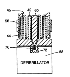
In Figures 3-7, a pair of the electrodes
sho~m in Figures 1 and 2 are mounted in a retainer for
use with a defibrillator system. Figures 3 and 4 show
the electrodes in a predeployment storage position.
In this position, the flexible substrate 42 of each
electrode is folded in an accordion fashion and placed
in retainer 60.
The portion of substrate 42 on which the
attachment pad 48 is located extends into a retainer
conn,ector area 70 for electrical attachment to a
corresponding connector 72 on the defibrillator 58.
Figure 5 shows the details of one embodiment of the
connectors. A metal crimp 74 at the end of substrate
42 makes electrical contact with attachment pad 48.
The crimp 74 partially extends through an opening 78
in the connector portion 70 of ret~;~er 60. When the
retainer connector portion is inserted into the
connector portion of the defibrillator 58, crimp 74
makes electrical contact with defibrillator contact
76. The resilient action of the crimps 74 also
W094/2~50 PCT~S94/05435 ~
3 ~ ~
provide the merh~nical attachment of retainer 60 to
defibrillator 58. Alternatively, other known
m~-hAn;cal attachment mechAnicms may be used to fasten
retA;n~r 60 to defibrillator 58. The contacts 76 for
each electrode are connected to the defibrillator
electronics in a known manner.
In the folded position, electrode disk 44,
the conductive gel covering the electrode disk, and
the adhesive surrounding the electrode disk lie
against an area 54 on the top surface of substrate 42.
The top surface of substrate 42 is coated with a
suitable release coating such as silicon in at least
release area 54. The release coating enables
electrode disk 44, its gel coating and the adhesive to
peel away from substrate 42 during deployment of the
electrode, as discussed below. The covering action of
the substrate over the conductive gel also helps keep
the conductive gel from drying out during storage. A
handle 56 attached to the back side of electrode body
45 lies in position in which it can be grasped by a
user during deployment of the electrodes.
Figures 6 and 7 demonstrate deployment and
placement of the electrodes on the patient. As shown
in Figure 4, the user pulls electrode body 45 up and
out of retainer 60 through openings 62 by grasping
handle 56. As it moves out of the retainer, the
electrode disk 44 and its conductive gel layer 51 peel
away from substrate surface 42. The pair of
electrodes in retainer 60 may be ext~n~ as far as
needed to reach the appropriate sites on the patient,
as shown in Figure 7. The c0~ ctors 46 and
attachment pads 48 on the substrates provide the
electrical connection between the electrodes and the
defibrillator for delivery of the defibrillating
voltage pulse and/or for monitoring of the electrical
activity of the patient's heart. After use, the
1 ~16334~
W094/2~S0 PCT~S94/05435
-7-
retainer and the pair of electrodes it houses can be
discarded and replaced with a new electrode set.
It may be necessary to store the electrodes
for an extended period prior to their deployment and
use. Figure 8 shows a possible protective covering
for preserving the integrity of the electrodes by, for
example, preventing the conductive gel from drying
out. The openings 62 of ret~i n~r 60 are covered with
a fLap 64. The flap may be formed from foil-backed
paper or plastic, such as Tyvek. Flap 64 is peeled
back from the retainer openings just prior to
deployment of the electrodes.
Figures 9-12 show an alternative embodiment
of 1:his invention. As shown in Figures 9 and 10, the
electrode apparatus 140 has a flexible body or
substrate 142, preferably formed from 1/16" closed
cell foam. A backing layer 182 is attached to the
underside of substrate 142 with a medical grade
adhesive. Backing layer 182 may be formed from Tyvek
or any other suitable material. The underside of
backing layer 182 is coated with a silicon release
material.
A pair of electrodes 144 are adhesively
attached to the top of substrate 142. Conductors 146
lead from electrodes 144 to attachment pads 148. Each
set of electrode, conductor and attachment pad is
preferably formed from a single piece of tin metal
foil 3 mils thick. The surface area of each electrode
is preferably 80 cm2. A layer of conductive gel 151
covers each electrode. The thickness of the
conductive gel layer is preferably 25 mils.
An insulating cover 147 is attached to the
top side of substrate 142 with medical grade adhesive.
Cover 147 has openings 180 for the electrodes and
openings 149 for the attachment pads. Openings 180
have diameters slightly smaller than the diameters of
their respective electrodes, and openings 149 have
W094/2~50 ~ 3 3 ~ -8- PCT~S9~/05435
diameters slightly smaller than the diameters of their
respective attachment pads. Medical grade adhesive
covers all of the top surface of cover 147 except for
handle area 156 and connector area 157 for attachment
of the electrode apparatus to a patient.
Figures 11 and 12 show the electrode
apparatus of this embodiment mounted in a retainer.
As seen in Figure 11, prior to deployment, the
electrode apparatus is wound around a spool-shaped
lo retainer 160 mounted on top of a defibrillator 158.
The portion of the electrode apparatus on which the
attachment pads 148 are located extend into the center
of the retainer spool where they make electrical
connection with conductors (not shown) that connect to
the defibrillator connector 172. A protective cover
164 may be kept over retainer spool 160 until the
electrodes are to be deployed.
In the undeployed position shown in Figure
11, the conductive gel layers 151 and the adhesive
coating on cover layer 147 face the inward toward the
center of the retainer spool and the release coating
on the underside of backing layer 182 faces outward
from the center. Thus, when the electrode apparatus
is wound about itself, the conductive gel layers 151
and the adhesive coating on the cover layer lie
against the silicon release coating of the backing
layer 182.
To deploy the electrode apparatus of this
embodiment, the protective cover 164 is removed, and
the electrode apparatus is unwound from ret~inPr spool
160 by pulling on handle or tab 156. The release
coating on backing layer 182 permits the conductive - ,
gel layers 151 and the adhesive on cover layer 147 to
peel away. The electrode apparatus is then applied to
the patient as shown in Figure 12.
The electrode apparatus and retainer spool
remain attached to the defibrillator during use. The
W094/26350 2 1 6 3 3 4 ~ PCT~S94/05435
conductors 146 and attachment pads 148 provide the
electrical connection between the electrodes 144 and
the defibrillator for delivery of the defibrillating
vol1:age pulse and/or for monitoring of the electrical
S activity of the patient's heart. After use, the
retainer spool and the electrode apparatus it houses
can be discarded and replaced with a new electrode
set
Other configurations are possible without
departing from the scope of the invention. For
example, other shapes of the retainer and the
protective covering may be used. In addition, a
batt:ery may be disposed in the electrode retainer to
provide the power to the defibrillator. On the other
hand, the retainer may be omitted altogether and the
electrodes attached directly to the defibrillator or
other instrument.
The electrically conductive traces on the
flexible substrate may be replaced in whole or in part
by wires or other conductors. Other materials and
dimensions may be employed. Finally, while this
invention has been described in the context of
defibrillators and defibrillator electrodes, it should
be understood that the invention applies to medical
electrodes used with other instruments, such as an ECG
monitor.
Other modifications will be apparent to
those skilled in the art.