Note: Descriptions are shown in the official language in which they were submitted.
~WO 94128418 . PCT/US94/06064
METHOD AND APPARATUS FOR
DESORPTION AND IONIZATION OF ANALYTES
» BACKGROUND OF THE INVENTION
This invention relates generally to methods and apparatus for
desorption and ionization of analytes for the purpose of subsequent scientific
analysis by such methods, for example, as mass spectrometry (MS) or
biosensors. Generally, analysis by mass spectrometry involves the
vaporization and ionization of a small sample of material, using a high energy
source, such as a laser, including a laser beam. The material is vaporized
from the surface of a probe tip into the gas or vapor phase by the laser beam,
and, in the process, some of the individual molecules are ionized by the gain
of a proton. The positively charged ionized molecules are then accelerated
through a short high voltage field and let fly (drift) into a high vacuum
chamber, at the far end of which they strike a sensitive detector surface.
Since the time-of flight is a function of the mass of the ionized molecule,
the
elapsed time between ionization and impact can be used to determine the
molecule's mass which, in turn, can be used to identify the presence or
absence of known molecules of specific mass.
» All known prior art procedures which present proteins or other large
biomolecules on a probe tip for laser desorption/ionization time-of flight
mass
p
spectrometry (TOF) rely on the preparation of a crystalline solid mixture of
-1-
PCT/LTS94106064~
WO 94128418
the protein or other analyte molecule in a large molar excess of acidic matrix
material deposited on the bare surface of a metallic probe tip. (The sample
probe tip typically is metallic, either stainless steel, nickel plated
material or
platinum). Embedding the analyte in such a matrix was thought to be
necessary in order to prevent the destruction of analyte molecules by the
laser
beam. The laser beam strikes the solid mixture on the probe tip and its
i
energy is used to vaporize a small portion of the matrix material along with
some of the embedded analyte molecules. Without the matrix, the analyte
molecules are easily fragmented by the laser energy, so that the mass, and
identity, of the original macromolecule is very difficult or impossible to
determine.
This prior art procedure has several limitations which have prevented
its adaptation to automated protein or other macrobiological molecular
analysis. First, in a very crude sample it is necessary to partially
fractionate
(or otherwise purify the sample as much as possible) to eliminate the presence
of excessive extraneous materials in the matrix/analyte crystalline or solid
mixture. The presence of large quantities of components may depress the ion
signal (either desorption, ionization and/or detection) of the targeted
analyte.
Such purification is time-consuming, expensive, typically results in low
recovery (or complete loss) of the analyte, and would be very difficult to do
in an automated analyzer.
Second, while the amount of analyte material needed for analysis by
the prior art method is not large (typically in a picomole range), in some
circumstances, such as tests on pediatric patients, analyte fluids are
available
only in extremely small volumes (microliters) and may be needed for
-2-
~WO 94/28418 , , . PCT/US94/06064
performing several different analyses. Therefore, even the small amount (i.e.,
volume) needed for preparation of the analyte/matrix crystalline mixture for
a single analysis may be significant. Also, only a tiny fraction (a few
thousandths or less) of analyte used in preparing the solid analyte/matrix
mixture for use on the probe tip is actually consumed in the desorption or
mass spectrometric analysis. Any improvement in the prior art procedure
which would make it possible to 1) use much less analyte, 2) to locate the
analyte or multiple analytes on the probe tip or surface in a predetermined
location, 3) to perform repeated analyses of the same aliquot of analyte
(e.g.,
before and after one or more chemical and or enzymatic reactions), and 4) to
conduct the test in a more quantitative manner, would be highly
advantageous in many clinical areas.
Third, the analyte protein, or other macromolecule, used in preparing
the solid solution of analyte/matrix for use on the probe tip is not suitable
for
any subsequent chemical tests or procedures because it is bound up (i.e.,
embedded) in the matrix material. Also, all of the matrix material used to
date is strongly acidic, so that it would adversely affect many chemical
reactions which might be attempted on the mixture in order to modify the
analyte molecules for subsequent examination. Any improvement in the
procedure which made it possible to conduct subsequent chemical
modifications or reactions on the analyte molecules, without removing them
from the matrix or the probe tip or without "matrix" altogether, would be of
enormous benefit to researchers and clinicians.
The first successful molecular mass measurements of intact peptides
and small proteins (only up to about 15 kDa) by any form of mass
-3-
I 'a P
WO 94/28418 ~ ~ ~ PCT/US94/06064
spectrometry were made by bombarding surfaces with high energy particles
(plasma desorption and fast atom bombardment mass spectrometry); this
breakthrough came in 1981 and 1982. Improvements came in 1985 and 1986,
however, yield (signal intensities), sensitivity, precision, and mass accuracy
remained relatively low. Higher molecular mass proteins (about 20 to 25
kDa) were not observed except on rare occasions; proteins representing
average molecular weights (approximately 70 kDa) were not ever observed
with these methods. Thus, evaluation of most proteins by mass spectrometry
remains unrealized.
In 1988, Hillenkamp and his coworkers used LTV laser desorption time-
of flight mass spectrometry and discovered that when proteins of relatively
high molecular mass were deposited on the probe tip in the presence of a very
large molar excess of an acidic, ITV absorbing chemical matrix (nicotinic
acid)
they could be desorbed in the intact state. This new technique is called
matrix-assisted laser desorption/ionization (MALDI) time-of flight mass
spectrometry. Note that laser desorption time-of flight mass spectrometry
(without the chemical matrix) had been around for some time, however, there
was little or no success determining the molecular weights of large intact
biopolymers such as proteins and nucleic acids because they were fragmented
(destroyed) upon desorption. Thus, prior to the introduction of a chemical
matrix, laser desorption mass spectrometry was essentially useless for the
detection of specific changes in the mass of intact macromolecules. Note that
the random formation of matrix crystals and the random inclusion of analyte
molecules in the solid solution is prior art.
_rI_
CA 02163426 2004-03-25
There are a number of problems and limitations with the prior art methods. For
example, previously, it has been found that it is difficult to wash away
contaminants
present in analyte or matrix. Other problems include formation of analyte-salt
ion
adducts, less than optimum solubility of analyte in matrix, unknown location
and
concentration of analyte molecules within the solid matrix, signal (molecular
ion)
suppression "poisoning" due to simultaneous presence of multiple components,
and
selective analyte desorption/ionization. Prior investigators, including Karas
and
Hillenkamp have reported a variety of techniques for analyte detection using
mass
spectroscopy, but these techniques suffered because of inherent limitations in
sensitivity
and selectivity of the techniques, specifically including limitations in
detection of
analytes in low volume, undifferentiated samples. (Hillenkamp, Bordeaux Mass
Spectrometry Conference Report, pp. 354-62 (1988); Karas and Hillenkamp,
Bordeaux
Mass Spectrometry Conference Report, pp. 416-17 (1988); Karas and Hillenkamp,
Analytical Chemistry, 60:2299 (1988); Karas, et al., Biomed. Environ. Mass
Spectrum (in
press).) The use of laser beams in time-of flight mass spectrometers is shown,
for
example, in U.S. Pat. Nos. 4,694,167; 4,686,366, 4,295,046, and 5,045,694.
The successful volatilization of high molecular weight biopolymers, without
fragmentation, has enabled a wide variety of biological macromolecules to be
analyzed
by mass spectrometry. More importantly perhaps, it has illustrated the
potential of using
mass spectrometry more creatively to solve problems routinely encountered in
biological
research. Most recent attention has been focused on the utility of matrix-
assisted laser
-5-
WO 94/28418 ~ ~ PCT/US94106064
desorption/ionization (MALDI) time-of flight (TOF) mass spectrometry (MS),
largely because it is rapid (min), sensitive (< pmol sample required), and
permits complex mixtures to be analyzed.
Although MALDI-TOF MS continues to be useful for the static
determinationJverification of mass for individual analytes, in the case of '
biopolymers, it is often differences in mass that provide the most important
information about unknown structures. Thus, for routine use in structural
biology, an unfortunate limitation of the MALDI-TOF MS technique relates
to sample preparation and presentation (deposition) on an inert probe
element surface, specifically, the requirement that analytes be embedded
(i.e.,
co-solidified) on the probe surface in a freshly prepared matrix of
crystalline
organic acid. The random distribution of analyte in a heterogeneous display
of crystal matrix on the probe element surface requires the deposition of far
more analyte or sample than is needed for the laser desorption process, even
for the collection of more than adequate mass spectra (e.g., multiple sets of
100 shots each). The remaining portion of the analyte is usually not
recovered for additional analyses or subsequent characterizations. Even
though 1 to 10 pmol (sometimes less) of analyte are typically required for
deposition on the probe surface, it has been estimated that less than a few
attomoles are consumed during laser desorption. Thus, only 1 part in 10~ or
lOg of the applied analyte may be necessary; the rest is lost.
Another important loss of potential data associated with the embedding
of analyte in a solid matrix is the reduction or the complete elimination of
ability to perform subsequent chemical and/or enzymatic modifications to the -
embedded analyte (e.g., protein or DNA) remaining on the probe surface.
-6-
WO 94/28418 ~ ~ PCT/US94/06064
Only another aliquot of analyte, or the ability to recover the embedded
analyte free of matrix (difficult with low recovery), allows what we now refer
to as differential mass spectrometry to be performed to derive structural
data.
In addition, there has been limited application of MS in biological
° 5 fields, likely due to the fact that many biologists and clinicians
are
intimidated by MS and/or skeptical in regard to its usefulness. Further, MS
is perceived as inaccessible or too costly, particularly because SDS
polyacrylamide gel electrophoresis is an adequate substitute in some instances
where MALDI would be applied (e.g., separation of crude biological fluids).
In addition, MALDI has had little exposure in biological and clinical
journals.
SUMMARY OF THE INVENTION
An object of the invention is to provide improved methods, materials
composition and apparatus for coupled adsorption, desorption and ionization
of multiple or selected analytes into the gas (vapor) phase.
Another object is to provide a method and apparatus for affinity-
directed detection of analytes, including desorption and ionization of
analytes
in which the analyte is not dispersed in a matrix solution or crystalline
structure but is presented within, on or above an attached surface of energy
absorbing "matrix" material through molecular recognition events, in a
position where it is accessible and amenable to a wide variety of chemical,
physical and biological modification or recognition reactions.
Another object is to provide such a method and apparatus in which the
analyte material is chemically bound or physically adhered to a substrate .
forming a probe tip sample presenting surface.
_7_
PCT/US94/06064
WO 94/28418
. . f: i
A further object is to provide means for the modification of sample
presenting surfaces with energy-absorbing molecules to enable the successful
desorption of analyte molecules without the addition of exogenous matrix
molecules as in prior art.
A further object is to provide the appropriate density of energy-
absorbing molecules bonded (covalently or noncovalently) in a variety of
geometries such that mono layers and multiple layers of attached energy-
absorbing molecules are used to facilitate the desorption of analyte molecules
of varying masses.
A further object is to provide multiple combinations of surfaces
modified with energy-absorbing molecules, affinity-directed analyte capture
devices, phototubes, etc.
An additional object is to provide such a method and apparatus in
which the substrate forming the probe tip or other sample presenting surface
is derivatized with one or more affinity reagents (a variety of densities and
degrees of amplification) for selective bonding with predetermined analytes
or classes of analytes.
A further object is to provide such a system in which the affinity
reagent chemically bonds or biologically adheres to the target analyte or
class
of analytes.
A still further object is to provide a method and apparatus for
desorption and ionization of analytes in which unused portion of the analytes
contained on the presenting surface remain chemically accessible, so that a
series of chemical, enzymatic or physical treatments of the analyte may be
conducted, followed by sequential analyses of the modified analyte.
_g_
~WO 94/28418 PCTIUS94/06064
A further object is to provide a method and apparatus for the combined
chemical or enzymatic modifications of target analytes for the purpose of
elucidating primary, secondary, tertiary, or quaternary structure of the
analyte and its components.
Another object is to provide a method and apparatus for desorption and
ionization of analyte materials in which cations other than protons (H+) are
utilized for ionization of analyte macromolecules.
Thus, in accomplishing the foregoing objects, there is provided in
accordance with the present invention, an apparatus for measuring the mass
of an analyte molecule of an analyte sample by means of mass spectrometry,
said apparatus comprising a spectrometer tube; a vacuum means for applying
a vacuum to the interior of said tube; electrical potential means within the
tube for applying an accelerating electrical potential to desorbed analyte
molecules from said analyte sample; sample presenting means removably
insertable into said spectrometer tube, for presenting said analyte sample in
association with surface associated molecule for promoting desorption and
ionization of said analyte molecules, wherein said surface molecule is
selected
from the group consisting of energy absorbing molecule, affinity capture
device, photolabile attachment molecule and combination thereof; an analyte
sample deposited on said sample presenting means in association with said
surface associated molecules, whereby at least a portion of said analyte
molecules not consumed in said mass spectrometry analysis will remain
accessible for subsequent chemical, biological or physical analytical
' procedures; laser beam means for producing a laser beam directed to said
analyte sample for imparting sufficient energy to desorb and ionize a portion
_g_
PCTIUS94/0606
WO 94!28418
of said analyte molecules from said analyte sample; and detector means
associated with said spectrometer tube for detecting the impact of accelerated
ionized analyte molecules thereon.
In addition, in accomplishing the foregoing objects, there is provided
in accordance with the present invention, a method in mass spectrometry to
measure the mass of an analyte molecule, said method comprising the steps
of: derivitizing a sample presenting surface on a probe tip face with an
affinity capture device having means for binding with an analyte molecule;
exposing said derivitized probe tip face to a source of said analyte molecule
so as to bind said analyte molecule thereto; placing the derivitized probe tip
with said analyte molecules bound thereto into one end of a time-of flight
mass spectrometer and applying a vacuum and an electric field to form an
accelerating potential within the spectrometer; striking at least a portion of
the analyte molecules bound to said derivitized probe tip face within the
spectrometer with one or more laser pulses in order to desorb ions of said
analyte molecules from.said tip; detecting the mass of the ions by their time
of flight within said mass spectrometer; and displaying such detected mass.
Further, in accomplishing the foregoing objects, there is provided in
accordance with the present invention, a method of measuring the mass of
analyte molecules by means of laser desorption/ionization, time-of flight mass
spectrometry in which an energy absorbing material is used in conjunction
with said analyte molecules for facilitating desorption and ionization of the
analyte molecules, wherein the improvement comprises presenting the analyte
molecules on or above the surface of the energy absorbing material, wherein '
at least a portion of the analyte molecules not desorbed in said mass
-10-
WO 94/28418 PCT/US94/06064
spectrometry analysis remain chemically accessible for subsequent analytical
procedures.
Additionally, in accomplishing the foregoing objects, there is provided
in accordance with the present invention, an apparatus for facilitating
desorption and ionization of analyte molecules, said apparatus comprising: a
sample presenting surface; and surface associated molecules, wherein said
surface associated molecules are selected from the group consisting of energy
absorbing molecule, affinity capture device, photolabile attachment molecule
and combination thereof, said surface associated molecules associated with
said sample presenting surface and having means for binding with said
analyte molecules.
Further, there is provided a method for capturing analyte molecules on
a sample presenting surface and desorbing/ionizing said captured analyte
molecules from said sample presenting surface for subsequent analysis, said
method comprising: derivitizing said sample presenting surface with an
affinity capture device or photolabile attachment molecule having means for
binding with said analyte molecules; exposing said derivitized sample present
surface to a sample containing said analyte molecules; capturing said analyte
molecules on said derivitized sample presenting surface by means of said
affinity capture device or photolabile attachment molecule; and exposing said
analyte molecules, while bound to said derivitized sample presenting surface
by means of said affinity capture device or photolabile attachment molecule,
to an energy or light source to desorb at least a portion of said analyte
- molecules from said surface.
-11-
WO 94/28418 PCTIUS94/06064
Additionally, in accordance with the present invention, there is
provided a method for preparing a surface for presenting analyte molecules
for analysis, said method comprising: providing a substrate on said surface
for
supporting said analyte; derivitizing said substrate with an affinity capture
device or photolabile attachment molecule having means for selectively '
bonding with said analyte; and a means for detecting said analyte molecules
bonded with said affinity capture device or photolabile attachment molecule.
Further, in accomplishing the foregoing objects, there is provided in
accordance with the present invention, a sample probe for promoting
desorption of intact analytes into the gas phase comprising: a sample
presenting surface; and an energy absorbing molecule associated with said
sample presenting surface, wherein said sample probe promotes desorption
of an intact analyte molecule positioned on, above or between the energy
absorbing molecules when said sample probe is impinged by an energy source.
Further, the energy absorbing molecule in the probe is selected from the
group consisting of cinnamamide, cinnamyl bromide, 2, 5-dihydroxybenzoic
acid and a-cyano-4-hydroxycinnamic acid.
Additionally, in accomplishing the foregoing objects, there is provided
in accordance with the present invention, a sample probe for desorption of
intact analyte into the gas phase, comprising: a sample presentation
surface; and a surface associated molecule wherein said surface associated
molecule is a photolabile attachment molecule having at least two binding
sites, wherein at least one site is bound to the sample presentation surface
and at least one site is available to bind an analyte and wherein the analyte
binding site is photolabile.
-12-
WO 94/28418 , . ~ ~ PCTIUS94106064
In addition, in accomplishing the foregoing objects there is provided in
accordance with the present invention, a sample probe for promoting
desorption of intact analytes into the gas phase comprising: a sample
presentation surface; and either
a mixture of at least two different molecules selected from the group
. consisting of an affinity capture device, an energy absorbing molecule and a
photolabile attachment molecule associated with said sample presentation
surface; wherein when an analyte is associated with said sample probe, said
sample probe promotes the transition of the analyte into the gas phase when
said sample probe is impinged by an energy source; or at least two different
affinity capture devices associated with said sample presentation surface;
wherein, when said sample probe is impinged by an energy source, said
sample probe promotes the transition of an analyte molecule into the gas
phase at different rates depending on the affinity capture device associated
with said analyte molecule.
In addition, in accomplishing the foregoing objects there is provided in
accordance with the present invention, a sample probe for promoting
desorption of intact analyte into the gas phase, comprising: a sample
presentation surface; and either a surface associated molecule, wherein said
surface associated molecule can function both as an energy absorbing
molecule and as an aff'mity capture device; or a surface associated molecule
wherein said surface associated molecule is a photolabile attachment molecule
having at least two binding sites, wherein at least one site is bound to the
' sample presentation surface and at least one site is available to bind an
analyte and wherein the analyte binding site is photolabile.
-13-
< A a
WO 94128418 PCT/US94106064
21~34r~
Additionally, there is provided in the present invention, a method in
mass spectrometry to measure the mass of an analyte molecule, said method
comprising the steps of: derivitizing a sample presenting surface on a probe
tip face with a photolabile attachment molecule (PAM), wherein said PAM
has at least two binding sites, one binding site binds to the sample
presenting
surface and at least one binding site is available for binding with an analyte
molecule; exposing said derivitized probe tip face to a source of said analyte
molecule so as to bind said analyte molecule thereto; placing the derivitized
probe tip with said analyte molecules bound thereto into one end of a time-of
flight mass spectrometer and applying a vacuum and an electric field to form
an accelerating potential within the spectrometer; striking at least a portion
of the analyte molecules bound to said derivitized probe tip face within the
spectrometer with one or more laser pulses in order to desorb ions of said
analyte molecules from-said tip; detecting the mass of the ions by their time
of flight within said mass spectrometer; and displaying such detected mass.
In addition, there is provided a method of measuring the mass of
analyte molecules by means of laser desorption/ionization, time-of flight mass
spectrometry in which a photolabile attachment molecule (PAM) is used in
conjunction with said analyte molecules for facilitating desorption and
ionization of the analyte molecules, the improvement comprising: presenting
the analyte molecules on or above the surface of the PAM, wherein at least
a portion of the analyte molecules not desorbed in said mass spectrometry
analysis remain chemically accessible for subsequent analytical procedures.
There is further provided in accordance with the present invention, a
sample probe for promoting of differential desorption of intact analyte into
-14-
WO 94128418 ~ ~ PCT/US94/06064
the gas phase, comprising: a sample presentation surface; and at least
two different photolabile attachment molecules associated with said sample
presentation surface; wherein, when said sample probe is impinged by an
energy source, said sample probe promotes the transition of an analyte
molecule into the gas phase at different rates depending on the photolabile
attachment molecule associated with said analyte molecule.
Additionally, there is provided in accordance with the present
invention, a sample probe for promoting desorption of intact analytes into the
gas phase comprising: a sample presenting surface; and a photolabile
attachment molecule associated with said sample presenting surface; wherein,
when said sample probe is impinged by an energy source, said sample probe
promotes the transition of an intact analyte molecule into the gas phase.
Further, there is provided in accordance with the present invention, a
method for biopolymer sequence determination comprising the steps of:
binding a biopolymer analyte to probe tip containing a sample presenting
surface having a surface selected molecule selected from the group consisting
of an energy absorbing molecule, an affinity capture device, a photolabile
attachment molecule and a combination thereof; desorption of biopolymer
analyte in mass spectrometry analysis, wherein at least a portion of said
biopolymer is not desorbed from the probe tip; analyzing the results of the
desorption modifying the biopolymer analyte still bound to the probe tip; and
repeating the desorption, analyzing and modifying steps until the biopolymer
is sequenced.
~ a
-15-
CA 02163426 2005-O1-19
Further, there is provided in accordance with the present invention a method
for
detecting an analyte comprising the steps of: (a) capturing the analyte from a
sample on a
sample presenting surface of a mass spectrometer probe, wherein the surface is
derivatized
with an affinity reagent that binds the analyte; (b) washing the sample
presenting surface
to remove unbound analyte; (c) applying a matrix to the analyte bound to the
affinity
reagent on the sample presenting surface; and (d) detecting the captured
analyte by laser
desorption/ionization mass spectrometry.
Additionally, there is provided in accordance with the present invention a
mass
spectrometer probe comprising: (a) a sample presenting surface derivatized
with an
affinity reagent that is capable of binding an analyte; (b) the analyte
captured by the
affinity reagent, wherein the analyte remains captured after washing; and (c)
a matrix
applied to the analyte captured by the affinity reagent.
In addition, there is provided in accordance with the present invention a mass
spectrometer comprising: (a) a mass spectrometer probe comprising: i. a sample
presenting surface derivatized with an affinity reagent that is capable of
binding an
analyte; ii. the analyte captured by the affinity reagent, wherein the analyte
remains
captured after washing; and iii. a matrix applied to the analyte captured by
the affinity
reagent; (b) a laser source that directs laser energy to the sample presenting
surface for
desorbing and ionizing the analyte; (c) means for applying an accelerating
electrical
potential to the desorbed, ionized analyte; and (d) a detector that detects
the desorbed,
ionized analyte.
-1Sa-
WO 94!28418 ~ PCTILT_594106064
Other and further objects, features and advantages will be apparent
and the invention more readily understood from a reading of the following
specification and by reference to the accompanying drawings forming a part
thereof, wherein the examples of the presently preferred embodiments of the
invention are given for the purposes of disclosure.
BRIEF DESCRIPTION OF THE DRAWINGS
The foregoing and other objects and advantages of the invention will
be apparent from the following specification and from the accompanying
drawings.
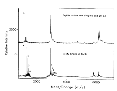
Figure lA (upper profile) shows the mass spectrum of the three
peptides (human histidine rich glycoprotein metal-binding domains
(GHHPH)aG (1206 Da), (GHHPH)5G (2904 Da), and human estrogen receptor
dimerization domain (D473-L525) (6168.4 Da)) desorbed in the presence of
neutralized energy absorbing molecules (sinapinic acid, pH 6.2). Figure 1B
(lower profile) shows the sequential in situ metal (Cu)-binding of the
peptides
in the presence of neutral energy absorbing molecules.
Figure 2A (top profile) shows the mass spectrum of the human casein
phosphopeptide (5P, 2488 Da) desorbed in the presence of neutralized energy
absorbing molecules (sinapinic acid, pH 6.5). Figure 2B (second from top
profile) shows the sequential in situ 5 min alkaline phosphatase digestion to
remove phosphate groups from the phosphopeptide. Figure 2C (third from
top profile) shows the mass spectrum of the phosphopeptide after further in
-16-
WO 94/28418 PCTIUS94106064
phosphatase digestion in the presence of acidic energy absorbing molecules
(2,5 dihydroxybenzoic acid, pH 2) as described in prior art.
Figure 3 shows a composite mass spectra of the (GHHPH)5G peptide
- (2904 Da) before (lower profile) and after (upper profile) in situ digestion
by
carboxypeptidase P in the presence of neutralized energy absorbing molecules
(sinapinic acid, pH 6.2).
Figure 4 shows a composite matrix-assisted laser desorption mass
spectra of peptide mixtures desorbed from solid glass, polypropylene-coated
stainless steel, polystyrene-coated stainless steel and solid nylon probe
elements.
SEAC
Figure 5, profile A shows the mass spectrum of sperm activating factor
(933 Da) and neurotensin (1655 Da) (and their multiple Na-adducts) in the
peptide solution unadsorbed by the IDA-Cu(II) surface. Figure 5, profile B,
shows the mass spectrum of angiotensin I (1296.5 Da) plus Na-adduct peaks
that were selectively adsorbed on the IDA-Cu(II) surface. Figure 5, profile C,
and Figure 6, profile C, show the mass spectrum of the same angiotensin I
adsorbed on IDA-Cu(II) after water wash. Figure 6, profile D, shows the
sequential an situ copper-binding (1 and 2 Cu) by affinity adsorbed
angiotensin I. Figure 6, profile E, shows the sequential in situ trypsin
- digestion of the affinity adsorbed angiotensin I.
-17-
~ru~ sw~~ ~RU~E ~s~
~.~~4~ 6 . .
WO 94/28418 . PCT/US94/06064~
Figure 7 shows the mass spectrum of myoglobin (4 to 8 fmole) affinity
adsorbed on IDA-Cu(II) surface.
Figure 8 (top profile) shows the mass spectrum of synthetic casein
peptide (1934 Da) with multiple phosphorylated forms affinity adsorbed from -
a crude mixture on TED-Fe(III) surface. After sequential in situ alkaline
phosphatase digestion, only the original nonphosphorylated form remained
(lower profile).
Figure 9, profile A, shows the mass spectrum of total proteins in infant
formula. Figure 9, profile B, shows the mass spectrum of phosphopeptides in
infant formula affinity adsorbed on TED-Fe(III) surface. Figure 9, profile C,
shows the mass spectrum of total proteins in gastric aspirate of preterm
infant obtained after feeding the infant formula. Figure 9, profile D, shows
the mass spectrum of phosphopeptides in the gastric aspirate affinity
adsorbed on TED-Fe(III) surface.
Figure 10 A shows the composite mass spectra of human and bovine
histidine-rich glycoprotein adsorbed on IDA-Cu(II) surface before and after
N-glycanase digestion. The mass shifts represent the removal of carbohydrate
from the respective glycoproteins. Figure 10 B shows the composite mass
spectra of trypsin digested peptides from the deglycosylated proteins of the
two species (top profile for human protein, second from bottom profile for
bovine protein) and in situ Cu(II)-binding of the trypsin digested peptides of
the two species (second from top profile for human protein, bottom profile for
-18-
SUBSTITUTE SHEET (RULE 26)
~WO 94/28418 . PCT/US94/06064
bovine protein; the numbers 1, 2 indicate the number of copper bound).
Figure 10C shows that one such Cu(II)-binding peptide (bottom profile) has
at least 4 His residues which are specifically modified by
diethylpyrocarbonate
to form 4 N-carbethoxy-histidyl adducts (1-4, top profile). Figure lOD shows
the partial C-terminal sequence of the major Cu-binding peptide in the bovine
histidine rich glycoprotein.
Figure 11 (bottom profile) shows the mass spectrum of rabbit anti-
human lactoferrin immunoglobulin alone (control) affinity adsorbed on sheep
anti-rabbit IgG paramagnetic surface. The top profile shows the mass
spectrum of human lactoferrin and rabbit anti-human lactoferrin
immunoglobulin complex affinity adsorbed on sheep anti-rabbit IgG
paramagnetic surface.
Figure 12 shows the mass spectrum of human lactoferrin affinity
adsorbed from preterm infant urine on a anti-human lactoferrin
immunoglobulin nylon surface. Figure 13 shows the equivalent mass
spectrum of whole preterm infant urine containing 1 nmole/ml of lactoferrin.
Figure 14 (lower profile) shows the mass spectrum of pure bovine
histidine rich glycoprotein. The upper profile shows the mass spectrum of
bovine histidine rich glycoprotein and fragments affinity adsorbed from
bovine colostrum on anti-bovine histidine rich glycoprotein immunoglobulin
- surface.
-19-
SUBSTITUTE SHEET (RULE 261
WO 94/28418 ~ PCT/US94/06064
Figure 15 shows the composite mass spectra of the peptides of follicle
stimulating hormone recognized by the different anti-follicle stimulating
hormone antibodies.
Figure 16 shows the mass spectrum of human lactoferrin affinity -
adsorbed on a single bead of single-stranded DNA agarose deposited on a 0.5
mm diameter probe element.
Figure 17 shows the mass spectrum of human lactoferrin affinity
adsorbed from preterm infant urine on single-stranded DNA surface
Figure 18A shows the composite mass spectra of the total proteins in
human duodenal aspirate (lower profile) and the trypsin affinity adsorbed
from the aspirate on a soybean trypsin inhibitor surface (upper profile).
Figure 18B shows the mass spectrum of trypsin affinity adsorbed from 1 ul
of aspirate on a soybean trypsin inhibitor nylon surface.
Figure 19A shows the mass spectrum of biotinylated insulin affinity
adsorbed from human urine on a Streptavidin surface. Figure 19B shows the
mass spectrum of biotinylated insulin affinity adsorbed from human plasma
on a Streptavidin surface.
Figure 20 (upper profile) shows the mass spectrum of total proteins in
human serum. Figure 20 (lower profile) shows the mass spectrum of -
-20-
S'UHSTfTUTE SHEET (RULE 26)
WO 94/28418 ~ PCT/US94/06064
serum albumin affinity adsorbed from human serum on a Cibacron-blue
surface.
SEND
- Figure 21 shows the molecular structure of surface bound
cinnamamide; R represents the surface plus cross-linker.
Figure 22 (upper profile) shows the mass spectrum of peptide mixtures
desorbed from surface bound cinnamamide. Figure 20B (lower profile) shows
the mass spectrum of the same peptide mixtures with free cinnamamide.
Figure 23 shows the molecular structure of surface bound cinnamyl
bromide; R represents the surface plus cross-linker.
Figure 24 (upper profile) shows the mass spectrum of peptide mixtures
desorbed from surface bound cinnamyl bromide. Figure 22B (lower profile)
shows the mass spectrum of the same peptide mixtures with free cinnamyl
bromide.
Figure 25 shows the molecular structure of surface bound MAP-
dihydroxybenzoic acid; R represents the surface plus cross-linker.
Figure 26 (upper profile) shows the mass spectrum of peptide mixtures
desorbed from surface bound MAP alone. Figure 26 (lower profile) shows the
-21-
SUBSTITUTE SHEET (RULE 26)
WO 94/28418 PCTIUS94/06064
mass spectrum of the same peptide mixtures desorbed from surface bound
MAP-dihydroxybenzoic acid.
Figure 27A shows the mass spectrum (1,200-50,000 m/z region) of
myoglobin desorbed from surface bound a-cyano-4-hydroxycinnamic acid.
Figure 25B shows the same mass spectrum in the low mass region (0-1200
m/z).
Figure 28 shows the molecular structure of energy absorbing molecules
bound to polyacrylamide or nylon or acrylic surface via glutaraldehyde
activation.
Figure 29 shows the molecular structure of energy absorbing molecules
bound to polyacrylamide or nylon or acrylic surface via divinyl sulfone
activation.
Figure 30 shows the molecular structure of energy absorbing molecules
bound to polyacrylamide or nylon or acrylic surface via
dicyclohexylcarbodiimide activation.
Figure 31 shows the molecular structure of energy absorbing molecules
bound to polyacrylamide or nylon or acrylic surface with multiple antigenic
peptide via dicyclohexylcarbodiimide activation.
-22-
SUBSTITUTE SHEET iRULE 261
WO 94/28418 , PCTIUS94/06064
Figure 32 shows the molecular structure of thiosalicylic acid bound to
iminodiacetate (IDA)-Cu(II) surface.
Figure 33 shows the mass spectrum of human estrogen receptor
- dimerization domain desorbed from thiosalicylic acid-IDA-Cu(II) surface.
Figure 34 shows the molecular structure of a-cyano-4-hydroxycinnamic
acid bound to DEAF surface.
Figure 35 shows the mass spectrum of human estrogen receptor
dimerization domain desorbed from sinapinic acid-DEAE surface. Figure 33B
shows the mass spectrum of myoglobin desorbed from a-cyano-4-
hydroxycinnamic acid DEAF surface.
Figure 36 shows the molecular structure of a-cyano-4-hydroxycinnamic
acid bound to polystyrene surface.
S~~AR
Figure 37 shows the C-terminal sequence analysis of surface
immobilized via photolytic bond histidine rich glycoprotein metal binding
domain.
-23-
StlHSTtTUTE SHEET (RULE 26)
WO 94/28418 . ', .' . ~ . ~ PCT/US94/06064~
~~.~3~~~
DETAILED DESCRIPTION OF THE INVENTION
It will be apparent to one skilled in the art that various substitutions
and modifications may be made to the invention disclosed herein without
departing from the scope and the spirit of the invention.
The development of new MS probe element compositions with surfaces
that allow the probe element to actively participate in the capture and
docking of specific analytes has recently defined several new opportunities in
the area now being described as Affinity Mass Spectrometry (AMS). In brief,
several types of new MS probe elements have been designed (Hutchens and
Yip, Rdpid Commun Mass Spectrom, 7: 576-580 (1993)) with Surfaces
Enhanced for Affinity Capture (SEAL). To date, SEAC probe elements have
been used successfully to retrieve and tether different classes of
biopolymers,
particularly proteins, by exploiting what is known about protein surface
structures and biospecific molecular recognition.
Progress in structural biology continues to be limited by the inability
to obtain biopolymer sequence information at an acceptable rate or level of
sensitivity. By utilizing the methods and apparatus of the present invention,
it has been demonstrated that AMS provides an opportunity to relieve this
limitation. Because the immobilized affinity capture devices on the MS probe
element surface (i.e., SEAC) determines the location and affinity
(specificity)
of the analyte for the probe surface, the subsequent analytical AMS process
is much more efficient for several reasons. First, the location of analyte on
the probe element surface is predetermined. Thus, the subsequent desorption
is no longer dependent on a random search of the probe surface matrix field _
with the incident laser beam. Second, analyte detection sensitivity (and
-24-
~WO 94/28418 . ~ ~ PCT/US94/06064
dynamic range) is increased because molecular ionization suppression effects
often observed with complex mixtures are eliminated. Third, the tethered
analyte that is not actually consumed by the initial laser-induced desorption
process remains available for subsequent analyses. If exogenous matrix was
used to promote analyte desorption, it is removed, in most cases, without loss
of the tethered analyte. The remaining analyte can then be chemically and/or
enzymatically modified directly in situ (i.e., while still on the probe
element).
When analyzed again by MS to determine differences in mass, specific
structural details are revealed. The entire process of analysis/modification
can be repeated many times to derive structural information while consuming
only very small quantities of analyte (sometimes only a few femtomoles or
less). The demonstrations of protein structure analysis based on AMS have
to date included both N- and C-terminal sequence analyses and verification
of several types of sequence-specific posttranslational modifications
including
phosphorylation and dephosphorylation, glycosylation, cysteine residue
reactivity, site-specific chemical modifications (e.g., Histidine residues),
and
ligand binding.
Beyond biopolymer sequence determinations and the solution of
individual biopolymer structures, is the ability to understand the structural
determinants of functional supramolecular assemblies. The opportunity to
investigate the structural determinants of higher order (e.g., quaternary)
structures is also presented by AMS. It has been demonstrated by using the
present invention that noncovalent molecular recognition events, some not
readily observed by more traditional bioanalytical procedures (often requiring
disruption of equilibrium and structure dissociating conditions), are
-25-
WO 94/28418 ~, PCT/US94/06064
,.
investigated directly by the evaluation of molecular associations (i.e.,
recognition) with macromolecular analytes that have been tethered, directly
or indirectly, to the probe element surface.
As used herein, "analyte" refers to any atom and/or molecule; including
their complexes and fragment ions. In the case of biological macromolecules,
including but not limited to: protein, peptides, DNA, RNA, carbohydrates,
steroids, and lipids. Note that most important biomolecules under
investigation for their involvement in the structure or regulation of life
processes are quite large (typically several thousand times larger than H2~).
As used herein, the term "molecular ions" refers to molecules in the
charged or ionized state, typically by the addition or loss of one or more
protons (H+).
As used herein, the term "molecular fragmentation" or "fragment ions"
refers to breakdown products of analyte molecules caused, for example, during
laser-induced desorption (especially in the absence of added matrix).
As used herein, the term "solid phase" refers to the condition of being
in the solid state, for example, on the probe element surface.
As used herein, "gas" or "vapor phase" refers to molecules in the
gaseous state (i.e., in vacuo for mass spectrometry).
As used herein, the term "analyte desorption/ionization" refers to the
transition of analytes from the solid phase to the gas phase as ions. Note
that the successful desorption/ionization of large, intact molecular ions by
laser desorption is relatively recent (circa 1988)--the big breakthrough was
the chance discovery of an appropriate matrix (nicotinic acid).
-26-
~WO 94/28418 _ ~ PCT/US94/06064
As used herein, the term "gas phase molecular ions" refers to those ions
that enter into the gas phase. Note that large molecular mass ions such as
proteins (typical mass = 60,000 to 70,000 times the mass of a single proton)
are typically not volatile (i.e., they do not normally enter into the gas or
vapor
- 5 phase). However, in the procedure of the present invention, large
molecular
mass ions such as proteins do enter the gas or vapor phase.
As used herein in the case of MALDI, the term "matrix" refers to any
one of several small, acidic, light absorbing chemicals (e.g., nicotinic or
sinapinic acid) that is mixed in solution with the analyte in such a manner so
that, upon drying on the probe element, the crystalline matrix-embedded
analyte molecules are successfully desorbed (by laser irradiation) and ionized
from the solid phase (crystals) into the gaseous or vapor phase and
accelerated as intact molecular ions. For the MALDI process to be successful,
analyte is mixed with a freshly prepared solution of the chemical matrix
(e.g.,
10,000:1 matrix:analyte) and placed on the inert probe element surface to air
dry just before the mass spectrometric analysis. The large fold molar excess
of matrix, present at concentrations near saturation, facilitates crystal
formation and entrapment of analyte.
As used herein, "energy absorbing molecules (EAM)" refers to any one
of several small, light absorbing chemicals that, when presented on the
surface of a probe element (as in the case of SEND), facilitate the neat
desorption of molecules from the solid phase (i.e., surface) into the gaseous
or vapor phase for subsequent acceleration as intact molecular ions. The
- term EAM is preferred, especially in reference to SEND. Note that analyte
desorption by the SEND process is defined as a surface-dependent process
-27-
WO 94/28418 PCT/US94106064
.
(i.e., neat analyte is placed on a surface composed of bound FA_.M_). In
contrast, MALDI is presently thought to facilitate analyte desorption by a
volcanic eruption-type process that "throws" the entire surface into the gas
phase. Furthermore, note that some EAM when used as free chemicals to
embed analyte molecules as described for the MALDI process will not work '
(i.e., they do not promote molecular desorption, thus they are not suitable
matrix molecules).
As used herein, "probe element" or "sample presenting device" refers to
an element having the following properties: it is inert (for example,
typically
stainless steel) and active (probe elements with surfizces enhanced to contain
EAM and/or molecular capture devices).
As used herein, "MALDI" refers to Matrix-Assisted Laser
Desorption/Ionization
As used herein, "TOF" stands for Time-of Flight.
As used herein, "MS" refers to Mass Spectrometry.
As used herein "MALDI-TOF MS" refers to Matrix-assisted laser
desorption/ionization time-of flight mass spectrometry.
As used herein, "ESI" is an abbreviation for Electrospray ionization.
As used herein, "chemical bonds" is used simply as an attempt to
distinguish a rational, deliberate, and knowledgeable manipulation of known
classes of chemical interactions from the poorly defined kind of general
adherence observed when one chemical substance (e.g., matrix) is placed on
another substance (e.g., an inert probe element surface). Types of defined
chemical bonds include electrostatic or ionic (+/ ) bonds (e.g., between a
positively and negatively charged groups on a protein surface), covalent bonds
-28-
",".WO 94/28418 _ ~ ~ PCT/LTS94/06064
(very strong or "permanent" bonds resulting from true electron sharing),
coordinate covalent bonds (e.g., between electron donor groups in proteins
and transition metal ions such as copper or iron), and hydrophobic
interactions (such as between two noncharged groups).
- 5 As used herein, "electron donor groups" refers to the case of
biochemistry, where atoms in biomolecules (e.g, N, S, O) "donate" or share
electrons with electron poor groups (e.g., Cu ions and other transition metal
ions).
The present invention uses a general category of probe elements (i.e.,
sample presenting means) with Surfaces Enhanced for Laser
Desorption/Ionization (SELDI), within which there are three (3) separate
subcategories. Surfaces Enhanced for Neat Desorption (SEND) where the
probe element surfaces (i.e., sample presenting means) are designed to contain
Energy Absorbing Molecules (FAM) instead of "matrix" to facilitate
desorption/ionizations of analytes added directly (neat) to the surface. Note
that this category 1 (SEND) is used alone or in combination with Surfaces
Enhanced for Affinity Capture (SEAC)(category 2), where the probe element
surfaces (i.e., sample presenting means) are designed to contain chemically
defined and/or biologically def"med affinity capture devices to facilitate
either
the specific or nonspecific attachment or adsorption (so-called docking or
tethering) of analytes to the probe surface, by a variety of mechanisms
(mostly noncovalent). Note that category 2 (SEAC) is used with added matrix
or it is used in combination with category 1 (SEND) without added matrix.
Thus, the combination of SEND and SEAC actually represents a distinctive
category.
-29-
WO 94!28418 ~ PCTIUS94/06064
Category 3 involves Surfaces Enhanced for Photolabile Attachment and
Release (SEPAR) where the probe element surfaces (i.e., sample presenting
means) are designed/modified to contain one or more types of chemically
defined crosslinking molecules to serve as covalent docking devices. These
Photolabile Attachment Molecules (PAM) are bivalent or multivalent in
character, that is, one side is first reacted so as to permanently attach the
PAM to the probe element surface of the sample presenting means, then the
other reactive sides) of the PAM is ready to be reacted with the analyte
when the analyte makes contact with the PAM-derivatized probe surface.
Such surfaces (i.e., sample presenting means) allow for very strong (i.e.,
stable, covalent) analyte attachment or adsorption (i.e., docking or
tethering)
processes that are covalent but reversible upon irradiation (i.e.,
photolabile).
Such surfaces represent platforms for the laser-dependent desorption of
analytes that are to be chemically and/or enzymatically modified in situ
(i.e.,
directly on the probe tip) for the purpose of structure elucidation. Only
those
analytes on the probe surface that are actually irradiated (small percentage
of total) is desorbed. The remainder of the tethered analytes remain
. covalently bound and is modified without loss due to some inadvertent
uncoupling from the surface. Note that the SEPAR category (category 3) is
characterized by analyte attachment processes that are reversible upon
exposure to light. However, the light-dependant reversal of the analyte
surface attachment bonds) does not necessarily enable analyte desorption
into the gas phase per se. In other words, the molecules responsible for the
photolabile attachment of the analytes to the probe surface are not
necessarily
the same as the Energy Absorbing Molecules (EAM) described for SEND. But
-30-
~O 94/28418 . ' PCTIUS94/06064
here, is an important exception: The present invention includes some hybrid
EAM/PAM chemicals that have dual functionality with respect to SEND and
SEPAR. That is, some EAM molecules presently used for SEND can be
modified to act as mediators of both the SEND and SEPAR processes.
Similarly, some hybrid affinity capture/PAM chemicals that have dual
functionality with respect to SEAL and SEPAR are provided. The present
invention uses some affinity capture devices, particularly those that are
biologically defined, that are modified to act as mediators of both the SEAC
and SEPAR processes.
The invention herein presents, a sample presenting means (i.e., probe
element surface) with surface-associated (or surface-bound) molecules to
promote the attachment (tethering or anchoring) and subsequent detachment
of tethered analyte molecules in a light-dependent manner, wherein the said
surface molecules) are selected from the group consisting of photoactive
(photolabile) molecules that participate in the binding (docking, tethering,
or
crosslinking) of the analyte molecules to the sample presenting means (by
covalent attachment mechanisms or otherwise). Further, a sample presenting
means (composed of one or more of the suitable probe element materials
described in previous claims), wherein analyte(s) are bound to the surface
said
sample presenting means by one or more photolabile bonds so that incident
pulses) of light (e.g., from one or more lasers) is used to break the
photolabile bonds) tethering the analyte(s) to the probe element surface in
a manner that is consistent with the subsequent desorption of the analyte
from the stationary (solid) phase surface of the probe into the gas (vapor)
phase is also presented.
-31-
PCT/US94I0606~
WO 94!28418
The chemical specificity(ies) determining the type and number of said
photolabile molecule attachment points between the SEPAR sample
presenting means (i.e., probe element surface) and the analyte (e.g., protein)
may involve any one or more of a number of different residues or chemical
structures in the analyte (e.g., His, Lys, Arg, Tyr, Phe, and Cys residues in
the case of proteins and peptides). In other words, in the case of proteins
and
peptides, the SEPAR sample presenting means may include probe surfaces
modified with several different types of photolabile attachment molecules to
secure the analyte(s) with a plurality of different types of attachment
points.
The wavelength of light or light intensity (or incident angle) required
to break the photolabile attachments) between the analyte and the probe
element surface mdy be the same or different from the wavelength of light or
light intensity (or incident angle) required to promote the desorption of the
analyte from the stationary phase into the gas or vapor phase.
The photolabile attachment of the analyte(s) to the probe element
surface (i.e., sample presenting means), particularly biopolymers such as
peptides, proteins, ribonucleic acid (RNA), deoxyribonucleic acids (DNA), and
carbohydrates (CHO), may involve multiple points of attachment between the
probe surface and the analyte macromolecule. Once the biopolymer is
attached via multiple points of attachment, different points in the backbone
of the biopolymer may be deliberately cut or fragmented by chemical and/or
enzymatic means so that many of the resulting fragments are now separate
and distinct analytes, each one still attached (tethered) to the probe surface
by one or more photolabile bonds, to be desorbed into the gas phase in -
parallel for simultaneous mass analyses with a time-of flight mass analyzer.
-32-
~O 94/28418
PCT/US94/06064
This process enables biopolymer (protein, peptides, RNA, DNA, carbohydrate)
sequence determinations to be made.
As used herein "affinity" refers to physical and/or chemical attraction
between two molecules. Typically used in nature for purposes of structure or
regulation of bioactivity (i.e., information transfer). Usually the affinity
of
one biomolecule for another is quite specific. Used in the present case to
describe principle by which molecular analytes of interest are captured. In
the case of SEAC, chemicals or biomolecules with a characteristic affinity for
the analyte(s) of interest are tethered (bound) to the surface of the probe
element to actively "seek" out and selectively bind the desired analyte.
As used herein, "molecular recognition" refers to the interaction event
between two molecules with a natural affinity for one another.
As used herein, "molecular capture" refers to the use of tethered
biomolecules to attract and bind (capture) other biomolecules for which a
specific affinity relationship exists.
As used herein, "passive adsorption" refers to the act of simply placing
the analyte (e.g., with matrix).
As used herein, "active docking" refers to the deliberate capture of
analyte molecules on the surface of an active probe element as in the case of
SEAC.
As referred to herein "stationary phase" means the same as solid phase.
In the present context either the probe element surface itself or one of the
"external" particulate SEND or SEAC devices used in conjunction with an
inert probe element surface.
-33-
WO 94/28418 ' PCTIiJS94/0606~
As used herein, "active surface area" refers to that area of the surface
thought or known to participate in the desired reaction or event (e.g., EAM
attachment or affinity capture). The active surface area may be significantly
less than the total surface area (due to physical effects such as steric
hinderance, some of the total area may not be available or useful).
As used herein, "ligand" refers to a typically relatively small molecule
(bait) that binds to a large biomolecule (fish). In the present case, ligands
are
attached (chemically bound) through a linker arm (fishing line) to the probe
element surface. This process allows the biomolecular capture event to be
localized on the surface (stationary or solid phase).
As used herein, "affinity reagent" refers to an analyte capture device,
viz., the class of molecules (both man made, unnatural, natural and
biological)
and/or compounds which have the ability of being retained on the presenting
surface (by~ covalent bonding, chemical absorption, etc.) while retaining the
ability of recognition and bonding to an analyte.
As used herein, "desorption" refers to the departure of analyte from the
surface and/or the entry of the analyte into a gaseous phase.
As used herein, "ionization" refers to the process of creating or
retaining on an analyte an electrical charge equal to plus or minus one or
more electron units.
As used herein, "adduct" refers to the appearance of an additional mass
associated with the analyte and usually caused by the reaction of excess
matrix (or matrix break-down products) directly with the analyte.
-34-
~O 94/28418 . '~ ~ ~ ~ . ' PCT/US94/06064
As used herein, "adsorption" - the chemical bonding (covalent and/or
noncovalent) of the energy-absorbing molecules, the affinity reagent (i.e.,
analyte capture device), and/or the analyte to the probe (presenting surface).
One embodiment of the present invention is an apparatus for
measuring the mass of an analyte molecule of an analyte sample by means of
mass spectrometry, said apparatus comprising: a spectrometer tube; vacuum
means for applying a vacuum to the interior of said tube; electrical potential
means within the tube for applying an accelerating electrical potential to
desorbed analyte molecules from said analyte sample;
sample presenting means removably insertable into said spectrometer tube,
for presenting said analyte sample in association with surface associated
molecule for promoting desorption and ionization of said analyte molecules,
wherein said surface molecule is selected from the group consisting of energy
absorbing molecule, affinity capture device, photolabile attachment molecule
and combination thereof; an analyte sample deposited on said sample
presenting means in association with said surface associated molecules;
whereby at least a portion of said analyte molecules not consumed in said
mass spectrometry analysis will remain accessible for subsequent chemical,
biological or physical analytical procedures; laser beam means for producing
a laser beam directed to said analyte sample for imparting sufficient energy
to desorb and ionize a portion of said analyte molecules from said analyte
sample; and detector means associated with said spectrometer tube for
detecting the impact of accelerated ionized analyte molecules thereon.
Another embodiment of the present invention is a method in mass
spectrometry to measure the mass of an analyte molecule, said method
-35-
WO 94/28418 ~ ~ PCT/US94/0606
.
comprising the steps of: derivitizing a sample presenting surface on a probe
tip face with an affinity capture device having means for binding with an
analyte molecule; exposing said derivitized probe tip face to a source of said
analyte molecule so as to bind said analyte molecule thereto; placing the
derivitized probe tip with said analyte molecules bound thereto into one end
of a time-of flight mass spectrometer and applying a vacuum and an electric ,
field to form an accelerating potential within the spectrometer; striking at
least a portion of the analyte molecules bound to said derivitized probe tip
face within the spectrometer with one or more laser pulses in order to desorb
ions of said analyte molecules from said tip; detecting the mass of the ions
by
their time of flight within said mass spectrometer; and displaying such
detected mass. In an preferred embodiment, this method further comprises
applying a desorption/ionization assisting matrix material to said probe tip
face in association with said affinity capture device. In a more preferred
embodiment, the method according further comprises removing said probe tip
from said mass spectrometer; performing a chemical or biological procedure
on said portion of said analyte molecules not desorbed to alter the
composition of said portion of said analyte molecules not desorbed;
reinserting said probe tip with said altered analyte molecules thereon; and
performing subsequent mass spectrometry analysis to determine the
molecular weight of said altered analyte molecules.
In an additional embodiment, said affinity capture device is chemically
bonded to said face of said probe tip, physically adhered to said face of said
probe tip, adapted to chemically bond to said analyte molecules, or adapted
, to biologically adhere to said analyte molecules. In a further embodiment,
-36-
~O 94/28418 PCT/US94/06064
said analyte molecules are biomolecules and said affinity reagent is adapted
to selectively isolate said biomolecules from an undifferentiated biological
sample. In a preferred embodiment, said matrix materials are in the weakly
acidic to strongly basic pH range. In a more preferred embodiment, said
' 5 matrix materials have a pH above 6Ø Further, an additional embodiment
presents the face of said probe tip formed of an electrically insulating
material.
An additional embodiment of the present invention is a method of
measuring the mass of analyte molecules by means of laser
desorption/ionization, time-of flight mass spectrometry in which an energy
absorbing material is used in conjunction with said analyte molecules for
facilitating desorption and ionization of the analyte molecules, wherein the
improvement comprises presenting the analyte molecules on or above the
surface of the energy absorbing material, wherein at least a portion of the
analyte molecules not desorbed in said mass spectrometry analysis remain
chemically accessible for subsequent analytical procedures.
A further embodiment of the present invention is an apparatus for
facilitating desorption and ionization of analyte molecules, said apparatus
comprising: a sample presenting surface; and surface associated molecules,
wherein said surface associated molecules are selected from the group
consisting of energy absorbing molecule, affinity capture device, photolabile
attachment molecule and combination thereof, said surface associated
molecules associated with said sample presenting surface and having means
for binding with said analyte molecules.
-37-
PCTIUS94/0606~
WO 94!28418
In a preferred embodiment, said sample presenting surface comprises
the surface of a probe tip for use in a time-of flight mass spectrometry
analyzer. In addition, the preferred embodiment presents an affinity capture
device or photolabile attachment molecule that is chemically bonded to said
sample presenting surface, physically adhered to said sample presenting
surface, chemically bonded to said analyte molecules, or is adapted to ,
biologically adhere to said analyte molecules. Further, the preferred .
embodiment presents analyte molecules are biomolecules and said affinity
capture device or photolabile attachment molecule is adapted to selectively
isolate said biomolecules from an undifferentiated biological sample.
In addition, the apparatus may have a matrix material deposited on
said sample presenting surface in association with said affinity capture
device
or photolabile attachment molecule. In a more preferred embodiment, the
matrix material is in the weakly acidic to strongly basic pH range. In a most
preferred embodiment, the matrix material has a pH above 6Ø Additionally,
a preferred embodiment includes a sample presenting surface formed of an
electrically insulating material.
In an additional embodiment of the present invention, there is
presented a method for capturing analyte molecules on a sample presenting
surface and desorbing/ionizing said captured analyte molecules from said
sample presenting surface for subsequent analysis, said method comprising:
derivitizing said sample presenting surface with an affinity capture device or
photolabile attachment'molecule having means for binding with said analyte
molecules; exposing said derivitized sample present surface to a sample
containing said analyte molecules; capturing said analyte molecules on said
-38-
~O 94/28418 . .
PCT/US94/06064
derivitized sample presenting surface by means of said affinity capture device
or photolabile attachment molecule; and exposing said analyte molecules,
while bound to said derivitized sample presenting surface by means of said
affinity capture device nr photolabile attachment molecule, to an energy or
' 5 light source to desorb at least a portion of said analyte molecules from
said
surface.
A further embodiment of the present invention is a method for
preparing a surface for presenting analyte molecules for analysis, said method
comprising: providing a substrate on said surface for supporting said analyte;
derivitizing said substrate with an affinity capture device or photolabile
attachment molecule having means for selectively bonding with said analyte;
and a means for detecting said analyte molecules bonded with said affinity
capture device or photolabile attachment molecule. In a preferred
embodiment, there is provided the additional step of applying a detection
material to said surface. In a more preferred embodiment, such detection
material comprises a fluorescing species, an enzymatic species, a radioactive
species, or a light-emitting species.
In an additional preferred embodiment, the step of depositing a
desorption/ionization assisting material to said sample presenting surface in
association with said affinity capture device or photolabile attachment
molecule is included. In a further preferred embodiment, the energy source
comprises a laser. In another preferred embodiment, an affinity capture
device is used and said energy source comprises an ion source. Further, a
preferred embodiment may include a portion of said analyte molecules
remaining bound to said sample presenting surface after exposure to said
-39-
PCT/US94/0606~
WO 94/28418
energy source. In a more preferred embodiment, the additional steps of
converting at least a portion of the analyte molecules remaining bound on
said derivitized sample presenting surface to modified analyte molecules by
a chemical, biological or physical reaction, wherein said analyte molecules
remain bound to said derivitized sample presenting surface by means of said
affinity capture device or photolabile attachment molecule; and exposing said
,
modified analyte molecules to an energy source so as to desorb at least a
portion of said modified analyte molecules from said surface are included.
In an embodiment of the present invention, a sample probe for
promoting desorption of intact analytes into the gas phase comprising: a
sample presenting surface; and an energy absorbing molecule associated with
said sample presenting surface, wherein said sample probe promotes
desorption of an intact analyte molecule positioned on, above or between the
energy absorbing molecules when said sample probe is impinged by an energy
source is provided. In a more preferred embodiment, the energy absorbing
molecule is selected from the group consisting of cinnamamide, cinnamyl
bromide, 2, 5-dihydroxybenzoic acid and cx-cyano-4-hydroxycinnamic acid.
Also in a preferred embodiment, one may utilize a sample presenting surface
selected from the group consisting of glass, ceramics, teflon coated magnetic
materials; organic polymers and native biopolymers.
In another embodiment of the present invention, there is provided a
sample probe for promoting desorption of intact analytes into the gas phase
comprising: a sample presenting surface; and an affinity capture device
associated with said sample presenting surface; wherein, when said sample
probe is impinged by an energy source, said sample probe promotes the
-40-
~O 94/28418 , . PCT/US94/06064
transition of an intact analyte molecule into the gas phase. In a preferred
embodiment, the affinity capture device is selected from the group consisting
of metal ions, proteins, peptides, imrnunoglobulins, nucleic acids,
carbohydrates, lectins, dyes, reducing agents and combination thereof. In
- 5 another preferred embodiment, the sample presenting surface is selected
from
the group consisting of glass, ceramics, teflon coated magnetic materials;
organic polymers and native biopolymers.
An additional embodiment presents a sample probe for desorption of
intact analyte into the gas phase, comprising: a sample presentation surface;
and a surface associated molecule wherein said surface associated molecule
is a photolabile attachment molecule having at least two binding sites,
wherein at least one site is bound to the sample presentation surface and at
least one site is available to bind an analyte and wherein the analyte binding
site is photolabile. , .
In another embodiment, there is provided a sample probe for
promoting desorption of intact analytes into the gas phase comprising: a
sample presentation surface; and either a mixture of at least two different
molecules selected from the group consisting of an affinity capture device, an
energy absorbing molecule and a photolabile attachment molecule associated
with said sample presentation surface; wherein when an analyte is associated
with said sample probe, said sample probe promotes the transition of the
analyte into the gas phase when said sample probe is impinged by an energy
source; or at least two different affinity capture devices associated with
said
- sample presentation surface; wherein, when said sample probe is impinged by
an energy source, said sample probe promotes the transition of an analyte
-41-
PCTlUS94/0606~
WO 94/28418
molecule into the gas phase at different rates depending on the affinity
capture device associated with said analyte molecule.
In a preferred embodiment, the analyte is selectively desorbed from the
mixture after impingement by the energy source. In another preferred
embodiment, the affinity devices are arranged in predetermined arrays. In
a more preferred embodiment, the arrays selectively absorb a plurality of ,
different analytes.
In a more preferred embodiment, an apparatus of the present invention
is used to quantitate an analyte, wherein the position and quantity of
affinity
capture devices determines the quantity of analyte absorbed. In another
preferred embodiment, the,binding may be selective or non-selective.
In an additional embodiment, a sample probe for promoting desorption
of intact analyte into the gas phase, comprising: a sample presentation
surface; and either a surface associated molecule, wherein said surface
associated molecule can function both as an energy absorbing molecule and
as an affinity capture device; or a surface associated molecule wherein said
surface associated molecule is a photolabile attachment molecule having at
least two binding sites, wherein at least one site is bound to the sample
presentation surface and at least one site is available to bind an analyte and
wherein the analyte binding site is photolabile.
A different embodiment of the present invention includes a method in
mass spectrometry to measure the mass of an analyte molecule, said method
H
comprising the steps of: derivitizing a sample presenting surface on a probe
tip face ' with a photolabile attachment molecule (PAM), wherein said PAM -
has at least two binding sites, one binding site binds to the sample
presenting
-42-
O 94/28418 . PCT/US94/06064
surface and at least one binding site is available for binding with an analyte
molecule; exposing said derivitized probe tip face to a source of said analyte
molecule so as to bind said analyte molecule thereto; placing the derivitized
probe tip with said analyte molecules bound thereto into one end of a time-of
- 5 flight mass spectrometer and applying a vacuum and an electric field to
form
an accelerating potential within the spectrometer; striking at least a portion
of the analyte molecules bound to said derivitized probe tip face within the
spectrometer with one or more laser pulses in order to desorb ions of said
analyte molecules from said tip; detecting the mass of the ions by their time
of flight within said mass spectrometer; and displaying such detected mass.
In a preferred embodiment, the step of applying a desorption/ionization
assisting matrix material to said probe tip face in association with said PAM
is included. In a more preferred embodiment, an additional steps of removing
said probe tip from said mass spectrometer; performing a chemical, biological
or physical procedure on said portion of said analyte molecules not desorbed
to alter the composition of said portion of said analyte molecules not
desorbed; reinserting said probe tip with said altered analyte molecules
thereon; and
performing subsequent mass spectrometry analysis to determine the
molecular weight of said altered analyte molecules are included. A preferred
embodiment may also include PAM being chemically bonded to said face of
said probe tip; PAM being chemically bonded to said analyte molecule,
wherein said bond between the PAM and the analyte molecule is broken and
the analyte molecule is released in a light dependent manner; or, where said
analyte molecules are biomolecules, said PAM is adapted to selectively isolate
-43-
.
WO 94/28418 PCT/US94/0606
said biomolecules from an undifferentiated biological sample. In another
preferred embodiment, said matrix materials are in the weakly acidic to
strongly basic pH range. In a more preferred embodiment, said matrix
materials have a pH above 6Ø A preferred embodiment may also include the
face of said probe tip being formed of an electrically insulating material.
A further embodiment presents a method of measuring the mass of
analyte molecules by means of laser desorption/ionization, time-of flight mass
spectrometry in which a photolabile attachment molecule (PAM) is used in
conjunction with said analyte molecules for facilitating desorption and
ionization of the analyte molecules, the improvement comprising: presenting
the analyte molecules on or above the surface of the PAM, wherein at least
a portion of the analyte molecules not desorbed in said mass spectrometry
analysis remain chemically accessible for subsequent analytical procedures.
Another embodiment of the present invention is a sample probe for
promoting of differential desorption of intact analyte into the gas phase,
comprising: a sample presentation surface; and at least two different
photolabile attachment molecules associated with said sample presentation
surface; wherein, when said sample probe is impinged by an energy source,
said sample probe promotes the transition of an analyte molecule into the gas
phase at different rates depending on the photolabile attachment molecule
associated with said analyte molecule. In a preferred embodiment, the
photolabile attachment molecules are arranged in predetermined arrays. In
a more preferred embodiment, the arrays selectively absorb a plurality of
different analytes.
-44-
'O 94!28418 _ PCT/US94/06064
An additional embodiment of the present invention includes a sample
probe for promoting desorption of intact analytes into the gas phase
comprising: a sample presenting surface; and a photolabile attachment
molecule associated with said sample presenting surface; wherein, when said
sample probe is impinged by an energy source, said sample probe promotes
the transition of an intact analyte molecule into the gas phase. In a
preferred
embodiment, and analyte is quantitated, wherein the position and quantity
of photolabile attachment molecule determines the quantity of analyte
absorbed.
Another embodiment shows a method for biopolymer sequence
determination comprising the steps of: binding a biopolymer analyte to probe
tip containing a sample presenting surface having a surface selected molecule
selected from the group consisting of an energy absorbing molecule, an
affinity capture device, a photolabile attachment molecule and a combination
thereof; desorption of biopolymer analyte in mass spectrometry analysis,
wherein at least a portion of said biopolymer is not desorbed from the probe
tip; analyzing the results of the desorption modifying the biopolymer analyte
still bound to the probe tip; and repeating the desorption, analyzing and
modifying steps until the biopolymer is sequenced. A preferred embodiment
presents the biopolymer selected from the group consisting of protein, RNA,
DNA and carbohydrate.
The following specific examples describe specific embodiments of the
present invention and its materials and methods, are illustrative of the
invention and are not intended to limit the scope of the invention.
_4,5_
WO 94/28418 PCT/LTS94/06064
~~fi~~~fi ~.
The examples of the present invention utilize a time-of flight mass
spectrometer with a high energy source, such as a laser beam, to vaporize the
analyte from the surface of a probe tip. In the process, some of the molecules
are ionized. The positively charged molecules are then accelerated through
a short high voltage field and enter into a field-free flight tube. A
sensitive
detector positioned at the end of the flight tube gives a signal as each
molecular ion strikes it. One skilled in the art recognizes that other modes
of detection and ionization can also be used.
EXAMPLE 1
Energy Absorbing Molecules in Aqueous, Neutralized Form
Prior art matrix material used in matrix-assisted laser desorption time-
of flight mass spectrometry are strongly acidic. One of the present
discoveries is that analytes is desorbed when mixed with neutralized energy
absorbing molecules dissolved in entirely aqueous solvents. By suitable
neutralization to pH 6.0 or above, the matrix material is made largely passive
to subsequent chemical or enzymatic reactions carried out on the analyte
molecules presented on the probe tip surfaces. Since only a small fraction of
the analyte molecules are used in each desorption/mass spectrometer
measurement, the samples on the probe tips are available for in situ
sequential chemical or enzymatic modifications. After modification the
samples are analyzed by mass spectrometry. Analysis on the same probe tips
provides a more accurate determination of the molecule and its
characteristics, including its structure. -
-46-
O 94/28418 PCT/US94/06064
Mass spectrometry is performed on a modified Vestec model VT2000
or a MAS model SELDI Research Linear time-of flight mass spectrometer
which uses a frequency-tripled output from a Q-switched neodymiumyttrium
aluminum garnet (Nd-YAG) pulsed laser (355 nm, 5 ns pulse). Ions desorbed
by pulsed laser irradiation are accelerated to an energy of 30 keV and allowed
to drift along a 2-meter field free drift region (maintained at 10~ torr). Ion
signals detected using a 20-stage discrete dynode electron multiplier are
amplified by a factor of 10 using a fast preamplifier prior to being recorded
using a 200 MS/s transient recorder (LeCroy TR8828D, 8-bit y-axis
resolution) or a Tektronix digitizer capable of fast signal averaging. The
laster irradiance is adjusted real-time, while monitoring the process on an
oscilloscope (Tektronix), in order to achieve optimum ion signal. Data
reduction (peak centroid calculations and time to mass/charge conversions)
are performed with PC-based software. A VG TOFSpec mass spectrometer
which uses a nitrogen laser generating pulsed laser at 335 nm. or a Linear
LDI 1700 mass spectrometer which uses a nitrogen laser generating pulsed
laser 335 nm. may also be used.
I. Specific Analysis
1. Sinapinic acid (Aldrich Chemical Co., Inc., Milwaukee, WI) is suspended
in water at 20 mg/ml (pH 3.88) and neutralized with triethylamine (Pierce,
Rockford, IL) to pH 6.2-6.5. An aqueous mixture (1 ~l) of synthetic peptides,
containing human histidine rich glycoprotein metal-binding domains
- (GHHPH)ZG (1206 Da), (GHHPH)bG (2904 Da), and human estrogen receptor
dimerization domain (D473-L525) (6168.4 Da) is mixed with 2 ~,l sinapinic
-47-
WO 94/28418 PCT/US94/0606~
acid (20 mg/ml water, pH 6.2) on a probe tip and analyzed by laser desorption
time-of flight mass spectrometry. After acquiring five spectra (average 100
laser shots per spectrum), the probe is retrieved, 2 ~l of 20 mM Cu(SO), is
added and the sample is reanalyzed by mass spectrometry. Figure lA (upper
profile) shows the mass spectrum of the three peptides desorbed in the '
presence of neutralized energy absorbing molecules. Figure 1P (lower profile)
A
shows the in situ metal-binding of the peptides in the presence of neutral
energy absorbing molecules. The (GHHPH)zG peptide can bind at least 4
Cu(II) , the (GHHPH)5G peptide can bind at least 5 Cu(II) and the
dimerization domain can bind at least 1 Cu(II) under the present
experimental conditions. Similar result is obtained with a-cyano-4-
hydroxycinnamic acid (20 mg/ml water) neutralized to pH 6.5.
2. An aliquot of 1 ~1 of human ~i casein phosphopeptide (R1-K18 + 5P)
(2488 Da) is mixed with 1 ~.1 of sinapinic acid (20 mg/ml water) neutralized
to pH 6.5, and analyzed by laser desorption time-of flight mass spectrometry.
After acquiring five spectra (average 100 laser shots per spectrum), the probe
is removed, the remaining phosphopeptide mixed with the neutralized
sinapinic acid is digested directly on the probe tip by 0.5 ~1 of alkaline
phosphatase (Sigma) and incubated at 23°C for 5 min. After acquiring
five
spectra (average 100 laser shots per spectrum), the probe is removed, further
digestion on remaining phosphopeptides is carried out by adding another
aliquot of 0.5 ~l of alkaline phosphatase and incubated at 23°C for 5
min.
The sample is re-analyzed by laser desorption mass spectrometry. Figure 2A
(top profile) shows the mass spectrum of the phosphopeptide desorbed in the
-48-
~O 94/28418 ' PCT/US94/06064
presence of neutralized energy absorbing molecules. Figure 2B (second from
top profile) shows the in situ 5 min alkaline phosphatase digestion to remove
phosphate groups from the phosphopeptide. The OP, 1P and 3P peaks
represent the products after removal of five, four and two phosphate groups
S respectively from the phosphopeptide. Figure 2C (third from top profile)
shows that further in situ digestion with alkaline phosphatase can result in
almost complete removal of all phosphate groups from the phosphopeptide.
In contrast, Figure 2D (bottom profile) shows that in the control experiment
where in situ alkaline phosphatase (0.5 ~,1) digestion is carried out in the
presence of energy absorbing molecules without prior neutralization (e.g.
sinapinic acid at pH 3.88 or dihydroxybenzoic acid at pH 2.07), very limited
digestion occurred in 10 min at 23°C.
3. An aliquot of 1 ~l of (GHHPH)5G peptide (2904 Da) is mixed with 2 ~1 of
sinapinic acid (20 mg/ml water) neutralized to pH 6.2, and analyzed by laser
desorption time-of flight mass spectrometry. After acquiring five spectra
(average 100 laser shotsper spectrum), the remaining peptides mixed with
neutralized sinapinic acid are digested directly on the probe tip by 1 ~,1 of
carboxypeptidase P (Boehringer Mannheim Corp, Indianapolis, IN) and
incubated at 23°C for 30 min. The sample is analyzed by mass
spectrometry.
Figure 3 shows a composite mass spectra of the peptide before (lower profile)
and after (upper profile) in situ digestion by carboxypeptidase P in the
presence of neutralized energy absorbing molecules. The decrease in mass
- represents the removal of a Gly residue from the C-terminal of the peptide.
-49-
. r :. : . . ,
WO 94/28418 ~ ~ - ' PCTlUS94106064~
These examples illustrate that neutralized energy absorbing molecules
in aqueous solutions are more biocompatible in preserving the structure and
function of the analytes even when added in large molar excess. Their
presence results in no inter~Ference to in situ sequential chemical or
enzymatic
reactions on the remaining analyte.
EXAMPLE 2
Nonmetallic Probe Elements (Sample Presenting Surfaces)
It has been found that the probe elements (probe tips or sample
presenting surfaces) used in the process of the invention need not be metal
or metal-coated, as described in prior art procedures. The sample presenting
surfaces are composed of a variety of materials, including porous or
nonporous materials, with the porous materials providing sponge-like,
polymeric, high surface areas for optimized adsorption and presentation of
analyte.
Polypropylene or polystyrene or polyethylene or polycarbonate are
melted in an open flame and deposited as a thin layer on a 2 mm diameter
stainless steel probe element so as to cover it completely. Solid glass rod or
solid nylon filaments (up to 1.5 mm diameter) or polyacrylamide rod are cut
into 1 cm segments and inserted into the stainless steel probe support.
Magnetic stir bars (1.5 x 8 mm, teflon-coated) are inserted into stainless
steel
probe tip support. An aliquot of 1 ~1 of peptide mixture containing
(GHHPH)5G and human estrogen receptor dimerization domain, is mixed
with 2 ~1 of dihydroxybenzoic acid (dissolved in 30% methanol, 0.1%
trifluoroacetic acid) on each of such probe elements and analyzed by laser
-50-
~O 94/28418 - PCT/US94/06064
desorption time-of flight mass spectrometry. Figure 4 shows that analytes
could be desorbed from several examples of insulating, biocompatible surfaces.
These surfaces can be derivatized (at varying densities) to bind by
chemical bonds (covalent or noncovalent) affinity adsorption reagents
(affinity
capture devices), energy absorbing molecules (bound "matrix"molecules) or
photolabile attachment molecules. The geometry of the sample presenting
surface is varied (i.e., size, texture, flexibility, thickness, etc.) to suit
the need
(e.g., insertion into a living organism through spaces of predetermined sizes)
of the experiment (assay).
EXAMPLE 3
Affnity-directed laser desorption
(Surface Enhanced Amity Capture, SEAC)
This example describes the use of existing and new solid phase affinity
reagents designed for the (1) capture (adsorption) of one or more analytes,
(2)
the preparation of these captured analytes (e.g., washing with water or other
buffered or nonbuffered solutions to remove contaminants such as salts, and
multiple cycles of washing, such as with polar organic solvent, detergent-
dissolving solvent, dilute acid, dilute base or urea), and (3) most
importantly,
the direct transfer of these captured and prepared analytes to the probe
surface for subsequent analyte desorption (for detection, quantification
and/or
mass analysis). Affinity capture devices are immobilized on a variety of
materials, including electrically insulating materials (porous and nonporous),
flexible or nonrigid materials, optically transparent materials (e.g., glass,
including glass of varying densities, thicknesses, colors and with varying
-51-
WO 94/28418 ~ PCT/US94/0606~
refractive indices), as well as less reactive, more biocompatible materials
(e.g.,
biopolymers such as agarose, dextran, cellulose, starches, peptides, and
fragments of proteins and of nucleic acids). The preferred probe tip, or
sample surface, for selective adsorption/presentation of sample for mass
analysis are (1) stainless steel (or other metal) with a synthetic polymer
coating (e.g., cross-linked dextran or agarose, nylon, polyethylene,
polystyrene) suitable for covalent attachment of specific biomolecules or
other
nonbiological affinity reagents, (2) glass or ceramic, and/or (3) plastics
(synthetic polymer). The chemical structures involved in the selective
immobilization of affinity reagents to these probe surfaces will encompass the
known variety of oxygen-dependent, carbon-dependent, sulfur-dependent,
and/or nitrogen-dependent means of covalent or noncovalent immobilization.
I Surface immobilized metal ion as the affinity capture device
1. Cu(II) ion is chelated by iminodiacetate (IDA) group covalently attached
to either porous agarose beads (Chelating Sepharose Fast Flow, Pharmacia
Biotech Inc., Piscataway, NJ, ligand density 22-30 ~,mole/ml gel) or solid
silica
gel beads (Chelating TSK-SW, ToyoSoda, Japan, ligand density 15-20
~mole/ml gel). A mixture of synthetic peptides containing neurotensin (1655
Da), sperm activating peptide (933 Da) and angiotensin I (1296.5 Da), is
mixed with 50 ~,l packed volume of TSK SW IDA-Cu(II) at pH 7.0 (20 mM
sodium phosphate, 0.5 M sodium chloride) at 23°C for 10 min. The gel is
separated from the remaining peptide solution by centrifugation and is then
washed with 200 wl sodium phosphate, sodium chloride buffer, pH 7.0 three
times to remove nonspecifically bound peptides. Finally, the gel is suspended
in 50 ~,1 of water. Aliquots of 2 ~1 gel suspension and nonadsorbed peptide
-52-
~O 94/28418 PCT/US94/06064
solution are mixed with 1 ~,l of sinapinic acid (dissolved in methanol) on a
stainless steel probe tip and analyzed by laser desorption time-of flight mass
spectrometry. After acquiring five spectra (average of 100 laser shots per
spectrum) on various spots of the probe tip, the sinapinic acid is removed by
- 5 methanol. An aliquot of 2 wl of 20 mM CuSO, is added, then mixed with 1
wl of sinapinic acid and reanalyzed by laser desorption time-of flight mass
spectrometry. After acquiring another five spectra (average of 100 laser shots
per spectrum) on various spots of the probe tip, the sinapinic acid is removed
by methanol. The remaining peptide adsorbed on IDA-Cu(II) gel beads is
then digested with 1 ~,1 of trypsin (Sigma) in 0.1 M sodium bicarbonate, pH
8.0 at 23°C for 10 min in a moist chamber. The gel beads are then
washed
with water to remove enzyme and salt before 1 ~l of sinapinic acid is added
and the sample analyzed by laser desorption time-of flight mass spectrometry.
Figure 5A shows the molecular ions (and multiple Na-adducts) of sperm
activating factor (933 Da) and neurotensin (1655 Da) in the remaining
peptide solution unabsorbed by the IDA-Cu(II). There is no significant peak
corresponding to angiotensin I (1296.5 Da). The mass spectrum in Figure 5B
shows the angiotensin I plus Na-adduct peaks that are selectively adsorbed
on the IDA-Cu(II) gel. When the IDA-Cu(II) gel is further washed with 500
~.1 of water two times, the resulting mass spectrum shows only the parent
angiotensin I ion and no other adduct peaks (Figures 5 and 6, profiles C).
Figure 6D shows the in situ copper binding (1 and 2 Cu) by the angiotensin
peptide. Figure 6E shows the in situ trypsin digestion of the angiotensin
peptide at the single Arg2 position in the sequence.
-53-
susmTUrE sN~~r ~RUU x~
PCT/LTS94/0606~
WO 94128418
This example illustrates that: a) laser desorption is successfully carried
out on analyte affinity adsorbed on surface-immobilized metal ion; b) once
bound, the surface is washed with various solvents to remove all
contaminating compounds in the sample to give a very clean mass spectrum
of the analyte; c) the affinity capture device selects only the analyte of
defined '
structure (in this case angiotensin I is selectively adsorbed from the peptide
mixture by IDA-Cu(II) because this peptide has a free N-terminal and two
histidine amino acid residues in the sequence, both properties are required
for
strong Cu(II)-binding; whereas both sperm activating factor and neurotensin
have blocked N-terminal and no histidine amino acid residues in their
sequences); d) structure and function analyses through sequential in situ
chemical or enzymatic modifications is carried out on the adsorbed analyte
with minimal loss at each step of reaction and wash; and e) a probe element
with surface bound substrate (e.g., angiotensin I) is used to monitor specific
enzyme activity (e.g., trypsin) in situ (e.g., inside the gastrointestinal
tract of
the human body).
2. A solution of horse heart myoglobin (325 pmole, 16,952 Da) is mixed with
50 ~1 of TSK SW IDA-Cu(II) at pH 7.0 (20 mM sodium phosphate, 0.5 M
sodium chloride) at 23°C for 10 min. The gel is separated from the
solution
by centrifugation and then washed with 500 ~1 of buffer two times and 500
~1 of water two times. The quantity of remaining myoglobin in all these
solutions are then estimated spectrophotometrically, the quantity adsorbed
on the gel can then be calculated. The gel is suspended in 50 ~1 of water and
then serially diluted into water. An aliquot of 0.5 ~1 of the diluted gel
-54-
~O 94!28418 . PCT/US94/06064
suspension is mixed with 1 wl of sinapinic acid (dissolved in 30% methanol,
0.1% trifluoroacetic acid) and analyzed by laser desorption time-of flight
mass
spectrometry. Figure 7 shows that a detectable signal (signal/noise=6, after
averaging 50 laser shots) of myoglobin is obtained with a calculated quantity
of 4 to 8 fmole deposited on the probe tip.
This example illustrates that affinity adsorbed analytes on a surface
are much more easier to transfer and are free from any loss by nonspecific
adsorption to container and transfer device surfaces. The adsorbed analyte
is sequestered on predetermined areas (that are even less than the laser spot
size) of the sample presenting surface in low (atto to femtomole) quantities
at a defined surface density or local concentration required for the efficient
detection by laser desorptionJionization time-of flight mass spectrometry.
3. The human [3 casein peptides (E2-K18) are synthesized on an Applied
Biosystem Model 430A Peptide Synthesizer using the NMP-HOBt protocol.
The Ser residues to be phosphorylated are coupled to the peptide chain
without side chain protecting group. The unprotected Ser are first
phosphinylated using di-t-butyl-N,N,-diisopropyl-phosphoramidite. The
phosphite ester is then oxidized with t-butyl peroxide, washed, and cleaved
from the resin. All the side chain protecting groups are removed with 95%
trifluoroacetic acid. The crude phosphopeptides are extracted with methyl t-
butyl ether and dried. This crude preparation of synthetic phosphopeptides
is dissolved in 50 mM MES, 0.15 M sodium chloride, pH 6.5 and mixed with
50 ~1 of tris(carboxymethyl)-ethylenediamine (TED)-Fe(III) immobilized on
porous Sepharose (synthesized as described by Yip, T.-T. and Hutchens, T.W.,
-55-
SUBSTITUTE SHEET (RULE Zb~
WO 94/28418 PCT/US94/0606~
Protein Expression and Purification 2: 355-362 (1991), ligand density 65
~,mole/ml) at 23°C for 15 min. The gel is washed with 500 ~l of the
same
buffer three times and then with 500 ~,l of water once. An aliquot of 1 wl of
gel is mixed with 1 ~1 of sinapinic acid (dissolved in 30% methanol, 0.1%
trifluoroacetic acid) on the probe tip and analyzed by laser desorption time-
of
flight mass spectrometry. After acquiring five spectra (average of 100 laser
shots per spectrum) on various spots of the probe tip, the sinapinic acid is
removed by methanol, and the remaining phosphopeptides adsorbed on TED-
Fe(III) is digested directly on the probe tip by 1 ~,l of alkaline phosphatase
(ammonium sulfate suspension, Sigma) in 50 mM HEPES pH 7.0 at 23°C for
10 min. in a moist chamber. The gel is washed with water to remove enzyme
and salt. Sinapinic acid is added and the sample is reanalyzed by laser
desorption time-of flight mass spectrometry. Figure 8 (top profile) shows the
distribution of casein peptide (1934 Da) with multiple phosphorylated forms.
After in situ alkaline phosphatase digestion, only the original
nonphosphorylated form remains (lower profile).
This example illustrates the application of SEAC as a quick monitor
of phosphopeptide synthesis in a crude mixture without prior cleanup. The
identity of the phosphopeptide is readily confirmed by in situ alkaline
phosphatase digestion.
4. Aliquots of 100 ~1 of preterm infant formula (SIMILAC, Meade Johnson)
and gastric content of preterm infant aspirated 90 min after feeding of the
formula are mixed with 50 ~1 of TED-Fe(III) Sepharose in 0.1 M MES, 0.15 _
M sodium chloride, pH 6.5 at 23°C for 15 min. The gel is washed with
500
-56-
SUBSTtT'UTE SHEET (RULE 2fi)
~WO 94/28418 . PCT/US94/06064
~,l of the same buffer three times and then with 500 ~,1 of water once.
Aliquots of 1 ~,1 of gel suspensions or preterm infant formula or gastric
aspirate are mixed with 2 ~,l of sinapinic acid (dissolved in 50%
acetonitrile,
0.1% trifluoroacetic acid) on the probe tip and analyzed by laser desorption
time-of flight mass spectrometry. Figure 9 shows that the mass spectrum of
whole gastric aspirate (second from top profile) is quite similar to that of
whole infant formula (bottom profile) in the 1,000-15,000 Da region.
However, the mass spectra of analytes selectively adsorbed by TED-Fe(III)
from the two samples are quite different, there are more low molecular
weight phosphopeptides (i.e., bound by TED-Fe(III)) present in the gastric
aspirate (top profile) than in the formula (second from bottom profile) due to
the gastric proteolytic digestion of phosphoproteins present in the formula.
This example illustrates that SEAC is particularly useful in analyzing
specific analytes in biological samples. Phosphopeptides are more difficult to
detect in the presence of other contaminating components in a complex
sample because they are less ionized in the positive ion mode. However,
when the phosphopeptides are selectively adsorbed and all other components
in the sample are removed, no such signal depression occurs.
5. Aliquots of 200 ~,1 of human and bovine histidine-rich glycoprotein are
mixed with 50 ~,l of IDA-Cu(II) Sepharose (Pharmacia) at pH 7.0 (20 mM
sodium phosphate, 0.5 M sodium chloride) at 23°C for 10 min. The gel is
washed with 500 ~,l buffer two times and 500 ~l water once. Aliquots of 1 wl
of gel are mixed with 2 wl of sinapinic acid (dissolved in 30% methanol 0.1%
trifluoroacetic acid) and analyzed by laser desorption time-of flight mass
-57-
SU~STIT(JTE SHEET (RULE 26)
WO 94/28418 ~'~ PCT/US94/0606~
spectrometry. After acquiring five spectra (average of 100 laser shots per
spectrum) on various spots of the probe tip, the sinapinic acid is removed by
methanol wash. The remaining glycoproteins adsorbed on the IDA-Cu(II) gel
is then digested with N-glycanase in 20 mM sodium phosphate, 0.5 M sodium
chloride, 3 M urea, pH 7.0 at 37°C overnight in a moist chamber. After
washing with water to remove enzyme and salt, 2 ~l of sinapinic acid is added
and the sample is analyzed by mass spectrometry. After acquiring five
spectra (average of 100 laser shots per spectrum) on various spots of the
probe tip, the sinapinic acid is removed by methanol. Aliquots of 2 ~1 of
trypsin in 0.1 M sodium bicarbonate are added and incubated at 37 °C
for 30
min in a moist chamber. After a water wash to remove enzyme and salt,
sinapinic acid is added and the sample is analyzed by mass spectrometry.
After acquiring five spectra (average of 100 laser shots per spectrum) on
various spots of the probe tip, the sinapinic acid is removed by methanol.
Aliquots of 2 ~l of 20 mM CuSO, is added. This is followed by addition of
2 ~,1 of sinapinic acid and then analyses by mass spectrometry. After
acquiring five spectra (average of 100 laser shots per spectrum) on various
. spots of the probe tip, the sinapinic acid is removed by methanol. Aliquots
of 2 ~1 of diethylpyrocarbonate (Sigma) in 5 mM HEPES, pH 6.5 are added
and incubated at 23°C for 30 min. After a water wash to remove
chemicals
and buffer salts, 2 ~,l of sinapinic acid is added and the sample is analyzed
by
mass spectrometry. To obtain a partial sequence of the metal-binding
peptides, instead of modifying the histidine residues with
diethylpyrocarbonate, add 1 uI of carboxypeptidase Y (Boehringer Mannheim)
to the tryptic digest adsorbed on the surface and incubate at room
_58_
~O 94/28418 , . PCTJUS94/06064
temperature in a moist chamber for 5 min. Wash away the enzyme and salt
with water, add 1 ul of sinapinic acid and analyze by mass spectrometry.
Figure 10A shows the composite mass spectra of human and bovine histidine-
rich glycoprotein adsorbed on IDA-Cu(II) Sepharose before and after N-
glycanase digestion. The mass shifts represent the removal of carbohydrate
from the respective glycoproteins. Figure 10B shows the composite mass
spectra of trypsin digested peptides from the deglycosylated proteins of the
two species (top profile for human protein, second from bottom profile for
bovine protein) and in situ Cu(II)-binding of the trypsin digested peptides of
the two species (second from top profile for human protein, bottom profile for
bovine protein; the numbers 1, 2 indicate the number of copper bound).
Figure 10C shows that one such Cu(II)-binding peptide (bottom profile) has
at least 4 His residues which are specifically modified by
diethylpyrocarbonate
to form 4 N-carbethoxy-histidyl adducts (1-4, top profile). Figure 10D shows
the partial C-terminal sequence of the major Cu-binding peptide in the bovine
histidine rich glycoprotein. This example illustrates the effective use of
SEAC
to probe the structure and function of metal-binding domains of proteins from
different species.
II. Surface immobilized antibody as the affinity capture device
1. Polyclonal rabbit anti-human lactoferrin antibody is custom generated
against purified human lactoferrin by Bethyl Laboratories (Montgomery, TX).
The antibody is affinity-purled by thiophilic adsorption and immobilized
lactoferrin columns. Sheep anti-rabbit IgG covalently attached to magnetic
beads are obtained from Dynal AS, Oslo, Norway (uniform 2.8 ~,m
-59-
SUBSTf TUTE SHEET (RULE 26)
PCT/US94/06064~
WO 94/28418
supermagnetic polystyrene beads, ligand density 10 ~,g sheep IgG per mg
bead). Human lactoferrin (1 nmole, 59Fe-labeled, 81,100 Da) is incubated with
rabbit anti-human lactoferrin antibody in 20 mM sodium phosphate, 0.15 M
sodium chloride, pH 7.0 at 37 °C for 30 min. Subsequently, 40 ~1 of
sheep
anti-rabbit IgG on Dynabeads (6-7 x l0a beads/ml) is added and incubated at
37°C for 30 min. The beads are washed with 500 ~.l of sodium phosphate
buffer three times and 500 ~.l water two times. The final amount of human
lactoferrin bound to the complex is estimated to be 4 pmole. Approximately
one-tenth of the beads is transferred to a teflon-coated magnetic probe tip,
mixed with 2 ~l of sinapinic acid (dissolved in 30% methanol, 0.1%
trifluoroacetic acid) and analyzed by laser desorption time-of flight mass
spectrometry. Figure 11 shows the presence of lactoferrin (81,143 Da) in the
antigen-primary antibody-secondary antibody complex (upper profile),
whereas the primary antibody-secondary antibody control (lower profile)
shows only the rabbit antibody signal (149,000 Da for singly charged, and
74,500 Da for the doubly charged).
This example illustrates that a) laser desorption is successfully carried out
on analyte affinity-adsorbed on surface immobilized antibody (if the analyte
signal is unambiguously identified in a mixture of primary antibody-analyte
complex, any capture device, e.g., surface immobilized secondary antibody,
Protein A, Protein G, Streptavidin, of the primary antibodies is used in this
method of identifying the analyte); b) the principle of protein discovery via
specific molecular recognition events where one of the analytes is detected
through its association with the primary target of capture; and c) the use of
_
magnetic surface as efficient capture device.
-60-
SUBSTITUTE SHEET (RULE 26)
~WO 94!28418 , PCTlUS94/06064
2. Affinity-purified rabbit anti-human lactoferrin is covalently bound to the
tip of an activated nylon probe element (2 mm diameter) via glutaraldehyde.
This is immersed in 1 ml of preterm infant urine, pH 7.0, containing 350
fmole of human lactoferrin and stirred at 4-8 °C for 15 hr. The nylon
probe
tip is removed and washed with 1 ml of 20 mM sodium phosphate, 0.5 M
sodium chloride, 3 M urea, pH 7.0 three times and 1 ml of water two times.
An aliquot of 2 ~,1 of sinapinic acid (dissolved in 30% methanol, 0.1%
trifluoroacetic acid) is added and the sample is analyzed by laser desorption
time-of flight mass spectrometry. Figure 12 shows the human lactoferrin
molecular ion (signal/noise=2.5, average of 25 laser shots) in the mass
spectrum. Figure 13 shows the equivalent mass spectrum of whole preterm
infant urine containing 1 nmole/ml of lactoferrin; the signal suppression
caused by the presence of other components in the urine sample is so severe
that even addition of several thousand fold excess over 350 fmole/ml of
lactoferrin as described for Figure 12 can not be detected.
This example illustrates the use of a SEAC device on a flat surface (a
two-dimensional configuration) of a flexible probe element. This SEAC device
may be used to isolate target analyte materials from undifferentiated
biological samples such as blood, tears, urine, saliva, gastrointestinal
fluids,
spinal fluid, amniotic fluid, bone marrow, bacteria, viruses, cells in
culture,
biopsy tissue, plant tissue or fluids, insect tissue or fluids, etc. The
specific
affinity adsorption step cleaned up the analyte from contamination by other
components in a complex sample and thus overcome the signal depression
_ effect especially when the analyte is present in very low concentration
(femtomole/ml).
-61-
SUBSTITUTE SHEET (RULE 26)
PCT/LJS94/0606~
WO 94/28418
. ~~.634~~
3. Further improvement of detection sensitivity by the SEAC technique is
achieved by amplification of a label bound to the analyte. One way of doing
this is by the combination of enzyme catalysis and the streptavidin-biotin
system. After capturing minute quantities of lactoferrin on a nylon probe
element as described in Example 3.IL2. biotinylated anti-lactoferrin antibody
or biotinylated single-stranded DNA is used to bind specifically to the ,
lactoferrin. Streptavidin is then added to bind specifically to the
biotinylated
label. Finally biotinylated alkaline phosphatase is added to bind specifically
to the streptavidin. Since several such biotinylated alkaline phosphatase can
bind to one streptavidin, there is a primary level of amplification. The
second
level of amplification comes from the enzyme catalysis where the enzyme can
achieve a turnover number of 102 to lOg miri 1. Assay of alkaline phosphatase
enzyme activity can easily be accomplished by using a low molecular weight
phosphorylated substrate such as ATP, NADPH or a phosphopeptide. The
efficiency of detecting the mass shift of a low molecular weight analyte is
much higher than that of a 80 kDa glycoprotein.
4. The ultimate improvement of detection at the present moment is achieved
by the amplification based on the polymerase chain reaction principle. After
capturing minute quantities of lactoferrin on a nylon probe element as
described in Example . 3.IL2. biotinylated anti-lactoferrin antibody or
biotinylated single-stranded DNA is used to bind specifically to the
lactoferrin. Streptavidin is then added to bind specifically to the
biotinylated
label. A piece of biotinylated linear DNA is finally added to bind to the .
streptavidin. This bound DNA label is amplified in a 30-cycle polymerase
-62-
~O 94/28418 ' , PCT/US94/06064
chain reaction procedure. Each cycle consists of a 1 min denaturation step
at 94°C, a 1 min annealing reaction at 58°C, and a 1 min primer
extension
reaction at 72°C. This technique provides amplification factors in the
lOB fold
range. The amplified DNA is detected directly by laser desorption mass
spectrometry using 3-OH picolinic acid as the matrix.
5. Polyclonal rabbit anti-bovine histidine rich glycoprotein antibody is
custom
generated against purified bovine histidine rich glycoprotein by Bethyl
Laboratories (Montgomery, TX). The antibody is affinity-purified by
thiophilic adsorption and immobilized bovine histidine rich glycoprotein
columns. The purified antibody is immobilized on AffiGel 10 (BioR,ad
Laboratories, Hercules, CA, ligand density 15 ~,mole/ml gel) according to
manufacturer's instruction. An aliquot of 200 ~1 of bovine colostrum is
diluted with 200 ~,l of 20 mM sodium phosphate, pH 7.0 and mixed with 50
~,1 of immobilized antibody at 23°C for 30 min. The gel is washed
with 500
~.1 of 20 mM sodium phosphate, 0.5 M sodium chloride, 3 M urea, pH 7.0
three times and 500 ~1 of water two times. An aliquot of 1~1 of the washed
gel is mixed with 2 wl of sinapinic acid (dissolved in 30% methanol, 0.1%
trifluoroacetic acid) on the probe tip and analyzed by laser desorption time-
of
flight mass spectrometry. Figure 14 shows the composite mass spectra of
purified bovine histidine rich glycoprotein (lower profile) and proteins
affinity
adsorbed from bovine colostrum (upper profile). The result indicates the
presence of intact histidine rich glycoprotein and its major proteolytic
fragments in bovine colostrum.
-63-
SUBSTITUTE SHEET (RULE 26)
WO 94/28418 ~ PCT/US94/0606~
This example illustrates the effective use of SEAC as a fast and simple
technique to detect and characterize new proteins in a small quantity of
biological fluid. This result supports the initial findings obtained by the
very
labor-intensive technique of immunoblotting of polyacrylamide gel
electrophoresis. '
6. Antibody epitope mapping is easily achieved with the SEAC technique.
Three different sources of anti-human follicle stimulating hormone (a
polyclonal specific against beta FSH from Chemicon International, Temecula,
CA, a monoclonal specific against beta 3 epitope from Serotec, Indianapolis,
IN, a monoclonal from Biodesign, Kennebunk, ME) are immobilized on
AffiGel 10 according to manufacturer's instruction. These immobilized
antibodies are alI tested to bind specifically the follicle stimulating
hormone
by incubating with two different preparations of follicle stimulating hormone
(a semipure preparation from Chemicon, and a crude preparation from
Accurate Chemical and Scientific Corp.) and then analyzed by mass
spectrometry in the presence of sinapinic acid. Then the semipure
preparation of human FSH (Chemicon) is digested with trypsin and separate
aliquots (7 ul) are reacted with the immobilized antibodies (10 ul of 1:1 gel
suspension) in phosphate-buffered saline at 4°C for 2 hr. After washing
with
phosphate-buffered saline and water, the adsorbed proteins are analyzed by
laser desorption mass spectrometry in the presence of sinapinic acid. Figure
15 shows the composite mass spectra of the peptides of follicle stimulating
hormone recognized by the different antibodies. The two monoclonal _
antibodies clearly recognize different epitopes, whereas the polyclonal
-64-
SUBSTIME SNEER (RULE 2S)
'VO 94/28418 . , ' PCTlUS94/06064
recognizes multiple epitopes common to those recognized by both
monoclonals.
III. Surface immobilized nucleic acid as the affinity capture device
1. Single-stranded DNA immobilized on 4% agarose beads are obtained from
GIBCO BRL (Gaithersburg, MD, ligand density 05-1.0 mg DNA/ml gel). An
aliquot of 1'~I-human lactoferrin (equivalent to 49 nmole) is mixed with 100
~,1 of immobilized single-stranded DNA in 20 mM HEPES, pH 7.0 at room
temperature for 10 min. The gel is washed with 500 ~1 of HEPES buffer five
times and then suspended in equal volume of water. The amount of
lactoferrin bound per bead is estimated to be 62 fmole by determining the
radioactivity and counting the number of beads per unit volume. Various
numbers of beads (from 1 to 12) are deposited on 0.5 mm diameter probe tips,
mixed with 0.2 ~,1 of sinapinic acid (dissolved in 30% methanol, 0.1%
trifluoroacetic acid) and analyzed by laser desorption time-of flight mass
spectrometry. Figure 16 shows the mass spectrum of lactoferrin affinity
adsorbed on a single bead of single-stranded DNA agarose. This is a
representative spectrum from a total of five (average of 100 laser shots per
spectrum) obtained from the single bead.
This example illustrates that laser desorption is successfully carried out
on analyte affinity adsorbed on surface immobilized biopolymer such as
nucleic acid. The specificity of interaction between human lactoferrin and
DNA has been documented and effectively exploited in capturing minute
quantities of lactoferrin from preterm infant urine. In this case, the
combination of the efficiency of transferring the lactoferrin affinity capture
-65-
SUBSTITUTE SHEET (RULE ~6)
WO 94/28418 ~ ~ PCTIUS94/0606
device with the sensitivity of laser desorption mass spectrometry greatly
increases the sensitivity of detection.
2. An aliquot of 1 ml of preterm infant urine containing 30 pmole of ~Fe-
human lactoferrin is mixed with 20 wl of single-stranded DNA agarose in 0.1
M HEPES pH 7.4 at 23 °C for 15 min. The gel is washed with 500 ~1
of
HEPES buffer two times and 500 ~.1 of water two times. The gel is suspended
in equal volume of water and 1 ~l of the suspension (containing not more
than 350 fmole of adsorbed lactoferrin as determined by radioactivity) is
mixed with 1 ~l of sinapinic acid (dissolved in 30% methanol, 0.1%
trifluoroacetic acid) on a probe tip and analyzed by laser desorption time-of
flight mass spectrometry. Figure 17 shows the mass spectrum of lactoferrin
extracted from urine by surface immobilized DNA as the affinity capture
device.
This example illustrates the efficiency and sensitivity of detecting
minute quantities of high molecular weight analyte in biological fluid with
the
DNA capture device.
1V Surface immobilized miscellaneous biomolecule as the affinity capture
devic
1. Soybean trypsin inhibitor (Sigma) is immobilized on AffiGel 10 (BioR,ad)
according to manufacturer's instructions. An aliquot of 100 ~1 of human
duodenal aspirate is mixed with 50 ~,1 of surface immobilized soybean trypsin
inhibitor at pH 7.0 (20 mM sodium phosphate, 0.5 M sodium chloride) at
23°C ,
for 15 min. The gel is then washed with 500 ~,1 of phosphate buffer three
-66-
SUBSTITUTE SHEET (RULE 26)
O 94/28418 PCT/US94/06064
times and 500 ~1 of water two times. Aliquots of 1 wl of gel suspension or the
original duodenal aspirate are mixed with 2 ~,1 of sinapinic acid (dissolved
in
50% acetonitrile, 0.1% trifluoroacetic acid) and analyzed by laser desorption
time-of flight mass spectrometry. Figure 18A shows the composite mass
spectra of the total duodenal aspirate (lower profile) and the proteins
adsorbed by surface immobilized soybean trypsin inhibitor (upper profile).
The major peak in the affinity captured sample represents trypsin. Similar
results are obtained with only 1 ~,1 of duodenal fluid deposited on a) the tip
of a nylon probe element coupled to soybean trypsin inhibitor via
glutaraldehyde and b) the tip of an acrylic probe element coated with
polyacrylamide coupled to soybean trypsin inhibitor via either glutaraldehyde
or divinyl sulfone (Figure 18B).
These results indicate a) the unambiguity in detecting and
characterizing a specific analyte in biological fluids and b) the feasibility
of
in situ sampling by inserting a flexible (e.g, nylon) probe element through an
endoscope directly into the human body (e.g. small intestine) for diagnostic
purposes.
2. Streptavidin immobilized on Dynabead (uniform, 2.8 ~,m,
superparamagnetic, polystyrene beads) is obtained Dynal, AS, Oslo, Norway.
Aliquots of 150 ~1 of human plasma or urine containing 18 pmole of
biotinylated insulin (Sigma) are mixed with 20 ~1 suspension of streptavidin
Dynabead at pH 7.0 (20 mM sodium phosphate, 0.5 M sodium chloride) at
23°C for 10 min. The beads are then washed with 500 ~,l buffer
containing
3M urea three times and 500 ~,1 water once. Aliquots of 0.5 ~1 of the bead
-67-
SUBSTfTUTE SHEET (RULE 26)
WO 94/28418 . , PCT/US94/0606~
suspension are mixed with 2 ~l of sinapinic acid (dissolved in 30% methanol,
0.1% trifluoroacetic acid) and analyzed by laser desorption time-of flight
mass
spectrometry. Figure 19A shows the mass spectrum of biotinylated insulin
affinity adsorbed from urine. The multiple peaks represent insulin
derivatized with one to three biotin groups. Figure 19B shows the mass
spectrum of biotinylated insulin affinity adsorbed from plasma.
This example illustrates that laser desorption is carried out on analyte
affinity adsorbed via the biotin-streptavidin binding. In view of the tight
binding between biotin and avidin (Ka=1015 M-1), this system serves as an
ideal SEAC device for proteins and nucleic acid on a probe surface where in
situ sequential chemical and enzymatic modifications are performed.
3. Human estrogen receptor DNA-binding domain (84 residues) is expressed
in bacteria. The plasmid expression vector pT7ERDBD (J. Schwabe, MRC
Laboratory of Molecular Biology, Cambridge, UI~ is transformed into E. coli
BL21(DE3)plyS cells (Novagene). Expression of the DNA binding domain is
induced by 1 mM isopropylthiogalactoside (GIBCO BRL) and the bacteria are
harvested after induction for 3 hr. Whole induced bacteria are analyzed
directly by matrix-assisted laser desorption/ionization mass spectrometry to
verify that the DNA-binding domain is the major peptide synthesized. The
peptide is purified by reverse phase HPLC from the bacterial lyzate, and
immobilized on AffiGel 10 (BioRad). A 30-by DNA sequence containing the
estrogen response element is synthesized by Genosys (Houston, TX).
Interaction of surface affinity adsorbed apo-, Zn- and Cu-bound forms of ,
DNA binding domain with sequence specific nucleic acid (estrogen response
-68-
S1JBSTIThTE SHEET (RULE 26)
O 94/28418 , ~ 2 ~ PCT/US94/06064
element) are studied on glass probe elements using 3-hydroxypicolinic acid as
the matrix.
This example illustrates the use of protein surface functional domain
as capture device in SEAC. The effect of metal-binding on the structure and
function of such protein surface domains can be investigated.
4. Different aliquots of lectins immobilized on surfaces (e.g., Con A-
Sepharose, wheat germ lectin-Sepharose, Pharmacia) are used to bind the
glycopeptides in human and bovine histidine-rich glycoprotein tryptic digests.
After washing with,buffers and water to remove unbound peptides, sequential
enzyme digestion are performed in situ with FUCase I, MANase I, HEXase
I , NANase III and PNGase (Glyko, Inc, Novato, CA). The samples are
analyzed with laser desorption time-of flight mass spectrometry to study the
carbohydrate composition of the glycopeptides in the two proteins. This
example illustrates the use of SEAC device to tether a glycopeptide, the
carbohydrate component of which can then be sequenced in situ.
V. Surface immobilized dve as the affinity capture device
Cibacron Blue 3GA-agarose (Type 3000, 4% beaded agarose, ligand
density 2-5 ~moles/ml gel) is obtained from Sigma. An aliquot of 200 ~,l of
human plasma is mixed with 50 ~,l of surface immobilized Cibacron Blue at
pH 7.0 (20 mM sodium phosphate, 0.5 M sodum chloride) at 23°C for 10
min.
The gel is then washed with 500 ~.l of buffer three times and 500 ~,l of water
two times. An aliquot of 1 ~1 of gel suspension is mixed with 2 ~l of
sinapinic
acid (dissolved in 50% acetonitrile, 0.1% trifluoroacetic acid) and analyzed
by
-69-
WO 94128418 ~ ~ ~ 3 4 ~ ~ . - ' . ' PCT/US94I0606
laser desorption time-of flight mass spectrometry. Figure 20 shows the
selective adsorption of human serum albumin (doubly charged ion [M+2H]2+,
32,000 m/z, singly charged ion [M+H]+, 64,000 m/z, dimer ion, 2[M+H]+,
128,000 m/z) from the serum sample by surface immobilized Cibacron Blue
(lower prof°~le). Other immobilized dyes tested included Reactive Red
120-
agarose, Reactive Blue-agarose, Reactive Green-agarose, Reactive Yellow-
agarose (all from Sigma) and each selects different proteins from human
plasma.
EXAMPLE 4
Surface Enhanced Neat Desorption (SEND)
This example describes the method for desorption and ionization of
analytes in which the analyte is not dispersed in a matrix crystalline
structure but is presented within, on or above an attached surface of energy
absorbing molecules in a position where it is accessible and amenable to a
wide variety of chemical, physical and biological modification or recognition
reactions. The surface is derivatized with the appropriate density of energy
absorbing molecules bonded (covalently or noncovalently) in a variety of
geometries such that mono layers and multiple layers of attached energy
absorbing molecules is used to facilitate the desorption of analyte molecules
of varying masses.
The Examples shown below (Groups I-I~ demonstrate the combined
SEND and SEAC where the adsorbed (bonded) energy absorbing molecules
also act as affinity adsorption reagents to enhance the capture of analyte ,
molecules.
-70-
SUBSTITUTE SHEET (RULE 26)
WO 94/28418 PCT/US94/06064
I. Energy absorbing molecules bound by covalent bond to the surface
1. Cinnamamide (Aldrich) (not a matrix at laser wavelength of 355 nm by
prior art) is dissolved in isopropanol: 0.5 M sodium carbonate (3:1) and mixed
with divinyl sulfone (Fluka, Ronkonkoma, N~ activated Sepharose
(Pharmacia) at 23°C for 2 hr. The excess energy absorbing molecules are
washed away with isopropanol. The proposed molecular structure is
presented in Figure 21. Aliquots of 2 ~,l of the bound or free molecules are
deposited on the probe tips, 1 ~,1 of human estrogen receptor dimerization
domain in 0.1°!o trifluoroacetic acid is added on top and analyzed by
laser
desorption time-of flight mass spectrometry. The result shows that peptide
ion signals are detected only on the bound energy absorbing molecule surface
(Figure 20, top profile), the free molecules are not effective (Figure 20,
bottom
profile).
2. Cinnamyl bromide (Aldrich) (not a matrix at laser wavelength of 355 nm
by prior art) is dissolved in isopropano1:0.5 M sodium carbonate (3:1) and
mixed with divinyl sulfone (Fluka) activated Sepharose at 23°C for 15
hr.
The excess energy absorbing molecules are washed away with isopropanol.
The proposed molecular structure is presented in Figure 23. Aliquots of 2 ~l
of the bound or free molecules are deposited on the probe tips, 1 ~l of
peptide
mixtures in 0.1 °lo trifluoroacetic acid is added on top and analyzed
by laser
desorption time-of flight mass spectrometry. The result shows that peptide
ion signals are detected only on the bound energy absorbing molecule surface
(Figure 24, top profile), the free molecules are not effective (Figure 24,
bottom
profile).
-71-
SUBSTITUTE SHEET (RULE 2fi)
WO 94128418 ~ PCT/US94/06064
. ~ _
3. Dihydroxybenzoic acid is activated by dicyclohexylcarbodiimide and mixed
with Fmoc-MAP 8 branch resin (Applied Biosystems, Forster City, CA) at
23°C for 15 hr. The excess energy absorbing molecules are washed away
by
methanol. The proposed molecular structure is presented in Figure 25.
Aliquots of 1 ~.1 of the MAP 8 branch surface with and without bound energy
absorbing molecules are deposited on the probe tips, 1 ~l of peptide mixtures
in 0.1% trifluoroacetic acid was added on top and analyzed by laser desorption
time-of flight mass spectrometry. The result shows that peptide ion signals
are detected only on the surface with bound energy absorbing molecules
(Figure 26, bottom profile), the control surface without any energy absorbing
molecules is not effective (Figure 24, top profile).
4. «-cyano-4-hydorxycinnamic acid is dissolved in methanol and mixed with
AffiGel 10 or AffiGel 15 (BioR,ad) at various pHs at 23°C for 2-24
hours. The
excess energy absorbing molecules are washed away by methanol. Aliquots
of 2 wl of the bound molecules are deposited on the probe tips, 1 wl of
peptide
mixtures or myoglobin, or trypsin or carbonic anhydrase is added on top and
analyzed by laser desorption time-of flight mass spectrometry. The result
shows that myoglobin ion signal is detected on the surface with bound energy
absorbing molecules (Figure 27A) with very little contaminating low mass ion
signals (Figure 27B).
5. A 40% polyacrylamide solution is prepared and cast into the desired shape
of a probe tip. The gel is allowed to air dry until no noticeable reduction in
size is observed. The tip is submerged into a 9% glutaraldehyde / buffer (v/v)
-72-
SUBSTIi'UTE SHEfT (RULE 261
~WO 94/28418 . PCTIUS94I06064
solution and incubated with gentle shaking at 37°C for 2 hours. After
incubation, buffer is used to rinse off excess glutaraldehyde. The activated
tip is added to a saturated buffered energy absorbing molecule solution and
incubated at 37°C (approx.) for 24 hours (approx.) with gentle shaking.
Organic solvents are used to solubilize the energy absorbing molecules in
situations that required it. The tip is rinsed with buffer and placed into a
9% ethanolamine/water (v/v) solution to incubate at 25°C with gentle
shaking
for 30 minutes. Next, the tip is rinsed with buffer and added to a 5 mg/mL
solution of sodium cyanoborohydride / buffer to incubate at 25°C for 30
minutes. Finally, the tip is rinsed well with buffer and stored until use. The
same reaction is carried out on nylon tips which is prepared by hydrolysis
with 6N HCl under sonication for 2 minutes and then rinsed well with water
and buffer. The same reaction is also performed on acrylic tips activated by
soaking in 20% NaOH for 7 days with sonication each day for 30-60 min and
then washed. The proposed general molecular structure of the surface is
shown in Figure 28.
6. A 40% polyacrylamide solution is prepared and cast into the desired shape
of a probe tip. The gel is air dried until no noticeable reduction in size is
observed. A 0.5 M sodium carbonate buffer with a pH of 8.8 is prepared as
rinsing buffer. The tip is next placed into a solution of divinyl sulfone
(Fluka) and buffer at a ratio of 10:1, respectively and incubated for 24
hours.
The tip is rinsed with buffer and placed into an energy absorbing molecule
buffered solution at a pH of 8 to incubate for 2 hours. The same reaction is
carried out on nylon tips which is prepared by hydrolysis with 6N HCl under
-73-
SUBSTITUTE SHEET (RULE 26)
PCT/US94/06064~
WO 94/28418
sonication for 2 minutes and then rinsed well with water and buffer. The
same reaction is also performed on acrylic tips activated by soaking in 20%
NaOH for 7 days with sonication each day for 30-60 min and then washed.
The proposed general molecular structure of the surface is shown in Figure
29.
7. A 40% polyacrylamide solution is prepared and cast into the desired shape
of a probe tip. The gel is air dried until no noticeable reduction in size is
observed. An energy absorbing molecule solution at 100 mg/mL in
dichloromethane / NMP (2:1 respectively) and a 1M dicyclohexylcarbodiimide/
NMP solution are mixed at a ratio of 1:2 (FA_M._ : DCC), respectively. The
FAM._ / DCC solution is next incubated at 25°C for 1 hour while
stirring. After
incubation, a white precipitate is observed. The white precipitate is filtered
in a sintered glass filter. The flow through is the DCC activated FA .M_.
Next,
the tip is placed into the DCC activated FA_M._ solution and incubated at
25°C
for 2 hours (approx.). The tip is finally rinsed with a variety of solvents
such
as acetone, dichloromethane, methanol, NMP, and hexane. The same reaction
is carried out on nylon tips which is prepared by hydrolysis with 6N HCl
under sonication for 2 minutes and then rinsed well with water and buffer.
The same reaction is also performed on acrylic tips activated by soaking in
20% NaOH for 7 days with sonication each day for 30-60 min and then
washed. The proposed general molecular structure of the surface is shown
in Figure 30.
-74-
SUBSTITUTE SHEET (RULE ,26j-
~1~~~2~
'WO 94/28418 ' ' ' PCT/US94/06064
8. A 40% polyacrylamide solution is prepared and cast into the desired shape
of a probe tip. The gel is air dried until no noticeable reduction in size was
observed. A 100 mg/mL solution of N-a-Fmoc-N-e-Fmoc-L-lysine in
dichloromethane / NMP (2:1 respectively) and a 1M DCC / NMP solution are
mixed at a ratio of 1:2 (lysine : DCC), respectively. The lysine / DCC
solution
is incubated at 25°C for 1 hour while stirring. After incubation, a
white
precipitate is observed and filtered with a sintered glass filter. The flow
through is DCC activated lysine. The tip is placed into the DCC activated
lysine solution and incubated at 25°C for 2 hours (approx.). The tip is
next
placed into 5mL of piperidine and incubated at 25°C for 45 minutes with
gentle stirring. DCC activated lysine is repeatedly reacted in consecutive
cycles with the tip until the desired lysine branching is attained. An EAM
solution at 100 mg/mL in dichloromethane / NMP (2:1 respectively) and a 1M
DCC/NMP solution are mixed at a ratio of 1:2 (EAM : DCC), respectively.
The FAIVf / DCC solution is incubated at 25°C for 1 hour while
stirring. After
incubation, a white precipitate is observed and filtered with a sintered glass
filter. The flow through is the DCC activated EAM. The FA .M_ contains an
acid functional group that reacts with the DCC. The tip is placed into the
DCC activated FA_M solution and incubated at 25_ °C for 2 hours
(approx.) with
gentle shaking. Finally, the tip is rinsed with excess dichloromethane, NMP,
and methanol before use. The same reaction is carried out on nylon tips
which is prepared by hydrolysis with 6N HCl under sonication for 2 minutes
and then rinsed well with water and buffer. The same reaction is also
a performed on acrylic tips activated by soaking in 20% NaOH for 7 days with
-75-
WO 94/28418 ~ ~ ' PCT/US94/0606~
sonication each day for 30-60 min and then washed. The proposed general
molecular structure of the surface is shown in Figure 31.
II. Energy absorbing molecules bound by co-ordinate covalent bond to the
surface
1. Thiosalicylic acid (Aldrich) is dissolved in either water or 50% methanol
in water or methanol. These solutions are either used as such or the pH of
the solutions is adjusted to 6.5 with 0.5 M sodium bicarbonate or ammonium
hydroxide or triethylamine. Cu(II) ion are chelated by either iminodiacetate
(IDA) (Chelating Sepharose Fast Flow, Pharmacia) or
tris(carboxymethyl)ethyleneidamine (TED) (synthesized as described by Yip
and Hutchens, 1991) immobilized on gel surface. The solutions of energy
absorbing molecule are mixed with the IDA-Cu(II) or TED-Cu(II) gel at
4° to
23°C for 5 min to 15 hours. The excess energy absorbing molecules are
washed away with either water or 50% methanol in water or methanol. The
proposed molecular structure of the surface is shown in Figure 32. Aliquots
of 1~,1 of the bound energy absorbing molecules are deposited on the probe
tips, 1 wl of peptide mixtures or estrogen receptor dimerization domain or
myoglobin in 0.1% trifluoroacetic acid is added on top and analyzed by laser
desorption time-of flight mass spectrometry. Figure 33 shows one
representative mass spectrum of estrogen receptor dimerization domain
desorbed from this surface.
2. Sequential in situ reactions are readily accomplished on samples deposited -
on top of an EAM surface. Thiosalicylic acid co-ordinate covalently bound to
-76-
SU8ST1TUTE SHEET (RULE 26)
~WO 94/28418 PCTIUS94/06064
IDA-Cu(II) on a probe surface is prepared as described in Section 2.1. An
aliquot of 1 ~,l of (GHHPH)5G peptide is deposited on the surface and
analyzed by laser desorption time-of flight mass spectrometry. After
obtaining several spectra (each an average of 50 laser shots), the sample is
- 5 removed. An aliquot of 2 ~1 of carboxypeptidase Y (Boehringer Mannheim)
is added directly on the surface and incubated at 37°C in a moist
chamber for
min to 1 hr. The in situ enzyme digestion is terminated by 1 ~,l of 0.1%
trifluoroacetic acid and the sample is reanalyzed by mass spectrometry.
3. Another illustration of sequential in situ reaction is trypsin digestion
followed by C-terminal sequencing. Thiosalicylic acid co-ordinate covalently
bound to IDA-Cu(II) on a probe surface is prepared as described in Section
2.1. An aliquot of 1 ~,l of estrogen receptor dimerization domain (6168.4 Da)
is deposited on the surface and analyzed by laser desorption time-of flight
mass spectrometry. After obtaining several spectra (each an average of 20
laser shots), the sample is removed. An aliquot of 2 ~,1 of trypsin (Sigma) in
O.1M sodium bicarbonate is added on the surface and incubated at 37°C
for
15 min. The in situ enzyme digestion is terminated by 1 ~,l of 0.1%
trifluoroacetic acid and the sample is reanalyzed by mass spectrometry. After
obtaining several spectra (each an average of 20 laser shots), the sample is
removed. An aliquot of 2 ~,l of carboxypeptidase Y (Boehringer Mannheim)
is added directly on the surface and incubated at 37°C in a moist
chamber for
1 hr. The in situ enzyme digestion is terminated by 1 ~,l of 0.1%
trifluoroacetic acid and the sample is reanalyzed by mass spectrometry.
-77-
WO 94/28418 ,~ ~ ~ . PCT/US94I06064~
III. Energy absorbing molecules bound by ionic bond to the surface
Sinnapinic acid or a-cyano-4-hydroxycinnamic acid are suspended in
water and the pH is adjusted to 6.6 with dilute sodium hydroxide. Tentacle
DEAF Fractogel (EM Separations, Gibbstown, NJ) is washed with 20 mM
HEPES, pH 6.0 and suction dried. The energy absorbing molecules solution
is mixed with the DEAF gel at 23°C for 15 hours. The gel is washed with
water until alI excess energy absorbing molecules were removed. The
proposed molecular structure of the surface is shown in Figure 34. An aliquot
of 0.5 ~l of the bound energy absorbing molecules is deposited on the probe
tips, 1 wl of estrogen receptor dimerization domain or myoglobin in 0.1%
trifluoroacetic acid is added on top and analyzed by laser desorption time-of
flight mass spectrometry. Figures 35 A and B show the mass spectra.
IV Energ~absorbing molecules bound by hydrophobic/Van der Waals bonds
to the surfaces
1. a-cyano-4-hydroxcinnamic acid is dissolved in 50% methanol in water
and dimethylsulfoxide. This is mixed with aminomethylated polystyrene at
23°C for 15 hours. The excess energy absorbing molecules are washed
away
with 50% methanol in water. The proposed molecular structure is shown in
Figure 36. An aliquot of 1 ~1 of the bound energy absorbing molecules is
deposited on the probe tip, 1 ~1 of peptide is added on top and analyzed by
laser desorption time-of flight mass spectrometry.
-78-
SUBSTITUTE SHEET (RULE 26)
~WO 94/28418 . PCT/US94/06064
EXAMPLE 5
Surfaces Enhanced for Photolabile Attachment and Release (SEPAR)
The linear assembly of individual building blocks (monomers) that
define the structure and characteristics of biopolymers such as DNA, RNA,
and protein are often unknown but are decoded or sequenced (in whole or in
part) with a method that involves differential mass determinations of
partially digested (i.e., chemical or enzymatic) biopolymer analytes by laser
desorption/ionization time-of flight (TOF) mass spectrometry (MS).
Given biopolymers are first coupled to the SELDI probe element
surface through one or more (multiple) covalent photolytic (i.e., light
sensitive) bonds. Next, various number of individual units (monomers) at the
ends of the biopolymer are cleaved (i.e., removed) in a single reaction by
enzymatic or chemical methods. The analytes remaining on the probe
element surface consist of a variety (population) of mass-defined biopolymers
with different numbers of their end monomer units missing. A small but
sufficient portion of the modified biopolymers are uncoupled (untethered)
from the probe element surface by laser light, that is, by cleavage of the
photolytic bonds with W light between 260 nm and 365 nm. The uncoupled
biopolymers are desorbed/ionized by time-of flight mass spectrometry.
I. Coupling of biopolymers to the SELDI surface
Three components are involved: 1) a surface that is activated to react
with either amine or carboxyl groups, or both; 2) photolytic compound,
- typically azo-based compound of the general formula Rl-N=N-R2, e.g., 5-(4-
aminophenylazo)salicylic acid (Aldrich), azodicarbonamide (Aldrich), or other
-79-
WO 94/28418 ~ ~, ~ PCT/US94/06064
mechanisms generating such photolytic bond such as the active hydrogen
reactive chemistries with diazonium compounds are used; and 3) biopolymer,
e.g., proteins, nucleic acids, carbohydrates.
A photolytic compound must first be attached to activated surface, e.g.,
azodicarbonamide to amine-reactive surfaces, aminophenylazosalicylic acid to
either amine or carboxyl reactive surfaces. Then activate either photolytic
compound or biopolymer by one of many conventional chemistries, e.g., amine
reactive chemistries - cyanogen bromide, N-hydroxysuccinimide esters, FMP
activation, EDC-mediated, divinyl sulfone; hydroxyl reactive chemistries -
epoxy activation, divinyl sulfone; sulfhydryl reactive chemistries -
iodoacetyl
activation, maleimide, divinyl sulfone, epoxy activation; carbonyl reactive
chemistries - hydrazide, reductive amination; active hydrogen reactive
chemistries - diazonium, which also generate a photolytic azo bond at the
same time. -Finally, attach the biopolymer to photolytic compound through
one or more (multiple) bonds. Wash away the excess chemicals with aqueous
and organic solvents, high ionic strength and low pH solvents in multiple
cycles.
II. Mass spectrometric analysis to verify structural integrity
UV laser from 260 to 365 nm will cleave the photolytic bond. The
uncoupled biopolymers are desorbed/ionized by MALDI TOF (one skilled in
the art knows that SEND, SEAC and SEPAR may also be used).
-80-
~WO 94/28418 PCT/US94/06064
III. In situ sequencing of biopolymer
This is accomplished by any of the well-known sequential degradation
with enzymatic or chemical methods, e.g., N-terminal sequencing of proteins
with aminopeptidase, C-terminal sequencing of proteins with
carboxypeptidase, N-terminal sequencing of proteins with Edman
degradation; sequencing of nucleic acids with exonuclease, sequencing of
nucleic acids with Sanger's method; sequencing of carbohydrate with specific
enzymes such as neuraminidase, mannase, fucase, galactosidase, glucosidase,
O- or N-glycanase. After washing to remove excess reagent and reaction
products, the analytes remaining tethered on the surface via multiple
photolytic bonds consisting of a population of mass-defined biopolymers with
different numbers of their end monomer missing are analyzed by MALDI
TOF MS (one skilled in the art knows that SEND, SEAL and SEPAR may
also be used).
Multiple internal sequencing with enzymatic or chemical methods, e.g.,
cleavage of proteins with endoprotease or cyanogen bromide followed by
sequential degradation of N- and/or C-terminal; cleavage of nucleic acids with
endonuclease followed by sequential degradation with exonuclease or chemical
method; cleavage of polysaccharide chains with endoglycosidase H or
endoglycosidase F followed by sequential cleavage with specific enzymes.
After washing to remove excess reagent and reaction products, the analytes
remaining on the surface consisting of multiple populations of mass-defined
biopolymers with different numbers of their end monomer missing are
analyzed by MALDI TOF MS (one skilled in the art knows).
-81-
WO 94/28418 PCT/LTS94/06064~
IV. Specific Examples of Sequencing
A demonstration of this principle is provided by the actual amino acid
sequence determination of a 26-residue peptide:
GHHPHGHHPHGHHPHGHHPHGHHPHGHHPHG.
This peptide (GHHPH)5G defines the metal-binding domain within the
intact sequence of the 80-kDa protein known as histidine-rich glycoprotein
(HRG).
Glass beads with surface arylamine groups as coupling ligands (Sigma) are
washed with and suspended in cold 0.3M HCI. A 50 mg/mL aqueous
solution of NaNO~ is added to the beads at a ratio of 1:5 (v/v)
(NaNOQ:HCl) and incubated at 4°C for 15 minutes with gentle shaking.
After incubation, the beads are washed with cold 0.3M HCl and 50 mM
sodium phosphate buffer pH 8Ø The peptide to be sequenced is added to
the beads in sodium phosphate buffer at pH 8.0 and incubated for 24 hrs.
at 4°C with gentle shaking. The beads with coupled peptides are washed
with sodium phosphate buffer, sodium phosphate buffer with high
concentration of salt (e.g., 1.0 M), dilute acid and organic solvent (e.g.,
methanol) until no peptide signal is detected in the supernate by MALDI-
TOF mass spectrometry (one skilled in the art knows SEND, SEAC, and
SEPAR may also be used) or by absorbance at 220 nm. An aliquot of 1 ~L
of the beads is then deposited on the probe tip, 1 ~.L of sinapinic acid
(dissolved in 50% methanol/0.1% trifluoroacetic acid) is mixed with the
beads and the sample was analyzed by laser desorption time-of flight mass
-82-
"'WO 94/28418 . PCT/US94/06064
spectrometry. After obtaining several spectra (each an average of 50 laser
shots), the remaining peptides on the surface are washed free of sinapinic
acid with methanol and then digested with carboxypeptidase Y (Boehringer
Mannheim) at 23°C in a moist chamber. The digested peptides are next
washed with phosphate buffered saline (PBS) pH 8Ø An aliquot of 1 ~L
r
of sinapinic acid is added to the surface and analyzed again by laser
desorption time-of flight mass spectrometry. The result of the C-terminal
sequence analysis of the GHHPHG sequence is shown in Figure 35. A
nascent sequence of the peptide is observed. The sequence is deduced by
the differences in the mass between two peaks.
The second example is the simultaneous sequencing of multiple
peptides covalently bound by photolytic bonds to a surface. Human
estrogen receptor dimerization domain (6168.4 Da) is tethered to the
surface via multiple photolytic bonds. The peptide has three methionine
residues in its sequence and are cleaved specifically by cyanogen bromide
to generate peptides of masses 2170.58 Da (D1-M18), 3118.77 Da (A19-
M45), 535.62 Da (S46-M50) and 397.fi2 Da (E51-L53). All these peptides
are bound to the surface via the photolytic bonds. Each of these are
subsequently digested in situ with carboxypeptidase Y to generate a
nascent sequence that is completely resolved from the other.
Another approach to protein structure determination is
simultaneous N-terminal sequencing of multiple peptides generated by
tryptic digest of a protein coupled to a surface by multiple photolytic
bonds. Insulin B chain is tethered to the surface via multiple photolytic
bonds. The peptide has two lysine/arginine residues in its sequence that
-83-
WO 94/28418 ~ PCT/US94/06064
are cleaved specifically by trypsin to generate peptides of masses 2585.9 Da
(F1-R22) and 859.0 Da (G23-K29), both of which are bound to the surface
via the photolytic bonds. Each of these are subsequently subjected in situ
to either aminopeptidase digestion or multiple cycles of Edman
degradation to generate a nascent sequence that is completely resolved -
from the other.
Coupling and sequencing of nucleic acids is performed with similar
procedure. Glass beads with surface arylamine groups as coupling ligands
(Sigma) are washed with and suspended in cold 0.3M HCI. A 50 mg/mL
aqueous solution of NaN02 is added to the beads at a ratio of 1:5 (v/v)
(NaN02:HC1) and incubated at 4°C for 15 minutes with gentle shaking.
After incubation, the beads are washed with cold 0.3M HCl and 50 mM
sodium phosphate buffer pH 8Ø The DNA (e.g., estrogen receptor
responsive element, a 30-base pair oligonucleotide) to be sequenced is
added to the beads in sodium phosphate buffer at pH 8.0 and incubated for
24 hrs. at 4°C with gentle shaking. The beads with coupled DNA are
washed with sodium phosphate buffer, sodium phosphate buffer with high
concentration of salt (e.g., 1.0 M), dilute acid and organic solvent (e.g.,
methanol) until no DNA signal is detected in the supernate by MALDI-
TOF mass spectrometry (one skilled in the art knows that SEND, SEAC
and SEPAR may also be used) or by absorbance at 260 nm. An aliquot of
1 ~L of the beads is then deposited on the probe tip, 1 ~L of 3-
hydroxypicolinic acid (dissolved in 50% methanol/0.1% trifluoroacetic acid)
is mixed with the beads and the sample is analyzed by laser desorption
time-of flight mass spectrometry. After obtaining several mass spectra
-84-
~O 94/28418 . ~ PCT/US94/06064
(each an average of 50 laser shots), the remaining DNA bound on the
surface are washed free of 3-hydroxypicolinic acid with methanol and
digested with exonuclease (Boehringer Mannheim) at 23°C in a moist
chamber. The digested DNA on the surface are next washed with
phosphate buffered saline (PBS) pH 8.0 to remove excess reagent and
reaction products. An aliquot of 1 ~,L of 3-hydroxypicolinic acid is added
to the surface and analyzed again by laser desorption time-of flight mass
spectrometry. A nascent sequence of the DNA is generated. The sequence
is deduced by the differences in the mass between two peaks.
Carbohydrate chains are oxidized by periodate and activated to be
specifically coupled to a photolytic compound on a surface. Sequencing of
the tethered carbohydrate with specific enzymes such as neuraminidase,
mannase, fucase, galactosidase, glucosidase, O- or N-glycanase is carried
out and determined by laser desorption time-of flight mass spectrometry.
5-(4-aminophenylazo)salicylic acid (Aldrich) is coupled to a carboxyl
reactive surface such as arylamine on controlled pore glass beads. The
carbohydrate moieties of human and bovine histidine rich glycoprotein are
oxidized by low concentration (0.2 M) of sodium meta-periodate in water at
23°C for 90 min. The excess reagents are washed away with water. Add
the proteins to the 5-(4-aminophenylazo)salicylic acid coupled to controlled
pore glass beads in phosphate buffer, pH 8Ø Then add sodium
cyanoborohydride (0.6 mg/100 ~,1) and stir in a fume hood at 23°C for
18
hr. Wash extensively with water, 1 M NaCl, and then water again to
remove excess reagents and unreacted proteins. An aliquot of 1 ~L of the
beads is then deposited on the probe tip, 1 ~L of sinapinic acid (dissolved
_85_
WO 94/28418 Z ~ ~ PCTIUS94106064~
in 50% methanol/0.1% trifluoroacetic acid) is mixed with the beads and the
sample is analyzed by laser desorption time-of flight mass spectrometry.
The remaining proteins bound on the surface are washed free of sinapinic
acid with methanol and incubated with 2 ~,l of trypsin in phosphate buffer
pH 8.0 at 37°C for 30 min. The surface with bound glycopeptides is
washed thoroughly with phosphate buffered saline and water to remove
excess reagent and unbound peptides. An aliquot of 1 ~,L of sinapinic acid
is mixed with the beads and the sample is analyzed by laser desorption
time-of flight mass spectrometry. After obtaining several mass spectra
(each an average of 50 laser shots), the remaining glycopeptides on the
probe surface are washed free of sinapinic acid with methanol and digested
in sequence or in combination with N-acetylneuraminidase (NANase III,
Glyko, 50 mM sodium phosphate buffer, pH 6.0, 37°C 1 hr),
mannosidase
(MANase I, Glyko, 50 mM sodium phosphate, pH 6.0, 37°C 18 hr),
fucosidase (FUCase I, Glyko, 50 mM sodium phosphate, pH 5.0, 37°C 18
hr), N-acetylglucosaminidase (HEXase I, Glyko, 50 mM sodium phosphate,
pH 5.0, 37°C 4 hr), O-glycosidase (Glyko, 50 mM sodium phosphate, pH
5.0, 37°C 18 hr) or N-glycanase (PNGase F, Glyko, 100 mM sodium
phosphate, pH 8.6, 37°C, 18 hr). The fragmented glycopeptides on the
surface are finally washed with phosphate buffered saline and water to
remove the reagents and reaction products. An aliquot of 1 wL of sinapinic
acid is added to the surface and analyzed again by laser desorption time-of
flight mass spectrometry. Nascent sequences of the glycopeptides are
observed. The sequences are deduced by the differences in the mass
between two peaks.
-86-
WO 94/28418 , ~ PCT/US94/06064
.. '
All patents and publications mentioned in this specification
are indicative of the levels of those skilled in the art to which the
invention pertains. All patents and publications are herein incorporated by
reference to the same extent as if each individual publication was
specifically and individually indicated to be incorporated by reference.
One skilled in the art will readily appreciate that the present
invention is well adapted to carry out the objects and obtain the ends and
advantages mentioned, as well as those inherent therein. The
oligonucleotides, compounds, methods, procedures and techniques
described herein are presently representative of the preferred
embodiments, are intended to be exemplary and are not intended as
limitations on the scope. Changes therein and other uses will occur to
those skilled in the art which are encompassed within the spirit of the
invention and are defined by the scope of the appended claims.
_87_