Note: Descriptions are shown in the official language in which they were submitted.
W094/297~ 2 ~ 7 PCT/CA94/00304
. ~
Title: MULTIPLE CAPILLARY
BIOCHEMICAL ANALYZER
FIELD OF THE lNV~NllON
This invention relates to apparatus used for
biochemical analysis.
~OUN~ AND SUMMARY OF THE lNV ~ lON
Simultaneous analysis of a large number of
biological samples is useful in flow cytometry, DNA
sequencing, liquid chromatography, oligonucleotide
analysis, zone electrophoresis of proteins, as well as
other electrophoretic techniques. In particular, rapid
DNA analysis is of importance in the Human Genome Project,
which is an attempt to identify the sequence of bases
(dideoxynucleotides) in human DNA.
One technique that has been applied to the
sequencing of DNA is capillary electrophoresis. In
capillary electrophoresis, an appropriate solution is
polymerized or gelled to form a porous matrix in a fused
silica capillary tube of internal dimensions in the order
of 50~m. An electric field is applied across the matrix.
Fragments of sample DNA injected into one end of the
capillary tube migrate through the matrix under the ef~ect
of the electric field at speeds that depend on the length
of the fragment. Hence, different length fragments arrive
at a detection part of the capillary at different times.
The dideoxynucleotide at one end of the fragment may be
labelled with a fluorescent marker during a reaction step.
The fluorescent marker is associated with the terminating
dideoxynucleotide. When the fragment passes through a
beam of light from a laser in the detection zone, the
fluorescent marker
8UB~ JTE SHEEI'
WO 94/2g712 PCT/CA94tO0304
%16~2~7
fluoresces and the fluorescence may be detected as
an electric signal. The intensity of the electric
signal depends on the amount of fluorescent marker
present in the matrix in the detection zone. The
dideoxynucleotide at the end of the fragment may
then be identified by a variety of methods. As
different length fragments migrate through the
matrix under the applied field, a profile of the
fragments may be obtained.
The use of three different DNA sequencing
techniques is set out in Swerdlow, H. et al, Three
DNA Sequencing Methods Using Capillary Gel
Electrophoresis and Laser Induced Fluorescence,
Anal. Chem., 53, 2835-2841, Dec. 15, 1991, and the
references cited therein. In the Tabor and
Richardson technique (one spectral channel
sequencing), a single fluorescent marker is used,
and the amount of dideoxynucleotide in the reaction
mixture is varied so that each base of the DNA
fragment may be identified with a particular
fluorescent peak height. For example, the
concentration of dideoxynucleotides might be varied
in the ratio of 8:4:2:1. The variation in
fluorescence intensity with time will then identify
the sequence of bases. In the DuPont system (two
spectral channel sequencing), succinylfluorescein
dyes are used to label four dideoxynucleotides. A
single wavelength (488nm) is used to excite
fluorescence from the dyes. Emission is distributed
between two spectral channels centered at 510 and
540 nm. The ratio of the fluorescent intensity in
the two spectral channels is used to identify the
terrnin~ting dideoxynucleotide. In the Applied
Biosystems system (four spectral channel
sequencing), four dyes (FAM, JOE, TAMRA and ROX) are
used to label primers to be used with each
WO94/29712 2 ~ ~ ~ 2 ~ 7 PCT/CA94/00304
.
dideoxynucleotide reaction. Two lines from an argon
laser (514.5 and 488 nm) are used to excite
fluGrescence. Interference filters sre used to
isolate emission at 540, 560, 580 and 610 nm and
peaks of the resulting four electrical signal
profiles are used to identify the bases.
Application of capillary electrophoresis
to DNA analysis is complicated by the scattering of
light from the porous matrix and the capillary
walls. For this reason, there has been proposed use
of a sheath flow cuvette in which a sample stream of
DNA is injected under l~m;n~r flow conditions in the
center of a surrounding sheath stream, generally of
the same refractive index. Such a cuvette is
described in Swerdlow H., et al, Capillary Gel
Electrophoresis for DNA Sequencing: Laser Induced
fluorescence detection with the sheath flow cuvette,
Journal of Chromatography, 516, 1990, 61-67.
However, the above described methods of
DNA sequencing using capillary electrophoresis have
usefl single capillaries and rapid DNA sequencing and
other biological process requiring simultaneous
ana:Lysis of sample streams require use of multiple
capillary systems.
One such multiple capillary system is
described in Huang et al, Capillary Array
Electrophoresis Using Laser Excited Confocal
Fluorescence Detection, Anal. Chem. 64, 967-972,
April 15, 1992. In the Huang device, multiple
capillaries lying side by side in a flat array
holder are sequentially scanned by a laser beam and
fluorescence detected from the capillaries using a
photomultiplier tube. Such a device suffers from the
same difficulties as with a single capillary that is
scanned with a laser, namely that there is light
4207
a flow chamber (26) haYing an interior cavity and a
det:ection region in said cavity, means (90) for
int:roducing a sheath fluid t96) into said flow chamber
(26), a plurality of c2pillary tubes ~14) for receLving
S sample~ to be analyzed, and means (303 for causing samples
in said capillary tu~es ~14) to ~ove therein,
characterized in that said means tg) for introduc~ng ~ald
sheath fluld ~96) cau~e5 said 6heath flui~ ~96) to flow
through said detection region as ~ common sheath fluid
stream, sald capillary tubes tl4) hav~ng ends ~18)
terminating in said flow chamber (26) up~tream of said
detection regicn ~n the direct~on of sheath ~luid ~low,
for deli~rering 6a~nple~ to be analy~ed into said detection
region, and said means (30) for causing said ~ample~ to
mo~e in said capi~lary tubes (14) cau~es said samples to
~ove from caid capillary end~ ~18) into said flow chamber
(26) so ~hat said samples are entrained as sa~ple streams
(58) in said common str~am of sheath fluid (96) in ~aid
~etection region, there being means for collecting ~a~d
en.trained ~ample ~trea~s (58) and said sheath fluid (96)
a~ a com~on co~bined atream and including a port ( 108)
downstrea~ of sa~d detection region for removlng ~aLd
common combined stream from sa~d ch~mber, an~ detection
m~ans (36, 38, 40, 42, 44, 46, 48, 50, 52, 54, 56 or 36,
136, 137, 138, 140 or 142) for detecting samples in said
entrained streams ~ 58~ .
In a further a~pect of the invent~on, the
capillary tu~es form a two dimen~ional array, such array
comprising plural rows of cap~llary tubes, each row of
c~pillary tubes terminat~ng at a different le~el from any
other row of capi~lary tube~.
. In a ~till further ~pect of the invention, the
means for remo~ing comprises a region in said chamber
downstream of sAid detection region for collectlng said
sample stre~s ~nd said sheath fluid as a co~mon stream,
and a port for removing said common stream from said
A~ilr-l'lr't~ SHEET
=
V ~3~ ~3 * 1~
216~07
chamber, and wherein ~aid capi~tary ~ubes ha~e inlet ends
for recei~ing sample, said ca~illary tu~es cont~nlng an
elactrophoretlc gel, ~a~d mean~ for causing sample
mi~ra~on includ~ng mean~ for apply~ng ~n electrophoretic
~ltage between said inlet en~s and said f ir~t mentioned
ends, s2id me~ns ~ar applying said volta~e ~ncluding ~eans
~or grounding sa~d sheath fluid.
In a still furth2r aspect, the inYenti~n provides a
~ethc~ of detect~ng substance5 in organic sample~l
comprising: directin~ æaid sam~le~ through 2 plurality of
capillary tu~es ~14) arranged side by s~do, ~d capill~ry
tubes haYing ends ~ , characterize~ in that a sheath
fluid ~96) is flow~d a3 a common shesth fluid strea~ past
ssid ends (18) of said C8p~ 112ry tubes ~14) and said
1~ sample9 are ~irected ~ut o~ ~aid capillary tuhes ~14~ into
said co~on sheath fluid stream so that ~a~d samples are
entra~ned ~ streams (~) in said common sheath fluld
s~ream, detec~g said subst~nces in said entrained s~mple
` streams l5~ and a~ter ~a~d detection~ collecting said
entrained sample stream~ ~5~) and said sheath ~lu d 8tream
~96~ as a common combined stre~ d removing said common
com~ined ~tream.
~ . .,
WO 94/29712 2 ~ ~ ~ 2 ~ 7 PCT/CA94/00304
l~RIEF DESCRIPTION OF THE DRAWIN~S
There will now be described a preferred
embodiment of the invention, with reference to the
drawings, by way of illustration, in which like
numerals denote like elements and in which:
Figure 1 is an isometric schematic view of
an exemplary biochemical analyzer according to the
in~ention showing a sheath flow cuvette, multiple
capillaries, a flow chamber and optical system;
Figure 2 is a section through the analyzer
of Figure 1 without optical components;
Figure 3A is a section through the chamber
of Figure 1 showing the passage of a laser beam
through the chamber;
Figure 3B is a schematic showing a light
collection and detection system for used with the
analyzer of Figure 2;
Figure 4 is a section through the multi-
capillary sheath flow cuvette of Figure 2 tthe
section is through the center but also shows
pedestals, which are off center, and appear behind
the chamber):
Figure 5 is a section at right angles to
the section of Figure 4 (the section is through the
center but also shows pedestals, which are off
center, and appear behind the ch~rh~r );
Figure 6 is a section along the line 6-6
in Figure 5 showing the inlet for sheath fluid;
Figure 7 is a section along the line 7-7
in Figure 5 showing the off center pedestals that
retain the flow chamber;
Figure 8 is a section along the line 8-8
in Figure 4 showing the base of the cuvette;
Figure 9 is a section along the line 9-9
in Figure 4 showing a split rod with a slot along
its central axis for ret~in;ng the capillary tubes;
W094/297~ 2 ~ ~ ~ 2 ~ 7 PCT/CA94/00304
, ~
Figure 10A is section through the top of
the sheath flow cuvette;
Figure 10B is longitudinal section through
the sheath flow cuvette;
Figure 10C is a section through the bottom
of the sheath flow cuvette;
Figures llA, llB and llC are graphs
showing the results of DNA sequencing using
apparatus according to the invention;
Figure 12 shows a schematic of a further
method of detecting analyte;
Figure 13 shows an apparatus for use for
the electrochemical detection of analyte;
Figure 14 is a schematic section of an
analyzer having a rectangular (in this case square)
array in a square flow chamber;
Figure 15 is a schematic view from the
bottom of the chamber of Figure 14; and
Figure 16 is an isometric view of a square
grid of capillaries for insertion in the chamber of
Figure 14.
DE~rATT.~n DESCRIPTION OF PREFERRED EMBODIMENTS
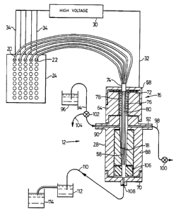
Referring to Figures 1 and 2, there is
shown an analyzer for analyzing a sample of DNA
including a sheath flow cuvette 12 enclosing the
ends of five capillary tubes 14 arrayed side by side
in a line like the teeth of a comb. The capillary
tubes 14 are held in a header 16 with their cleaved
ends 18 ter~in~ting inside the chamber 12. The other
ends 20 of the capillary tubes 14 terminate in five
of the wells 22 of a conventional microtiter plate
24. The capillary tubes 14 are conventional fused
silica capillaries, with about 50~m ID and 150~m OD,
available from Polymicro. The cuvette 12 is formed
of a quartz chamber 26 secured within a stainless
W094/297~ PCT/CA94/00304
216~ 7
steel holder 28, the design of which is shown in
Figures 4 - 9 in more detail. A high voltage source
30, such as a Spellman RHR-30PN60 30 KV power
supply, is connected to the stainless steel holder
28 through a first electrode 32 (grounded) and also
through five second electrodes 34 to fluid in the
wells 22. Thus, when the capillary tubes 14 and
chamber 12 are filled with conducting material, a
high voltage may be applied across the material in
the capillary tubes using the high voltage source
30. The circuit is formed by the grounded electrode
32, the stainless steel holder 28 (formed of cap 68,
capillary retainer 64, chamber retainer 66 and cap
70), fluid in the cuvette 12 and in the chamber 26,
matrix in the capillary tubes, including sample
buffer if present, buffer solution in the wells 22
and the electrodes 34.
A laser 36 or other source of collimated
electromagnetic radiation provides a collimated beam
38 of light that is aligned to pass through a
focusing lens 40 into the chamber 12 along a
projection of the capillary tubes into the chamber,
as close as possible to the ends of the capillary
tubes 14, as shown in Figure 3A. The wavelength of
the laser 36 is chosen to excite fluorescence in the
sample being analyzed, as for example DNA reacted
with a fluorophor. An appropriate choice for DNA
analysis is an Innova 70-4 argon ion laser available
from Coherent Inc. of Palo Alto, California. Such
a laser may be operated with multiple wavelength
mirrors (488 and 514.5 nm), with appropriate
selection of the wavelength depending on the method
used for sequencing the DNA.
Fluorescence from the sample in the
chamber 12 is detected through a collection lens 42
that images the fluorescence on to a plurality of 1
WO 941297~ 7, ~ 7 PCT/CA94/00304
r ~
mm aperture GRIN (gradient index) lenses 44
(available from Nipon Scientific Glass through
Precision Cells, Inc. of Farmingdale, NY) which are
affixed to receiving ends 46 of fiber optics 48.
The fibre optics 48 may be secured in known r~nner
as for example to a Melles Griot optical bread board
(not shown). Transmitting ends 50 of the fiber
optics 48 lead into avalanche photodiodes 52 or
other individual photon detectors, one for each
capillary tube 14, and whose output is connected
through an interface 54 to a computer 56. Exemplary
photodiodes 48 are RCA (EG&G) C30902S photodiodes,
powered by a PS310 Stanford Research System high
voltage power supply, or model SPCM 100 photodiodes
available from EG&G Canada Ltd.. Fluorescence is
transmitted along the fiber optics 48 to the
photodiodes 52 whose electrical output is
proportional to the intensity of the fluorescence.
Electrical signals output from the photodiodes 52
are passed through a data acquisition board 54 (such
as may be obtained from National Instruments or from
Data Translation, model DT2221-G) to a computer 56
such as a Macintosh II computer for processing
according to known techniques. Such processing
includes filtering the signal to give a desired
frequency response, and a second filter or phase
lock loop to identify the position of the peak
centers. For interface boards from National
Instruments, it may be necessary to decrease
illumination intensity to avoid over saturation of
the photon detectors. Alternatively, light collected
in individual GRIN lenses 44 may be passed through
a bundle of optical fibres and imaged onto or
abutted against an array detector. However, CCD
cameras are not believed to be fast enough for high
speed DNA sequencing.
W094/297~ PCT/CA94/00304
7 ~
As shown in Figures 3A and 3B, if the
fluorescence emitted from the DNA sample has a
spectrum centered on more than one wavelength of
light, then a means of dividing the spectrum of the
received light may be used. Light from laser 36
passes through focusing optic 40 and passes through
the sample streams 58. Fluorescence from the sample
streams is collected by optic 42 and passed through
a spectral filter 60 (for filtering scattered light)
to GRIN lenses 44 on the ends of fibre optics 48.
Light in the fibre optics 48 is passed through
wavelength division demultiplexers 62 where light
from different spectral bands is separated into two
sets 48a and 48b of fibre optics and two sets of
avalanche photodiodes 52a and 52b.
The selection of the filter 60 and the
optical system depends on the sequencing reaction to
be performed. For a single codor sequencer, using
the sequencing method of Richardson-Tabor, a single
spectral filter 60 with a bandwidth of 45 nm
centered at 530 nm may be used to detect fluorescein
labeled products. The filter should be selected to
rinimize background signals due to Raman and Raleigh
scatter of the excitation beam 38. For the DuPont
sequencing system, two detection channels are
required, one detector channel to image light in a
band centered at 510 nm and the other to image light
centered at 540 nm. Light collected from the
collection optic is split into two paths using the
wavelength division demultiplexers 62, one path
leading to one set of photon detectors 52a and the
other leading to the other set of photon detectors
52b. Other methods of wavelength division
demultiplexing may be used as for example rapidly
switching a filter wheel so that the light from the
sample stream is time division demultiplexed. For
W094/297~ ~ 7 PCT/CA94/00304
sequencing using the method developed by Applied
Biosystems Inc. (see the Swerdlow article), four
channels are required. As with the DuPont system,
two detector systems are used, and a filter wheel
may be used as the spectral filter 60 to rotate two
selected filters across the path of the light
collected by the collection optic. By alternating
the two filters in the two detection systems, a
signal from four spectral channels may be generated.
The collection optic 42 should be selected
to provide an image that is matched in size to the
aperture of the GRIN lenses 44, such as may be
provided by a flat field high numerical aperture
mic:roscope objective, for example as made by
Leitz/Wild (0.40 NA achromat objective). With a
sample stream diameter of 50 ~m and a GRIN lens
diameter of 1 mm, for example, the magnification
should be about 20x, generating spots several
millimeters apart. Since the light from the
co]lection optic tends to expand with a curved
wavefront, the GRIN lenses should be arranged to
have their collection faces perpendicular to radii
of the wavefront.
Referring to Figures 4 - 9, the chamber 26
is held in a stainless steel holder 28 to form a
sheath flow cuvette. The holder 28 includes an upper
section or capillary retainer 64 and a lower section
or chamber ret~;ner 66 each machined from individual
pieces of steel rod. The retainers 64 and 66 are
threaded together at 65 (threads not shown). A top
cap 68 is threaded onto the upper end of retAiner
64. A bottom cap 70 is threaded onto the lower end
of retA;ner 66. An upper seal 72 made of plastic
forms a seal between the cap 68 and retainer 64. A
like seal (not shown) may be used to seal the cap 70
to the retainer 66. An O-ring (not shown) or other
W094/297~ PCT/CA94/00304
~ 1 642~7
12
suitable seal should be provided to ensure that the
retainers 64, 66 are sealed together to prevent
leakage at 65. The cap 68 has a central hole for
receiving the capillary tubes 14. A plastic sleeve
74 into which the capillaries are threaded has epoxy
applied to it to form a seal around the capillary
tubes 14 as they enter the cap 68. The capillary
ret~;ner 64 includes a hollow bore lined with a
plastic cylindrical and annular spacer 76. Filling
out the hollow bore of the retainer 64 are two
facing semi-circular metal rods 78 each with a
groove machined into their facing flat faces to form
a rectangular slot 80. The slot 80 is dimensioned to
receive the capillaries 14 snugly and hold them
against each other in a line.
The chamber ret~iner 66 includes two
circular sections 82 and 84 and a pedestal section
86 in which the metal of the rod has been machined
away to form four pedestals 88 in which the chamber
26 is securely retained. Metal in the chamber
ret~iner 66 is machined away in the pedestal section
86 to form cavities 89. Removal of the metal in this
section 86 allows a microscope objective to be
placed close to the chamber 26 (within a few
millimetres). Upper circular section 82 includes a
sheath fluid inlet 90 and a bubble removal port 92.
The sheath fluid inlet 90 is connected via Teflontm
tubing 94 (see Figure 2) to a source of sheath fluid
96 (not shown to scale). The bubble removal port 92
is connected by Teflontm tubing 98 to a valve 100.
The tubing 94 may include a three way valve 102 with
waste line 104 for removing bubbles from the sheath
fluid. In the chamber ret~iner 66, at the base of
the chamber 26 is a plastic bottom plug 106 that
holds the chamber 26 in place. The cap 70 is
provided with a waste outlet port 108 that is
W094/297~ 2 ~ PCT/CA94/00304
connected to Teflontm tubing 110 to a waste beaker
112.
As shown in Figure 2, sheath fluid is
provided through inlet 90. The sheath fluid enters
the top of the chamber 26 and moves as a syphon flow
under gravity from the top of the chamber to the
bottom, past the ends 18 of the capillary tubes 14.
The fluid should be provided in a steady, non-pulsed
flow, and should be filtered and purified to avoid
any background signal passing due to particles
passing through field of view of the collection
optics. The fluid is chosen to have similar index of
refraction as the fluid carrying the sample DNA to
avoid reflection and refraction at interfaces
between fluids of different in~e~es of refraction.
The simplest way to achieve this is to use the same
fluid for the sheath fluid as carries the sample
DNA, as for example lxTBE. The volumetric flow of
the sheath stream is low, in the order of less than
lOmL/hr, which for the embodiment described is in
the order of 4mm/s, though it may be as much as lOx
less for some applications. The fluid is drained to
waste after exiting the chamber 12 through port 108.
The waste beaker 112 should be kept half-filled with
buffer. If the waste stream forms drops, the sample
stream profile is distorted when the drop detaches.
A periodic noise results from the periodic
detachment of the drops. The beaker 112 preferably
has a small hole drilled in it with a tube leading
to a larger beaker 114. The level of the first
beaker 112 rem~in~ constant, so that the sheath flow
velocity under conditions of syphon flow changes
slowly. A constant syphon head may also assist in
ensuring constant sheath flow rate. For the
apparatus described a 5 cm syphon head has been
found adequate. Bubbles should not be present in
W094/297~ PCT/CA94/00304
2 ~
14
the sheath flow. These can be eliminated by visual
inspection and eliminated using the three way valve
102 (by switching the fluid cont~;n;ng the bubble to
waste).
5Referring to Figures lOa, lOb and lOc, the
chamber 26 includes end walls 122a, 122b, side walls
124a, 124b, top 126 and bottom 128. The walls need
not be planar but may contain projections to align
the capillaries. Each wall is 1 mm thick at the top
10and made of high quality optical quartz, or such
other inert material as is transparent to the
selected electromagnetic radiation emitted by either
the laser 36 or the sample passing out of the
capillary tubes 14. The side walls 124a, 124b are
15constant thickness from top to bottom, while the end
walls esch thicken inward towards the bottom by
50~m. The interior of 130 of the chamber 26 has the
same ~i ?~ion X laterally as the thickness of the
capillary tube used (150~m in the exemplary
20embodiment) and the dimension Y1 from end wall to
end wall a little more (50 ~m more in the exemplary
embodiment) than the sum of the thicknesses of the
capillary tubes 14. The interior 132 at the bottom
of the chamber has the same dimension X laterally as
25the thickness of the capillaries used and the
m~ion Y2 from end wall to end wall a little less
(50~m in the exemplary embodiment) than the sum of
the thicknesses of the capillary tubes 14. The
capillary tubes 14 should be snugly fit in the
30interior of the chamber 26, with their ends
t~rmi n~ting adjacent each other. It is preferable
that the capillary tubes 14 be placed in the chamber
26 before they are filled with matrix material.
Particularly if capillary tubes are re-
35used, the collection optics, including the GRIN
lenses 44, will ~e fixed and the capillary tubes 14
W094/297~ ~ 7 PCT/CA94100304
must be aligned with the collection optic so that
fluorescence from the sample stream irradiated by
the laser beam 38 is imaged onto the GRIN lenses 44.
The capillary tubes 14 are first inserted through
the cap 68 and retainer 64 into the slot 80 formed
by the two rods 78. The capillary tubes 14 may be
loaded together or one by one. The capillary tubes
14 are inserted into the chamber 26 in this m~nner
and pushed together into the chamber 26 until they
are firmly held in the chamber 26. With the chamber
26 of the ~ ions stated, the capillary tubes 14
will ter~in~te about half way through the chamber
26. The top of the chamber 26 thus encompasses the
capillary tubes 14 with the capillary tubes 14
abutting the interior walls of the chamber at the
ends near the center of the chamber and at the sides
throughout the length of the capillary tubes within
the chamber 26. Abutment of the capillary tubes
against the interior walls of the chamber seals any
gaps between the capillary tubes at the center of
the chamber 26. Unless such gaps are sealed, non-
uniformities in the sheath flow can result which can
affect the signal quality. The capillary tubes 14
are preferably cleaved at their ends using well
known techniques employed in the manufacture of
fiber optics in order to obtain a smooth and flat
end. The capillary tubes 14 will therefore extend
into the interior of the chamber 26 an amount that
is dependent on the rate of decrease of the end wall
to end wall dimension of the chamber, and will
typically be 1 cm for the exemplary embodiment
described. The chamber 26 has height H about 2 cm
from top to bottom as shown in the example. Such
chambers may be purchased from Nipon Scientific
Glass through Precision Cells, Inc. of Farmingdale,
NY, to order. The height H of the chamber is
W094/297~ PCT/CA94/00304
16
somewhat arbitrary, sufficient to allow both fixture
of the capillary tubes and to allow the light beam
to pass through the chamber below the capillary
ends. 2 cm is chosen to allow addition of a second
laser beam below the first if two lasers are used
for analysis. The top of the side walls 124a, 124b
should be slightly bevelled to ease insertion of the
capillary tubes 14. The construction of the chamber
is quite important, particularly when the capillary
tubes are not electrically isolated from the high
voltage applied across the porous matrix material in
the capillary tubes. If the capillary tubes are not
isolated electrically, repulsive forces between them
can create forces which if not evenly distributed,
can shatter the capillary tubes. The capillary tubes
14 should therefore all be held securely in the
chamber to prevent these stresses from concentrating
at one tube.
The capillary tubes 14 should te in~te
within about lO~m from each other. The laser beam 38
should entirely pass within about 100 ~m from the
ends of the capillary tubes. Careful alignment of
the capillary tubes is required so that the image of
the fluorescence falls directly on the GRIN lenses.
This can be checked by passing light backward
through the GRIN lenses. The light should pass
through the sample stream exactly at the same point
that fluorescence due to the laser beam occurs.
Visual inspection can be used to verify the correct
alignment of the capillary tubes, with appropriate
safety precautions due to the use of laser light.
The length of the flow cell (distance
between the end walls 124a and 124b) and the number
of capillaries that can be detected in a single flow
cell are determined by the distance over which laser
beam size can be matched to the sample stream radius
W094/297~ PCT/CA94100304
17
as it exits the capillary. To optimize sensitivity,
the laser beam should be located as near as possible
to the ends of the capillaries to m; n i m i ze effects
of diffusion of the sample into the sheath fluid.
The laser beam should therefore pass through the
acceleration region of the sample flow. At this
point, faster moving sheath fluid draws the sample
fluid from the matrix. Since the entire cuvette is
grounded (through electrode 32), there is very
little electric field inside the cuvette, and the
sample fluid is not drawn by the electric field out
of the capillaries. Thus it is the sheath flow that
draws the sample fluid from the matrix in the
capillaries. As the sample fluid moves away from the
end of the capillary its cross-section contracts,
and then exr~n~ due to diffusion of the sample
fluid into the sheath fluid. The laser beam should
pass through a point above the point of maximum
contraction, thus before the diffusion zone.
A single laser beam is aligned to be
parallel with the long axis of the cuvette (end wall
122a to end wall 122b) simultaneously exciting
fluorescence from each sample stream in turn. The
size of the laser beam should be selected to ensure
similar illumination of each sample stream. With a
lens (for example a microscope objective with lx
magnification) between the laser 36 and the chamber
26 a beam waist can be located in the center of the
chamber. The beam spot size at the center of the
chamber should be eclual to the sample stream
diameter at that point. Nith 50 ~m ID capillary
tubes, this is about 50 ~m. The beam diameter will
be larger in both directions away from this point,
but with this arrangement, the fluorescence is close
to optimum.
-
W094/297~ PCT/C~94/00304
For setting up the analyzer for DNA
analysis, care must be taken as is known for
capillary electrophoresis. Thus, the matrix material
must be selected for stability, for discrimination
of longer base lengths and for speed of sequencing.
No one matrix is suitable for all applications. For
DNA sequencing, a 0%C (non-cross-linked), 5-6%T
acrylamide gel has found to be adequate and has the
added advantage of low viscosity which allows it to
be readily replaced, without removal of the
capillary tubes 14 from the chamber 26. A
proprietary gel, Long-Rangertm from AT Biochemicals,
has been found useful for applications using high
voltage in the order of 800V/cm, such as in
diagnostic applications. Long-Rangertm gel allows
sequencing rates in the order of 200 bases in 3
minutes with greater than 95% accuracy. 0%C gels
provide sequencing rates in the order of 600 bases
in two hours at 200V/cm. Gel temperatures between
20C and 35C have been found to give good results.
The Long-Ranger gel is prepared within a
50 ~m ID capillary by polymerization of a carefully
degassed 5~ solution of Long-Ranger in a 7 M urea,
O . 6 x TBE buffer. Polymerization is initiated with
0.4 parts per thousand (V/V) TENED and o.4 parts per
thousand (W/V) ammonium persulphate. Such a gel is
stable and may be used for three separations. Use of
Long-Ranger gel with a single 50~m ID capillary has
yielded sequencing rates of 3200 bases per hour at
800V/cm.
The gel may include 0 - 20% of formamide.
Addition of formamide in this range decreases
compressions, particularly in the range 10 - 20~,
thereby increasing resolution in regions of
compression. However, it has been found that too
much (20% or more) formamide reduces the separation
W094/297~ ~ 2~ PCT/CA94/00304
' ~
19
rate, theoretical plate count, and resolution for
normally migrating fragments without a concomitant
decrease in compressions. An optimum concentration
of 10% formamide improves resolution of compressed
regions without degrading other characteristics of
the gel. It has also been found that operating the
gel at room temperature is adequate and simplifies
the engineering of the analyzer. Results of using
formamide have been described in Rocheleau, M.J., et
al, Electrophoresis, 13, 484-486, 1992.
The gel should be established in the
capillary tubes 14 without voids or bubbles forming
during polymerization of the acrylamide due to
shrinkage, which may be particularly acute if a
bifunctional silane reagent is used to bind the gel
to the capillary wall. Such bubbles can be
eliminated by use of low percent acrylamide, short
columns, adding polyethylene glycol to the monomer
mixture (though this is not desired for DNA
fragments longer than about 100 bases since it
degxades the separation) or by allowing
pol~merization to occur in a pressured vessel or
other methods known in the art.
Also, defects in the gel at the ends 20
may occur when loading samples of DNA into the
capillary tubes 14. Such defects are particularly of
concern when the capillary tubes 14 are reused. It
is therefore desirable to cut off a portion (several
millimetres) of the capillary tube 14 after a run.
Also, such a defect can be minimized by loading
smaller amounts of DNA sample, as much as five times
lower, as compared with conventional electrophoresis
sequencing of DNA. Thus for example the sample using
the apparatus disclosed should be loaded at 150V/cm
for 60 s.
W094l297~ PCT/CA94100304
Flaws in the gel can be inspected by
visual inspection in a microscope or by passing two
laser light beams at an angle through the gel to
intersect each other in the gel. Modulated light
scatter of the laser light from flaws in the gel may
be detected using a collection optic and
photomultiplier tube.
Loading of the gel into the capillary
tubes 14 also requires care. It is desirable that
gel characteristics be uniform from capillary tube
to capillary tube. If the capillary tubes are loaded
with gel sequentially, differences in the gel may
severely degrade the analysis. It is preferable to
= load the gel monomer into a single container and to
= 15 fill the capillaries with the gel from the single
cont~;ner simultaneously, as by vacuum syphoning the
gel. At high electric fields (in the order of
800V/cm), the gel can extrude about 50 ~m from the
detection end of the capillary. To elim;n~te
extrusion, about 2 cm of the gel at the detection
end is covalently bonded to the interior walls of
t h e c a p i l l a r y t u b e s w i t h y -
methacryloxypropyltrimethoxysilance. Such known
methods for establishing a gel as described in
United States patents 4,865,706 and 4,865,707 to
Karger et al and 4,810,456 to Bente et al may also
be used.
Data has been collected from the system of
Figure 1 with detection at three capillaries using
the Tabor and Richardson sequencing technique. An
M13mpl8 template was used to generate fragments of
DNA. Manganese was used instead of magnesium in the
sequencing buffer. Sequenase was used for chain
extension. A FAM labeled primer is used and a single
sequencing reaction is performed with ddATP, ddCTP,
ddGTP and ddTTP present in a 8:4:2:1 ratio. A 50~m
W094/297~ ~ 2 ~ 7 PCT/CA94/00304
capillary was filled with 4%T, 5%C gel and operated
at 200V/cm. For a run of 330 bases in 70 minutes,
comparable data was obtained as for single capillary
systems, although the throughput was 850 bases/hour
for a 3 capillary system. Figures lla, llb and llc
show the résults of the sequencing.
Resolution is limited to fragments less
than 300 bases in length at high voltages near
800V/cm. Generally speaking, retention time
increases linearly with fragment length for a given
high V/cm until the mobility of the fragments
approaches a limiting value and no separation is
achieved. This is called biased reptation. As the
electric field increases, the transition to biased
reptation moves to shorter fragments. Biased
reptation is highly undesirable since it causes
sequencing fragments to coelute, destroying the
separation resolution. Hence for longer fragments
(in the order of 600 bases), the electric field can
be decreased to about 140 V/cm, with an increase in
separation time. Noderate gel temperature (in the
order of 20 to 35C) can assist in improving
sequencing rate, though it does not appear to
strongly affect the transition from reptation to
biased reptation. Lower %T acrylamide gels can also
assist in the sequencing of longer fragments.
The analyzer described here has utility
for a wide variety of applications, with some
modifications. In each case there is some means to
force analyte through the capillaries, the
capillaries are held in the chamber as shown in
Fi~ure 1 and 2 for example, and sheath fluid is
supplied through the cuvette, with the sheath fluid
preferably having the same index of refraction as
the fluid carrying the analyte.
W094/297~ PCT/CA94/0030
r~
22
The detection of analyte may also be
accomplished using thermooptical absorption. In this
technique, the laser 36 is used to excite the
analyte which tends to heat the analyte and change
the index of refraction of the fluid by which it is
carried. As shown in Figure 12, the deflection of
the beams 138 from a second laser 136 after
collimating with an appropriate optic 137 by the
sample fluid emerging from the ends of the capillary
tubes 14 is then detected by the optical system 140,
which may be designed as shown in Figure 1.
An analyzer for use as an electrochemical
detector is shown in Figure 13. Electrodes 142 enter
the chamber 26 (made of an inert non-conducting
material such as quartz) from the bottom end 132 Of
the chamber. Each electrode 142 is connected to an
amplifier (not shown), and the output of the
= amplifier is provided to a processor, for example a
computer, through an interface for analysis in
accordance with known principles (similar to the
optical processing of the signals). In such a case,
the laser 36 is not required, since the
identification of the sample is by electrochemical
analysis. Multiple capillaries allow for rapid
analysis.
The analyzer may also be used to detect
impurities in fluids by detecting light scatter. In
such a case, the high voltage source 30 is not
required, since the fluid may be pumped directly as
a fluid through the capillary tubes, nor is the
spectral filter 60 required since the total
intensity of the scattered light may be detected.
The GRIN lenses 44 and detectors 52 detect
variations in the scatter of light resulting from
particles or impurities in the fluid.
W094/297~ 2 1 6 4 2 0 ~ PCT/CA94/00304
23
The analyzer is also useful for the
detection of organic contAmin~ntS~ for example the
fluorescent detection of polycyclic aromatic
hydrocarbons. In such detection, the capillary tubes
are filled with chromatographic packing material
(coated silica beads) instead of a polymer and the
analyte sample is forced through the capillary tubes
using a pump instead of the high voltage source 30.
The laser 36 should emit radiation at about 330 nm
or such other appropriate wavelength for detection
of organic cont~min~nts. Fluorescence emitted by the
sample of contAmin~nt is detected through an
appropriate spectral filter 60 and the optical
apparatus shown for example in Figure 1.
In a further example, the analyzer may be
used for flow cytometry. In flow cytometry a sample
cont~ining cells taken from an ~ni~l or human body
by fine needle aspiration is stained using a
fluorescent reagent such 8S a nucleic acid stain or
antibodies. With the present analyzer, the sample is
forced under air pressure by a pump that replaces
the high voltage source 30 through the capillary
tubes 26 and the laser beam 38 is passed through the
sample as it emerges from the capillary tubes 26
into the sheath flow. The intensity of the
fluorescence from the fluorescent reagent is
detected using the optical system of Figure 1 and
used to estimate the number of sets of chromosomes
in the cells, and this is useful, in accordance with
known procedures in the diagnosis and prognosis of
cancer.
Multiple capillary tubes may also be used
to spray analyte into a mass spectrometer. In such
a case, the capillaries are bundled within a
circular or polyhedral cuvette with sheath flow
about the capillaries. The bundle of capillaries is
WO 94/29712 PCT/CA94/00304
0 ~
24
inserted into the ionization chamber of a mass
spectrometer such as the triple quadrupole mass
spectrometer sold by Sciex Division of MDS Health
Group Limited, of Thornhill, Ontario, C~nA-l~, under
its trademark TAGA 6000E. For electrospray of
analyte, the capillary tubes are made conducting at
the end that extends into the ionization chamber.
Electrical potential is applied to the ends of the
capillaries in known r~nner.
A square, rectangular or other suitable
polyhedral array of capillary tubes may also be used
as well, as shown in Figures 14, 15 and 16 for the
case of a square capillary array. The array may be
rectangular as well. Other polyhedral arrays could
be used in principal, but this complicates the
optics. The array of capillary tubes 14 is formed
from five rows 144 of five capillary tubes 14 each,
all bound within a square chamber 146 forming part
of a square sheath flow cuvette. The cuvette is
5i~ r to the cuvette shown in Figures 4 - 9 only
the central chamber is square. An optical system 148
disposed adjacent the cuvette includes a collection
optic 154, GRIN lenses 156, and optic fibres 158
leading to photodiodes and the balance of the
optical system as shown in Figure 1.
Each row 144 of capillary tubes is similar
to the row shown in Figure 1, but succeeding rows in
the direction of the optical system 148 terrin2~te
higher in the sheath flow cuvette as shown at 160.
All capillary tubes 14 in a row terminate adjacent
each other. Sheath flow is provided about all of the
tubes 14 within the sheath flow cuvette. All four
walls 150 of the sheath flow cuvette taper inward
towards the bottom 152 of the chamber. The ends of
the capillary tubes 14 define a sloping plane P,
sloping downward and away from the optical system
wo 94~29712 2 ~. 6 ~ 2 3 ~ PCT/CA94/00304
148~ An elliptical or other linear cross-section
laser beam 162 oriented at the same slope as the
sloping plane (or close to it) is directed just
below the ends of the capillary tubes 14.
Fluorescence of the samples forms a sloping square
array of fluorescent spots 164 that appears as a
square grid of spots 166 from a view at right angles
to the cuvette.
Fluorescence from sample streams emerging
from the capillary tubes 14 is collected by an optic
154 and imaged on to the square array of GRIN lenses
156, which lie in the image plane of the fluorescent
spots produced by the optic 154. The GRIN lenses 156
are oriented with their faces perpendicular to the
wavefront from the collection optic 154. Light
collected by the GRIN lenses is transmitted through
optic fibres to photodetectors of the type shown in
Figure 1.
It is possible to operate the cuvette
upside down to allow bubbles in the sheath stream to
move upward with the stream to waste.
A person skilled in the art could make
immaterial modifications to the invention described
and claimed in this patent document without
departing from the essence of the invention.
s.