Note: Descriptions are shown in the official language in which they were submitted.
WO 94/29436 2164226 PCT/US94/06255
METHODS FOR SELECTIVELY STIMULATING
PROLIFERATION OF T CELLS
Background of the Invention
The development of techniques for propagating T cell populations in vitro has
been
crucial to many of the recent advances in the understanding of T cell
recognition of antigen
and T cell activation. The development of culture methods for the generation
of human
antigen-specific T cell clones has been useful in defining antigens expressed
by pathogens
and tumors that are recognized by T cells to establish methods of
immunotherapy to treat a
variety of human diseases. Antigen-specific T cells can be expanded in vitro
for use in
adoptive cellular immunotherapy in which infusions of such T cells have been
shown to have
anti-tumor reactivity in a tumor-bearing host. Adoptive immunotherapy has also
been used
to treat viral infections in immunocompromised individuals.
Techniques for expanding human T cells in vitro have relied on the use of
accessory
cells and exogenous growth factors, such as IL-2. The use of IL-2 and, for
example, an anti-
CD3 antibody to stimulate T cell proliferation is known to expand the CD8+
subpopulation
of T cells. The requirement for MHC-matched antigen presenting cells as
accessory cells
presents a significant problem for long-term culture systems. Antigen
presenting cells are
relatively short lived. Thus, in a long-term culture system, antigen
presenting cells must be
continuously obtained from a source and replenished. The necessity for a
renewable supply
of accessory cells is problematic for treatment of immunodeficiencies in which
accessory
cells are affected. In addition, when treating viral infection, accessory
cells which may carry
the virus may result in contamination of the entire T cell population during
long term culture.
An alternative culture method to clone and expand human T cells in vitro in
the absence of
exogenous growth factor and accessory cells would be of significant benefit.
Summary of the Invention
This invention pertains to methods for selectively inducing ex vivo expansion
of a
population of T cells in the absence of exogenous growth factors, such as
lymphokines, and
accessory cells. In addition, T cell proliferation can be induced without the
need for antigen,
thus providing c:- expanded T cell population which is polyclonal with respect
to antigen
reactivity. The method provides for sustained proliferation of a selected
population of CD4+
or CD8+ T cells over an extended period of time to yield a multi-fold increase
in the number
of these cells relative to the original T cell population.
According to the method of the invention, a population of T cells is induced
to
proliferate by activating the T cells and stimulating an accessory molecule on
the surface of
the T cells with a ligand which binds the accessory molecule. Activation of a
population of T
cells is accomplished by contacting the T cells with a first agent which
stimulates a
CA 02164226 2004-05-12
WO 94/29436 PCT/US94/06255
-2-
TCR/CD3 complex-associated signal in the T cells. Stimulation of the TCR/CD3
complex-
associated signal in a T cell is accomplished either by ligation of the T cell
receptor
(TCR)/CD3 complex or the CD2 surface protein, or by directly stimulating
receptor-coupled
signaling pathways. Thus, an anti-CD3 antibody, an anti-CD2 antibody, or a
protein kinase
C activator in conjunction with a calcium ionophore is used to activate a
population of T
cells.
To induce proliferation, an activated population of T cells is contacted with
a second
agent which stimulates an accessory molecule on the surface of the T cells.
For example, a
population of CD4+ T cells can be stimulated to proliferate with an anti-CD28
antibody
directed to the CD28 molecule on the surface of the T cells. Proliferation of
a population of
CD8+ T cells is accomplished by use of a monoclonal antibody ES5.2D8 which
binds to an
accessory molecule having a molecular weight of about 27 kD present on
activated T cells.
Alternatively, proliferation of an activated population of T cells can be
induced by
stimulation of one or more intracellular signals which result from ligation of
an accessory
molecule, such as CD28.
Following activation and stimulation of an accessory molecule on the surface
of the T
cells, the progress of proliferation of the T cells in response to continuing
exposure to the
ligand or other agent which acts intracellularly to stimulate a pathway
mediated by the
accessory molecule is monitored. When the rate of T cell proliferation
decreases, the T cells
are reactivated and restimulated, such as with additional anti-CD3 antibody
and a co-
stimulatory ligand, to induce further proliferation. In one embodiment, the
rate of T cell
proliferation is monitored by examining cell size. Alternatively, T cell,
proliferation is
monitored by assaying for expression of cell surface molecules in response to
exposure to the
ligand or other agent, such as B7-1 or B7-2. The monitoring and restimulation
of the T cells
can be repeated for sustained proliferation to produce a population of T cells
increased in
number from about 100- to about 100,000-fold over the original T cell
population.
The method of the invention can be used to expand selected T cell populations
for use
in treating an infectious disease or cancer. The resulting T cell population
can be genetically
transduced and used for immunotherapy or can be used for in vitro analysis of
infectious
agents such as HIV. Proliferation of a population of CD4+ cells obtained from
an individual
infected with HIV can be achieved and the cells rendered resistant to HIV
infection.
Following expansion of the T cell population to sufficient numbers, the
expanded T cells are
restored to the individual. Similarly, a population of tumor-infiltrating
lymphocytes can be
obtained from an individual afflicted with cancer and the T cells stimulated
to proliferate to
sufficient numbers and restored to the individual. In addition, supernatants
from cultures of T
cells expanded in accordance with the method of the invention are a rich
source of cytokines
and can be used to sustain T cells in vivo or ex vivo.
WO 94/29436 2164226 PCT/US94/06255
-3-
Brief Description of the Drawings
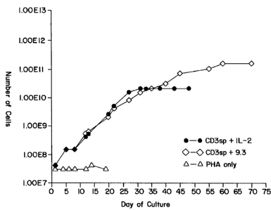
Figure 1 depicts in vitro growth curves of CD4+ peripheral blood T cells in
response
to culture with either an anti-CD3 antibody and interleukin-2 (IL-2) (=-=), an
anti-CD3
antibody and an anti-CD28 antibody mAb 9.3 (0-0) or PHA only
(A-A).
Figure 2 depicts the growth curve of CD4+ peripheral blood T cells cultured in
fetal
calf serum and either anti-CD3 antibodies and IL-2 (=-=) or an anti-CD3
antibody and an
anti-CD28 antibody, mAb 9.3 (0-0).
Figure 3 depicts the growth curves of CD4+ peripheral blood T cells cultured
in the
presence of phorbol myristic acid (PMA) and ionomycin with or without IL-2, or
with an
anti-CD28 antibody, mAb 9.3. The symbols are as follows: PMA and ionomycin
(P+I) is
represented by (0); PMA, ionomycin and IL-2 (P+I+IL-2) is represented by (=);
and PMA,
ionomycin and anti-CD28 antibody (P+I+9.3) is represented by (=).
Figure 4 is a schematic representation of the selective expansion of CD4+ T
cells
following CD28 stimulation in comparision to T cell stimulation with IL-2.
Figure 5 depicts fluorescent activated cell sorter analysis (FACS) in which
cells were
stained after isolation (day 0, panel A), or after 26 days in culture with
either CD28
stimulation (panel B) or IL-2 culture (panel C), with phycoerythrin conjugated
anti-CD3,
CD4, CD8 or with an IgG2a control monoclonal antibody and fluorescence
quantified with a
flow cytometer.
Figure 6 shows FACS analysis of the EX5.3D10 monoclonal antibody depicting
reactivity with CD28 in comparison to an anti-CD28 monoclonal antibody 9.3.
The
following cell lines were tested: Panel A, untransfected CHO-DG44 cells; Panel
B, CHO-
HH cells; Panel C, unactivated peripheral blood lymphocytes; and Panel D,
Jurkat No. 7
cells.
Figure 7 shows FACS analysis of the ES5.2D8 monoclonal antibody depicting the
binding reactivity with the following cell lines: Panel A, CHO-DG44 cells;
Panel B, CHO-
105A cells; Panel C, unactivated human peripheral blood lymphocytes; and Panel
D, PMA
activated peripheral blood lymphocytes.
Figure 8 is a photograph depicting immunoprecipitation analysis of detergent
lysates
of surface labeled human activated T cells indicating that monoclonal antibody
ES5.2D8
reacts with a 27 kD cell surface protein.
Figure 9 depicts the increases in mean cell volume of CD4+ T cells following
stimulation (Si, S2, S3, S4, S5 and S6) with an anti-CD3 monoclonal antibody
and an anti-
CD28 monoclonal antibody over days in culture.
Figure 10 depicts the cyclic expression of B7-1 on CD4+ T cells following
stimulation (Si, S2, S3, S4, S5 and S6) with an anti-CD3 monoclonal antibody
and an anti-
CD28 monoclonal antibody over days in culture.
WO 94/29436 PCTIUS94/06255
2164226
-4-
Figure 11 is a bar graph depicting the amount of IL-2 produced by CD4+ T cells
following stimulation with an anti-CD3 monoclonal antibody and an anti-CD28
monoclonal
antibody or IL-2 over days in culture.
Figure 12 is a bar graph depicting the amount of granulocyte-macrophage colony-
stimulating factor (GM-CSF) produced by CD4+ T cells following stimulation
with an anti-
CD3 monoclonal antibody and an anti-CD28 monoclonal antibody or IL-2 over days
in
culture.
Figure 13 is a bar graph depicting the amount of tumor necrosis factor (TNF)
produced by CD4+ T cells following stimulation with an anti-CD3 monoclonal
antibody and
an anti-CD28 monoclonal antibody or IL-2 over days in culture.
Figure 14 is a bar graph depicting the T cell receptor (TCR) diversity in CD4+
T cells
following stimulation with an anti-CD3 monoclonal antibody and an anti-CD28
monoclonal
antibody at day 1 and day 24 of culture.
Figure 15 depicts cell surface staining of CD4+ T cells obtained from an HIV
seronegative individual following stimulation (Si, S2 and S3) with an anti-CD3
monoclonal
antibody and an anti-CD28 monoclonal antibody over days in culture.
Figure 16 depicts cell surface staining of CD4+ T cells obtained from an HIV
seropositive individual following stimulation (Si, S2 and S3) with an anti-CD3
monoclonal
antibody and an anti-CD28 monoclonal antibody over days in culture.
Figure 17 depicts expansion of CD8+ T cells following stimulation with an anti-
CD3
monoclonal antibody and an monoclonal antibody ES5.2D8 at day 4 and day 7 of
culture.
Detailed Description of the Invention
The methods of this invention enable the selective stimulation of a T cell
population
to proliferate and expand to significant numbers in vitro in the absence of
exogenous growth
factors or accessory cells. Interaction between the T cell receptor (TCR)/CD3
complex and
antigen presented in conjunction with either major histocompatibility complex
(MHC) class I
or class II molecules on an antigen-presenting cell initiates a series of
biochemical events
termed antigen-specific T cell activation. The term "T cell activation" is
used herein to
define a state in which a T cell response has been initiated or activated by a
primary signal,
such as through the TCR/CD3 complex, but not necessarily due to interaction
with a protein
antigen. A T cell is activated if it has received a primary signaling event
which initiates an
immune response by the T cell.
T cell activation can be accomplished by stimulating the T cell TCR/CD3
complex or
via stimulation of the CD2 surface protein. An anti-CD3 monoclonal antibody
can be used to
activate a population of T cells via the TCR/CD3 complex. Although a number of
anti-
human CD3 monoclonal antibodies are commercially available, OKT3 prepared from
hybridoma cells obtained from the American Type Culture Collection or
monoclonal
TWO 94/29436 216 4 2 2 6 PCTIUS94/06255
-5-
antibody G19-4 is preferred. Similarly, binding of an anti-CD2 antibody will
activate T cells.
Stimulatory forms of anti-CD2 antibodies are known and available. Stimulation
through
CD2 with anti-CD2 antibodies is typically accomplished using a combination of
at least two
dit::rent anti-CD2 antibodies. Stimulatory combinations of anti-CD2 antibodies
which have
been described include the following: the T11.3 antibody in combination with
the T11.1 or
T11.2 antibody (Meuer, S.C. et al. (1984) Cell 3..:897-906) and the 9.6
antibody (which
recognizes the same epitope as Ti 1.1) in combination with the 9-1 antibody
(Yang, S. Y. et
al. (1986) J. Immunol. 137:1097-1100). Other antibodies which bind to the same
epitopes as
any of the above described antibodies can also be used. Additional antibodies,
or
combinations of antibodies, can be prepared and identified by standard
techniques.
A primary activation signal can also be delivered to a T cell through use of a
combination of a protein kinase C (PKC) activator such as a phorbol ester
(e.g., phorbol
myristate acetate) and a calcium ionophore (e.g., ionomycin which raises
cytoplasmic
calcium concentrations). The use of these agents bypasses the TCR/CD3 complex
but
delivers a stimulatory signal to T cells. These agents are also known to exert
a synergistic
effect on T cells to promote T cell activation and can be used in the absence
of antigen to
deliver a primary activation signal to T cells.
Although stimulation of the TCR/CD3 complex or CD2 molecule is required for
delivery of a primary activation signal in a T cell, a number of molecules on
the surface of T
cells, termed accessory or costimulatory molecules have been implicated in
regulating the
transition of a resting T cell to blast transformation, and subsequent
proliferation and
differentiation. Thus, in addition to the primary activation signal provided
through the
TCR/CD3 complex, induction of T cell responses requires a second,
costimulatory signal.
One such costimulatory or accessory molecule, CD28, is believed to initiate or
regulate a
signal transduction pathway that is distinct from those stimulated by the TCR
complex.
Accordingly, to induce an activated population of T cells to proliferate
(i.e., a
population of T cells that has received a primary activation signal) in the
absence of
exogenous growth factors or accessory cells, an accessory molecule on the
surface of the T
cell, such as CD28, is stimulated with a ligand which binds the accessory
molecule or with an
agent which acts intracellularly to stimulate a signal in the T cell mediated
by binding of the
accessory molecule. In one embodiment, stimulation of the accessory molecule
CD28 is
accomplished by contacting an activated population of T cells with a ligand
which binds
CD28. Activation of the T cells with, for example, an anti-CD3 antibody and
stimulation of
the CD28 accessory molecule results in selective proliferation of CD4+ T
cells. An anti-
CD28 monoclonal antibody or fragment thereof capable of crosslinking the CD28
molecule,
or a natural ligand for CD28 (e.g., a member of the B7 family of proteins,
such as B7-
1(CD80) and B7-2 (CD86) (Freedman, A.S. et al. (1987) J. Immunol. J.3J:3260-
3267;
Freeman, G.J. et al. (1989) J. Immunol. 433:2714-2722; Freeman, G.J. et al.
(1991) J. Exp.
WO 94/29436 PCT/US94/06255
2164226
-6-
Med.174:625-63 1; Freeman, G.J. et at. (1993) Science 2L2:909-91 1; Azuma, M.
et al. (1993)
Nature 3f¾:76-79; Freeman, G.J. et al. (1993) J. Exp. Med. m:2185-2192)) can
be used to
induce stimulation of the CD28 molecule. In addition, binding homologues of a
natural
ligand, whether native or synthesized by chemical or recombinant technique,
can also be used
in accordance with the invention. Ligands useful for stimulating an accessory
molecule can
be used in soluble form or immobilized on a solid phase surface as described
herein. Anti-
CD28 antibodies of fragments thereof useful in stimulating proliferation of
CD4+ T cells
include monoclonal antibody 9.3, an IgG2a antibody (Dr. Jeffery Ledbetter,
Bristol Myers
Squibb Corporation, Seattle, WA), monoclonal antibody KOLT-2, an IgGI
antibody, 15E8,
an IgGI antibody, 248.23.2, an IgM antibody and EX5.3D10, an IgG2a antibody.
A preferred anti-CD28 antibody is monoclonal antibody 9.3 or EX5.3D10. The
EX5.3DI0 monoclonal antibody was derived from immunizing a Balb/c mouse with
CHO
(Chinese hamster ovary) cells transfected with the human CD28 gene (designated
CHO-hh).
Hybridomas from the fusion were selected by whole cell ELISA screening against
Jurkat
(human T leukemia) CD28 tranfectants designated Jurkat #7. Reactivity of the
EX5.3D10
with CD28 was further confirmed by fluorescent activated cell sorter analysis
(FACS)
analysis in which it was tested side by side with the monoclonal 9.3 (Figure
6). Neither
antibody bound to untransfected CHO-DG44 cells and their binding profiles were
nearly
identical for the two CD28 transfectant lines, CHO-hh and Jurkat #7, as well
as normal
human peripheral blood lymphocytes. A hybridoma which produces the monoclonal
antibody EX5.3D10 has been deposited with the American Type Culture Collection
on June
4, 1993, at ATCC Deposit No. HB 11373.
In another embodiment of the invention, an activated population of CD4+ T
cells is
stimulated to proliferate by contacting the T cells with an agent which acts
intracellularly to
stimulate a signal in the T cell mediated by ligation of an accessory
molecule, such as CD28.
The term "agent", as used herein, is intended to encompass chemicals and other
pharmaceutical compounds which stimulate a costimulatory or other signal in a
T cell
without the requirement for an interaction between a T cell surface receptor
and a
costimulatory molecule or other ligand. For example, the agent may act
intracellularly to
stimulate a signal associated with CD28 ligation. In one embodiment, the agent
is a non-
proteinaceous compound. As the agent used in the method is intended to bypass
the natural
receptor:ligand stimulatory mechanism, the term agent is not intended to
include a cell
expressing a natural ligand. Natural ligands for CD28 include members of the
B7 family of
proteins, such as B7-1(CD80) and B7-2 (CD86).
It is known that CD28 receptor stimulation leads to the production of D-3
phosphoinositides in T cells and that inhibition of the activity of
phosphatidylinositol 3-
kinase (P13K) in a T cell can inhibit T cell responses, such as lymphokine
production and
cellular proliferation. Protein tyrosine phosphorylation has also been shown
to occur in T
WO 94/29436 2164226 PCT/US94/06255
-7-
cells upon CD28 ligation and it has been demonstrated that a protein tyrosine
kinase
inhibitor, herbimycin A, can inhibit CD28-induced IL-2 production
(Vandenberghe, P. et al.
(1992) J. Exp. Med. J:951-960; Lu, Y. et al. (1992) J. Immunol.1_2:24-29).
Thus, to
selectively expand a population of CD4+ T ,cells, the CD28 receptor mediated
pathway can
be stimulated by contacting T cells with an activator of P13K or an agent
which stimulates
protein tyrosine phosphorylation in the T cell, or both. An activator of P13K
can be identified
based upon its ability to stimulate production of at least one D-3
phosphoinositide in a T cell.
The term "D-3 phosphoinositide" is intended to include derivatives of
phosphatidylinositol
that are phosphorylated at the D-3 position of the inositol ring and
encompasses the
compounds phosphatidylinositol(3)-monophosphate (PtdIns(3)P),
phosphatidylinositol(3,4)-
bisphosphate (Ptdlns(3,4)P2), and phosphatidylinositol(3,4,5)-trisphosphate
(PtdIns(3,4,5)P3). Thus, in the presence of a P13K activator, the amount of a
D-3
phosphoinositide in the T cell is increased relative to the amount of the D-3
phosphoinositide
in the T cell in the absence of the substance. Production of D-3
phosphoinositides (e.g.,
Ptdlns(3)P, Ptdlns(3,4)P2 and/or PtdIns(3,4,5)P3) in a T cell can be assessed
by standard
methods, such as high pressure liquid chromatography or thin layer
chromatography, as
discussed above. Similarly, protein tyrosine phosphorylation can be stimulated
in a T cell,
for example, by contacting the T cell with an activator of protein tyrosine
kinases, such as
pervanadate (see O'Shea, J.J. et al. (1992) Proc. Natl. Acad. Sci. USA
$2:10306-103101; and
Secrist, J.P. (1993) J Biol. Chem. 20:5886-5893). Alternatively, the T cell
can be contacted
with an agent which inhibits the activity of a cellular protein tyrosine
phosphatase, such as
CD45, to increase the net amount of protein tyrosine phosphorylation in the T
cell. Any of
these agents can be used to expand an activated population of CD4+ T cells in
accordance
with the methods described herein.
In order to induce proliferation and expand a population of CD8+ T cells, an
activated
population of T cells is stimulated through a 27 kD accessory molecule found
on activated T
cells and recognized by the monoclonal antibody ES5.2D8. As described in
Example 9, a
population of CD8+ T cells was preferentially expanded by stimulation with an
anti-CD3
monoclonal antibody and the ES5.2D8 monoclonal antibody. The monoclonal
antibody
ES5.2D8 was produced by immunization of mice with activated human blood
lymphocytes
and boosted with recombinant human CTLA4 protein produced in E. coll.. The
ES5.2D8
monoclonal antibody is of the IgG2b isotype and specifically binds to cells
transfected with
human CTLA4. Hybridomas producing CTLA4-specific antibody were identified by
screening by ELISA against human CTLA4 protein as well as by differential FACS
against
wild type CHO-DG44 cells vs. CHO-105A cells, which are transfected with the
human
CTLA4 gene. As shown in Figure 7, the ES5.2D8 clone reacts strongly with both
activated
human T cells and CHO-105A cells but not with CHO-DCA4 cells, indicating that
it does
indeed bind to CTLA4. Immunoprecipitation of detergent lysates of surface
labeled activated
WO 94/29436 PCTIUS94/06255
2164226
-8-
human T cells revealed that ES5.2D8 also reacts with a 27 kD cell surface
protein (Figure 8).
A hybridoma which produces the monoclonal antibody ES5.2D8 was deposited on
June 4,
1993 with the American Type Culture Collection at ATCC Deposit No. HB 11374.
Accordingly, to expand a population of CD8+ T cells, an antibody, such as
monoclonal antibody ES5.2D8, or other antibody which recognizes the same 27 kD
ligand as
ES5.2D8 can be used. As described in Example 10, the epitope recognized by the
monoclonal antibody ES5.2D8 was identified by screening a phage display
library (PDL).
Antibodies which bind to the same epitope as the monoclonal antibody ES5.2D8
are within
the scope of the invention. Such antibodies can be produced by immunization
with a peptide
fragment including the epitope or with the native 27 kD antigen. The term
"epitope", as used
herein, refers to the actual structural portion of the antigen that is
immunologically bound by
an antibody combining site. The term is also used interchangeably with
"antigenic
determinant". A preferred epitope which is bound by an antibody or other
ligand which is to
be used to stimulate a CD8+ T cell population includes or encompasses, an
amino acid
sequence:
(Xaa1)n-Gly-Xaa2-Trp-Leu-Xaa3-Xaa4-Asp(Glu)-(Xaa5)n (SEQ ID NO: 5),
wherein Xaa4 may or may not be present, Xaal, Xaa2, Xaa3, Xaa4 and Xaa5 are
any amino
acid residue and n = 0-20, more preferably 0-10, even more preferably 0-5, and
most
preferably 0-3. In a preferred embodiment, Xaa2 is Cys or Ile, Xaa3 is Leu or
Arg and Xaa4,
if present, is Arg or Pro. Typically, Xaal and Xaa4 are additional amino acid
residues found
at either the amino or carboxy side, or both the amino and carboxy sides, of
the core epitope
in the native 27 kD protein. It will be appreciated by those skilled in the
art that in the native
protein, additional non-contiguous amino acid residues may also contribute to
the
conformational epitope recognized by the antibody. Synthetic peptides
encompassing the
epitope can be created which includes other amino acid residues flanking the
core seven
amino acid residues (i.e., Xaa can alternatively be other amino acid residues
than those found
in the native protein). These flanking amino acid residues can function to
alter the properties
of the resulting peptide, for example to increase the solubility, enhance the
immunogenicity
or promote dimerization of the resultant peptide. When the peptide is to be
used as an
immunogen, one or more charged amino acids (e.g., lysine, arginine) can be
included to
increase the solubility of the peptide and/or enhance the immunogenicity of
the peptide.
Alternatively, cysteine residues can be included to increase the dimerization
of the resulting
peptide.
Other embodiments of the invention pertain to expansion of a population of
CD8+ T
cells by use of an agent which acts intracellularly to stimulate a signal in
the T cell mediated
by ligation of the 27 kD protein. As used herein the term "agent" encompasses
chemicals and
WO 94/29436 216 4 2 2 6 PCT/US94/06255
-9-
other pharmaceutical compounds which stimulate a signal in a T cell without
the requirement
for an interaction between a T cell surface receptor and a ligand. Thus, this
agent does not
bind to the extracellular portion of the 27 kD protein, but rather mimics or
induces an
intracellular signal (e.g., second messenger) associated with ligation of the
protein by an
appropriate ligand. The ligands described herein (e.g., monoclonal antibody
ES5.2D8) can
be used to identify an intracellular signal(s) associated with T cell
expansion mediated by
contact of the 27 kD protein with an appropriate ligand (as described in the
Examples) and
examining the resultant intracellular signalling that occurs (e.g., protein
tyrosine
phosphorylation, calcium influx, activation of serine/threonine and/or
tyrosine kinases,
phosphatidyl inositol metabolism, etc.). An agent which enhances an
intracellular signal
associated with the 27 kD protein can then be used to expand CD8+ T cells.
Alternatively,
agents (e.g., small molecules, drugs, etc.) can be screened for their ability
to enhance T cell
expansion using a system such as that described in the Examples.
In yet another aspect of the invention, methods for expanding a population of
antigen
specific T cells are provided. To produce a population of antigen specific T
cells, T cells are
contacted with an antigen in a form suitable to trigger a primary activation
signal in the T
cell, i.e., the antigen is presented to the T cell such that a signal is
triggered in the T cell
through the TCR/CD3 complex. For example, the antigen can be presented to the
T cell by
an antigen presenting cell in conjuction with an MHC molecule. An antigen
presenting cell,
such as a B cell, macrophage, monocyte, dendritic cell, Langerhan cell, or
other cell which
can present antigen to a T cell, can be incubated with the T cell in the
presence of the antigen
(e.g., a soluble antigen) such that the antigen presenting cell presents the
antigen to the T cell.
Alternatively, a cell expressing an antigen of interest can be incubated with
the T cell. For
example, a tumor cell expressing tumor-associated antigens can be incubated
with a T cell
together to induce a tumor-specific response. Similarly, a cell infected with
a pathogen, e.g. a
virus, which presents antigens of the pathogen can be incubated with a T cell.
Following
antigen specific activation of a population of T cells, the cells can be
expanded in accordance
with the methods of the invention. For example, after antigen specificity has
been
established, T cells can be expanded by culture with an anti-CD3 antibody and
an anti-CD28
antibody according to the methods described herein.
The term "antibody" as used herein refers to immunoglobulin molecules and
immunologically active portions of immunoglobulin molecules, i.e., molecules
that contain
an antigen binding site which specifically binds (immunoreacts with) an
antigen, such as
CD3, CD28. Structurally, the simplest naturally occurring antibody (e.g., IgG)
comprises
four polypeptide chains, two heavy (H) chains and two light (L) chains inter-
connected by
disulfide bonds. It has been shown that the antigen-binding function of an
antibody can be
performed by fragments of a naturally-occurring antibody. Thus, these antigen-
binding
fragments are also intended to be designated by the term "antibody". Examples
of binding
WO 94/29436 PCT/US94/06255
2164226 -10-
fragments encompassed within the term antibody include (i) an Fab fragment
consisting of
the VL, VH, CL and CH1 domains; (ii) an Fd fragment consisting of the VH and
CHI
domains; (iii) an Fv fragment consisting of the VL and VH domains of a single
arm of an
antibody, (iv) a dAb fragment (Ward et al., (1989) Nature 3-4jL:544-546 )
which consists of a
VH domain; (v) an isolated complimentarity determining region (CDR); and (vi)
an F(ab')2
fragment, a bivalent fragment comprising two Fab fragments linked by a
disulfide bridge at
the hinge region. Furthermore, although the two domains of the Fv fragment are
coded for by
separate genes, a synthetic linker can be made that enables them to be made as
a single
protein chain (known as single chain Fv (scFv); Bird et al. (1988) Science
242:423-426; and
Huston et al. (1988) PNAS$J:5879-5883) by recombinant methods. Such single
chain
antibodies are also encompassed within the term "antibody". Preferred antibody
fragments
for use in T cell expansion are those which are capable of crosslinking their
target antigen,
e.g., bivalent fragments such as F(ab')2 fragments. Alternatively, an antibody
fragment
which does not itself crosslink its target antigen (e.g., a Fab fragment) can
be used in
conjunction with a secondary antibody which serves to crosslink the antibody
fragment,
thereby crosslinking the target antigen. Antibodies can be fragmented using
conventional
techniques as described herein and the fragments screened for utility in the
same manner as
described for whole antibodies. An antibody of the invention is further
intended to include
bispecific and chimeric molecules having a desired binding portion (e.g.,
CD28).
The language "a desired binding specificity for an epitope", as well as the
more
general language "an antigen binding site which specifically binds
(immunoreacts with)",
refers to the ability of individual antibodies to specifically immunoreact
with a T cell surface
molecule, e.g. CD28. That is, it refers to a non-random binding reaction
between an antibody
molecule and an antigenic determinant of the T cell surface molecule. The
desired binding
specificity is typically determined from the reference point of the ability of
the antibody to
differentially bind the T cell surface molecule and an unrelated antigen, and
therefore
distinguish between two different antigens, particularly where the two
antigens have unique
epitopes. An antibody which binds specifically to a particular epitope is
referred to as a
"specific antibody".
"Antibody combining site", as used herein, refers to that structural portion
of an
antibody molecule comprised of a heavy and light chain variable and
hypervariable regions
that specifically binds (immunoreacts with) antigen. The term "immunoreact" or
"reactive
with" in its various forms is used herein to refer to binding between an
antigenic determinant-
containing molecule and a molecule containing an antibody combining site such
as a whole
antibody molecule or a portion thereof.
Although soluble forms of antibodies may be used to activate T cells, it is
preferred
that the anti-CD3 antibody be immobilized on a solid phase surface (e.g.,
beads). An
antibody can be immobilized directly or indirectly by, for example, a
secondary antibody, to
WO 94/29436 2164226 PCT/US94/06255
-11-
a solid surface, such as a tissue culture flask or bead. As an illustrative
embodiment, the
following is a protocol for immobilizing an anti-CD3 antibody on beads. It
should be
appreciated that the same protocol can be used to immobilize other antibodies
or fragments
thereof (e.g., an anti-CD28 antibody) to beads.
Protocols
1. Pre-absorbing Goat anti-mouse IgG with OKT-3
A) BioMag Goat anti-Mouse IgG (Advanced Magnetics, Inc., catalog
number 8-4340D) is incubated with at least 200 g of OKT-3 per 5 x 10⁸
magnetic particles in PBS for 1 hour at 5 C.
B) Particles are washed three time in PBS with the aid of a magnetic
separation unit.
Note: Advanced Magnetics also has an anti-Human CD3 directly conjugated
(Catalog number 8-4703N) which will induce T-cell stimulation.
II. Pre-labeling Lymphocytes with OKT-3
A) 1 x 10⁶ cells (PBMC) are incubated in PBS with 10 g/ml of OKT-3
for 15 minutes at room temperature.
B) Cells are washed twice with PBS.
III. Binding Magnetic Particles to PBMC for Stimulation
A) PBMC surface labeled with OKT-3 are cultured with Goat anti-Mouse
IgG (see above) at one bead per cell following a 30 minute incubation at 20 C
with gentle agitation.
B) Goat anti-Mouse IgG beads which were previously absorbed to OKT-3
are incubated with PBMC (1:1) for 30 minutes at 20 C with gentle agitation
and cultured.
IV. Binding Magnetic Particles to PBMC for Separation
Same as above (Part III) except the bead to cell ratio is increased to 20:1
rather
than 1:1.
To practice the method of the invention, a source of T cells is obtained from
a subject.
The term subject is intended to include living organisms in which an immune
response can be
elicited, e.g., mammals. Examples of subjects include humans, dogs, cats,
mice, rats, and
transgenic species thereof. T cells can be obtained from a number of sources,
including
peripheral blood leukocytes, bone marrow, lymph node tissue, spleen tissue,
and tumors.
Preferably, peripheral blood leukocytes are obtained from an individual by
leukopheresis. To
isolate T cells from peripheral blood leukocytes, it may be necessary to lyse
the red blood
WO 94/29436 PCT/US94/06255
216422G -12-
cells and separate peripheral blood leukocytes from monocytes by, for example,
centrifugation through a PERCOLLTM gradient. A specific subpopulation of T
cells, such as
CD4+ or CD8+ T cells, can be further isolated by positive or negative
selection techniques.
For example, negative selection of a T cell population can be accomplished
with a
combination of antibodies directed to surface markers unique to the cells
negatively selected. *
A preferred method is cell sorting via negative magnetic immunoadherence which
utilizes a
cocktail of monoclonal antibodies directed to cell surface markers present on
the cells
negatively selected. For example, to isolate CD4+ cells, a monoclonal antibody
cocktail
typically includes antibodies to CD 14, CD20, CD I lb, CD 16, HLA-DR, and CD8.
Additional monoclonal antibody cocktails are provided in Table 1.
The process of negative selection results in an essentially homogenous
population of
CD4+ or CD8+ T cells. The T cells can be activated as described herein, such
as by contact
with a anti-CD3 antibody immobilized on a solid phase surface or an anti-CD2
antibody, or
by contact with a protein kinase C activator (e.g., bryostatin) in conjunction
with a calcium
ionophore. To stimulate an accessory molecule on the surface of the T cells, a
ligand which
binds the accessory molecule is employed. For example, a population of CD4+
cells can be
contacted with an anti-CD3 antibody and an anti-CD28 antibody, under
conditions
appropriate for stimulating proliferation of the T cells. Similarly, to
stimulate proliferation of
CD8+ T cells, an anti-CD3 antibody and the monoclonal antibody ES5.2D8 can be
used.
Conditions appropriate for T cell culture include an appropriate media (e.g.,
Minimal
Essential Media or RPMI Media 1640) which may contain factors necessary for
proliferation
and viability, including animal serum (e.g., fetal bovine serum) and
antibiotics (e.g.,
penicillin streptomycin). The T cells are maintained under conditions
necessary to support
growth, for example an appropriate temperature (e.g., 37 C) and atmosphere
(e.g., air plus
5% C02).
To maintain long term stimulation of a population of T cells following the
initial
activation and stimulation, it is necessary to separate the T cells from the
activating stimulus
(e.g., the anti-CD3 antibody) after a period of exposure. The T cells are
maintained in
contact with the co-stimulatory ligand throughout the culture term. The rate
of T cell
proliferation is monitored periodically (e.g., daily) by, for example,
examining the size or
measuring the volume of the T cells, such as with a Coulter Counter. A resting
T cell has a
mean diameter of about 6.8 microns. Following the initial activation and
stimulation and in
the presence of the stimulating ligand, the T cell mean diameter will increase
to over 12
microns by day 4 and begin to decrease by about day 6. When the mean T cell
diameter
decreases to approximately 8 microns, the T cells are reactivated and
restimulated to induce
further proliferation of the T cells. Alternatively, the rate of T cell
proliferation and time for
T cell restimulation can be monitored by assaying for the presence of cell
surface molecules,
such as B7-1, B7-2, which are induced on activated T cells. As described in
Example 5, it
WO 94/29436 21.64? 2 6 PCTIUS94/06255
-13-
was determined that CD4+ T cells do not initially express the B7-1 receptor,
and that with
culture, expression is induced. Further, the B7-1 expression was found to be
transient, and
could be re-induced with repeated anti-CD3 restimulation. Accordingly, cyclic
changes in
B7-1 expression can be used as a means of monitoring T cell proliferation;
where decreases
in the level of B7-1 expression, relative to the level of expression following
an initial or
previous stimulation or the level of expression in an unstimulated cell,
indicates the time for
restimulation.
For inducing long term stimulation of a population of CD4+ or CD8+ T cells, it
may
be necessary to reactivate and restimulate the T cells with a anti-CD3
antibody and an anti-
CD28 antibody or monoclonal antibody ES5.2D8 several times to produce a
population of
CD4+ or CD8+cells increased in number from about 10- to about 1,000-fold the
original T
cell population. Using this methodology, it is possible to get increases in a
T cell population
of from about 100- to about 100,000-fold an original resting T cell
population. Moreover, as
described in Example 6, T cells expanded by the method of the invention
secrete high levels
of cytokines (e.g., IL-2, IFNy, IL-4, GM-CSF and TNFa) into the culture
supernatants. For
example, as compared to stimulation with IL-2, CD4+ T cells expanded by use of
anti-CD3
and anti-CD28 costimulation secrete high levels of GM-CSF and TNFa into the
culture
medium. These cytokines can be purified from the culture supernatants or the
supernatants
can be used directly for maintaining cells in culture. Similarly, the T cells
expanded by the
method of the invention together with the culture supernatant and cytokines
can be
administered to support the growth of cells in vivo. For example, in patients
with tumors, T
cells can be obtained from the individual, expanded in vitro and the resulting
T cell
population and supernatant, including cytokines such as TNFa, can be
readministered to the
patient to augment T cell growth in vivo.
Although the antibodies used in the methods described herein can be readily
obtained
from public sources, such as the ATCC, antibodies to T cell surface accessory
molecules, the
CD3 complex, or CD2 can be produced by standard techniques. Methodologies for
generating antibodies for use in the methods of the invention are described in
further detail
below.
1. Antibody Production
A. The Immmunogen. The term "immunogen" is used herein to describe a
composition
containing a peptide or protein as an active ingredient used for the
preparation of antibodies
against an antigen (e.g., CD3, CD28). When a peptide or protein is used to
induce antibodies
it is to be understood that the peptide can be used alone, or linked to a
carrier as a conjugate,
or as a peptide polymer.
To generate suitable antibodies, the immunogen should contain an effective,
immunogenic amount of a peptide or protein, optionally as a conjugate linked
to a carrier.
WO 94/29436 PCTIUS94/06255
2164226 -14-
The effective amount of peptide per unit dose depends, among other things, on
the species of
animal inoculated, the body weight of the animal and the chosen immunization
regimen as is
well known in the art. The immunogen preparation will typically contain
peptide
concentrations of about 10 micrograms to about 500 milligrams per immunization
dose,
preferably about 50 micrograms to about 50 milligrams per dose. An
immunization
preparation can also include an adjuvant as part of the diluent. Adjuvants
such as complete
Freund's adjuvant (CFA), incomplete Freund's adjuvant (IFA) and alum are
materials well
known in the art, and are available commercially from several sources.
Those skilled in the art will appreciate that, instead of using natural
occurring forms
of the antigen (e.g., CD3, CD28) for immunization, synthetic peptides can
alternatively be
employed towards which antibodies can be raised for use in this invention.
Both soluble and
membrane bound forms of the protein or peptide fragments are suitable for use
as an
immunogen and can also be isolated by immunoaffmity purification as well. A
purified form
of protein, such as may be isolated as described above or as known in the art,
can itself be
directly used as an immunogen, or alternatively, can be linked to a suitable
carrier protein by
conventional techniques, including by chemical coupling means as well as by
genetic
engineering using a cloned gene of the protein. The purified protein can also
be covalently or
noncovalently modified with non-proteinaceous materials such as lipids or
carbohydrates to
enhance immunogenecity or solubility. Alternatively, a purified protein can be
coupled with
or incorporated into a viral particle, a replicating virus, or other
microorganism in order to
enhance immunogenicity. The protein may be, for example, chemically attached
to the viral
particle or microorganism or an immunogenic portion thereof.
In an illustrative embodiment, a purified CD28 protein, or a peptide fragment
thereof
(e.g., produced by limited proteolysis or recombinant DNA techniques) is
conjugated to a
carrier which is immunogenic in animals. Preferred carriers include proteins
such as
albumins, serum proteins (e.g., globulins and lipoproteins), and polyamino
acids. Examples
of useful proteins include bovine serum albumin, rabbit serum albumin,
thyroglobulin,
keyhole limpet hemocyanin, egg ovalbumin and bovine gamma-globulins. Synthetic
polyamino acids such as polylysine or polyarginine are also useful carriers.
With respect to
the covalent attachment of CD28 protein or peptide fragments to a suitable
immunogenic
carrier, there are a number of chemical cross-linking agents that are known to
those skilled in
the art. Preferred cross-linking agents are heterobifunctional cross-linkers,
which can be used
to link proteins in a stepwise manner. A wide variety of heterobifunctional
cross-linkers are
known in the art, including succinimidyl 4-(N-maleimidomethyl) cyclohexane- 1-
carboxylate
(SMCC), m-Maleimidobenzoyl-N- hydroxysuccinimide ester (MBS); N-succinimidyl
(4-
iodoacetyl) aminobenzoate (SIAB), succinimidyl 4-(p-maleimidophenyl) butyrate
(SMPB),
1-ethyl-3-(3-dimethylaminopropyl) carbodiimide hydrochloride (EDC); 4-
succinimidyl-
oxycarbonyl-a-methyl-a-(2-pyridyldithio)-tolune (SMPT), N-succinimidyl 3-(2-
WO 94/29436 216422 6 PCT/US94/06255
-15-
pyridyldithio) propionate (SPDP), succinimidyl 6-[3-(2-pyridyldithio)
propionate] hexanoate
(LC-SPDP).
In may also be desirable to simply immunize an animal with whole cells which
express a protein of interest (e.g., CD28) on their surface. Various cell
lines can be used as
immunogens to generate monoclonal antibodies to an antigen, including, but not
limited to T
cells. For example, peripheral blood T cells can be obtained from a subject
which
constituitively express CD28, but can be activated in vitro with anti-CD3
antibodies, PHA or
PMA. Alternatively, an antigen specific (e.g., alloreactive) T cell clone can
be activated to
express CD28 by presentation of antigen, together with a costimulatory signal,
to the T cell.
Whole cells that can be used as immunogens to produce CD28 specific antibodies
also
include recombinant transfectants. For example, COS and CHO cells can be
reconstituted by
transfection with a CD28 cDNA to produce cells expressing CD28 on their
surface. These
transfectant cells can then be used as immunogens to produce anti-CD28
antibodies. Other
examples of transfectant cells are known, particularly eukaryotic cells able
to glycosylate the
CD28 protein, but any procedure that works to express transfected CD28 genes
on the cell
surface could be used to produce the whole cell immunogen.
Alternative to a CD28-expressing cell or an isolated CD28 protein, peptide
fragments
of CD28 or other surface antigen such as the 27 kD antigen can be used as
immunogens to
generate antibodies. For example, the epitope bound by the ES5.2D8 monoclonal
antibody
comprises an amino acid sequence: (Xaal)n Gly-Xaa2-Trp-Leu-Xaa3-Xaa4-Asp(Glu)-
(5)n (SEQ ID NO: 5), wherein Xaa4 may or may not be present, Xaal, Xaa2, Xaa3,
Xaa4
and Xaa5 are any amino acid residue and n = 0-20, more preferably 0-10, even
more
preferably 0-5, and most preferably 0-3. In a preferred embodiment, Xaa2 is
Cys or Ile, Xaa3
is Leu or Arg and Xaa4, if present, is Arg or Pro. Thus, a peptide having the
amino acid
sequence of SEQ ID NO: 5 can be used as an immunogen. Accordingly, the
invention further
encompasses an isolated peptide comprising an amino acid sequence: (Xaal)õGly-
Xaa2-
Trp-Leu-Xaa3-Xaa4-Asp(Glu)-(Xaa5)n (SEQ ID NO: 5), wherein Xaa4 may or may not
be
present, Xaal, Xaa2, Xaa3, Xaa4 and Xaa5 are any amino acid residue and n = 0-
20, more
preferably 0-10, even more preferably 0-5, and most preferably 0-3. In a
preferred
embodiment, Xaa2 is Cys or Ile, Xaa3 is Leu or Arg and Xaa4, if present, is
Arg or Pro.
Alternatively, it has been found that the ES5.2D8 monoclonal antibody cross-
reacts with a
number of other peptide sequences (determined by phage display technology as
described in
Example 3). Examples of these other peptide sequences are shown below:
2D8#2 (SEQ ID NO: 1) HQFCDHWGCWLLRETHIFTP
2D8#4(SEQIDNO:2) HQFCDHWGCWLLRETHIFTP
2D8#10(SEQIDNO:3) HQFCDHWGCWLLRETHIFTP
2D8#6(SEQIDNO:4) LRLVLEDPGIWLRPDYFFPA
WO 94/29436 PCT/US94/06255
2164226 -16-
Any of these peptides, or other peptides containing a stretch of seven amino
acids bracketed
in bold type (representing the epitope bound by the antibody) possibly flanked
by alternative
amino acid residues, can also be used as immunogens to produce an antibody for
use in the
methods of the invention and are encompassed by the invention. For use as
immunogens,
peptides can be modified to increase solubility and/or enhance immunogenicity
as described
above.
B. Polvconal Antibodies. Polycolonal antibodies to a purified protein or
peptide
fragment thereof can generally be raised in animals by multiple subcutaneous
(sc) or
intraperitoneal (ip) injections of an appropriate immunogen, such as the
extracellular domain
of the protein, and an adjuvant. A polyclonal antisera can be produced, for
example, as
described in Lindsten, T. et al. (1993) J. Immunol. M:3489-3499. In an
illustrative
embodiment, animals are typically immunized against the immunogenic protein,
peptide or
derivative by combining about 1.tg to 1 mg of protein with Freund's complete
adjuvant and
injecting the solution intradermally at multiple sites. One month later the
animals are boosted
with 1/5 to 1/10 the original amount of immunogen in Freund's complete
adjuvant (or other
suitable adjuvant) by subcutaneous injection at multiple sites. Seven to 14
days later, the
animals are bled and the serum is assayed for anti-protein or peptide titer
(e.g., by ELISA).
Animals are boosted until the titer plateaus. Also, aggregating agents such as
alum can be
used to enhance the immune response.
Such mammalian-produced populations of antibody molecules are referred to as
"polyclonal" because the population comprises antibodies with differing
immunospecificities
and affinities for the antigen. The antibody molecules are then collected from
the mammal
(e.g., from the blood) and isolated by well known techniques, such as protein
A
chromatography, to obtain the IgG fraction. To enhance the specificity of the
antibody, the
antibodies may be purified by immunoaffinity chromatography using solid phase-
affixed
immunogen. The antibody is contacted with the solid phase-affixed immunogen
for a period
of time sufficient for the immunogen to immunoreact with the antibody
molecules to form a
solid phase-affixed immunocomplex. The bound antibodies are separated from the
complex
by standard techniques.
C. Monoclonal Antibodies. The term "monoclonal antibody" or "monoclonal
antibody composition", as used herein, refers to a population of antibody
molecules that
contain only one species of an antigen binding site capable of immunoreacting
with a
particular epitope of an antigen. A monoclonal antibody composition thus
typically displays
a single binding affinity for a particular protein with which it immunoreacts.
Preferably, the
WO 94/29436 2164226 PCTIUS94/06255
-17-
monoclonal antibody used in the subject method is further characterized as
immunoreacting
with a protein derived from humans.
Monoclonal antibodies useful in the methods of the invention are directed to
an
epitope of an antigen(s) on T cells, such that complex formation between the
antibody and the
antigen (also referred to herein as ligation) induces stimulation and T cell
expansion. A
monoclonal antibody to an epitope of an antigen (e.g., CD3, CD28) can be
prepared by using
a technique which provides for the production of antibody molecules by
continuous cell lines
in culture. These include but are not limited to the hybridoma technique
originally described
by Kohler and Milstein (1975, Nature 256:495-497), and the more recent human B
cell
hybridoma technique (Kozbor et al. (1983) Immunol Today 4:72), EBV-hybridoma
technique
(Cole et al. (1985), Monoclonal Antibodies and Cancer Therapy, Alan R. Liss,
Inc., pp. 77-
96), and trioma techniques. Other methods which can effectively yield
monoclonal
antibodies useful in the present invention include phage display techniques
(Marks et al.
(1992) JBiol Chem 16007-16010).
In one embodiment, the antibody preparation applied in the subject method is a
monoclonal antibody produced by a hybridoma cell line. Hybridoma fusion
techniques were
first introduced by Kohler and Milstein (Kohler et al. Nature (1975) 2¾:495-
97; Brown et al.
(1981) J. Immunol 122:539-46; Brown et al. (1980) JBiol Chem 255:4980-83; Yeh
et al.
(1976) PNAS Z6:2927-31; and Yeh et al. (1982) Int. J. Cancer 22:269-75). Thus,
the
monoclonal antibody compositions of the present invention can be produced by
the following
method, which comprises the steps of:
(a) Immunizing an animal with a protein (e.g., CD28) or peptide thereof. The
immunization is typically accomplished by administering the immunogen to an
immunologically competent mammal in an immunologically effective amount, i.e.,
an
amount sufficient to produce an immune response. Preferably, the mammal is a
rodent such
as a rabbit, rat or mouse. The mammal is then maintained for a time period
sufficient for the
mammal to produce cells secreting antibody molecules that immunoreact with the
immunogen. Such immunoreaction is detected by screening the antibody molecules
so
produced for immunoreactivity with a preparation of the immunogen protein.
Optionally, it
may be desired to screen the antibody molecules with a preparation of the
protein in the form
in which it is to be detected by the antibody molecules in an assay, e.g., a
membrane-
associated form of the antigen (e.g., CD28). These screening methods are well
known to
those of skill in the art, e.g., enzyme-linked immunosorbent assay (ELISA)
and/or flow
cytometry.
(b) A suspension of antibody-producing cells removed from each immunized
mammal
secreting the desired antibody is then prepared. After a sufficient time, the
mouse is
sacrificed and somatic antibody-producing lymphocytes are obtained. Antibody-
producing
cells may be derived from the lymph nodes, spleens and peripheral blood of
primed animals.
WO 94/29436 PCT/US94/06255
2164226 -18-
Spleen cells are preferred, and can be mechanically separated into individual
cells in a
physiologically tolerable medium using methods well known in the art. Mouse
lymphocytes
give a higher percentage of stable fusions with the mouse myelomas described
below. Rat,
rabbit and frog somatic cells can also be used. The spleen cell chromosomes
encoding
desired immunoglobulins are immortalized by fusing the spleen cells with
myeloma cells,
generally in the presence of a fusing agent such as polyethylene glycol (PEG).
Any of a
number of myeloma cell lines may be used as a fusion partner according to
standard
techniques; for example, the P3-NS1/1-Ag4-1, P3-x63-Ag8.653 or Sp2/O-Ag14
myeloma
lines. These myeloma lines are available from the American Type Culture
Collection
(ATCC), Rockville, Md.
The resulting cells, which include the desired hybridomas, are then grown in a
selective medium, such as HAT medium, in which unfused parental myeloma or
lymphocyte
cells eventually die. Only the hybridoma cells survive and can be grown under
limiting
dilution conditions to obtain isolated clones. The supernatants of the
hybridomas are
screened for the presence of antibody of the desired specificity, e.g., by
immunoassay
techniques using the antigen that has been used for immunization. Positive
clones can then
be subcloned under limiting dilution conditions and the monoclonal antibody
produced can
be isolated. Various conventional methods exist for isolation and purification
of the
monoclonal antibodies so as to free them from other proteins and other
contaminants.
Commonly used methods for purifying monoclonal antibodies include ammonium
sulfate
precipitation, ion exchange chromatography, and affinity chromatography (see,
e.g., Zola et
al. in Monoclonal Hybridoma Antibodies: Techniques And Applications, Hurell
(ed.) pp. 51-
52 (CRC Press 1982)). Hybridomas produced according to these methods can be
propagated
in vitro or in vivo (in ascites fluid) using techniques known in the art.
Generally, the individual cell line may be propagated in vitro, for example in
laboratory culture vessels, and the culture medium containing high
concentrations of a single
specific monoclonal antibody can be harvested by decantation, filtration or
centrifugation.
Alternatively, the yield of monoclonal antibody can be enhanced by injecting a
sample of the
hybridoma into a histocompatible animal of the type used to provide the
somatic and
myeloma cells for the original fusion. Tumors secreting the specific
monoclonal antibody
produced by the fused cell hybrid develop in the injected animal. The body
fluids of the
animal, such as ascites fluid or serum, provide monoclonal antibodies in high
concentrations.
When human hybridomas or EBV-hybridomas are used, it is necessary to avoid
rejection of
the xenograft injected into animals such as mice. Immunodeficient or nude mice
may be used
or the hybridoma may be passaged first into irradiated nude mice as a solid
subcutaneous
tumor, cultured in vitro and then injected intraperitoneally into pristane
primed, irradiated
nude mice which develop ascites tumors secreting large amounts of specific
human
monoclonal antibodies.
WO 94/29436 2164226 PCTIUS94/06255
-19-
Media and animals useful for the preparation of these compositions are both
well
known in the art and commercially available and include synthetic culture
media, inbred mice
and the like. An exemplary synthetic medium is Dulbecco's minimal essential
medium
(DMEM; Dulbecco et al. (1959) Virol. 8:396) supplemented with 4.5 gm/1
glucose, 20 mM
glutamine, and 20% fetal caf serum. An exemplary inbred mouse strain is the
Balb/c.
D. Combinatorial Antibodies. Monoclonal antibody compositions of the invention
can also be produced by other methods well known to those skilled in the art
of recombinant
DNA technology. An alternative method, referred to as the "combinatorial
antibody display"
method, has been developed to identify and isolate antibody fragments having a
particular
antigen specificity, and can be utilized to produce monoclonal antibodies (for
descriptions of
combinatorial antibody display see e.g., Sastry et al. (1989) PNASM:5728; Huse
et al.
(1989) Science 24¾:1275; and Orlandi et al. (1989) PNAS$¾:3833). After
immunizing an
animal with an appropriate immunogen (e.g., CD3, CD28) as described above, the
antibody
repertoire of the resulting B-cell pool is cloned. Methods are generally known
for directly
obtaining the DNA sequence of the variable regions of a diverse population of
immunoglobulin molecules by using a mixture of oligomer primers and PCR. For
instance,
mixed oligonucleotide primers corresponding to the 5' leader (signal peptide)
sequences
and/or framework 1 (FR1) sequences, as well as primer to a conserved 3'
constant region
primer can be used for PCR amplification of the heavy and light chain variable
regions from
a number of murine antibodies (Larrick et al. (1991) Biotechniques 11:152-
156). A similar
strategy can also been used to amplify human heavy and light chain variable
regions from
human antibodies (Larrick et al. (1991) Methods: Companion to Methods in
Enzymology
2:106-110).
In an illustrative embodiment, RNA is isolated from activated B cells of, for
example,
peripheral blood cells, bone marrow, or spleen preparations, using standard
protocols (e.g.,
U.S. Patent No. 4,683,202; Orlandi, et al. PNAS (1989) ${:3833-3837; Sastry et
al., PNAS
(1989) ${2:5728-5732; and Huse et al. (1989) Science 2x¾:1275-1281.) First-
strand cDNA is
synthesized using primers specific for the constant region of the heavy
chain(s) and each of
the x and X light chains, as well as primers for the signal sequence. Using
variable region
PCR primers, the variable regions of both heavy and light chains are
amplified, each alone or
in combinantion, and ligated into appropriate vectors for further manipulation
in generating
the display packages. Oligonucleotide primers useful in amplification
protocols may be
unique or degenerate or incorporate inosine at degenerate positions.
Restriction endonuclease
recognition sequences may also be incorporated into the primers to allow for
the cloning of
the amplified fragment into a vector in a predetermined reading frame for
expression.
The V-gene library cloned from the immunization-derived antibody repertoire
can be
expressed by a population of display packages, preferably derived from
filamentous phage, to
WO 94/29436 PCT/US94/06255
2164226 -20-
form an antibody display library. Ideally, the display package comprises a
system that allows
the sampling of very large variegated antibody display libraries, rapid
sorting after each
affinity separation round, and easy isolation of the antibody gene from
purified display
packages. In addition to commercially available kits for generating phage
display libraries
(e.g., the Pharmacia Recombinant Phage Antibody System, catalog no. 27-9400-
01; and the
Stratagene SurJZAPTM phage display kit, catalog no. 240612), examples of
methods and
reagents particularly amenable for use in generating a variegated antibody
display library can
be found in, for example, Ladner et al. U.S. Patent No. 5,223,409; Kang et al.
International
Publication No. WO 92/18619; Dower et al. International Publication No. WO
91/17271;
Winter et al. International Publication WO 92/20791; Markland et al.
International
Publication No. WO 92/15679; Breitling et al. International Publication WO
93/01288;
McCafferty et al. International Publication No. WO 92/01047; Garrard et al.
International
Publication No. WO 92/09690; Ladner et al. International Publication No. WO
90/02809;
Fuchs et al. (1991) BiolTechnology 2:1370-1372; Hay et al. (1992) Hum Antibod
Hybridomas
3-:81-85; Huse et al. (1989) Science 24¾:1275-1281; Grifflhs et al. (1993)
EMBO J12:725-
734; Hawkins et al. (1992) JMo1 Biol22 :889-896; Clackson et al. (1991) Nature
3:624-
628; Gram et al. (1992) PNAS$2:3576-3580; Garrad et al. (1991) Bio/Technology
2:1373-
1377; Hoogenboom et al. (1991) Nuc Acid Res 12:4133-4137; and Barbas et al.
(1991) PNAS
$$:7978-7982.
In certain embodiments, the V region domains of heavy and light chains can be
expressed on the same polypeptide, joined by a flexible linker to form a
single-chain Fv
fragment, and the scFV gene subsequently cloned into the desired expression
vector or phage
genome. As generally described in McCafferty et al., Nature (1990) 241:552-
554, complete
VH and VL domains of an antibody, joined by a flexible (GlY4-Ser)3 linker can
be used to
produce a single chain antibody which can render the display package separable
based on
antigen affinity. Isolated scFV antibodies immunoreactive with the antigen can
subsequently
be formulated into a pharmaceutical preparation for use in the subject method.
Once displayed on the surface of a display package (e.g., filamentous phage),
the
antibody library is screened with the protein, or peptide fragment thereof, to
identify and
isolate packages that express an antibody having specificity for the protein.
Nucleic acid
encoding the selected antibody can be recovered from the display package
(e.g., from the
phage genome) and subcloned into other expression vectors by standard
recombinant DNA
techniques.
E. Hybridomas and Methods of Preparation. Hybridomas useful in the present
invention are those characterized as having the capacity to produce a
monoclonal antibody
which will specifically immunoreact with an antigen of interest (e.g., CD3,
CD28). Methods
for generating hybridomas that produce, e.g., secrete, antibody molecules
having a desired
2164226
WO 94/29436 PCT/US94/06255
-21-
immunospecificity, e.g., having the ability to immunoreact with the CD28
antigen, and/or an
identifiable epitope of CD28 are well known in the art. Particularly
applicable is the
hybridoma technology described by Niman et al. (1983) PNAS 80:4949-4953; and
by Galfre
et al. (1981) Meth. Enzymol. fl:3-46.
II. Uses of the Methods of the Invention
The method of this invention can be used to selectively expand a population of
CD4+
or CD8+ T cells for use in the treatment of infectious disease, cancer and
immunotherapy.
As a result of the method described herein, a population of T cells which is
polyclonal with
respect to antigen reactivity, but essentially homogeneous with respect to
either CD4+ or
CD8+ can be produced. In addition, the method allows for the expansion of a
population of
T cells in numbers sufficient to reconstitute an individual's total CD4+ or
CD8+ T cell
population (the population of lymphocytes in an individual is approximately
1011). The
resulting T cell population can be genetically transduced and used for
immunotherapy or can
be used in methods of in vitro analyses of infectious agents. For example, a
population of
tumor-infiltrating lymphocytes can be obtained from an individual afflicted
with cancer and
the T cells stimulated to proliferate to sufficient numbers. The resulting T
cell population can
be genetically transduced to express tumor necrosis factor (TNF) or other
factor and restored
to the individual.
One particular use for the CD4+ T cells expanded by the method of the
invention is in
the treatment of HIV infection in an individual. Prolonged infection with HIV
eventually
results in a marked decline in the number of CD4+ T lymphocytes. This decline,
in turn,
causes a profound state of immunodeficiency, rendering the patient susceptible
to an array of
life threatening opportunistic infections. Replenishing the number of CD4+ T
cells to normal
levels may be expected to restore immune function to a significant degree.
Thus, the method
described herein provides a means for selectively expanding CD4+ T cells to
sufficient
numbers to reconstitute this population in an HIV infected patient. It may
also be necessary is
to avoid infecting the T cells during long-term stimulation or it may
desirable to render the T
cells permanently resistant to HIV infection. There are a number of techniques
by which T
cells may be rendered either resistant to HIV infection or incapable of
producing virus prior
to restoring the T cells to the infected individual. For example, one or more
anti-retroviral
agents can be cultured with CD4+ T cells prior to expansion to inhibit HIV
replication or
viral production (e.g., drugs that target reverse transcriptase and/or other
components of the
viral machinery, see e.g., Chow et al. (1993) Nature 361, 650-653).
Several methods can be used to genetically transduce T cells to produce
molecules
which inhibit HIV infection or replication. For example, in one embodiment, T
cells can be
genetically transduced to produce transdominant inhibitors, which are mutated,
nonfunctional
forms of normal HIV gene products. Transdominant inhibitors function to
oligomerize or
WO 94/29436 PCTIUS94/06255
2164226
-22-
compete for binding with the wild type HIV proteins. Several transdominant
inhibitors have
been. derived from HIV proteins including tat, rev, and gag. The function of
tat is to enhance
the transcription of viral genes by binding to the trans activation response
element (tar) found
in the promoter region of most HIV genes. Rev, through binding to the rev
response element
(RRE) found at the 5' end of unspliced HIV transcripts, facilitates the
transport of
unprocessed mRNA from the nucleus to the cytoplasm for packaging into virions.
Gag is
first synthesized as a single polypeptide and subsequently cleaved by a virus-
encoded
protease to yield three structural proteins, p15, p17, and p24. Transdominant
inhibitors
derived from these gene products have been demonstrated to inhibit infection
of cells
cultured with lab pet HIV isolates. One example of a transdominant inhibitor
which appears
to act by forming nonfunctional multimers with wild-type Rev is RevM10. RevM10
construct has blocked infection of CEM cells by HTLV-IIIB for up to 28 days
(Malim et al.
JEM 176:1197, Bevec et al. PNAS 89:9870). In these studies, RevM10 failed to
demonstrate
adverse effect on normal T cell function as judged by the criteria of growth
rate and IL-2
secretion.
In another approach T cells can be transduced to produce molecules known as
"molecular decoys" which are binding elements for viral proteins critical to
replication or
assembly, such as TAR. High level retrovirus-mediated expression of TAR in CEM
SS cells
has been found to effectively block the ARV-2 HIV isolate, as measured by RT
assay
(Sullenger et al. Cell 63:601). Importantly, it also blocked SIV (SIVmac251)
infection,
suggesting that inhibition of HIV infection with molecular decoys may be
generally
applicable to various isolates and thereby alleviate the problem of
hypervariability. Further,
it has been demonstrated that TAR expression has no discernible effects on
cell viability
(Sullenger et al. J Virol. 65:6811). Another "molecular decoy" which T cells
can be
transduced to produce is a soluble CD4 tagged at the carboxy terminus with a
KDEL (lysine-
aspartic acid-glutamic acid-leucine) sequence, a signal for ER retention
(Buonocore and
Rose, PNAS 90:2695)(Nature 345:625). The sCD4-KDEL gene expression is driven
by the
HIV LTR. H9 cells transduced with the sCD4-KDEL construct show up regulation
of
expression of intracellular CD4 upon HIV infection. This strategy effectively
blocked
production of HIV MN for up to 60 days post infection. The proposed advantage
of this
inhibitor is that the virus should not be able to escape it's effect by
mutating because CD4
binding is essential for HIV infectivity.
T cells can also be transduced to express antisense molecules and ribozyme
which
block viral replication or infection. Viral replication can be inhibited with
a variety of
antisense strategies. One particular ribozyme which cleaves HIV integrase
(Sioud and Drlica,
PNAS 88:7303), has been developed and may offer an approach to blocking
infection as
opposed to merely viral production.
WO 94/29436 2164226 PCT/US94/06255
-23-
Another approach to block HIV infection involves transducing T cells with HIV-
regulated toxins. Two examples of this type of approach are the diphtheria
toxin A gene
(Harrison et al. AIDS Res. Hum. Retro. 8:39) and the herpes simplex virus type
1 thymidine
kinase gene (HSV TK) (Caruso and Klatzmann, PNAS 89:182). In both cases,
transcription
was under the control of HIV regulatory sequences. While the diphtheria toxin
is itself toxic,
the HSV TK requires the addition of acyclovir to kill infected cells. For
example the use of
HSV TK followed by the addition of 10 gm acyclovir for 17 days totally blocks
HIV
infection of HUT 78 cells for up to 55 days of culture.
The methods for stimulating and expanding a population of antigen specific T
cells
are useful in therapeutic situations where it is desirable to upregulate an
immune response
(e.g., induce a response or enhance an existing response) upon administration
of the T cells to
a subject. For example, the method can be used to enhance a T cell response
against tumor-
associated antigens. Tumor cells from a subject typically express tumor-
associated antigens
but may be unable to stimulate a costimulatory signal in T cells (e.g.,
because they lacks
expression of costimulatory molecules). Thus, tumor cells can be contacted
with T cells from
the subject in vitro and antigen specific T cells expanded according to the
method of the
invention and the T cells returned to the subject. Alternatively, T cells can
be stimulated and
expanded as described herein to induce or enhance responsiveness to pathogenic
agents, such
as viruses (e.g., human immunodeficiency virus), bacteria, parasites and
fungi.
This invention is further illustrated by the following examples which should
not be
construed as limiting. The contents of all references and published patent
applications cited
throughout this application are hereby incorporated by reference. The
following
methodology described in the Materials and Methods section was used throughout
the
examples set forth below.
METHODS AhM MATERIALS
Preparation of Immobilized Anti-CD3 Antibody
Tissue culture flasks were coated with anti-CD3 monoclonal antibody. Although
a
number of anti-human CD3 monoclonal antibodies are available, OKT3 prepared
from
hybridoma cells obtained from the American Type Culture Collection was used in
this
procedure. For any anti-CD3 antibody the optimal concentration to be coated on
tissue
cultured flasks must be determined experimentally. With OKT3, the optimal
concentration
was determined to be typically in the range of 0.1 to 10 micrograms per
milliliter. To make
coating solution, the antibody was suspended in 0.05 M tris-HCI, pH 9.0 (Sigma
Chemical
Co., St. Louis, MO). Coating solution sufficient to cover the bottom of a
tissue culture flask
was added (Falcon, Nunc or Costar) and incubated overnight at 40 C. The flasks
were
washed three times with phosphate buffered saline without calcium or magnesium
(PBS w/o
Ca or Mg) and blocking buffer (PBS w/o Ca or Mg plus 5% bovine serum albumin)
added to
WO 94/29436 PCT/US94/06255
2164226
-24-
cover the bottom of the flask and were incubated two hours at room
temperature. After this
incubation, flasks were used directly or frozen for storage, leaving the
blocking solution on
the flask.
Isolation of Peripheral Blood Leukoc es(PBLs)
Samples were obtained by leukopheresis of healthy donors. Using sterile
conditions,
the leukocytes were transferred to a T800 culture flask. The bag was washed
with Hanks
balanced salt solution w/o calcium or magnesium (HBSS w/o) (Whittaker
Bioproducts, Inc.,
Walkersville, MD). The cells were diluted with HESS w/o and mixed well. The
cells were
then split equally between two 200 milliliter conical-bottom sterile plastic
tissue culture
tubes. Each tube was brought up to 200 ml with HBSS w/o and spun at 1800 RPM
for 12
minutes in a Beckman TJ-6 centrifuge. The supernatant was aspirated and each
pellet
resuspended in 50 ml HBSS w/o. The cells were transferred to two 50 ml conical
bottom
tubes and spun at 1500 RPM for eight minutes. Again the supernatant was
aspirated.
To lyse the red blood cells, the cell pellets were resuspended in 50 ml of ACK
lysing
buffer (Biofluids, Inc., Rockville MD, Catalog #304) at room temperature with
gentle mixing
for three minutes. The cells were again pelleted by spinning at 1500 RPM for 8
minutes.
After aspirating the supernatant, the pellets were combined into one 50 ml
tube in 32 ml
HBSS w/o.
Separation of the PBLs from monocytes was accomplished by centrifugation
through
a PERCOLLTM gradient. To prepare 1 liter of PERCOLLTM solution (PERCOLLTM-MO),
716 ml of PERCOLLTM (Pharmacia, Piscataway, NJ, Catalog #17-0891-01) was
combined
with 100 ml 1.5 M sodium chloride, 20 ml 1M sodium-HEPES, and 164 ml water.
All
reagents must be tissue culture grade and sterile filtered. After mixing, this
solution was
filtered through a sterile 0.2 m3 filter and stored at 40 C. 24 ml of
PERCOLLTM-MO was
added to each of two 50m1 conical bottom tubes. To each tube 16 ml of the cell
suspension
was added. The solution was mixed well by gently inverting the tubes. The
tubes were spun
at 2800 RPM for 30 minutes without a brake. The tubes were removed from the
centrifuge,
being careful not to mix the layers. The PBLs were at the bottoms of the
tubes. Then, the
supernatant was aspirated and the PBLs were washed in HBSS w/o by centrifuging
for 8
minutes at 1500 RPM.
Cell Sorting Via Negative Magnetic Immunoadherence
The cell sorting via negative magnetic immunoadherence must be performed at 40
C.
The washed cell pellets obtained from the PERCOLLTM gradients described above
were
resuspended in coating medium (RPMI-1640 (BioWhittaker, Walkersville, MD,
Catalog #
12-167Y), 3% fetal calf serum (FCS) (or 1% human AB- serum or 0.5% bovine
serum
albumin) 5 mM EDTA (Quality Biological, Inc., Gaithersburg, MD, Catalog # 14-
117-1), 2
WO 94/29436 2164226 PCT/US94/06255
-25-
mM L-glutamine (BioWhittaker, Walkersville, MD, Catalog # 17-905C), 20 mM
HEPES
(Bio)Whittaker, Walkersville, MD, Catalog # 17-757A), 50 g/ml gentamicin
(Bio)Whittaker,
Walkersville, MD, Catalog # 17-905C)) to a cell density of 20 x 106 per ml. A
cocktail of
monoclonal antibodies directed to cell surface markers was added to a final
concentration of
1 g/ml for each antibody. The composition of this cocktail is designed to
enrich for either
CD4+ or CD28+ T cells. Thus, the cocktail will typically include antibodies to
CD14, CD20,
CD1lb, CD16, HLA-DR, and (for CD4+ cells only) CD8. (See Table 1 for a list of
sorting
monoclonal antibody cocktails.) The tube containing cells and antibodies was
rotated at 40
for 30-45 minutes. At the end of this incubation, the cells were washed three
times with
coating medium to remove unbound antibody. Magnetic beads coated with goat
anti-mouse
IgG (Dynabeads M-450, Catalog #11006, P&S Biochemicals, Gaithersburg, MD) and
prewashed with coating medium were added at a ratio of three beads per cell.
The cells and
beads were then rotated for 1-1.5 hours at 40 C. The antibody-coated cells
were removed
using a magnetic particle concentrator according to the manufacturer's
directions (MPC- 1,
Catalog # 12001, P&S Biochemicals, Gaithersburg, MD). The nonadherent cells
were
washed out of the coating medium and resuspended in an appropriate culture
medium.
TABLE 1: Sorting Monoclonal Antibody Cocktails:
(Italicized mAbs are available from the ATCC)
Cocktail Targets Representative mAbs
rt-A CD14 63D3 (IgGI), 20.3 (IgM)
CD20 1F5 (IgG2a), Leu-16 (IgGI)
CD16 FC-2.2 (IgG2b), 3G8 (IgGI)
HLA-DR 2.06 (IgGI), HB IOa (IgG)
Cocktail Targets Representative mAbs
rT-B CD 14 63D3 (IgGI), 20.3 (IgM)
CD21 HBS (IgG2a)
CD16 FC-2.2 (IgG2b), 3G8 (IgGI)
HLA-DR 2.06 (IgGI), HB 1 Oa (IgG)
Cocktail Targets Representative mAbs
r9.3-A CD 14 63D3 (IgGI), 20.3 (Iglu)
CD20 1F5 (IgG2a), Leu-16 (IgGI)
CD1 lb OKMI (IgG2b), 60.1 (IgG2b)
CD16 FC-2.2 (IgG2b), 3G8 (IgGI)
HLA-DR 2.06 (IgGI), HB 1 Oa (IgG)
WO 94/29436 PCT/US94/06255
2164226 -26-
Cocktail Targets Representative mAbs
r9.3-B CD14 63D3 (IgGl), 20.3 (IgM)
CD21 HB5 (IgG2a)
CD11b OKMI (IgG2b), 60.1 (IgG2b)
CD16 FC-2.2 (IgG2b), 3G8 (IgGJ)
HLA-DR 2.06 (IgG1), HB I Oa (IgG)
Cocktail Targets Representative mAbs
rCD4-A CD14 63D3 (IgGl), 20.3 (IGM)
CD20 IF5 (IgG2a), Leu-16 (IGgl)
CD11b OKMI (IgG2b), 60.1 (IgG2b)
CD16 FC-2.2 (IgGb), 3G8 (IgGl)
HLA-DR 2.06 (IgGI), HB I Oa (IgG)
CD8 51.1(IgG2), GIO-1.1(IgG2a),
OKT8 (IgG2a)
Cocktail Targets Representative mAbs
rCD8-B CD14 63D3 (IgGI), 20.3 (IgM)
CD20 IF5 (IgG2a), Leu-16 (IGgI)
CD! 1 b OKMI (IgG2b), 60.1 (IgG2b)
CD16 FC-2.2 (IgG2b), 3G8 (IgGl)
HLA-DR 2.06 (IgGI), HB I Oa (IgG)
CD4 G17-2.8 (IgGl)
Cocktail Targets Representative mAbs
rMO CD2 35.1 (IgG2a), 9.6 (IgG2a)
CD20 IF5 (IgG2a), Leu-16 (IGgl)
Cocktail Targets Representative mAbs
rB CD2 35.1 (IgG2a), 9.6 (IgG2a)
CD 14 63D3 (IgGi), 20.3 (IgM)
CDl lb OKMI (IgG2b), 60.1 (IgG2b)
CD16 FC-2.2 (IgG2b), 3G8 (IgG1)
"vO 94/29436 1 6 4 2 2 6 PCT/US94/06255
-27-
Long Term Stimulation:
Tissue culture flasks precoated with anti-CD3 monoclonal antibody were thawed
and
washed three times with PBS. The purified T cells were added at a density of 2
X 106/ml.
Anti-CD28 monoclonal antibody mAb 9.3 (Dr. Jeffery Ledbetter, Bristol Myers
Squibb
Corporation, Seattle, WA) or EX5.3D10, ATCC Deposit No. HB11373 (Repligen
Corporation, Cambridge, MA) was added at a concentration of 1 gg/ml and cells
were
cultured at 37 C overnight. The cells were then detached from the flask by
forceful pipetting
and transferred to a fresh untreated flask at a density of 0.5 x 10⁶/ml.
Thereafter, the cells
were resuspended every other day by forceful pipetting and diluted to 0.5 x
106/ml. The
mean diameter of the cells was monitored daily with a Coulter Counter 2M
interfaced to a
Coulter Channelyzer. Resting T cells have a mean diameter of 6.8 microns. With
this
stimulation protocol, the mean diameter increased to over 12 microns by day 4
and then
began to decrease by about day 6. When the mean diameter decreased to about 8
microns,
the cells were again stimulated overnight with anti-CD3 and anti-CD28 as
above. It was
important that the cells not be allowed to return to resting diameter. This
cycle was repeated
for as long as three months. It can be expected that the time between
restimulations will
progressively decrease.
Example 1: Long Term Growth of CD4+ T cells With Anti-CD3 and
Anti-CD28 Antibodies
Previous known methods to culture T cells in vitro require the addition of
exogenous
feeder cells or cellular growth factors (such as interleukin 2 or 4) and a
source of antigen or
mitogenic plant lectin. Peripheral blood CD28+ T cells were isolated by
negative selection
using magnetic immunobeads and monoclonal antibodies as described in the
Methods and
Materials section above. CD4+ cells were further isolated from the T cell
population by
treating the cells with anti-CD8 monoclonal antibody and removing the CD8+
cells with
magnetic immunobeads. Briefly, T cells were obtained from leukopheresis of a
normal
donor, and purified with FICOLLTM density gradient centrifugation, followed by
magnetic
immunobead sorting. The resulting CD28+, CD4+ T cells were cultured in defined
medium
(X-Vivo10 containing gentamicin and L-glutamine (Whittaker Bioproducts) at an
initial
density of 2.0 x 10⁶/ml by adding cells to culture dishes containing plastic-
adsorbed Goat
anti-mouse IgG (Kirkegaard and Perry Laboratories, Gaithersburg, MD) and anti-
CD3 mAb
G19-4. After 48 hours, the cells were removed and placed in flasks containing
either hIL-2
(5%, CalBiochem) or anti-CD28 mAb (500 ng/ml). The cells cultured with IL-2
were fed
with fresh IL-2 at 2-day intervals. Fresh medium was added to all cultures as
required to
maintain a cell density of 0.5 x 10⁶/ml. Cells were restimulated at
approximately weekly
intervals by culture on plastic-adsorbed anti-CD3 mAb for 24 hours, the cells
removed and
placed at 1.0 x 10⁶/ml in fresh medium in flasks containing either IL-2 or
anti-CD28 mAb.
WO 94/29436 PCT/US94/06255.
2164226 -28-
In the example shown in Figure 1, the culture vessel initially contained 50 x
106
cells, and the cells were cultured in an optimal amount of mitogenic lectin
PHA, or cultured
with cyclic stimulation of plastic immobilized anti-CD3 mAb in the presence of
interleukin 2
or anti-CD28 mAb 9.3. The cells cultured in PHA alone did not proliferate,
with all cells
dying by about day 20 of culture, demonstrating the functional absence of
accessory cells. In
contrast, the cells grown in anti-CD3 with IL-2 or anti-CD28 entered a
logarithmic growth
phase, with equal rates of growth for the first three weeks of culture.
However, the anti-CD3
cultures began to diverge in growth rates during the fourth week of culture,
with the IL-2 fed
cells entering a plateau phase after a "'2.8log10 expansion. In contrast, the
cultures grown in
the presence of anti-CD28 remained in logarithmic growth until the sixth week
of culture, at
which time there had been a -3.8log10 expansion. Thus, CD28 receptor
stimulation, perhaps
by anti-CD28 crosslinking, is able to stimulate the growth of CD4+ T cells in
the absence of
fetal calf serum or accessory cells, and furthermore, about 10-fold more cells
can be obtained
using anti-CD28 as opposed to addition of exogenous IL-2. In repeated
experiments, CD4+
T cell expansion using anti-CD28 antibody consistently yielded more CD4+ T
cells than
expansion using IL-2 (e.g., up to 1000-fold more cells). This system has the
added advantage
of not requiring the presence of accessory cells which may be advantageous in
clinical
situations where accessory cells are limiting or defective.
Example 2: Long Term Growth of Anti-CD28-Treated T cells In Medium
Containing Fetal Calf Serum
Another series of experiments tested whether the growth advantage of CD28
receptor
stimulation was due to replacement of factors normally present in fetal calf
serum. T cells
were obtained from leukopheresis of a normal donor, and purified with FICOLLTM
density
gradient centrifugation, followed by magnetic immunobead sorting. The
resulting CD28+,
CD4+ T cells were cultured at an initial density of 2.0 x 10⁶/ml in medium
(RPMI-1640
containing 10% heat-inactivated fetal calf serum [Hyclone, Logan, Utah] and
gentamicin and
L-glutamine) by adding cells to culture dishes containing plastic-adsorbed
OKT3. After 48
hours, the cells were removed and placed in flasks containing either hIL-2
(10% final
concentration, CalBiochem) or anti-CD28 mAb 9.3 (800 ng/ml). The cells were
fed with
fresh medium as required to maintain a cell density of 0.5 x 10⁶/ml, and
restimulated at
approximately weekly intervals by culture on plastic adsorbed anti-CD3 mAb for
24 hours.
As shown in Figure 2, the cells entered logarithmic growth phase, with equal
rates of
growth for the first three weeks of culture. However, the anti-CD3 cultures
began to diverge
in growth rates during the fourth week of culture, with the IL-2 fed cells
entering a plateau
phase after a -4.0log10 expansion. In contrast, the cultures grown in the
presence of anti-
CD28 remained in logarithmic growth until the fifth week of culture, at which
time there had
'V0 94/29436 2164226 PCTIUS94/06255
-29-
been a -5.llog10 expansion. Thus, CD28 stimulation resulted in a 125,000-fold
expansion
of the initial culture while IL-2 feeding resulted in a 10,000-fold expansion
of cells.
Example 3: Long Term Growth of T cells in Phorbol Ester, lonomycin
and Anti-CD28-Stimulated T cells
Further experiments tested whether alternative methods of activating T cells
would
also permit CD28 stimulated growth. Pharmacologic activation of T cells with
PMA and
ionomycin is thought to mimic antigen receptor triggering of T cells via the
TCR/CD3
complex. T cells were obtained from leukopheresis of a normal donor, and
purified with
sequential FICOLLTM and PERCOLLTM density gradient centrifugations, followed
by
magnetic immunobead sorting. The resulting CD28+, CD4+ T cells were cultured
at an
initial density of 2.0 x 10⁶/ml by adding cells to culture dishes containing
phorbol myristic
acid (PMA 3 ng/ml, Sigma) and ionomycin (120 ng/ml, Calbiochem, lot #3710232).
After
24 hours, the cells were diluted to 0.5 x 10⁶/ml and placed in flasks
containing either rIL-2
(50 IU/ml, Boerhinger Mannheim, lot #11844900)) or anti-CD28 mAb (1 ug/ml).
The cells
were fed with fresh medium as required to maintain a cell density of 0.5 x
106/ml, and
restimulated cyclically at approximately weekly intervals by readdition of PMA
and
ionomycin. Fresh IL-2 was added to the IL-2 containing culture at daily
intervals.
The results of this experiment are shown in Figure 3. T cells that were
purified of
accessory cells did not grow in cell numbers in the presence of PMA ("P" in
the Figure) and
ionomycin ("I" in the Figure), with or without IL-2. The cells clumped and
enlarged, as
indicated by size analysis, indicating the cells had been induced to enter the
G1 phase of the
cell cycle but did not progress to DNA synthesis and cell division. In
contrast, addition of
CD28 mAb to PMA plus ionomycin treated cells resulted in logarithmic cell
growth. Thus,
anti-CD3 mAb is not required to provide T cell activation. It should be
appreciated that other
activators of protein kinase C, such as bryostatin or diacylglycerol can be
used in place of
PMA.
Example 4: mono eno a of Cells Cultured with Anti-CD3
Stimulation and Addition of IL-2 or Anti-CD28 mAb
To examine the subsets of T cells that are expanded, PBL were propagated for
16
days using either anti-CD3 and IL-2 or anti-CD3 and anti-CD28. Figure 4
demonstrates the
selective enrichment of CD4 cells from peripheral blood lymphocytes.
Mononuclear cells
were isolated from blood by ficoll hypaque density gradient centrifugation.
The cells were
stained with CD4 and CD8 monoclonal antibodies, and analyzed for the percent
positive cells
on day 0. The cells were then cultured on plastic immobilized anti-CD3
monoclonal
antibody G19-4 plus IL-2 or plastic immobilized anti-CD3 monoclonal antibody
G19-4 plus
anti-CD28 monoclonal antibody 9.3 (0.5 g/ml). The cells were isolated from
culture on day
WO 94/29436 PCT/US94/06255
2164226 -30-
16, and repeat staining for CD4 and CD8 antigens was done by flow cytometry.
Data was
gated on the lymphocyte population by forward angle light scatter and side
scatter. By this
analysis, the % CD4 and CD8 cells were 8.0% and 84.5% in the cells grown in IL-
2, and
44.6% and 52.5% in the cells grown in CD28. These results suggest that CD28
expansion
favors the CD4+ cell, in contrast to the well-established observation that
CD8+ cells
predominate in cells grown in IL-2 (for example, see D.A. Cantrell and K.A.
Smith, (1983),
J. Exp. Med 158:1895 and Gullberg, M. and K.A. Smith (1986) J. Exp. Med. 163,
270).
To further test this possibility, CD4+ T cells were enriched to 98% purity
using
negative selection with monoclonal antibodies and magnetic immunobeads as
described
above. Fluorescent Activated Cell Sorter (FACS) Analysis was used to examine
the
phenotype of the T cells cultured with anti-CD3 and anti-CD28. Cells were
pelleted by
centrifugation and resuspended in PBS/1% BSA. The cells were then washed by
repeating
this procedure twice. The cells were pelleted and resuspended in 100 l of
primary antibody
solution, vortexed, and kept on ice for one hour. After washing twice in
PBS/1% BSA, the
cells were resuspended in 100 l of fluorescein-labeled goat-anti-mouse IgG
and incubated
for 30 minutes on ice. At the end of this incubation, the cells were washed
twice in PBS and
resuspended in 500 l 1% paraformaldehyde in PBS. The labeled cells were
analyzed on an
Ortho Cytofluorograph. Cells were stained after isolation, or after 26 days in
culture, with
phycoerythrin conjugated anti-CD3 (Leu-4), CD4 (Leu-3A), CD8 (OKT8) or with
IgG2a
control monoclonal antibodies and fluorescence quantified with a flow
cytometer. The cells
were cultured for one month using anti-CD3 and either IL-2 or anti-CD28 to
propagate the
cells. There was equal expansion of the cells for the first 26 days of the
culture (not shown),
however, as can be seen in Figure 5, the phenotype of the cells diverged
progressively with
increasing time in culture so that at day 26 of culture, the predominant cell
in anti-CD28
culture was CD4+ while the cells in the IL-2 culture were predominantly CD8+.
Thus, CD28
receptor stimulation, perhaps by crosslinking, is able to selectively expand T
cells of the CD4
phenotype while the conventional method of in vitro T cell culture yields
cells of the CD8
phenotype. Additional experiments have been conducted with similar results,
indicating that
CD28 stimulation of initially mixed populations of cells is able to yield
cultures containing
predominately or exclusively CD4 T cells, and thus one can expand and "rescue"
the CD4
cells that were initially present in limiting amounts.
Example 5: Use of Cell Sizing or Cyclic Expression of B7 on CD4+ T cells to
Monitor
T Cell Expansion
To determine the time of T cell restimulation, changes in cell volume were
monitored
using a Coulter Counter ZM interfaced with a Coulter. CD28+, CD4+ T cells were
isolated
as described by magnetic immunoselection, and cultured in the presence of anti-
CD28 mAb
9.3 (0.5 gg/ml) and restimulated with plastic immobilized anti-CD3 monoclonal
antibody
WO 94/29436 214226 PCTIUS94/06255
-31-
G19-4 as indicted. Figure 9 demonstrates the cyclic changes in cell volume
during six
consecutive restimulations ("S I" to "S6") performed essentially as described
in Example 1.
Briefly, cells were expanded with anti-CD3 and anti-CD28 over three weeks in
culture. Cells
were changed to fresh medium at each restimulation with anti-CD3 antibody.
Stimulations
were spaced at ten day intervals. The cells were restimulated whenever cell
volume
decreased to <400 fl.
In another experiment, cyclic expression of the B7-1 antigen was used to
determine
the time for T cell restimulation. The cells obtained from the experiment
shown in Figure 10
were stained with a CTLA-4Ig fusion protein (obtained from Repligen
Corporation; see also
Linsley P.S. et al. (1991) J. Exp. Med. 174, 561-569) and analyzed by flow
cytometry to
measure B7-1 receptor expression. It was determined that CD4+ T cells do not
initially
express the B7-1 receptor, and that with culture, expression is induced.
Further, the B7-1
expression was found to be transient, and to be re-induced with repeated anti-
CD3
restimulation.
Example 6: Production of Cytokines T Cells Following Anti-CD28 Stimulation
Experiments were conducted to analyze the cytokines produced by T cells
following
anti-CD28 stimulation. CD28+/CD4+ T cells were isolated as described in the
previous
examples. The cells were stimulated with plastic immobilized anti-CD3 mAb and
IL-2 (200
U/ml), or anti-CD3 and anti-CD28 without added lymphokine. The cells were
restimulated
with anti-CD3 antibody as determined by changes in cell volume as described in
Example 5.
Cell culture supernatant was removed at the time points indicated and analyzed
for IL-2
(Figure 11), GM-CSF (Figure 12), and TNF-a (Figure 13). IL-2 was determined by
bioassay
on CTLL-2 cells while TNF-a and GM-CSF were measured by ELISA according to
manufacturers instructions (TNFa, GMCSF:R&D Systems, Minneapolis, W. The data
shown for the various cytokines are from separate experiments. In other
experiments (not
shown) anti-CD3 plus anti-CD28 stimulation was shown to cause high levels of
IL-4 and IL-
5 in culture supernatants after approximately day 10 of culture, although only
small amounts
of these cytokines were present during the early period of culture.
The patterns of cytokine secretion with cells expanded by several
restimulations
according to the protocol described in the examples was compared to cells
expanded with
anti-CD3 plus IL-2 over three weeks in culture. Cells were changed to fresh
medium at each
restimulation with anti-CD3 antibody. Stimulations were spaced at ten day
intervals. After
24 hours of further culture, an aliquot of cell culture supernatant was
removed for assay.
ELISA assays for individual cytokines were performed with kits from various
suppliers (IL-
2:T Cell Diagnostics, Cambridge, MA; IFN-y Endogen, Inc., Boston, MA; IL-4,
TNFa,
GMCSF:R&D Systems, Minneapolis, MN) according to directions supplied with the
kits. As
can be seen from the results of a representative experiment shown in Table 2,
the two
WO 94/29436 PCT/US94/06255
2164226
-32-
protocols result in very similar levels of IL-2 and IL-4 secretion. The higher
levels of GM-
CSF and TNFa secretion with anti-CD3 and anti-CD28 costimulation suggests that
the
proliferative capacity of this combination of stimuli may be due in part to
its ability to
stimulate an autocrine loop.
Table 2
Comparison of cytokines secreted by T cells expanded with anti-CD3
and IL-2 versus T cells expanded with anti-CD3 and anti-CD28.
Concentration of lymphokine in pg/ml
Stimulation Costimulus IL-2 IFN-y IL-4 GM-CSF TNFa
cycle
Si IL-2 20714 1458 16 2303 789
aCD28 13794 2211 14 3812 3387
S2 IL-2 20250 16600 964 51251 3221
aCD28 28411 56600 1030 138207 13448
S3 IL-2 21282 8617 1153 86418 2899
aCD28 14129 12583 1044 120418 5969
Example 7: Polyclonality of T Cells Following Anti-CD28 Stimulation
The polyclonality of a population of T cells following stimulation with an
anti-CD3
and an anti-CD28 antibody as described in the preceding examples was
determined.
CD28+/CD4+ T cells were isolated as described in the previous examples. The
cells were
stimulated with plastic immobilized anti-CD3 mAb and anti-CD28 mAb and FACS
analysis
conducted essentially as described in Example 4 using a panel of anti-TCR
antibodies (Vp5a,
V 05b, V(35c, V(36a, V(38a, V(312a and Va2a) obtained from Pharmingen. The
polyclonality
of the T cell population was determined before (Day 1) and after stimulation
(Day 24). As
shown in Figure 14, the TCR diversity of a population of T cells stimulated
through CD28 is
maintained at day 24.
CA 02164226 2004-05-12
WO 94/29436 PCT/US94/06255
-33-
Example 8: Comparison of Cell Surface Staining of T Cells from HIV+ and HIV-
Individuals Following Anti-CD28 Stimulation
Another series of experiments was conducted to determine the expression of
various T
cell surface markers on cells from HIV seropositive and seronegative
individuals expanded
according to the procedures described in the previous examples. CD28+/CD4+ T
cells were
obtained as described herein. In these experiments, the anti-CD3 mAb was
labeled with a
first label (e.g., rhodamine) and the appropriate second antibody (e.g., anti-
CD28, anti-CD4,
anti-CD8) was labeled with a second label (e.g., fluorescein). T cells were
stimulated with
plastic immobilized anti-CD3 mAb and anti-CD28 mAb as described herein and the
percent
of T cells expressing a variety of cell surface markers at differenct
stimulations (i.e., S 1, S2
and S3) determined by FACS analysis. As shown in Figures 15 and 16, the
overall cell
surface marker distribution on T cells obtained from HIV seropositive and
seronegative
individuals is approximately the same throughout the stimulation assay. It is
noteworthy that
the presence of one cell surface marker, CD45RA, which is a marker for naive T
cells,
declines over the course of CD28 stimulated T cell expansion. In contrast, the
percent of T
cells expressing the memory T cell surface marker, CD45RO, increases with CD28
stimulation. Thus, T cell expansion through CD28 stimulation preferentially
expands
memory T cells or converts naive T cells to memory T cells. It should be noted
that the
decline in the percent of T cells expressing CD28 is an artifact of the
experiment due to the
presence of anti-CD28 antibody in the T cell culture throughout the assay. The
presence of
anti-CD28 antibody prevents staining of the CD28 antigen.
Example 9: Long Term Growth of CD8+ T cells With Anti-CD3 and
Monoclonal Antibody 2D8
Experiments were conducted to determine whether a population of CD8+ T cells
could be preferentially expanded by stimulation with an anti-CD3 mAb and a
monoclonal
antibody ES5.2D8. CD28+ T cells were obtained essentially as described in
Example 1. To assay
for CD8 expression, a primary anti-CD8 antibody and a labeled appropriate
secondary
antibody were used in FACS analysis to determine the percent positive cells.
As shown in
Figure 17, at day 7 following stimulation of T cells with the anti-CD3 mAb G19-
4sp and the
niAb ES5.2D8, the CD8+ fraction had increased from approximately 20% to over
40%. Another
monoclonal antibody ER4.7G11 (referred to as 7G11) was also found to stimulate
CD8+ T
cells. This antibody was raised against recombinant human CTLA4 and has been
deposited
with the ATCC on June 3, 1994 at Accession No. HB 11642. This result indicates
that binding of
either a distinct region of CTLA4 or of a cross-reactive cell surface protein
selectively
activates CD8+ T cells.
CA 02164226 2004-05-12
WO 94/29436 PCT/US94/06255
-34-
Example 10: Defining the Epitope of the Monoclonal Antibody 2D8
To detemine the epitope of the monoclonal antibody ES5.2D8, epitope mapping
was
performed by phage display library (PDL) screening and was confirmed using
synthetic
peptides. A random 20 amino acid PDL was prepared by cloning a degenerate
oligonucleotide into the fUSE5 vector (Scott, J.K. and Smith, G.P. (1990)
Science 24:386-
390) as described in Cwirla, S.E. et al. (1990) Proc. Natl. Acad. Sci. USA
87:6378-6382. The
PDL was used to identify short peptides that specifically bound mAb ES5.2D8 by
a micropanning
technique described in Jellis, C.L. et al. (1993) Gene .137:63-68. Individual
phage clones
were purified from the library by virtue of their affinity for immobilized mAb
and the random
peptide was identified by DNA sequencing. Briefly, mAb ES5.2D8 was coated onto
Nunc
Maxisorp 96 well plates and incubated with 5 x 10i0 phage representing 8 x 10⁶
different
phage displaying random 20 amino acid peptides. Specifically bound phage were
eluted,
amplified, then incubated with the antibody a second time. After the third
round, 7 phage
were isolated, and DNA was prepared for sequencing.
Sequence analysis of these clones demonstrated that three of the seven
sequences
were identical and a fourth was similar:
ES5.2D8#2 (SEQ ID NO: 1) HQFCDHWGCWLLRETHIFTP
ES5.2D8#4(SEQIDNO:2) HQFCDHWGCWLLRETHIFTP
ES5.2D8#10(SEQIDNO:3) HQFCDHWGCWLLRETHIFTP
ES5.2D8#6(SEQIDNO:4) LRLVLEDPGIWLRPDYFFPA
Based on this data an epitope of G X W L X DIE (SEQ ID NO: 5) was proposed.
WO 94/29436 216422 6 PCT/US94/06255
-35-
EQUIVALENTS
Those skilled in the art will recognize, or be able to ascertain using no more
than
routine experimentation, many equivalents to the specific embodiments of the
invention
described herein. Such equivalents are intended to be encompassed by the
following claims.