Note: Descriptions are shown in the official language in which they were submitted.
2 1 ~5557
WO 95/01710 PCT/US94/07283
IMPLANTABLE MAGNETIC HEARING AID TRANSDUCER
FIELD OF THE INVENTION
The present invention relates to the field of devices and methods for
improving hearing in hearing impaired persons and particularly to the field of
implantable tr~n.~d~lcers for vibrating the bones of the middle ear.
BACKGROUND OF THE INVENTION
A number of auditory system defects are known to impair or prevent
hearing. To illustrate such defects, a schematic representation of part of the human
auditory system is shown in Figure 9. The auditory system is generally comprisedof an external ear AA, a middle ear JJ, and an internal ear FF. The external earAA includes the auditory canal BB and the tympanic membrane CC, and the
int~rn~l ear FF includes an oval window EE and a vestibule GG which is a
passageway to the cochlea (not shown). The middle ear JJ is positioned between
the external ear and the middle ear, and includes an ell~t~chi~n tube KK and three
bones called ossicles DD. The three ossicles DD: the malleus LL, the incus MM,
and the stapes HH, are positioned b~lweel~ and connected to the tympanic
membrane CC and the oval window EE.
In a person with normal hearing, sound enters the external ear AA where it
is slightly amplified by the resollalll characteristics of the auditory canal BB of the
external ear. The sound waves produce vibrations in the tympanic membrane CC,
part of the çxt~rn~l ear that is positioned at the proximal end of the auditory canal
BB. The force of these vibrations is m~gnified by the ossicles DD.
Upon vibration of the ossicles DD, the oval window EE, which is part of
the internal ear FF, conducts the vibrations to cochlear fluid (not shown) in the
inner ear FF thereby stimulates receptor cells (not shown), or hairs, within thecochlea. In response to the stimulation, the hairs generate an electrochemical signal
which is delivered to the brain via one of the cranial nerves and which causes the
brain to perceive sound.
wo 95/01710 2 1 6 5 5 5 7 PCT/US94/07283
Some patients with hearing loss have ossicles that lack the resiliency
necessary to increase the force of vibrations to a level that will adequately stimulate
the receptor cells in the cochlea. Other patients have ossicles that are broken, and
which therefore do not conduct sound vibrations to the oval window.
Prostheses for ossicular reconstruction are sometimes implanted in patients
who have partially or completely broken ossicles. These prostheses are normally
cut to fit snugly between the tympanic membrane CC and the oval window EE or
stapes HH. The close fit holds the implants in place, although gelfoam is
sometimes packed into the middle ear to ensure against loosening. Two basic
forms are available: total ossicle replacement prostheses (TORPs) which are
connected between the tympanic membrane CC and the oval window EE; and
partial ossicle replacement prostheses (PORPs) which are positioned between the
tympanic membrane and the stapes HH.
Although these prostheses provide a mech~ni~m by which vibrations may be
con~ cted through the middle ear to the oval window of the inner ear, additionaldevices are frequently neces~ry to ensure that vibrations are delivered to the inner
ear with sufficient force to produce high quality sound perception. Even when a
prosthesis is not used, disease and the like can result in hearing hl,pai~ ent.
Various types of hearing aids have been developed to restore or improve
hearing for the hearing impaired. With conventional hearing aids, sound is
detected by a microphone, amplified using amplification cir~iuiL-y, and transmitted
in the form of acoustical energy by a speaker or tr~n~ cer into the middle ear by
way of the ~y~lpanic membrane. Often the acoustical energy delivered by the
speaker is detected by the microphone, causing a high-pitched feedb~ whistle.
Moreover, the amplified sound produced by conventional hearing aids normally
includes a significant amount of distortion.
Attempts have been made to elimin~te the feedback and distortion problems
associated with conventional hearing aid systems. These ~lleln~ have yielded
devices which convert sound waves into electromagnetic fields having the same
frequencies as the sound waves. A microphone detects the sound waves, which are
both amplified and converted to an electrical current. The current is delivered to a
coil winding to generate an electrom~gn~tic field which interacts with the magnetic
WO 9S/01710 2 1 6 5 5 5 7 PCT/US94/07283
field of a magnet positioned in the middle ear. The magnet vibrates in response to
the interaction of the magnetic fields, causing vibration of the bones of the middle
ear or the skull.
Fxi~ting electromagnetic transducers present several problems. Many are
5 installed using complex surgical procedures which present the usual risks associated
with major surgery and which also require disarticulating (disconnecting) one ormore of the bones of the middle ear. Disarticulation deprives the patient of anyresidual hearing he or she may have had prior to surgery, placing the patient in a
worsened position if the implanted device is later found ineffective in improving
the patient's hearing.
Existing devices also are incapable of producing vibrations in the middle ear
which are substantially linear in relation to the current being conducted to the coil.
Thus the sound produced by these devices includes significant distortion becausethe vibrations con-lucted to the inner ear do not precisely correspond to the sound
waves detecte~ by the microphone.
An easily implantable electr m~gn~tic tr~n~d~lcer is therefore needed which
will conduct vibrations to the oval window with sufficient force to stimulate
hearing ,o~ plion and with minim~l distortion.
SUMMARY OF THE INVENTION
The present invention relates to the field of devices and methods for
improving hearing in hearing h,l~ailed persons and particularly to the field of
imrl~nt~hle tr~n~ cers for vibrating the bones of the middle ear. In general, the
imrl~nt~ble m~En~tic tr~n~d~lcer of the present invention includes a magnet
positioned inside a housing that is proportioned to be disposed in the ear and in
contact with middle ear or internal ear structure such as the ossicles or the oval
window. A coil is also disposed inside the housing. The coil and magnet are eachconnected to the housing, and the coil is more rigidly connected to the housing
than the magnet.
When altern~tin~ current is delivered to the coil, the magnetic field
generated by the coil interacts with the m~gn~tic field of the magnet c~ ing both
the magnet and the coil to vibrate. As the current altern~1es, the magnet and the
2 1 65557
WO 95/01710 PCTIUS94/07283
coil and housing alternately move towards and away from each other. The
vibrations produce actual side-to-side displacement of the housing and thereby
vibrate the structure in the ear to which the housing is connected.
In one embodiment, the present invention contemplates an apl)ald~ls for
S improving hearing by generating mechanical vibrations in a middle ear, the
app~dlus comprising: a) a sealed housing disposed in the middle ear; b) an
electrically conductive coil disposed within the housing; c) a magnet assembly,
including a magnet, disposed within the housing; and d) mounting means for
mounting the coil and the magnet to the housing, the coil and magnet arranged soas to move relative to each other when alternating current is passed through the coil
thereby causing vibration of the housing. In one embodiment, the a~paldlus further
comprises conduction means for conducting the vibrations to an oval window of the
ear, such as ~tt~l~.hment means for ~tt~hing the housing to an ossicle of the middle
ear. In one embodiment, the ~tt~r.hment means includes a clip connected to the
housing and gripping the ossicle. In another embodiment, the ~tt~rhment means
includes an adhesive on the housing and the ossicle.
In one embo-lim~nt the conduction means comprises an ossicular prosthesis
attached to the housing and positioned between a tympanic membrane and the oval
window of the middle ear. In yet another embodiment, the conduction means
comprises an ossicular prosthesis ~tt~.h~d to the housing and positioned between a
tympanic membrane and an ossicle of the middle ear. In still another embodiment,the conduction means comprises an ossicular prosthesis attached to the housing and
positioned between two ossicles of the middle ear.
A variety of housings are possible. In one embodiment, the housing
includes a hole passing the~lhrough and wherein an ossicle is positioned in the
opening such that the housing completely encircles the ossicles. What is important
is that there is a linear relationship between the current in the coil and displacement
of the housing.
As noted above, a magnet and a coil are positioned in the housing. While
various arrangements are possible, it is preferred that the housing and coil have a
combined mass such that the mass of the magnet is higher than the combined mass.
wo 95/01710 2 1 6 5 5 5 7 PCT/US94/07283
Various mounting arrangements are also possible. In one embodiment, the
mounting means includes first supporting means for supporting the coil within the
housing and second supporting means for supporting the magnet within the housingwherein the relative support provided by the first and second supporting means is
such that the magnet is capable of moving more freely within the housing than the
coil. In one embodiment, the second supporting means comprises a gelatinous
medium disposed within the housing such that the magnet floats within the
gelatinous medium. In yet another embodiment, the second supporting means
comprises a membrane att~r.hing the magnet to the housing.
The present invention also conlelllplates, in one embodiment, an app~allls
for improving hearing by producing mechanical vibrations in a middle ear, the
a~p~dLLIs comprising: a) a sealed housing proportioned to be disposed within a
middle ear; b) an electrically conductive coil disposed within the housing; c) amagnet assembly, including a magnet, disposed within the housing; and
d) mounting means for mounting the coil and magnet to the housing wherein the
coil and magnet are arranged so as to move relative to each other when alternating
current is passed through the coil, thereby causing vibration of the housing.
In one embodiment, the ~pa~dlus further comprises conduction means for
con~ cting vibrations from the housing to an oval window of the ear while
isolating the vibrations from the surrounding region. In one embodiment, the
conduction means includes attachment means for ~tt~rhing the housing subst~nti~lly
exclusively to a tympanic membrane and the oval window of the ear. In another
embodiment, the conduction means includes ~ h".rnt means for ~tt~rhing the
housing subst~nti~lly exclusively to a tympanic membrane and an ossicle of the
middle ear. In still another embo-limrnt the conduction means includes attachment
means for ~tt~rhing the housing s lbst~nti~lly exclusively between two ossicles of
the middle ear. In yet another embodiment, the conduction means includes
~tt~rhmrnt means for ~tt~rhing the housing subst~nti~lly exclusively to an ossicle
of the middle ear.
The present invention further contemplates an a~paldllls for improving
hearing by mechanically vibrating an ossicle in a middle ear, the ~a~dllls
comprising: a) a sealed housing plol)ollioned to be disposed in the middle ear and
WO 9~/01710 2 1 6 5 5 5 7 PCT/US94/07283
secured to the ossicle; b) a magnet assembly, including a magnet, disposed within
the housing, the magnet having a mass; c) an electrically conductive coil disposed
within the housing, wherein the coil is configured to receive electrical currentgenerated by a sound tr~n.cdl1cer; and d) mounting means for mounting the magnetand the coil to the housing such that the coil is more rigidly secured to the housing
than the magnet.
The present invention further contemplates an ~ypa~aLus for improving
hearing by delivering vibrations to an oval window of an ear, the apparatus
comprising: a) a sealed housing; b) a magnet assembly, including a magnet,
disposed within the housing; c) an electrically conductive coil disposed within the
housing, the coil configured to receive electrical current generated by a sound
tr~n.cd~lcer; d) mounting means for mounting the magnet and the coil to the
housing; and e) conduction means for conducting vibrations from the housing to
the oval window of an ear while subst~nti~lly isolating the vibrations from the
surrounding region. Again, the mounting means includes first ~ul,po~ g means forsupporting the magnet within the housing and second supporting means for
supporting the coil within the housing and wherein the relative support provided by
the first and second supporting means is such that the magnet is capable of moving
more freely within the housing than the coil. In one embodiment the conduction
means comprises: a) an ossicular prosthesis capable of being secured between a
tympanic membrane and an oval window of the ear; and b) means for securing the
housing to the ossicular prosthPci~. In another embodiment, the conduction meanscomprises: a) an ossicular prosthesis capable of being secured between a tympanic
membrane and a malleus of the ear; and b) means for securing the housing to the
ossicular prosthesis. In still another embodiment, the conduction means comprises
r.hmrnt means for ~tt~rhing the housing substantially exclusively to an ossicle
of the middle ear, such as a clip secured to the housing and gripping the ossicle.
The present invention further contemplates an appal~allls for improving
hearing by producing vibrations in a middle ear, the a~al~lus comprising: a) a
sealed housing; b) a magnet assembly, including a magnet, disposed within the
housing, the magnet assembly generating a subst~nti~lly uniform flux field; c) an
electrically conductive coil disposed within the housing; d) mounting means for
wo 95/01710 2 1 6 5 5 5 7 PCT/US94/07283
mounting the magnet and the coil within the coil such that the coil and magnet are
capable of moving relative to each other when alternating current is passed through
the coil and such that the movement of the coil is subst~nti~lly limited to within the
uniform flux field and is thereby substantially linear in relation to the current in the
S coil.
The present invention also contemplates a method of improving hearing by
oscillating an ossicle of a middle ear, comprising the steps of: a) producing a first
magnetic field within the middle ear using a magnet disposed within and ~tt~ch~dto a housing secured to the ossicle; and b) conducting an alternating electricalcurrent to a coil winding disposed within and attached to the housing to produce a
second magnetic field which interacts with the first magnetic field, causing thehousing to oscillate.
The present invention further contemplates a method of improving hearing
by delivering vibrations to an oval window of an ear, the method comprising the
steps of: a) detecting a sound wave having a frequency; b) converting the sound
wave to alternating electrical current having the same frequency as the sound wave;
c) producing a first relatively uniform magnetic field within the middle ear using a
magnet; d) producing a second magnetic field using the allr. ..~ g electrical
current; e) converting the first and second magnetic fields to mechanical vibrations;
and f) con~ rting the vibrations to an oval window of the ear while subst~nti~lly
isolating such vibrations from surrounding regions of the middle ear, wherein step
(e) includes the step of interacting the first and second magnetic fields. In one
embodiment the method further comprises the step of: a) providing a housing, a
coil disposed within and ~tt~rh~d to the housing, and a magnet disposed within and
~tt~ched to the housing; b) con~ cting the all~ dlillg electrical current through the
coil to produce the second magnetic field; c) interacting the first and second
magnetic fields to produce the mechanical vibrations; and d) securing the housing
to the oval window of the ear.
The present invention also contemplates a method of improving hearing by
producing mechanical vibrations within the middle ear comprising the steps of:
a) providing a magnet and a coil disposed within and secured to a housing;
wo 95/01710 2 1 6 5 5 5 7 PCTIUS94/07283
b) securing the housing substantially exclusively to a structure in the ear; andc) delivering an alternating current to the coil to produce relative movement of the
magnet and coil and to thereby cause the housing the vibrate. It is pler~lled that
the securing step comprises the steps of: a) providing an ossicular prosthesis;
S b) securing the housing to the prosthesis; and c) fixing the prosthesis between a
tympanic membrane and a malleus of the ear. Alternatively, the securing step
comprises the steps of: a) providing an ossicular prosthesis; b) securing the
housing to the prosthesis; and c) fixing the prosthesis between a tympanic
membrane and an oval window of the ear. The securing step may also simply
comprise the securing the housing substantially exclusively to an ossicle.
The present invention also col~le~ ,lates a method of improving hearing by
delivering vibrations to an oval window of an ear in response to sound waves, the
method comprising the steps of: a) converting the sound waves to altern~ting
current; b) converting the alt~rn~ting current to mechanical vibrations within the
middle ear; and c) conducting the mechanical vibrations to the oval window. It is
c;fc~led that the cond-lctinp step further comprises the steps of: a) conducting the
mechanical vibrations to an ossicle of the middle ear; and b) isolating the
mechanical vibrations from a surrounding region of the middle ear. Alternatively,
the conducting step further comprises the steps of: a) conductin~ the mechanicalvibrations to an ossicular prosthesis positioned in the middle ear; and b) isolating
the mechanical vibrations from a surrounding region of the middle ear.
The present invention contemplates a method of improving hearing in a
subject, comprising the steps of: a) providing a device comprising i) a tr~n~clucer
comprising a magnet and a first coil disposed within and ~tt~rh,od to a housing,ii) a receiving coil, and iii) leads connecting, and allowing for current between,
said tr~n~ducer and said receiving coil; b) providing a hearing hllpaired subject;
c) surgically implanting said tr~n~-lucer in the middle ear of said subject and said
receiving coil external to said middle ear; and d) co~llucting current from saidreceiving coil to said implanted tr~n.~ducer so as to cause said housing to oscillate.
It is preferred that the surgical implanting comprises creating a channel in thetemporal bone of said subject and inserting said tr~n~d~lcer through said channel
into the middle ear. In one embodiment, implanting further comprises securing
WO 95/01710 2 1 6 5 5 5 7 PCT/US94/07283
said housing substantially exclusively to an ossicle in said middle ear. In one
embodiment, the securing comprising ~tt~ching the housing to the long process ofthe incus. Alternatively, the implanting comprises securing said housing betweenthe incus and malleus of said middle ear. It is preferred that implanting further
comprises shaping a concave portion of the mastoid and subcutaneously placing
said receiving coil in said concave portion. It is also preferred that implanting
- further comprises placing said leads in said channel.
The present invention also contemplates a method of improving hearing in a
subject, comprising the steps of: a) providing a device comprising i) a tr2n~ducer
- 10 comprising a first coil rigidly attached to a housing and a magnet suspended within
said first coil, ii) a receiving coil, and iii) leads connecting, and allowing for
current between, said tr~n~dllcer and said receiving coil; b) providing a hearing
impaired subject; c) surgically implanting said tr~ncd~1cer in the middle ear of said
subject and said receiving coil external to said middle ear; and d) cond-lcting
current from said receiving coil to said implanted tr~n~d~lcer so as to cause said
housing to oscillate. Again, it is ynefclled that the surgical implanting comprises
creating a channel in the telllyol~l bone of said subject and inserting said tr~n~ lcer
through said çh~nnel into the middle ear. It is also prer~lled that the implanting
further comprises securing said housing substantially exclusively to an ossicle in
said middle ear.
BRIEF DESCRIPTION OF THE DRAWINGS
Figure l is a cross-sectional side view of a transducer according to the
present invention.
Figure 2 is a partial y~l~ye~;live view of a tr~n.cdl~lcer according to the
present invention.
Figure 3a is a sch~m~tic representation of a portion of the auditory system
showing a tr2n~d~lcer connected to a malleus of the middle ear.
Figure 3b is a perspective view of a tr~n~d~lc~r according to the present
invention.
- 30 Figure 4 is a cross-sectional side view of an alternate embodiment of a
tr~n~d~lcer according to the invention.
wo gStOl710 2 l 6 5 5 5 7 PCTtUS94/07283
Figure 5 is a schematic representation of a portion of the auditory system
showing the embodiment of Figure 4 positioned around a portion of a stapes of the
middle ear.
Figure 6 is a schematic representation of a portion of the auditory system
showing a tr~n~ducer of the present invention and a total ossicular replacement
prosthesis secured within the ear.
Figure 7 is a sçhem~tic representation of a portion of the auditory system
showing a tr~n~duçer of the present invention and a partial ossicular replacement
prosthesis secured within the ear.
Figure 8 is a schematic representation of a portion of the auditory system
showing a tr~n~duçer of the present invention positioned for receiving alternating
current from a subcutaneous coil inductively coupled to an external sound
tr~n~ducer positioned outside a patient's head.
Figure 9 is a sçh~m~tic repl.,sellLalion of a portion of the human auditory
1 5 system.
Figure 10 is an illustration of the system that incc,l~ ales a laser Doppler
velocimeter (LDV) to measure vibratory motion of the middle ear.
Figure 11 depicts, by means of a frequency-l~ollse curve, the vibratory
motion of the live human eardrum as a function of the frequency of sound waves
delivered to it.
Figure 12 is a cross-sectional view of a tr~n~d~lcer (Tr~n~ducer 4b) placed
between the incus and the malleus during cadaver e~ ;lllentation.
Figure 13 illu~LIales through a frequency-response curve that the use of
Tr~n~clucer 4b resulted in gain in the high frequency range above 2 kHz.
Figure 14 illu~LIaleS through a frequency-response curve that the use of
Tr~n~duc~r 5 resulted in marked improvement in the frequencies between 1 and 3.5kHz with m~xi.~l.... output e~ceeding 120dB SPL equivalents when compared with
a baseline of stapes vibration when driven with sound.
Figure 15 illustrates through a frequency-response curve that the use of
Tr~n.cducer 6 resulted in marked improvement in the frequencies above 1.5 kHz
with m~hllulll output ~xcee-ling 120dB SPL equivalents when colllpared with a
baseline of stapes vibration when driven with sound.
- 10 -
WO 95/01710 2 1 6 5 5 57 PCT~US94/07283
GENERAL DESCRIPTION OF THE INVENTION
The present invention relates to the field of devices and methods for
improving hearing in hearing impaired persons and particularly to the field of
implantable tr~n~ducers for vibrating the bones of the middle ear. To employ thedevices and methods of the present invention with the greatest success, it is
necessary to understand: i) the characteristics of the magnetic tr~n.cd~lcer itself and
the mech~ni~m of its function; ii) the process of selecting hearing-impaired patients
most likely to benefit from the implantation of the trAn.cd~lcer; iii) the surgical
procedure used to implant the tr~n~dl~cer into the middle ear; and iv) post-operative
treatment and other procedures. Each of these points is described below in the
following order: I) The Magnetic Tr~n~ lcer; II) Pre-Operative Procedure; III)
Surgical Procedure; and IV) Post-Operative Procedure.
I. THE MAGNETIC TRANSDUCER
The invention includes a magnetic tr~n.~ lcer comprised of a magnet
assembly and a coil secured inside a sealed housing. The housing is proportionedto be affixed to an ossicle within the middle ear. While the present invention is
not limited by the shape of the housing, it is preferred that the housing is of a
cylindrical capsule shape. Slmilarly, it is not int~n~led that the invention be limited
by the composition of the housing. In general, it is plefell~d that, the housing is
composed of a biocon,palible material.
The housing colll~il1s both the coil and the magnet assembly. The magnet
assembly is positioned in such a manner that it can oscillate freely without
colliding with either the coil or the interior of the housing itself. When plopc;lly
positioned, a permanent magnet within the assembly produces a predomin~ntly
~1~ irO~ flux field. Although the plere.led embodiment of the invention involvesuse of perm~n~nt magnets, electrom~gnçt~ may also be used.
Various components are involved in delivering the signal derived from
çxtern~lly-generated sound to the coil affixed within the middle ear housing. First,
an external sound tr~n~ lcer similar to a conventional hearing aid tr~n~dncer ispositioned on the skull. This çxt~rn~l tr~n~d~1cer processes the sound and transmits
a signal, by means of magnetic induction, to a subcutaneous tr~n.~dllcer. From a
WO 95/01710 2 1 6 5 5 5 7 PCT/US94/07283
coil located within the subcutaneous transducer alternating current is conducted by
a pair of leads to the coil of the tr~n.cd-lcer implanted within the middle ear. That
coil is more rigidly affixed to the housing's interior wall than is the magnet also
located therein.
When the altçrn~ting current is delivered to the middle ear housing,
attractive and repulsive forces are generated by the interaction between the magnet
and the coil. Because the coil is more rigidly ~ttarhrd to the housing than the
magnet assembly, the coil and housing move together as a unit as a result of theforces produced. The vibrating tr~ncducer triggers sound perception of the highest
quality when the relationship between the housing's displacement and the coil's
current is substantially linear. Such linearity is best achieved by positioning and
m~int~ining the coil within the substantially uniform flux field produced by themagnet assembly.
For the tr~ncd~lcer to operate effectively, it must vibrate the ossicles with
enough force so that the vibrations are transferred to the cochlear fluid within the
inner ear. The force of the vibrations created by the tr~ncd~lrPr can be optimized
by m~ximi7.in~ both the mass of the magnet assembly relative to the combined
mass of the coil and the housing, and the energy product (EP) of the permanent
m~gn.ot.
The tr~nc~ucer is preferably affixed to the ossicles or to the oval window.
Attachment in those locations prevents the tr~ncdur,er from cont~r,ting bone andtissue, which would absorb the mechanical energy it produces. When the
tr~ncducer is ~tt~rh~cl to the ossicles, a bioco~llp~lible clip is generally used.
However, in an alternate tr~ncd-lrer design, the housing colllains an opening that
results in it being armular in shape; such a design allows the housing to be
positioned around the stapes or the malleus. In other embodiments, the tr~n~-inrer
is attached to total ossicular replacement prostheses (TORPs) or partial ossicular
replacement prostheses (PORPs).
II. PRE-OPERATIVEPROCEDURE
Presently, patients with hearing losses above 50dB are thought to be the best
candidates for the device; however, deaf patients are not potential candidates.
WO 95/01710 2 1 h 5 5 5 7 PCT/US94/07283
Patients suffering from mild to mild-to-moderate hearing losses may, in the future,
be found to be potential candidates for the device. Extensive audiologic pre-
operative testing is essential both to identify patients who would benefit from the
device and to provide baseline data for comparison with post-operative results. In
addition, such testing may allow identification of patients who could benefit from
an additional procedure at the time that the device is surgically implanted.
Following identification of a potential recipient of the device, ~plopl;ate
patient counseling should ensue. The goal of such counseling is for the surgeon
and the audiologist to provide the patient with all the information needed to make
an informed decision regarding whether to opt for the device instead of
conventional tre~tnnent. The ultimate decision as to whether a patient might
subst~nti~lly benefit from the invention includes both the patient's audiometric data
and medical history and the patient's feelings regarding implantation of such a
device. To assist in the decision, the patient should be informed of potential
adverse effects, the most common of which is a slight shift in residual hearing.More serious adverse effects include the potential for full or partial facial paralysis
resulting from damage to the facial nerve during surgery. In addition, the inner ear
may also be damaged during pl~rçrnent of the device. Although uncommon due to
the use of biocompatible materials, immunologic rejection of the device could
conceivably occur.
Prior to surgery, the surgeon needs to make several patient-management
decisions. First, the type of anesthetic, either general or local, needs to be chosen;
a local ~n~sth~tic Pnh~nre~ the op~OlLu~ y for intra-operative testing of the device.
Second, the particular tr~n~ cçr embodiment (e.g, attachment by an incus clip ora PORP) that is best suited for the patient needs to be ascertained. However, other
embodiments should be available during surgery in the event that an alternative
embodiment is required.
III. SURGICAL PROCEDURE
The surgical procedure for implantation of the implantable portion of the
device can be reduced to a seven-step process. First, a modified radical
mastoidectomy is performed, whereby a channel is made through the temporal bone
wo 95/01710 2 1 6 5 5 5 7 PCT/US94/07283
to allow for adequate viewing of the ossicles, without disrupting the ossicular
chain. Second, a concave portion of the mastoid is shaped for the placement of the
receiver coil. The middle ear is further prepared for the in~t~ tion of the implant
embodiment, if required; that is to say, other n~ces~ry surgical procedures may
also be performed at this time. Third, the device (which comprises, as a unit, the
tr~n.~d~lcer connected by leads to the receiving coil) is inserted through the
surgically created channel into the middle ear. Fourth, the tr~n~chlcer is installed in
the middle ear and the device is crimped or fitted into place, depending on which
tr~n~dllcer embodiment is lltili7.P~l As part of this step, the leads are placed in the
channel. Fifth, the receiver coil is placed within the concave portion created in the
mastoid. (See step two, above.) Sixth, after reviving the patient enough to provide
responses to audiologic stimuli, the patient is tested intra-operatively following
placement of the external amplification system over the implanted receiver coil. In
the event that the patient fails the intra-operative tests or complains of poor sound
quality, the ~ulgeon must determine whether the device is correctly coupled and
plopelly placed. Generally, unfavorable test results are due to poor in~t~ tion, as
the device le.l~lires a snug fit for O~ti~ ll performance. If the device is
~etçrmined to be non-operational, a new implant will have to be installed. Finally,
antibiotics are ~rlmini~tçred to reduce the likelihood of infection, and the patient is
closed.
IV. POST-OPERATIVE PROCEDURE
Post-operative tre~tm~nt entails those procedures usually employed after
similar types of surgery. Antibiotics and pain medications are prescribed in thesame manner that they would be following any mastoid surgery, and norrnal
activities that will not impede proper wound healing can be resumed within a 24 -
48 hour period after the operation. The patient should be seen 7 - 10 days
following the operation in order to evaluate wound healing and remove stitches.
Following proper wound healing, fitting of the external amplification system
and testing of the device is conducted by a dispensing audiologist. The audiologist
adjusts the device based on the patient's subjective evaluation of that positionwhich results in optimal sound perception. In addition, audiological testing should
- 14 -
wo 95/01710 2 1 6 5 5 5 7 PCT/US94/07283
be performed without the external amplification system in place to determine if the
surgical implantation affected the patient's residual hearing. A final test should be
conducted following all adjuskments in order to compare post-operative audiological
data with the pre-operative baseline data.
The patient should be seen about thirty days later to measure the device's
performance and to make any necessary adjustments. If the device performs
significantly worse than during the earlier post-operative testing session, the
patient's progress should be closely followed; surgical adjustment or replacement
may be required if audiological results do not improve. In those patients where the
device performs .s~ticf~ctQrily, semi-annual testing, that can eventually be reduced
to annual testing, should be conducted.
DETAILED DESCRIPTION OF THE PREFERRED EMBODIMENTS
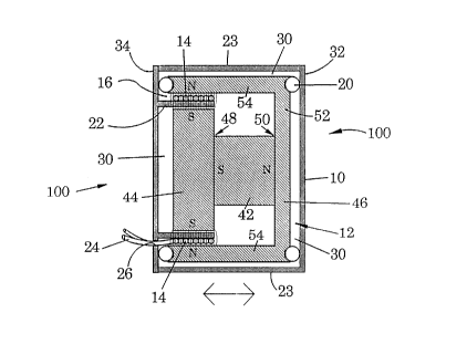
The skucture of an exemplary embodiment of a k~n~d~lcer according to the
present invention is shown in Figures 1 and 2. The implantable k~n~d~lcer 100 ofthe present invention is generally comprised of a sealed housing 10 having a
magnet assembly 12 and a coil 14 disposed inside it. The magnet assembly is
loosely suspended within the housing, and the coil is rigidly secured to the housing.
As will be described, the magnet assembly 12 preferably includes a p~ ntont
magnet and associated pole pieces. When alternating current is conducted to the
coil, the coil and magnet assembly oscillate relative to each other and cause the
housing to vibrate. The housing 10 is propolLioned to be att~r.ll~d within the
middle ear JJ, which comprises the malleus LL, the incus MM, and the stapes HH,
collectively known as the ossicles DD, and the region surrounding the ossicles.
The exemplary housing is preferably a cylindrical capsule having a diameter of 1mm and a thickness of 1 mm, and is made from a biocompatible material, such as
ll. The housing has first and second faces 32, 34 that are subst~nti~lly
parallel to one another and an outer wall 23 which is substantially perpendicular to
the faces 32, 34. Affixed to the interior of the housing is an interior wall 22 which
defines a circular region and which runs subst~nti~lly parallel to the outer wall 23.
The magnet assembly 12 and coil 14 are sealed inside the housing. Air
spaces 30 surround the magnet assembly so as to separate it from the interior of the
21 65557
WO 95/01710 PCT/US94/07283
housing and to allow it to oscillate freely without colliding with the coil or
housing. The magnet assembly is connected to the interior of the housing by
flexible membranes such as silicone buttons 20. The magnet assembly may
alternatively be floated on a gelatinous medium such as silicon gel which fills the
air spaces in the housing. A substantially uniform flux field is produced by
configuring the magnet assembly as shown in Figure l. The assembly includes a
permanent magnet 42 positioned with ends 48, 50 CO~ g the north and south
poles subst~nti~lly parallel to the circular faces 32, 34 of the housing. A first
cylindrical pole piece 44 is connected to the end 48 cont~ining the south pole of
the magnet and a second pole piece 46 is connected to the end 50 cont~ining the
north pole. The first pole piece 44 is oriented with its circular faces parallel to the
circular faces 32, 34 of the housing 10. The second pole piece 46 has a circularface which has a rectangular cross-section and which is parallel to the circular faces
32, 34 of the housing. The second pole piece 46 additionally has a pair a wall 54
which is parallel to the wall 23 of the housing and which surrounds the first pole
piece 44 and the perm~n~nt magnet 42.
The pole pieces must be m~nl-fActllred out of a m~gnetic material such as
SmCo. They provide a path for the m~gnPtic flux of the permanent magnet 42
which is less resistive than the air surrounding the perm~nent magnet 42. The pole
pieces conduct much of the magnetic flux and thus cause it to pass from the second
pole piece 46 to the first pole piece 44 at the gap in which the coil 14 is
positioned.
For the device to operate properly, it must vibrate the ossicles with
sufficient force to transfer vibrations to the cochlear fluid. The force of vibrations
are best m~ximi7Pcl by m~ximi7.ing two parameters: the mass of the magnet
assembly relative to the combined mass of the coil and housing, and the energy
product (EP) of the permanent magnet 42.
The ratio of the mass of the magnet assembly to the combined mass of the
magnet assembly, coil and housing is most easily maximized by constructing the
housing of a thinly machined, lightweight material such as titanium and by
configuring the magnet assembly to fill a large portion of the space inside the
housing, although there must be adequate spacing between the magnet assembly
- 16 -
wo 95/01710 2 1 6 5 5 5 7 PCTrUS94/07283
and the housing and coil for the magnet assembly to swing freely within the
housing.
The magnet should preferably have a high energy product. NdFeB magnets
having energy products of thirty-four and SmCo magnets having energy products oftwenty-eight are presently available. A high energy potential m~ximi7Ps the
attraction and repulsion between the magnetic fields of the coil and magnet
assembly and thereby maximizes the force of the oscillations of the tr~n~dllcer.Although it is preferable to use permanent magnets, electromagnets may also be
used in carrying out the present invention.
The coil 14 partially encircles the magnet assembly 12 and is fixed to the
interior wall 22 of the housing 10 such that the coil is more rigidly fixed to the
housing than the magnet assembly. Air spaces separate the coil from the magnet
assembly. A pair of leads 24 are connected to the coil and pass through an
opening 26 in the housing to the exterior of the tr~n~dllcer, through the surgically-
created channel in the t~ )oldl bone (indicated as CT in Figure 8), and attach to a
subcutaneous coil 28. The subcutaneous coil 28, which is preferably implanted
beneath the skin behind the ear, delivers alternating current to the coil 14 via the
leads 24. The opening 26 is closed around the leads 24 to form a seal (not shown)
which prevents cont~min~ntc from entering the tr~n~d~lcer.
The perception of sound which the vibrating tr~n.cclllcer ultimately triggers isof the highest quality when the relationship between the displAcem~nt of the
housing 10 and the current in the coil 14 is subst~nti~lly linear. For the
relationship to be linear, there must be a CG". ~onding displ~çment of the housing
for each current value reached by the ~ltern~ting current in the coil. Linearity is
most closely approached by positioning and m~ i,.g the coil within the
subst~nti~lly uniform flux field 16 produced by the magnet assembly.
When the magnet assembly, coil, and housing are configured as in Figure 1,
~lt~rn~ting current in the coil causes the housing to oscillate side-to-side in the
directions indicated by arrows Figure 1. The tr~n~ducer is most efficient when
positioned such that the side-to-side movement of the housing produces side-to-side
movement of the oval window EE as indicated by arrows in Figure 3.
wo 9S/01710 2 1 6 5 5 5 7 PCT/US94107283
The tr~ncducer may be affixed to various structures within the ear. Figure
3a shows a tr~ncducer 100 attached to an incus MM by a biocompatible clip 18
which is secured to one of the circular faces 32 of the housing 10 and which at
least partially surrounds the incus MM. The clip 18 holds the tr~n.cducer firmly to
the incus so that the vibrations of the housing which are generated during operation
are conducted along the bones of the middle ear to the oval window EE of the
inner ear and ultimately to the cochlear fluid as described above. An exemplary
clip 18, shown in Figure 3b, includes two pairs of lil~~ prongs 52 which have a
substantially arcuate shape and which may be crimped tightly around the incus.
The tr~ncducer 100 must be connected substantially exclusively to the
ossicles DD or the oval window EE. The tr~nc~lucer must be mechanically isolatedfrom the bone and tissue which surrounds the middle ear since these structures will
tend to absorb the mechanical energy produced by the tr~ncducer. It is thereforepreferable to secure the tr~n.cchlcer 100 to only the ossicles DD or oval window EE
and to therefore isolate it from the surrounding region NN. For the purposes of
this description, the surrounding region consists of all structures in and surrounding
the external, middle, and internal ear other than the ossicles DD, tympanic
membrane CC, oval window EE and any structures connecting them with each
other.
An alternate tr~ncducer 100a having an alternate mech~nicm for fixing the
tr~ncdu(er to structures within the ear is shown in Figures 4 and 5. In this
alternate tr~ncducPr 100a, the housing 10a has an opening 36 passing from the first
face 32a to the second face 34a of the housing and is thereby annular shaped.
When implanted, a portion of the stapes HH is positioned within the opening 36.
This is accomplished by sep~ali~lg the stapes HH from the incus MM and slipping
the O-shaped tr~n~ucer around the stapes HH. The separated ossicles are then
returned to their natural position and where the connective tissue between them
heals and causes them to reconnect. This embodiment may be secured around the
malleus in a similar fashion.
Figures 6 and 7 illustrate the use of the tr~ncducer of the present invention
in combination with total ossicular replacement prostheses (TORPs) or partial
ossicular replacement prostheses (PORPS). These illustrations are merely
- 18 -
~ 2165557
WO 95/01710 PCT/US94/07283
representative: other designs incorporating the transducer into TORPs and PORPs
may be easily envisioned.
TORPs and PORPs are constructed from biocompatible materials such as
titanium. Often during ossicular reconstruction surgery the TORPs and PORPs are
formed in the operating room as needed to accomplish the reconstruction. As
shown in Figure 6, a TORP may be comprised of a pair of members 38, 40
conn~ctçd to the circular faces 32b, 34b of the tr~n~d~cer 100b. The TORP is
positioned between the tympanic membrane CC and the oval window EE and is
preferably of sufficient length to be held into place by friction. Referring to Figure
7, a PORP may be comprised of a pair of members 38c, 40c connected to the
circular faces 32c, 34c of the tr~n~d~lcer a positioned between the malleus LL and
the oval window EE.
Figure 8 shows a schematic replese~ ion of a tr~n.cducer 100 and related
components positioned within a patient's skull PP. An external sound transducer
200, is subst~nti~lly id~rltic~l in design to a conventional hearing aid tr~n~d~lrer and
is comprised of a microphone, sound l.roce~shlg unit, amplifier, battery, and
external coil, none of which are depicted in detail. The external sound tr~n~ cçr
200 is positioned on the exterior of the skull PP. A subcutaneous sound tr~n~ .er
28 is connected to the leads 24 of the tr~n~d~cer 100 and is positioned under the
skin behind the ear such that the external coil is positioned directly over the
location of the subcutaneous coil 28.
Sound waves are ~etectçd and converted to an electrical signal by the
microphone and sound processor of the external sound tr~n~dllc~r 200. The
amplifier amplifies the signal and delivers it to the external coil which subsequently
delivers the signal to the subcutaneous coil 28 by magnetic induction. When the
al~ 1;..g current replesç~-ting the sound wave is delivered to the coil 14 in the
implantable tr~n~du~er 100, the m~gn~tic field produced by the coil interacts with
the m~gn-otic field of the magnet assembly 12.
As the current alternates, the magnet assembly and the coil alternately
attract and repel one another and, with the alternate attractive and repulsive forces
c~ ing the magnet assembly and the coil to alternately move towards and away
from each other. Because the coil is more rigidly att~ch~d to the housing than is
- 19 -
WO 95/01710 2 l 6 5 5 5 7 PCT/US94/07283
the magnet assembly, the coil and housing move together as a single unit. The
directions of the alternating movement of the housing are indicated by arrows inFigure 8. The vibrations are conducted via the stapes HH to the oval window EE
and ultimately to the cochlear fluid.
EXPERIMENTAL
The following examples serve to illustrate certain plefelled embodiments
and aspects of the present invention and are not to be construed as limiting thescope thereof. The ~A~ ental disclosure which follows is divided into: I) In
Vivo Cadaver Examples; and II) In Vivo Subjective Evaluation of Speech and
Music. These two sections summarize the two approaches employed to obtain in
vivo data for the device.
I. IN VIVO CADAVER EXAMPLES
When sound waves strike the tympanic membrane, the middle ear structures
vibrate in response to the intensity and frequency of the sound. In these examples,
l 5 a laser Doppler velocimeter (LDV) was used to obtain curves of device
performance versus pure tone sounds in human cadaver ears. The LDV tool that
was used for these examples is located at the Veterans .Admini~tration Hospital in
Palo Alto, CA. The tool, illustrated in Figure lO, has been used extensively forme~cllring the middle ear vibratory motion and has been described by Goode et al.
Goode et al. used a similar system to measure the vibratory motion of the live
human eardrum in response to sound, the results of which are depicted in Figure
l l, in order to demonstrate the method's validity and to validate the cadaver
temporal bone model.
In each of the three examples that follow, dissection of the human temporal
bone included a facial recess approach in order to gain access to the middle ear.
After removal of the facial nerve, a small target 0.5 mm by 0.5 mm square was
placed on the stapes footplate; the target is required in order to facilitate light
return to the LDV sensor head.
Sound was presented at 80dB sound pressure level (SPL) at the eardrum in
each example and measured with an ER-7 probe microphone 3 mm away from the
- 20 -
2 t 65557
WO 95/01710 PCT/US94/07283
eardrum. An ER-2 earphone delivered pure tones of 80dB SPL in the audio range.
The sound level was kept constant for all frequencies. The displacement of the
stapes in response to the sound was measured by the LDV and recorded digitally
by a computer which utilizes FFT (Fast Fourier Transform); the process has been
automated by a commercially available software program (Tymptest), written for
the applicant's lab, exclusively for testing human temporal bones.
In each example, the first curve of stapes vibration in response to sound
served as a baseline for comparison with the results obtained with the device.
EXAMPLE 1
Tr~n~ducer 4b
Tr~n.cdllcer Construction: A 4.5 mm diameter by 2.5 mm length tr~n.~d~lcer,
illustrated in Figure 12, used a 2.5 mm diameter NdFeB magnet. A mylar
membrane was glued to a 2 mm length by 3 mm diameter plastic drinking straw so
that the magnet was inside the straw. The tension of the membrane was tested forwhat was expected to be the le.luiled tension in the system by p~lp~ting the
structure with a tooth pick. A 5 mm biopsy punch was used to punch holes into anadhesive-backed piece of paper. One of the resulting paper-backed adhesive diskswas placed, adhesive side down, on each end of the assembly m~king sure the
assembly was centered on the adhesive paper structure. A camel hair brush was
used to carefully apply white acrylic paint to the entire outside surface of thebobbin-shaped structure. The painted bobbin was allowed to dry between multiple
coats. This process strengthened the structure. Once the structure was completely
dry, the bobbin was then carefully wrapped with a 44 gauge wire. After an
adequate amount of wire was wrapped around the bobbin, the resulting coil was
also painted with the acrylic paint in order to prevent the wire from spilling off the
structure. Once dried, a thin coat of five-minute epoxy was applied to the entire
outside surface of the structure and allowed to dry. The resulting leads were then
stripped and coated with solder.
Methodology: The tr~n~ cer was placed between the incus and the malleus
- 30 and moved into a "snug fit" position. The tr~n~d-lcer was connected to the Crown
amplifier output which was driven by the computer pure-tone output. The current
- 21 -
21 65557
WO 95/01710 PCT/US94/07283
was recorded across a 10 ohm resistor in series with Transducer 4b. With the
transducer in place, the current to the kansducer was set at 10 milli~mps (mA) and
the measured voltage across the tr~n.ed~lcer was 90 millivolts (mV); the values were
constant throughout the audio frequency range although there was a slight variation
S in the high frequencies above 10 kHz. Pure tones were delivered to the tr~nednrer
by the computer and the LDV measured the stapes velocity resulting from
tr~ned~lcer excitation. The resulting figure was later converted into displacement
for purposes of graphical illustration.
Results: As Figure 13 depicts, the tr~ned~lcer resulted in a gain in the high
frequencies above 2 kHz, but little improvement was observed in the low
frequencies below 2 kHz. The data marked a first successful attempt a
m~nnf~r,tllring a k~ned~lcer small enough to fit within the middle ear and
demonstrated the device's potential for high fidelity-level performance. In addition,
the tr~nerlllcer is designed to be ~ rhPd to a single ossicle, not held in place by
the tension between the incus and the malleus, as was required by the crude
prototype used in this example. More advanced prototypes affixed to a single
ossicle are ~;Ape~iled to result in improved perform~nce.
EXAMPLE 2
Tr~n.ed~lcer 5
Tr~ne-l~lc~r Construction: A 3 mm diameter by 2 mm length tr~n.ed~lcer
(similar to Tr~ned~lc~r 4b, Figure 12) used a 2 mm diameter by 1 mm length
NdFeB m~gn~t A mylar membrane was glued to a 1.8 mm length by 2.5 mm
diameter plastic drinking straw so that the magnet was inside the straw. The
rem~ining description of Tr~ned~cer 5's construction is analogous to that of
Tr~neducer 4b in Example 1, supra, except that: i) a 3 mm biopsy punch was used
instead of a S mm biopsy punch; and ii) a 48 gauge, 3 litz wire was used to wrapthe bobbin structure instead of a 44 gauge wire.
Methodology: The tr~ned~lcPr was glued to the long process of the incus
with cyanoacrylate glue. The tr~ned~lcer was connected to the Crown amplifier
which was driven by the computer pure-tone output. The current was recorded
across a 10 ohm resistor in series with Tr~ned~lcer S. The current to the tr~ned~lcer
- 22 -
WO 95/01710 2 1 6 5 5 5 7 PCT/US94107283
-
was set at 3.3 mA, 4 mA, 11 mA, and 20 mA and the measured voltage across the
transducer was 1.2 V, 1.3 V, 1.2 V, and 2.5 V, respectively; the values were
constant throughout the audio frequency range although there was a slight variation
in the high frequencies above 10 kHz. Pure tones were delivered to the transducer
by the computer, while the LDV measured stapes velocity, which was subsequently
converted to displacement for graphical illustration.
Results: As Figure 14 shows, Tr~n~d~lrer 5, a much smaller transducer than
Tr~n~d~lcer 4b, demonstrated marked improvement in frequencies between 1 and
3.5 kHz, with m~imnm output excee~ing 120dB SPL equivalents when compared
to stapes vibration when driven with sound.
EXAMPLE 3
Tr~n~d~lcer 6
Transducer Construction: A 4 mm diameter by 1.6 mm length transducer
used a 2 mm diameter by 1 mm length NdFeB magnet. A soft silicon gel material
(instead of the mylar membrane used in Examples 1 and 2) held the magnet in
position. The magnet was placed inside a 1.4 mm length by 2.5 mm diameter
plastic drinking straw so that the magnet was inside the straw and the silicon gel
material was gingerly applied to hold the m~gn~t The tension of the silicon gel
was tested for what was expected to be the required tension in the system by
palp~ting the structure with a tooth pick. The rem~ining description of the
Tr~n~-hlc~r 6's construction is analogous to that of Tr~n~ducer 4b in Example 1,supra, except that: i) a 4 rnm biopsy punch was used instead of a 5 mm biopsy
punch; and ii) a 48 gauge, 3 litz wire was used to wrap the bobbin structure instead
of a 44 gauge wire.
Methodology: The tr~n~-lucPr was placed between the incus and the malleus
and moved into a "snug fit" position. The tr~n.echlcer's lead were connected to the
output of the Crown amplifier which was driven by the computer pure-tone output.The current was recorded across a 10 ohm precision resistor in series with
Tr~n~d-.cçr 6. In this example, the current to the tr~n~-l--cer was set at 0.033 rnA,
- 30 0.2 mA, 1 mA, 5 mA and the measured voltage across the tr~n.cdllcer was 0.83
mV, 5 mV, 25 mV, 125 mV, respectively; these values were constant throughout
- 23 -
wo 9S/01710 2 1 6 5 5 5 7 PCT/US94/07283
the audio frequency range although there was a slight variation in the high
frequencies above 10 kHz. Pure tones were delivered to the tr~n.cducer by the
computer, while the LDV measured the stapes velocity, which was subsequently
converted to displacement for graphical illustration.
Results: As Figure 15 depicts, the tr~n~dllcer resulted in marked
improvement in the frequencies above 1.5 kHz, with maximum output excee~ling
120dB SPL equivalents when compared to the stapes vibration baseline driven withsound. The crude prototype demonstrated that the device's potential for significant
sound improvement, in terms of gain, could be expected for those suffering from
severe hearing hllpahlllent. As was stated in Example 1, the tr~n~ducer is designed
to be attached to a single ossicle, not held in place by the tension between the incus
and the malleus, as was required by the prototype used in this example. More
advanced prototypes affixed to a single ossicle are expected to result in improved
performance.
II. IN VIVO SUBJECTIVE EVALUATION OF SPEECH AND
MUSIC
This example, con-lucted on living human subjects, resulted in a subjective
measure of tr~n~ducer performance in the areas of sound quality for music and
speech. Tr~n~ducer 5, used in Example 2, supra, was used in this example.
EXAMPLE 4
Methodology: A soft silicon gel hllplession of a tympanic membrane,
resembling a soft contact lens for the eye, was produced, and the tr~n~ducer wasglued to the concave surface of this i",plession. The tr~n~duc~r and the connected
silicon h"plession were then placed on the subject's tympanic membrane by an
otologic surgeon while looking down the subject's extern~l ear canal with a Zeiss
OPMI-l stereo surgical microscope. The device was centered on the tympanic
membrane with a non-magnetic suction tip and was held in place with mineral oil
through surface tension between the silicon gel membrane and the tympanic
membrane. After in~t~ tion, the tr~n~ducer's leads were taped against the skin
- 24 -
WO 95/01710 2 1 6 5 5 5 7 PCT/US94/07283
posterior to the auricle in order to prevent dislocation of the device during testing.
The tr~n.c~ cer's leads were then connected to the Crown D-75 amplifier output.
The input to the Crown amplifier was a common portable compact disk (CD)
player. Two CDs were used, one featuring speech and the other featuring music.
The CD was played and the output level of the tr:~n~d~lcer was controlled with the
Crown amplifier by the subject. The subject was then asked to rate the sound
quality of the device.
Results: The example was conducted on two subjects, one with normal
hearing and one with a 70 dB "cookie-bite" sensori-neural hearing loss. Both
subjects reported excellent sound quality for both speech and music, no distortion
was noticed by either subject. In addition, the hearing-impaired subject indicated
that the sound was better than the best hi-fidelity equipment that he had heard.One should recall that the tr~n~ cer is not design~l to be implanted in a silicon
gel membrane att~r~Pd to the subject's tympanic membrane. The method described
was utilized because the crude tr~n~dllcer prototypes that were tested could never
be used in a live human in implanted form, the method was the closest
approximation to actually implanting a tr~n~ cer at the time the test was
pel~o~ ed, and the applicant needed to validate the results observed from the InVivo Cadaver Examples with a subjective evaluation of sound quality.
From the above, it should be clear that the present invention provides an
easily implantable electromagnetic tr~n~dl~cer. The ~p~dlus conducts vibrations to
the oval window with sufficient force to stim~ te hearing perception with minim~l
distortion.
- 25 -