Note: Descriptions are shown in the official language in which they were submitted.
095/0WK4 216 5 S 6 2 PCT~S94/0~2
--1--
1 Specification
3 APPARATUS AND NETHOD FOR INNEDIATE DIAGNOSIS
4 OF VAGINAL YEAST INFECTIONS
6 R~r,Rr~OUND OF THE lNv~:N-llON
7 Field of the Invention
8 The present invention relates generally to a method
9 and apparatus for immediate diagnosis of exudative vaginal
yeast infections. In particular, this invention relates
11 to a method and apparatus which permit diagnosis or
12 ruling-out of vaginal yeast infections without culturing
13 yeast, thereby permitting immediate diagnosis through
14 fluorescence of a dyed sample of the vaginal exudate.
16 Brief Description of the Prior Art
17 Vaginal yeast infections are a common problem
18 affecting women of all ages. Vaginal anti-yeast
19 prescriptions numbered 13 million in 1990 in the U.S. The
infection results from the ove,yLowLh of yeast which are
21 often normally present but in much smaller numbers. The
22 symptoms of a yeast infection include vaginal itching,
23 discharge, soreness, irritation or burning. Since a
24 vaginal yeast infection is strictly due to the presence of
large numbers of the responsible organism, diagnosis and
26 treatment would seem to be simple. However, other more
27 serious vaginal infections can present a similar clinical
28 picture which usually includes local itching, a vaginal
29 discharge, and possibly abdominal pain and fever. For
example, bacterial vaginosis, bacterial trichomoniasis,
31 chlamydial infections, and gonorrhea can resemble yeast
32 infections, especially to women without medical training.
33 Presently, over-the-counter anti-yeast medications
34 have been approved for treating vaginal yeast infections.
35 The availability of these medications encourages women to
36 self-diagnose and self-treat a potentially serious medical
37 problem, without a medical ex~m-n~tion, based upon a hope
38 that the problem may simply be due to yeast. Self-
39 diagnosis without diagnostic data is dangerous, since
~l6ss62
W095/~W64 PCT~S94106862
2--
1 proper treatment may be delayed or the wrong treatment may
2 be undertaken, possibly leading to invasive infections or
3 sterility due to pelvic inflammatory disease.
4 The prior art teaches confirmation of the presence of
an infectious organism through the use of a variety of
6 culture techniques. U.S. Patents Nos. 3,368,569 and
7 4,953,560 teach use of a swab including a culture medium.
8 U.S. Patents No. 3,616,265, 4,653,510, and 4,485,824 teach
9 a variety of swabs and culture mediums for simplification
of transferring a vaginal secretion to a culture medium.
11 All of these techniques require that the yeast be
12 cultured, a technique that requires incubation of the
13 yeast for 12 to 24 hours under aseptic conditions with
14 complex agar media by medical personnel. Therefore, none
of these devices provide an immediate method for diagnosis
16 of the presence of the high number of yeast associated
17 with a vaginal yeast infection, and none of them are
18 applicable to a test which may be used at home by persons
19 who are not medically trained.
21 SUMMARY OF THE INVENTION
22 It is a primary object of the present invention to
23 provide a method and an apparatus for immediate and
24 inexpensive determination of whether the large numbers of
yeast associated with vaginal yeast infections are
26 present.
27 It is a further object of the present invention to
28 provide a method and an apparatus which are easy to use so
29 that a woman may immediately determine whether or not a
high number of vaginal yeast are present, indicating a
31 possible yeast infection, or alternatively whether
32 symptoms associated with a yeast infection may be due to
33 some other more serious problem with similar symptoms.
34 Another object of the present invention is to provide
a simple, self-administered, inexpensive, accurate and
36 reliable test system that allows a woman to immediately
37 determine if she has an overgrowth of vaginal yeast to
38 insure that diagnostic data is available before treatment
39 is begun.
O95/OWK4 21 6 5 5 6 2 PCT~S94/06~2
--3
1 A further object of the present invention is to
2 provide a test that indicates whether treatment for yeast
3 infection is inappropriate, thereby saving critical time
4 and indicating that a medical visit is necessary instead.
Briefly, the preferred embodiment of the present
6 invention is a method and apparatus for immediately
7 detecting the presence of high numbers of vaginal yeast
8 which are associated with yeast infections through a
9 method and apparatus which use a sample of vaginal
discharge, dyes the yeast present in the discharge with a
11 fluorescent dye specifically sensitive to yeast, and
12 subjects the dye to ultraviolet radiation to determine the
13 level of visible fluorescence. The level of fluorescence
14 indicates the presence or absence of the high number of
yeast associated with a vaginal infection.
16 The att~;nment of the foregoing and related objects,
17 advantages and features of the invention should be more
18 readily apparent to those skilled in the art after review
19 of the following more detailed description of the
invention.
21
22 IN THE DRAWING
23 Fig. 1 is a plan view of the preferred embodiment of
24 a kit for immediate diagnosis of vaginal yeast infections
in accordance with this invention; and
26 Fig. 2 is a perspective view of a specimen slide
27 designed for use with this invention.
28
29 DET~T~-~n DESCRIPTION OF THE ~K~KK~ EMBODIMENT
This invention constitutes a method and an apparatus
31 for immediate diagnosis of vaginal yeast infections. With
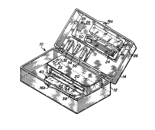
32 reference to Fig. 1, a yeast detection test kit 10 is
33 shown. Kit 10 includes a kit body with lower test kit
34 body 12 and upper test kit body 14. The test kit body is
35 preferably formed of an inexpensive but durable material,
36 such as plastic. For reasons discussed below concerning
37 the fluorescence method of detection used, the test kit
38 body is preferably black to absorb any stray light and to
39 make the detection method more sensitive by making
WO95/OWK4 ~6s5 6~ PCT~S94/06862
1 fluorescence easier to perceive. The test kit body
2 includes a closing tab 16 which attaches a locking piece
3 (not shown) on lower test kit body 12.
4 Lower test kit body 12 contains the elements of the
test kit used in conducting the yeast infection test.
6 These include a fluorescence light housing 18 which
7 includes a fluorescence light bulb 20, on/off switches 22,
8 and a battery (not shown) to supply power to the
9 fluorescence light bulb 20. The preferred embodiment uses
near-ultraviolet light to diagnose yeast infections
11 through visible fluorescence of a dyed yeast sample.
12 Therefore, a battery-powered near-W ultraviolet light is
13 used. An example of a suitable battery-powered
14 fluorescent lamp which has proved suitable is Radio Shack
Catalog No. 61-2734, using a black light W bulb No.
16 F4T5BLB made by WKO, in Japan.
17 Lower test body 12 also includes a snap-in holder 38
18 which is used for accurately positioning a specimen slide
19 40 relative to fluorescence light bulb 20.
Upper test kit body 14 includes the materials
21 necessary for obtaining and preparing a vaginal exudate
22 specimen for testing. Test equipment compartment 28 holds
23 specimen slides 26, cover slips 25, and swabs 24. Cover
24 slips 25 are preferably glass, and swabs 24 are preferably
composed of cotton or some other absorbent material. A
26 more detailed view of slides 26 is shown in lower test kit
27 body 12 as slide 40 and in Fig. 2. Specimen slide 40 has
28 an area for a control portion 42 and an area for an
29 exudate portion 44. Preparation of the specimen slide 40
is described in further detail below.
31 With reference to Fig. 2, an embodiment of a specimen
32 slide 40 is shown in perspective view. A glass or black
33 plastic slide is the present preferred embodiment, but an
34 absorptive slide as shown in Fig. 2 may be used. The
absorptive slide includes a top layer 52 which is
36 comprised of a 1 micrometer milipore non-cellulose filter
37 paper, which is dyed black so that fluorescence from the
38 slide is easier to detect. The middle layer 54 is
OgS/00064 21 ~SS PCT~S94/06862
1 comprised of absorptive filter paper, and the bottom layer
2 56 is comprised of a rigid fiberboard support.
3 Referring again to Fig. 1, upper test kit body 14
4 also includes a solution compartment 30 which holds three
5 bottles of test solution: an alkaline treatment solution
6 32, a dye solution 34, and a rinse solution 36.
7 Yeast infection test kit 10 is designed to facilitate
8 simple and immediate diagnosis of yeast infections. The
9 kit allows a woman to perform a test in a quick, simple
and private manner.
11 The following criteria have been established for
12 vaginal yeast infections. Vaginal yeast at levels of less
13 than 103 colony-forming units/milliliter of secretion are
14 normal vaginal flora, not representing yeast infection and
15 are present in up to 50~ of the female post-puberty
16 population. Only levels of yeast of 104 colony-forming
17 units/milliliter of secretion or higher constitute the
18 overgrowth of yeast diagnosed as a yeast infection.
19 Therefore, vaginal yeast infection is a relative
20 concentration diagnosis. It takes approximately 103 total
21 yeast/milliliter as determined by direct microscopic
22 hemocytometer count to give one colony-forming unit on a
23 culture plate. Therefore, yeast infection is only present
24 when the total concentration of all yeast cells exceeds
25 approximately 107 yeast/milliliter. Recent studies have
26 measured infection concentrations exceeding 109 colony-
27 forming units/millimeter of exudate.
28 Specimen slides 26 are packaged in a protective
29 packet, such as plastic or foil, which is preferably
30 airtight and purged with nitrogen to insure a non-
31 contaminated and non-oxidizing environment for the
32 specimen slide 26. The presence of a non-contaminated,
33 non-oxidizing environment is important because specimen
34 slide 26 contains a control portion, as shown in the
35 detail of specimen slide 40 in Figs. 1 and 2. Control
36 portion 42 of specimen slide 40 contains a concentration
37 of yeast which correlates with the minimum concentration
38 found in vaginal yeast infections. As noted above, a
39 yeast infection is only present when the total
W095/0HK4 ~ ~6~S PCT~S94/0~K2
--6--
1 concentration of yeast cells exceeds approximately 107
2 yeast/milliliter. In general, higher numbers of yeast
3 correlate with more severe clinical symptoms, i.e. higher
4 levels of yeast cause a more severe infection. Therefore,
control portion 42 holds a standardized yeast sample of
6 approximately 107 yeast/milliliter. The yeast need not be
7 alive. We have found that a control specimen has
8 sufficient lifetime to permit use of a pre-packaged
9 control over an extended time period (exceeding several
months), which may be lengthened by use of packaging to
11 retard contamination and aging of the control specimen.
12 Slide 40 preferably includes a notation (on the slide
13 itself or on its packaging) of the expiration date for the
14 control portion 42 of the slide.
This test method measures total yeast concentration
16 by staining the yeast with a dye, Calcofluor White, which
17 forms a specific chemical bond to cellulose and chitin in
18 the yeast cell wall. Other biological agents present in
19 vaginal discharges are dissolved in the preparation of the
20 specimen slide. When yeast stained with calcofluor white
21 are exposed to ultraviolet light, a green fluorescence is
22 emitted. At the level of 107 total yeast/millimeter (the
23 concentration of the preferred standardized sample), the
24 fluorescence of the stained sample is easily visible to
25 the naked eye. Higher concentrations, as would occur in
26 more severe yeast infections, are brighter.
27 The test for yeast infection is conducted as follows.
28 First, a slide 26 iS removed from its package and placed
29 on paper towels. A sample of vaginal discharge is
30 obtained with a swab 24, and then the swab is dabbed or
31 rolled over the exudate sample portion 44 of slide 40.
32 This is preferably done 1 or 2 times with the slide, at
33 two-minute intervals. Exudate portion 44 is allowed to
34 dry for approximately four or five minutes.
The exudate portion 44 and control portion 42 of
36 specimen slide 40 are prepared by first applying several
37 drops of an alkaline treatment and wash solution 32 to
38 exudate portion 44 and control portion 42. The alkaline
39 treatment solution preferably consists of approximately
095/~WK4 21 ~SS6 PCT~S94/06862
--7--
1 10~ by weight potassium hydroxide in water, or a similar
2 alkaline solution such as 10~ sodium hydroxide in water.
3 The potassium hydroxide solution dissolves only non-yeast
4 structures. The potassium hydroxide solution is allowed
to sit on the sample areas for about thirty seconds. The
6 slide is then tipped on its side to allow any excess
7 solution to run off onto the paper towel.
8 Next, several drops of dye solution 34, preferably
9 calcofluor white, is added to exudate portion 44 and
control portion 42 of specimen slide 40 and allowed to sit
11 for about 30 seconds. Calcofluor white, an optical
12 brightener, is a colorless dye used as a whitening agent
13 in the textile and paper industry. Because it binds to
14 cellulose, chitin, and fungal elements, and fluoresces
when exposed to long wavelength W and short wavelength
16 visible light, it has been used to demonstrate cellulose
17 in microorganisms, stain the cell walls of plants, and to
18 screen specimens for fungal elements. The preferred dye
19 solution utilizes 0.1 gram calcofluor white M2R (Poly-
sciences, Inc., Warrington, PA, or Sigma Chemical Co., St.
21 Louis, MO) and 0.05 gram Evans Blue (Sigma Chemical)
22 dissolved in 100 ml distilled water.
23 After the dye solution has been applied, the slide is
24 again tipped to allow excess solution to run off onto the
paper towel. Now, several drops of rinse solution 36,
26 preferably approximately 10~ KOH in water (which does not
27 affect calcofluor's binding to yeast), are gently placed
28 on the slide sample areas and allowed to stand flat and
29 still for about thirty seconds. The slide is then tipped
on its side again to allow any excess solution to run off
31 onto the paper towel. This further removes any dye not
32 bound to yeast. A cover slip 25 is placed directly on top
33 of the sample portions of specimen slide 40 and allowed to
34 sit for about 5 seconds. The specimen slide 40 is then
turned over and gently pressed down on the paper towel to
36 express any excess solution.
37 Slide 40 is turned right side up and placed into
38 snap-in holder 38, which positions the slide exactly with
39 respect to fluorescence light bulb 20.
WO95/~WK4 ~ ~6~ 6 PCT~S94/06862
1 Once specimen slide 40 has been prepared, the kit is
2 taken into a darkened room, and the fluorescence light
3 bulb 20 is turned on. The room is preferably as dark as
4 possible so that the eye-sensitivity in viewing the
5 fluorescence of the specimen slide 40 will be as sensitive
6 as possible. Any bluish-green glow coming from the
7 exudate portion 44 of specimen slide 40 is compared
8 visually to the glow coming from the control portion 42 of
9 the specimen slide 40. When the exudate portion 44 and
control portion 42 of specimen slide 40 are compared for
11 relative fluorescence, a fluorescence in the exudate
12 portion 44 greater than or equal to the fluorescence of
13 the control portion 42 of the specimen slide indicates the
14 presence of a concentration of yeast which indicates a
15 yeast infection. If the exudate glow is less bright than
16 the glow from the standard sample area, then a diagnosis
17 of vaginal yeast cannot be made with certainty, and the
18 woman should consult a doctor as soon as possible to
19 determine whether or not a more serious infection is
present.
21 The present invention uses both positive and negative
22 results to obtain useful information concerning the
23 possible causes of the vaginal discharge or discomfort.
24 In particular, if a yeast infection is diagnosed, over-
25 the-counter anti-yeast medication can be used. If the
26 test is negative, the woman has been able to eliminate a
27 yeast infection as the cause for a problem and will not be
28 tempted to self-treat for a yeast infection
29 inappropriately, and will be on notice that a more
30 thorough medical test is required.
31 The test kit 10 of this invention may be used for
32 multiple diagnosis provided that additional specimen
33 treatment solutions 32, 34 and 36, specimen slides 26,
34 swabs 24, and cover slips 25 are provided as required.
We have compared the results obtained with the method
36 and kit of this invention with the results obtained in a
37 medical clinic, which utilize microscopic evaluation of
38 slides to determine the presence of yeast or other agents.
39 In approximately 98~ of the cases, we find agreement
95/00064 9 1 6SS62 PCT/US94/06862
1 between our results in diagnosing yeast infection and the
2 clinical microscopic test results.
3 This invention has been described in terms of a
4 specific dye, calcofluor white, which stains yeast to
~ 5 fluoresce blue-green under ultraviolet light. This system
6 is particularly useful because the excitation light (near
7 W and short wavelength visible (purple)) is easily
8 distinguished from the fluorescence (blue-green) with the
9 naked eye. However, any dye fluorescence test which
correlates specifically with yeast concentration will work
11 suitably well in this method. For example, yeast
12 concentration could be correlated with fluorescence from
13 fluorescein-labeled anti-yeast antibodies in an immuno-
14 fluorescence microscopic technique.
This invention has been described in terms of finding
16 concentrations of yeast in vaginal exudates. However, it
17 is equally applicable to finding concentrations of yeast
18 in any fluids or semi-solid matter. The test would be
19 performed in the matter described above for the
fluid/semi-solid matter of interest.
21 For example, if urine were used in the sample slide
22 portion instead of vaginal exudate, the results would be
23 indicative of a bladder yeast infection. The fluorescent
24 intensity for such infections would be similar to those
associated with vaginal yeast infections.
26 Another example is oral yeast infections (thrush).
27 If the white, semi-solid curd-like exudate of thrush were
28 used in the sample area, the kit and method described
29 above would easily show very bright fluorescence in this
essentially solid yeast culture. Use of the kit and
31 method would be identical, with Q-tip application of the
32 sample material adjacent to the control sample. The
33 concentration and amount of yeast in the control portion
34 would be the controlled variable for various diagnostic
- 35 conclusions.
36 Thus, the amount of yeast in the control portion
37 could be used for semi-quantitative comparison and
38 measurement of the yeast levels in any fluid or semi-
39 solid, whether or not the material is biological or
W095/~64 ~6~ PCT~S94/~62
--10--
1 infection exudate, where one wants a semi-quantitative
2 measure of yeast amount or concentration. Adjustment of
3 the yeast concentration in the control specimen allows
4 calibration of the method.
The visibility of fluorescence to the naked eye
6 starts at a yeast concentration of one million yeast per
7 milliliter. For use with the naked eye, the control
8 portion concentration useful for comparison to samples
9 must be above this one million level. Below this level
concentrations cannot be determined by the naked eye.
11 Fortuitously, vaginal and bladder yeast infections begin
12 at this level. Moreover, in samples of solid exudate, as
13 in thrush and some vaginal yeast infections, the exudates
14 are essentially of infinite yeast concentration since they
15 consist of basically solid yeast culture.
16 If an electro-optical fluorescent reader and
17 comparator is employed, yeast concentrations below the one
18 million yeast per milliliter level can be detected, and
19 quantitative measurement of both higher and lower levels
20 is straightforward with use of calibrated control
21 specimens.
22 A restriction that applies is that the sample
23 material must be free of "interfering substances". These
24 would consist of any plant-derived materials such as
25 paper, cotton, cork or cellulose-related products. With
26 regard to the use of Calcofluor White dye, this dye
27 (Calcofluor White) stains the cellulose and chitin of
28 fungal cell walls. Therefore, any non-fungal animal or
29 plant product containing these chemicals would be an
30 interfering substance.
31 In body exudates, such as vaginal exudates, urine, or
32 thrush plaques, the only living source of this cellulose-
33 chitin material is yeast. In this way, interfering
34 substances are ruled out by the source of the sample as
35 well as by the pre-treatment and final rinse of the
36 materials with 10~ KOH. Interfering non-living
37 substances, such as cotton fibers from clothes or Q-tips,
38 would either not be present or occur in amounts too low to
39 be visibly fluorescent to the naked eye. Interfering
095/O~K4 S~S2 PCT~S94/06862
1 substances can occur in feces due to ingestion. In the
2 absence of large amounts of interfering substances, the
3 kit and method described above may be used to detect the
4 amount of yeast present in any fluid/semi-solid material.
Although the present invention has been described in
6 terms of a specific embodiment, it is anticipated that
7 alterations and modifications thereof will no doubt become
8 apparent to those skilled in the art. It is therefore
9 intended that the following claims be interpreted as
covering all such alterations and modifications as fall
11 within the true spirit and scope of the invention.
12 What is claimed is: