Note: Descriptions are shown in the official language in which they were submitted.
2168713
PATENT
P-2962
5 CONTROL FORWARD/FLASHBACK FORWARD ONE HAND INTRODUCER NEEDLE
AND CATHETER ASSEMBLY
Back~round of the Invention
This invention relates to a catheter and introducer needle assembly. Catheters,
particularly intravenous (IV) catheters, are used for directing~fluid into or withdrawing fluid
from a patient. The most common type of IV catheter is an over-the-needle IV catheter. As
its name implies, an over-the-needle IV catheter is mounted over an introducer needle having
a sharp distal tip. With the distal tip of the introducer needle extending beyond the distal tip
of the IV catheter, the assembly is inserted through the patient's skin into a vein. Once
placement of the assembly in the vein is verified by flashback of blood in the needle, the
needle is withdrawn leaving the IV catheter in place. The proximal end of the IV catheter
typically has a hub that is designed to be connectable to an IV fluid supply line after insertion
of the IV catheter in a patient.
Although typical IV catheter and introducer needle assemblies generally perform their
functions satisfactorily, they do have certain drawbacks. For example, the entire procedure
of properly placing an IV catheter into a patient can be cumbersome and awkward and
require the use of both hands of the healthcare worker. In addition, prior IV catheter and
introducer needle assemblies require a flashback chamber located on the proximal end of the
needle. This location is inconvenient for the healthcare worker because during insertion of
the assembly into a patient, the healthcare worker's attention is directed to the distal tip of
the needle. Thus in order to determine if the needle is properly placed in a vein, the
healthcare worker has to divert his attention away from the point of insertion of the IV
catheter and introducer needle assembly into the patient. Furthermore, prior IV catheter and
introducer needle assemblies do not provide a simple means of breaking adhesion between
the needle and IV catheter prior to insertion of the assembly into the patient. Adhesion can
~16~713
occur because the inside diameter of the catheter is typically less than the outer diameter of
the needle to provide a smaller profile for the assembly and facilitate insertion of the IV
catheter into the patient. Thus, when the assembly is sterilized and is not used for a period
of time after sterilization, the catheter can adhere to the introducer needle. In addition, the
lubricant that is used on the introducer needle and catheter can contribute to tip adhesion.
Finally, prior IV catheter and introducer needle assemblies do not allow for the flexibility of
allowing an IV fluid supply line to be connected to the IV catheter prior to insertion of the IV
catheter and introducer needle assembly into a patient.
Summarv of the Invention
It is therefore an object of this invention to provide an IV catheter and introducer
needle assembly that is easy to control and insert into a patient.
It is another object of this invention to provide an IV catheter and introducer needle
assembly that allows the healthcare worker to determine if the assembly is properly placed in
a patient's vein without the need for the healthcare worker to divert his attention away from
the insertion site.
It is yet a further object of this invention to provide an IV catheter and introducer
needle assembly that allows an IV fluid supply line to be connected to the IV catheter prior to
insertion of the assembly into a patient.
It is still a further object of this invention to provide an IV catheter and introducer
needle assembly that allows adhesion between the IV catheter and the needle to be broken
prior to insertion of the assembly into a patient.
It is yet another object of this invention to provide an IV catheter and introducer needle
assembly that allows the healthcare worker to withdraw the needle into the IV catheter by
using only one hand.
The above and other objects are satisfied by the IV catheter and introducer needle
assembly of this invention. The IV catheter includes a hub at its proximal end having a wing
2~G8713
radially extending from the IV catheter hub. This catheter wing is generally aligned with the
longitudinal axis of the catheter. The IV catheter hub has a side port in fluid communication
with the IV catheter. The side port is preferably located distal of the catheter wing. The
needle is connected at its proximal end to a needle hub and has a pair of notches formed
5 therein. The notches are formed in the needle such that blood can flow from the needle tip
inside the needle through the notches outside the needle into the catheter lumen and through
the side port into a translucent extension tube that extends along the distal end of the
catheter wing. In this way, the healthcare worker can visualize flashback of blood when the
needle has been inserted into a patient's vein.
The needle hub has a radially extending fin formed thereon and is generally aligned
with the longitudinal axis of the needle. The needle fin and catheter wing are arranged so
that they can rotate with respect to one another. This allows the needle to rotate with respect
to the catheter to break any adhesion therebetween. In addition, the needle fin extends
forward of the catheter wing when the needle hub is fully engaged with the IV catheter hub.
This arrangement allows the distal tip of the needle to be retracted into the catheter with a
simple proximal motion by the healthcare worker on the needle fin.
Brief Description of the Drawin~s
The above and other objects and advantages will be apparent upon consideration of
the following drawings and detailed description. The preferred embodiments of the present
invention are illustrated in the appended drawings in which like reference numbers refer to
like elements and in which:
FIG. 1 is a left perspective view of the IV catheter and introducer needle of this
invention;
FIG. 2 is a cross-sectional view of the invention taken along line 2-2 in FIG. 1 showing
the assembly prior to insertion into a patient with the needle in the forward position;
216871 3
-
FIG.3is a right perspective view of this invention showing the rotation of the needle
with respect to the catheter;
FIG.4is a cross-sectional view of the invention similar to the view of FIG.2 after the
distal tip of the needle has been retracted into the catheter;
FIG.5is a left side elevation view of the invention after the distal tip of the needle has
been retracted;
FIG.6is a right side elevation view of the invention showing the side opposite to the
side shown in FIG.5;
FIG.7is an enlarged view of the forward portion of the invention shown in FIG.2;FIG.8is similar to the view shown in FIG.7 but with the needle retracted;
FIG.9is an enlarged view of the distal tip of the needle and catheter with the needle
in the forward position;
FIG.10is an enlarged view of the distal tip of the needle and catheter with the needle
rotated with respect to the catheter; and
FIG.11is an enlarged view of the distal tip of the needle and catheter with the needle
withdrawn into the catheter.
Detailed Description of the Invention
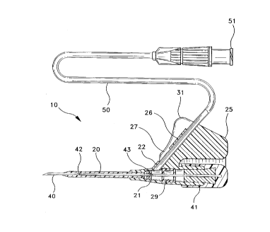
The catheter and introducer needle assembly 10 of this invention is shown in FIG.1.
This assembly includes catheter 20 affixed to catheter hub 21 and needle 40 affixed to
needle hub 41.
Catheter hub 21 includes a side port 22 which has an extension tube 50 connectedthereto located toward the distal portion of catheter hub 21. The proximal end of extension
tube 50 includes a standard luer lock adaptor 51 to allow the connection of an IV fluid supply
line to extension tube 50. Such an IV fluid supply line can be connected to extension tube 50
prior to insertion of assembly 10 into a patient. Side port 22is in fluid communication with
the lumen of catheter 20 so that fluid infused through extension tube 50 will pass into the
21G87~ 3
patient once catheter 20 is properly positioned in the patient. Conversely, blood exiting a
patient's vein through catheter 20 can travel through extension tube 50. The proximal end of
catheter hub 21 is sealed with an elastomeric plug 29, such as silicone, (See FIGS. 2 and 4)
to ensure that fluid does not leak out of the proximal end of catheter hub 21. Catheter hub 21
5 also includes a wing 25 that extends radially from catheter hub 21. Wing 25 is generally
aligned with the longitudinal axis of catheter 20. Wing 25 should be large enough to be
grasped easily by a healthcare worker.
The forward or distal end of wing 25 defines a tubular member 26. Preferably tubular
member 26 does not extend the full length of the distal end of wing 25. Extension tube 50 fits
10 through tubular member 26 and into communication with side port 22. The space between
side port 22 and tubular member 26 defines a window 27 that allows the healthcare worker to
observe the distal portion of extension tube 50~ At least that part of extension tube 50 that
extends past window 27 is formed from polyvinyl chloride so that it is translucent or
transparent in window 27. In this manner, flashback of blood can be observed in window 27
15 once needle 40 has entered a patient's vein. Window 27 is not necessary where tubular
member 26 is made of a clear material such as polycarbonate. In such a case, flashback of
blood through extension tube 50 can be seen through tubular member 26.
Needle 40 has its proximal end connected to needle hub 41. Radially extending fin 31
is formed on needle hub 41 and is generally longitudinally aligned with needle 40. Needle 40
20 includes a pair of notches 42 and 43 formed therein. One notch 42 is preferably located
adjacent to the distal tip, while the other notch 43 is positioned on needle 40 such that when
needle 40 is inserted into catheter 20 in the fully forward position, see FIG. 2, notch 43 is
distal of elastomeric plug 29 and is in fluid communication with extension tube 50. Notch 42
is preferably located within about 1/2 inch of the heel of the needle. Notch 43 is preferabiy
25 located within 1/2 inch of side port 22.
When needle 40 is inserted completely into catheter 20 in the forward most position so
that needle hub 41 is engaged with catheter hub 21, the distal tip of needle 40 extends
~(i8713
beyond the distal tip of catheter 20 as shown in FIG. 2. In addition, the distal end of fin 31
extends beyond the distal end of wing 25 as shown in FIG. 2. In this arrangement and with
fin 31 adjacent to wing 25, catheter and introducer needle assembly 10 is ready for insertion
into a patient. The healthcare worker grips both wing 25 and fin 31 to insert catheter and
5 introducer needle assembly 10 into a patient~ Because wing 25 and fin 31 are close to the
distal tip of needle 40, insertion of catheter and introducer needle assembly 10 into a patient
is relatively simple and the healthcare worker can control catheter and introducer assembly
10 much easier than with prior catheter and needle assemblies. The outer surface of fin 31
and wing 25 may each be formed with an impression therein to making it even easier for the
10 healthcare worker to grasp fin 31 and wing 25. In addition, the inner surface of wing 25 may
have a bump 88 that mates with a notch 89 formed on the inner surface of fin 31. The bump
88 and notch 89 are positioned in such a way that they align fin 31 forward of wing 25 so that
the distal tip of needle 40 extends beyond the distal tip of catheter 20. Of course, bump 88
could be formed on fin 31 while mating notch 89 could be formed on wing 25. Prior to
15 insertion of catheter and introducer needle assembly 10 into a patient, fin 31 and wing 25 can
be rotated away from each other, see FIG. 3? to thereby cause rotation of needle 40 with
respect to catheter 20 and break any adhesion therebetween.
After catheter and introducer needle assembly 10 has been inserted into a vein7
flashback of blood in needle 40 will be observed through notch 42 if catheter 20 is
20 transparent or translucent. In addition, flashback of blood will be observed through the part
of extension tube 50 that is in window 27 as long as at least this portion of extension tube 50
is transparent or translucent. Flashback of blood will be observed in window 27 because
blood will exit notch 43 and travel up extension tube 50 past window 27. At this point, the
healthcare worker can simply push the distal end of fin 31 in the proximal direction to shield
25 the distal tip of needle 40 inside catheter 20. See FIGS. 4, 5, 6, 8 and 11. By shielding the
distal tip of needle 40 inside catheter 20, catheter 20 can be advanced into the vein without
needle 40 puncturing the vein again. Needle 40 provides column strength to catheter 20 as it
21G~713
is advanced into the vein. Needle 40 can then be removed from catheter hub 21 leaving
catheter 20 in place in the patient.
Plug 29 may be filled with gel, such as silicone gel. This gel would seal the hole left
by needle 40 when it is removed from catheter hub 21. In addition, this gel would fill notches
5 42 and 43 when needle 40 passes through plug 29. This would prevent fluid from leaking
through notches 42 and 43 while needle 40 is being removed from catheter hub 21.Thus, it is seen that an IV catheter and introducer needle assembly is provided that is
easy to control and insert into the patient, that allows adhesion between the IV catheter and
the introducer needle to be broken prior to insertion of the assembly into a patient, that
10 allows the healthcare worker to determine if the assembly is properly placed in a patient's
vein without the need for the healthcare worker to divert his attention away from the insertion
site, that allows an IV fluid supply line to be connected to the catheter prior to insertion of the
assembly into a patient and that allows the healthcare worker to withdraw the needle into the
catheter by using only one hand.