Note: Descriptions are shown in the official language in which they were submitted.
~ 95/04568216 ~ 7 2 ~ PCT~S94/08922
DescriPtion
INFECTION RESISTANT MEDICAL DEVICES
SPecification
This application is a continuation-in-part of
United States Patent Application Serial No. 08/103,087,
filed August 6, 1993 by Modak and Sampath.
1. INTRODUCTION
5The present invention relates to medical
devices which have been rendered infection resistant by
impregnating their surfaces with antiinfective agents
and/or by creating an antiinfective barrier at portals of
entry for pathogens.
102. BACKGROUND OF THE INVENTION
The present invention is directed toward medical
devices which minimize the risk of infection by
inhibiting the entry, adherence, and/or proliferation of
microbes.
15Whenever a medical device comes in contact with
a patient, a risk of infection is created. Thus, a con-
r taminated examination glove, tongue depressor, or stetho-
scope could transmit infection. The risk of infection
dramatically increases for invasive medical devices, such
20 as intravenous catheters, arterial grafts, intrathecal or
intracerebral shunts, which not only are, themselves, in
intimate contact with body tissues and fluids, but also
create a portal of entry for pathogens. A number of
W095/~68 ~6~ PCT~S94/0892
methods for reducing the risk of infection have been
developed, none of which have been clinically proven to
be completely satisfactory.
United States Patent No. 3,566,874, issued
March 2, 1971, by Shepherd et al. discloses the coating
of catheters, especially urinary catheters, with hydro-
philic acrylate and methacrylate polymer, which are,
themselves, said to be associated with antimicrobial
activity. Shepherd et al. indicates that their anti-
infective activity may be enhanced by incorporatingantibiotic into the polymer coating. The soaking of
polymer-coated catheters in aqueous solutions of anti-
biotics is disclosed.
United States Patent No. 4,612,337, issued
September 16, 1986, by Fox, Jr. et al., teaches soaking a
polymeric material with a solution of an antimicrobial
agent dissolved in an organic solvent, soaking the poly-
meric material with an organic solvent for a metal salt,
and resoaking the polymeric material with the solution of
the antimicrobial in organic solvent. Acetic acid,
chloroform, ethanol, acetone and ether are disclosed as
suitable organic solvents.
United States Patent No. 4,925,668, issued May
15, 1990, by Khan et al. discloses a substantially hydro-
philic polymeric medical article having a coating o~chlorhexidine and a silicone on its surface, and may have
chlorhexidine bulk distributed throughout the article.
Solutions of chlorhexidine and silicone oil in ethanol-
Freon TF are disclosed.
~ 95/04568 2 I S 8 7 2 ~ PCT~Sg4/089~
United States Patent No. 5,013,717, issued May
7, 1991, by Solomon et al. discloses medical articles
coated with silicone and antithrombotic agent, using
solvent systems such as toluene, petroleum ether,
methylene chloride or fluorinated hydrocarbons, which may
further comprise polar solvents such as ethanol or
isopropanol.
United States Patent No. 5,013,306, issued May
7, 1991, by Solomon et al., relates to polymeric medical
articles steeped in a solution of chlorhexidine (pre-
ferably 5-15%) in water, methylene chloride or methanol.
United States Patent No. 5,019,096, issued May
28, 1991, by Fox, Jr. et al., teaches the coating of
medical articles with solutions of polymer and anti-
infective agent to form a layer of polymer containingantiinfective agent on the surface of the article.
Suitable solvents include acetic acid, methyl acetate,
ethyl acetate, hexane, N-N-dimethylacetamide, tetra-
hydrofuran, alcohols, water, N-ethyl-2-pyrrolidone, n-
cyclohexyl-2-pyrrolidone and mixtures thereof.
Certain devices have incorporated antiinfective
agents in proximity to portals of entry.
United States Patent No. 4,432,766, issued
February 21, 1984, and United States Patent No.
4,738,668, issued April 19, 1988, both by Bellotti et
al., disclose a pair of separate conduits, each having an
internal seal zone. The ends of the conduits may be
joined, antiseptic (e.g. chlorine gas) may be instilled,
W095l04568 PCT~S94/0892 ~
2~8729
and then the internal seals may be opened to permit fluid
flow.
United States Patent No. 4,623,329, issued
November 18, 1986, by Drobish et al., discloses a
concentric, replenishable fluid antimicrobial agent
reservoir about the shaft or drainage tube of a catheter.
Segura et al., 1989, J. Clin. Microbiol. 27:
2656-2659 discloses an intravascular catheter having a
hub comprising an antiseptic chamber containing a
solution of antiinfective agent, such as 3% iodinated
alcohol.
Messing et al., 1990, Clinical Nutrition 9:
220-225 discloses the injection of antibiotic solution
into an intravenous catheter, and retaining that solution
for 12 hours per day, as a way of diminishing catheter-
related sepsis in patients receiving parenteral nutri-
tion.
United States Patent No. 5,176,665, issued
January 5, 1993, by Watanabe et al. discloses a device
which may be passed into a urine collecting container,
and through which antiinfective agent may be introduced
into the container. The device eliminates or prevents
the proliferation of pathogens in the container.
United States Patent No. 5,236,422, issued
August 17, 1993, by Eplett et al., discloses a cylin-
drical antiseptic cuff with an inner shaft to be placed
along a urinary catheter within a patient's distal
urethra. In alternative embodiments, the cuff may be
charged with antimicrobial agent (e.g.antibiotic fluid),
~ 95/04568 1~8 72~ PCT~S94/08922
or may be constructed of concentric layers of material
which may be successively removed once colonized with
bacteria.
; United States Patent No. 5,263,930, issued
November 23, 1993, by Ensminger, discloses an implantable
patient access port which provides a percutaneous route
for access using an external filament such as an external
catheter, needle, wire or optical fiber. The access port
incorporates an internal reservoir for the retention of
an antibacterial fluid and further includes a means for
refilling the fluid chamber.
In contrast to the medical articles disclosed
in the prior art, the present invention relates to an
improved method of impregnating an article surface with
antiinfective agent, and provides antiinfective activity
in hubs and ports without the use of instilled fluids or
gases.
3. SUMMARY OF THE INVENTION
The present invention provides for medical
devices which are antiinfective as a result of anti-
infective agents impregnated onto their surfaces and/or
antiinfective activity incorporated into their access
sites. It is based, at least in part, on the discovery
that certain combinations of antimicrobial agents and
solvents change the surface characteristics of polymeric
medical devices, thereby facilitating the retention of
antimicrobial agents. It is further based on the dis-
co~ery that the incorporation of antiinfective polymeric
WOg~/04568 ~ ~ PCT~S94/08922
~ 6
inserts into the access site of a medical device providessubstantially improved antiinfective activity.
3.1. ABBREVIATIONS
AgNO3 silver nitrate
AgSD silver sulfadiazine
CHA chlorhexidine acetate
HBC heparin benzalkonium chloride
LTSB lecitin containing trypticase
soy broth
THF tetrahydrofuran
TSB trypticase soy broth
4. DETAILED DESCRIPTION OF THE INVENTION
For clarity of disclosure, and not by way of
limitation, the detailed description of the invention is
divided into the following subsections:
(a) impregnation of medical devices with
antiinfective agent;, and
(b) antiinfective access sites.
4.1. IMPREGNATION OF MEDICAL DEVICES
WITH ANTIINFECTIVE AGENT
The present invention provides for medical
devices impregnated with antiinfective agent, as well as
for methods of impregnating medical articles with anti-
infective agent.
The present invention relates to a wide variety
of medical devices, including devices fabricated from
natural or synthetic polymers. For example, and not by
way of limitation, the present invention relates to
O95/045~ 21 ~ ~ PCT~594/089
intravenous, intraarterial, intracerebral, intrathecal,
and urinary catheters, arterial and venous grafts,
stents, wound dressings, Omaya reservoirs, heart valves,
artifical organs, prostheses, wound drainage bags, ostomy
bags, urine collection bags, and containers for medical
substances, such as blood storage bags, ampules and
similar containers for injectable substances, etc.. In
preferred embodiments, the present invention provides for
catheters, especially intravenous catheters (e.q. central
venous catheters). The term "catheter assembly", as used
herein, refers to an apparatus comprising a catheter
body, connected to an extension line by a hub, in which
the extension line is further connected to an injection
port by another hub. Alternatively, the catheter body
may be connected, via a hub, directly to an injection
port. In most conventional catheter assemblies, the hub
and injection port have threaded ends which may be joined
by screwing them together. The medical devices may be
fabricated, for example, and not by limitation, from
natural and/or synthetic polymers, including hydrophilic
as well as hydrophobic materials. Such materials
include, but are not limited to, polyurethanes, nylon,
Dacron, silicone, polytetrafluoroethylene and poly-
vinylchloride.
Antiinfective agents useful according to the
invention include, but are not limited to, biguanides
such as chlorhexidine and pharmaceutically acceptable
salts thereof, antimicrobial metals, particularly silver
and salts thereof (including silver nitrate (AgNO3) and
WO95/04568 ~ ~G~ 8 PCT~S94/08922
silver sulfadiazine (AgSD)), heparin, benzalkonium
chloride, and antibiotics such as penicillins, cephal-
osporins, aminoglycosides, quinolones and glycopeptide
antibiotics. Combinations of agents may also be used.
According to particular embodiments of the
invention, the surface of a medical device may be
impregnated with antiinfective agent using a solution
comprising antiinfective agent and one or more solvents
which alter the surface characteristics of the device.
This surface alteration is preferably minor, so as to
improve impregnation of antiinfective agent without
rendering the surface of the device rough or sticky. The
composition of the solution may vary depending upon the
physical characteristics of the device to be impregnated.
For example, but not by way of limitation,
suitable solvent systems for polyurethane devices (such
as polyurethane Triple Lumen catheters manufactured by
Arrow International) include the following: (1) 20% THF /
60% EtOH/ 20% NH40H, in which the antiinfective agent
added may be, but is not limited to 1% CHA + 0.2% AgSD
(it is noted that in this and similar solvent systems,
AgSD may preferably be solubilized first, prior to the
addition of additional antiinfective agent); (2) 20% THF
/ 80% EtOH, in which the antiinfective agent added may
be, but is not limited to, 1% CHA + 0.2% benzalkonium
chloride; (3) 20% NH40H / 80% EtOH, in which the anti-
infective agent added may be, but is not limited to, 1%
CHA + 0.2% AgSD; (4) 100% EtOH, in which the antiin-
fective agent added may be, but is not limited to, 1% CHA
~ 95/04568 168 729 PCT~S94/08922
+ 0.2% benzalkonium chloride; (S) 20% NH40H / 40% EtOH /
40% THF, in which the antiinfective agent added may be,
but is not limited to, 5% CHA + O.S% AgSD or 1% CHA +
0.5% AgSD; (6) 50% EtOH /50% H20, in which the anti-
infective agent added may be, but is not limited to, 2-5%
CHA; (7) 70% THF / 30% EtOH, in which the antiinfective
agent added may be, but is not limited to 1% CHA; (8) 60%
THF / 40% EtOH, in which the antiinfective agent added
may be, but is not limited to, 1% CHA; (9) 50% THF / 50%
EtOH, in which the antiinfective agent added may be, but
is not limited to, 1-5% CHA; (10) 40% THF / 60% EtOH, in
which the antiinfective agent added may be, but is not
limited to, 1% CHA; (11) 30% THF / 70% EtOH, in which the
antiinfective agent added may be, but is not limited to,
1% CHA; (12) 20% THF / 80% EtOH, in which the anti-
infective agent added may be, but is not limited to, 1%
CHA; (13) 40% THF / 60% MeOH, in which the antiinfective
agent added may be, but is not limited to, 0.5% CHA; (14)
20% NH40H / 40% THF / 40% MeOH, in which the antiin-
fective agent added may be, but is not limited to, 0.1%
AgSD + 0.5% CHA; (15) 40% THF / 60% MeOH, in which the
antiinfective agent added may be, but is not limited to,
0.1% benzalkonium chloride + 0.5% CHA; (16) 2M ammonia in
20% MeOH / 80% H20, in which the antiinfective agent
added may be, but is not limited to, 2-5% CHA: (17) 20%
NH40H / 20% MeOH / 60% H20, in which the antiinfective
agent added may be, but is not limited to, 2-5% CHA +
0.5% AgSD; (18) 10% NH40H / 10% MeOH / 80% THF, in which
the antiinfective agent added may be, but is not limited
wo 95~s68 ~6~ PCT~S94/08922 -
to, 0.5% AgSD + 1% CHA; (19) 5% MDX silicone in 90% THF /
10% MeOH, in which the antiinfective agent added may be,
but is not limited to, 3% CHA; (20) methylene chloride
(up to 20%, and especially 10% in any miscible solvent);
(21) dimethylacetamide (up to 20% and especially 10% in
any miscible solvent); and (22) methyl-ethyl-ketone (up
to 60%). It should be noted that, as used herein, the
specification of a solution which is, for example, X%
solvent A / Y% solvent B / Z% solvent C, to which the
antiinfective agent may be P% agent Q, where X + Y + Z =
100, indicates that concentration of agent Q is P% of the
combined solution of A, B and C. Of the foregoing, the
preferred solvent systems for altering the surface char-
acteristics of articles fabricated from polyurethane are
systems (10) and (21). In a preferred embodiment, a
solution of 20% methylene chloride, 80% isopropanol, and
.5-1% CHA and/or .5-1% Benzalkonium chloride is used.
- For example, but not by way of limitation,
suitable solvent systems for impregnation of silicone
devices include the following: (1) 20% NH40H / 20% MeOH /
60% THF, in which the antiinfective agent added may be,
but is not limited to, 5% CHA + 1% AgSD; (2) 50% ÉtOH /
50% THF, in which the antiinfective agent added may be,
but is not limited to, 2-5% CHA; (3) 20% 2M ammonia in
MeOH / 80% THF, in which the antiinfective agent added
may be, but is not limited to, 2-5% CHA; (4) 20% NH40H /
20% MeOH / 60% THF, in which the antiinfective agent
added may be, but is not limited to, 2-5% CHA + 0.5%
AgSD; (5) 10% NH40H / 10% MeOH / 80% THF; (6) methylene
~ 95/~68 ll 16~7~ PCT~594/089n
chloride, to which antiinfective agent dissolved in a
miscible solvent (e.a., isopropanol) is added; (7)
dimethylacetamide, to which antiinfective agent dissolved
in a miscible solvent (e.~., EtOH) is added; (8) methyl
ethyl ketone, to which antiinfective agent dissolved in a
miscible solvent (e.q., EtOH) is added; and (9) THF, to
which antiinfective agent dissolved in a miscible solvent
(e.a., EtOH) is added. Of the foregoing, the preferred
solvent systems for altering the surface characteristics
of articles fabricated from silicone are systems (6) and
(9) -
Antiinfective agent is preferably present inthe soaking solution (i.e. a solvent system which alters
the surface characteristics of the device) in relatively
high concentrations so as to achieve rapid impregnation
of high levels of agent into the device surface. For
biguanides, suitable concentrations are 0.5% to 5%. For
silver salts, such as silver sulfadiazine, silver
carbonate, silver oxide and silver nitrate, suitable
concentrations are 0.05 to 1.0%. In certain circum-
stances, where a patient may be sensitive to an anti-
infective agent or where an interaction between the
antiinfective agent impregnated into a device and a
component of a solution to be stored in or passed through
the device would be undesireable, the amount of anti-
infective agent impregnated may be limited accordingly.
The medical device may then be soaked in the
antiinfective agent-containing solution. If all surfaces
of the device are to be impregnated, the device may be
W095/O~C8 ~6~ ~ 12 PCT~S94/089
immersed in the solution. If only the interior of the
device is to be impregnated, the device may be filled
with the solution. If only the exterior of the device is
to be coated, the access ports of the device (e.a. the
ends of a catheter) may be sealed, and the body of the
device washed or immersed with the solution. All of the
foregoing methods are referred to, herein, as "soaking".
After soaking for a period of time sufficient
to achieve the desired level of impregnation ( generally
5 minutes to 24 hours), the device may be removed from
contact with the solution and dried.
For surfaces which are refractory to impreg-
nation, such as silicone, the surface may preferably be
pre-treated prior to the use of impregnation methods dis-
cussed above. Pre-treatment of the surface may be
achieved using, for example, strong acids and bases,
including, but not limited to, NaOH, KOH, H2P04 (between
10-60% in aqueous solution). The silicone surface may be
exposed to such agents for 5-30 minutes, washed, and
dried, and then may be impregnated as set forth above.
If a polyurethane surface is refractory to
impregnation, it may be pretreated by short-term exposure
(e.g.,- a brief dip) to higher concentrations of organic
solvents than those used for impregnation, or exposure to
low concentrations (1-10%) of acids and alkali as set
forth above.
In further embodiments of the invention, dual
coatings of a medical device are provided. According to
these embodiments, a solvent system which alters the
~ 9S/04568 1 68 72~ PCT~Sg4/08922
surface characteristics of the device is used on one
surface and a polymeric coating, which may comprise an
antiinfective agent and/or antithrombotic agent (see
below) may be used on the other surface. For example,
and not by way of limitation, a solvent system as des-
cribed above may be used to impregnate the internal sur-
face of an device (e.a. the lumenal surface of a cath-
eter) with antiinfective agent, and a polymeric coating
may be applied to the external surface of the device
(e.a. the external surface of a catheter). Suitable
polymeric coatings include polyurethane, polylactic acid,
polyethylene oxide, or silicone (including oils as well
as cured silicone rubbers). The polymeric coating may be
applied in a manner as set forth for possible means of
impregnating the device. For example, and not by way of
limitation, once the internal surface of the device is
impregnated with antiinfective agent, the access site of
the device may be closed and the polymeric coating
applied to the exterior of the device. As a specific
example, the lumenal surface of a catheter may be
impregnated by filling the catheter with solution con-
taining antiinfective agent, "soaking", then removing the
solution and allowing the device to dry. Then, the ends
of the catheter may be sealed (e.a. by heating, by the
insertion of a rod which fills the lumen, or by blowing
air through the device) and then the catheter may be
dipped in a solution comprising polymer as well as
antiinfective agent. Suitable coating methods are set
forth in U.S. Patent No. 5,133,090. For other specific,
wo 95/04568 ~6~ PCT~S94/0892 ~
nonlimiting examples of catheters having dual coatings,
see Sections 11, 13, 15 and 16, below.
In particular embodiments of the invention,
bacterial adherence to the interior surface of a medical
device, and, preferably, a catheter lumen may be pre-
vented by impregnating the interior surface of the
article with silver. The quantity of silver present may
be insufficient to interact with medications, yet able to
prevent adherence of bacteria to the impregnated surface.
For example, and not by way of limitation, a polyurethane
medical article such as a catheter may be soaked in a
solution prepared by mixing equal portions of EtOH and a
10% solution of AgNO3 in water. See Section 6, below. An
impregnated device is considered to inhibit adherence of
bacteria when adherence of bacteria is decreased by at
least 50%.
Certain catheter materials appear to also
affect polymorphonuclear phagocytosis. Some invest-
igators have reported a correlation between catheter
thrombogeneity and vulnerability to microbial colon-
ization. Thrombus formation is promoted by host-derived
matrix proteins such as fibronectin deposited on the
catheter surface.
Coating the catheter surface with heparin has
been previously shown by Kido, et al., 1982, Amer. J.
Radiol. 139:957-961 to reduce thrombus formation during
the early hours of insertion. Heparin is bonded to the
catheter surface with benzalkonium chloride; a cationic,
95/04568 63 72~ PCT~S94/08922
quaternary ammonium surfactant which has antiinfective
properties.
Medical devices, such as catheters, prepared
according to the invention, may further comprise coatings
of heparin-benzalkonium chloride in order to improve
antiinfective activity by inhibiting thrombus formation.
For a specific, nonlimiting example, see Section 12,
below.
4.2. ANTIINFECTIVE ACCESS SITES
Because access sites of medical devices, such
as the hubs and injection ports of catheters and exten-
sion lines, have been shown to be a foremost source of
bacterial colonization, development of infection-resis-
tant access sites are warranted.
The term "access site", as used herein, refers
to that portion of the device which serves as a boundary
between the environment and the interior of the device.
The environment is a potential source of pathogens. For
example, the access sites of a catheter assembly are the
hub and injection ports; the hub and injection ports are
exposed to the environment external to the patient.
Another access site of a catheter assembly is the cath-
eter tip; it is exposed to the internal environment of
the patient. Examples of other access sites associated
with other medical devices include the portion of an
ostomy bag which comes in contact with a patient stoma,
the injection chamber of an Omaya reservoir (even though
it is subcutaneous), and the soft polymeric seal of a
container of injectable material through which a needle
WOg5/04568 ~ ~ PCT~S94/08922
16
is passed in order to access the injectable material, to
name but a few.
In some cases the antiinfective agents used to
render an access site infection resistant may not, them-
selves, be suitable to coat the inner surface of thearticle itself. For example, the antiinfective agent may
have a propensity to react with administered pharma-
ceuticals, or have toxic side effects. If used to coat
the interior of the entire device, undesireable conse-
quences may occur. However, such agents, comprised onlyin the relatively limited area of the access site(s),
would be unlikely to me deleterious. Thus, more potent
antiinfective agents may be incorporated into the access
sites. This is advantageous, as pathogens must traverse
the access site(s) in order to produce infection. By
rendering the access site(s) antiinfective, a first line
of defense against infection is created where it is most
effective.
Suitable antiinfective agents include those set
forth above (as well as increased concentrations of such
agents), and also parachlorometaxylene (PCMX), Triclosan,
and povidoneiodine. For example, and not by way of limi-
tation, access sites may be dipped in solutions com-
prising 0.5-2% AgSD and O.5-4% CHA.
According to the present invention, antiin-
fective activity can be created at an access site by (1)
impregnating the access site with antiinfective agent
and/or (2) incorporating an antiinfective insert into the
access site.
~ 95t04568 2 t ~ ~ 72 9 PCT~S94/08922
The access site may be impregnated with anti-
infective agent using solvent systems and methods as set
forth in the preceding section. For example, and not by
way of limitation, polyurethane catheter hubs and ports
(e.g. as produced by Arrow, International) may be dipped
into solutions of (1) 1% CHA / 0.2% AgSD / 20% THF / 60%
EtOH / 20% NH40H; (2) 1% CHA / O.2% benzalkonium chloride
/ 20% THF / 80% EtOH; (3) 1% CHA / 0.2% AgSD / 20% NH40H
/ 80% EtOH; (4) 1% CHA / 0.2% benzalkonium chloride /
100% EtOH; (5) 0.25% AgSD / 1% CHA / 50% NH40H / 50%
MeOH; (6) 0.25% AgSD / 1% CHA / 30% NH40H / 50% MeOH /
20% THF; or (7) 0.5% AgSD / 1% CHA / 30% NH40H / 60% MeOH
/ 10% THF. The external surfaces or internal surfaces of
the access sites may be impregnateded exclusively, or
both may be impregnated.
In further embodiments of the invention, an
antiinfective disc, ring, seal or patch may be
incorporated into the access site. Optionally, such
disc, ring, seal or patch may be introduced either
permanently or temporarily into the access site. If
temporary, the antiinfective disc, ring, seal or patch
may be exchanged while the device is in use.
For example, but not by way of limitation, an
antiinfective disc or ring may be incorporated into the
injection port or hub of a catheter assembly. If per-
manent, the disc or ring will remain intact throughout
the use of the catheter or injection port. If temporary,
the disc or ring may be changed, for example, each time
the assembly is opened to the external environment.
wo gs/04s68 6~9 PCT~S94108922 ~
18
Discs, rings, seals, or patches according to
the invention may be fabricated, for example and not by
way of limitation, from polyurethane, silicone, Dacron,
Teflon, biodegradable polymers such as polylactic acid or
polyglycolic acid, etc.
A disc may be fit into the access site, and may
either serve as at least a partial barrier or, for exam-
ple in the case of a catheter, may permit the flow of
fluids through its substance. Alternatively, the disc
may allow the passage of a needle.
A ring may function similarly to a disc, but
would not serve a barrier function. It would provide for
better transit of fluids.
A seal, as contemplated herein, would include a
means for covering an access site. For example, but not
by way of limitation, the soft polymer that allows pas-
sage of a needle into an injection port, and which is
present on the exterior of the device, would be con-
sidered a seal.
A patch would include a flexible membrane which
could be moved into position, on a catheter assembly, to
cover the area where the catheter enters the skin of a
patient. Similarly, a moveable antiinfective ring could
be placed around the outside of a catheter (such as an
intravenous catheter, urinary catheter, etc.) and moved
into position and afixed at the point where the catheter
passes into the body of the patient.
The antiinfective disc, ring, seal or patch may
be impregnated with antiinfective agent using solvents
95/04568 ~ 6~ 72~ PCT~S94/08922
19
systems and antiinfective agents as set forth above. In
specific, non-limiting embodiments of the invention, a
dacron or polyurethane ring may be dipped in one of the
following solutions: 1% CHA / 0.2% AgSD / 20% THF / 60%
EtOH / 20% NH40H; (2) 1% CHA / 0.2% Benzalkonium Chloride
/ 20% THF / 80% EtOH; (3) 1% CHA / 0.2% AgSD / 10% NH40H
/ 80% EtOH; (4) 0.5% CHA / 40% THF / 60% MeOH or (5) 1%
CHA / 0.2% Benzalkonium Chloride / 100% EtOH. For a
specific, nonlimiting example of a Dacron disc prepared
according to the invention, see Section 10, below.
A disc, ring, etc. may also be prepared, by
specific, non-limiting, embodiments of the invention, by
impregnating with antiinfective solution (as set forth
above), such that the disc, ring, etc. retains fluid, so
that upon connection with the hub, the fluid containing
the antiinfective agent is expressed from the disc, ring,
etc. and sterilizes the hub.
In one specific, non-limiting embodiment of the
present invention, the luer-lock area of intravenous
catheters may be rendered antiinfective. Solution con-
tA;ning various antiinfective agents, as set forth above,
can be used for this purpose. The luer-lock area of the
hub and the injection port may be dipped in the solu-
tions and dried.
wo 95~04s68 ~6~ ~9 PCT~S94/0892 ~
5. EXAMPLE: RENDERING CA~ ~K HUBS AND PORTS
ANTIINFECTIVE BY DIPPING IN SELECT SOLVENTS
CONTAINING ANTIINFECTIVE AGENT
5.1. MATERIALS AND METHODS
Catheter hubs and ports constructed of poly-
urethane (Arrow International, Inc.) were dipped in one
of the following solutions:
(1) 1% CHA / 0.2% AgSD / 20% THF / 60% EtOH / 20%
NH40H;
(2) 1% CHA / 0.2% Benzalkonium Chloride / 20% THF /
80% EtOH;
(3) 1% CHA / 0.2% AgSD / 20% NH40H / 80% EtOH; or
(4) 1% CHA / 0.2% Benzalkonium Chloride / 100% EtOH.
The hubs and ports were partially coated on
both surfaces to an extent corresponding to area covered
by the threads.
These were air dried and stored.
5.2. RESULTS
The threaded areas of ten hubs and injection
ports, treated as above, as well as untreated hubs and
ports (control) were placed in 0.2 ml of StaPhYlococcus
aureus culture (104 CFU/ml). After 24 hours, they were
transferred to fresh culture. This procedure was
repeated until the cultures showed presence of bacterial
growth. Each day for seven days, growth in culture and
adherence to the device were measured (0.1 ml of the
culture was mixed with 0.1 ml of drug inactivating LTSB
media and was subcultured on trypticase soy agar;
adherence was tested as set forth in Section 6.2.1,
below).
~1~7~
95tO4568 -~ PCT/US94/08922
For untreated (control) hubs and ports, heavy
growth in culture and heavy bacterial adherence were
observed within twenty four hours.
For hubs and ports treated with any of the four
solutions set forth above, there was no bacterial growth
or adherence observed for four days. After five, six,
and seven days, relatively light growth in culture, but
no bacterial adherence, were observed.
6. EXAMPLE: PREPARATION OF ANTIINFECTIVE
CA~ ~ ASSEMBLIES
6.1. MATERIALS AND METHODS
6.1.1. PREPARATION OF ANTIINFECTIVE DACRON AND
POLYURETHANE RINGS FOR INSERTION
INTO THE INJECTION PORT CAVITY
Dacron and polyurethane rings were prepared as
follows: Dacron graft material manufactured by Meadox
Inc. and trimmed to form rings (approximately 1 cm
diameter, 1-2 cm length) and polyurethane rings prepared
using 80A polyurethane pellets (Thermedics) were dipped
for five seconds at room temperature in one of the fol-
lowing solutions:
(A) 1% CHA / 0.5% AgSD / 20% NH40H / 80% EtOH;
(B) 1% CHA / 0.5% Benzalkonium Chloride / 100%
EtOH;
(C) 1% AgSD in Ethanol;
(D) 1% AgSD / 20% NH40H / 80% EtOH;
(E) 1% AgSD / 10% Phenoxyethanol / 90% EtOH;
(F) 1% CHA / 10% Phenoxyethanol / 90% EtOH; or
W095/04568216 ~ 7 ~ 9 PCT~S94/0892 ~
(G) 1% AgSD / 0.5% CHA / 5% Phenoxyethanol /
20%
NH40H / 75% EtOH and dried.
6.1.2. PREPARATION OF ANTIINFECTIVE
HUBS AND PORTS
The methods and solutions set forth in Example
Section 5, above, were used to prepare antiinfectives
hubs and ports.
6.1.3. PREPARATION OF ANTIINFECTIVE CA~ KS
10The interior surfaces of a polyurethane Triple
Lumen catheters (Arrow, Int'l.), polyurethane extension
lines (Arrow, Int'l.), polyurethane hub (Arrow, Int'l.)
and a polyurethane injection ports (Arrow, Int'l.) were
rendered antiinfective by filling with a solution which
is 50% EtOH and 50% (10% AgN03 in water), and therefore
is 5% AgN03 / 50% EtOH / 50% H20, removing the solution
after four hours, and then allowing the articles to air
dry.
6.2. RESULTS
6.2.1. LACK OF BACTERIAL ADHERENCE TO SURFACES
IMPREGNATED WITH SILVER
The ability of bacteria to adhere to the
luminal surfaces of extension lines impregnated with
silver by filling with AgN03 solution, as set forth
above, was tested as follows.
The inner surface of 6 cm segments of extension
lines, which were either impregnated with silver or
untreated (control), were exposed to a culture of
Staphylococcus aureus (104 CFU/ml) by filling the seg-
ments and then incubating at 37 degrees Centigrade for 24
~ 95/~568 21~8 ~9 PCT~S94/08922
hours. The culture was then forced out into a culturetube and diluted 1:1 with drug inactivating media (LTSB),
and subcultured.
After expelling the initial culture, the exten-
sion lines were refilled with fresh culture and the pro-
cess repeated. After 24 hours, the culture was expelled,
the catheters were flushed with media once, and then
filled with media and vortexed to collect adherent organ-
isms. The presence or absence of adherent organisms was
determined by culturing. In each instance, for nine
days, cultures obtained from control catheter segments
tested positive for the presence of adherent bacteria,
whereas cultures of silver-impregnated catheter segments
were negative.
6.2.2. ANTIINFECTIVE CA~ K ASSEMBLIES
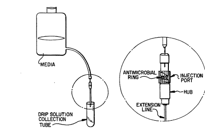
An in vitro model of a continuous flow system
(See Figure 1) was used to test the antiinfective
activity of catheter assemblies comprising hubs, ports,
discs, and extension lines as set forth above. Twice a
day, in the morning and in the evening, the hub and port
were infected with 106 CFU S. aureus. After ten days of
continuous flow the bacterial adherence on the hub,
injection port and extension lines was determined.
Untreated assemblies were found to yield
greater than 106 CFU associated with the hub and port and
greater than 107 CFU associated with the extension line.
For an assembly consisting of (1) a hub and
port dipped in 1% CHA / 0.2% AgSD / 10% NH40H / 80% EtOH
and (2) an untreated extension line, although no bac-
W095/04568 2 ~ 6 ~ ~ ~ PCT~S94/08922
24terial growth was observed from the hub and port, the
extension line yielded 1. 8 X 10³ CFU. This growth could
be reduced to zero by incorporating an antimicrobial
Dacron disc, dipped in 1% CHA / 0.5% AgSD / 20% NH40H /
80% EtOH, into the injection port of the assembly. Alter-
natively, growth could be reduced to zero by impregnating
the luminal surface of the extension line with silver.
Similarly, an assembly consisting of (1) a hub
and port dipped in 1% CHA / 0.2% Benzalkonium Chloride /
100% EtOH and (2) an untreated extension line, although
no bacterial growth was observed from the hub and port,
the extension line yielded 1. 5 X 10³ CFU. This growth was
reduced to zero in an assembly further containing an anti-
microbial Dacron disc, dipped in 1% CHA / 0.5% Benzal-
konium Chloride / 100% EtOH in the injection port. Growthwas also reduced to zero in an assembly which did not
contain a disc, but in which the luminal surface of the
extension line was impregnated with silver.
The foregoing results demonstrate the advan-
tages of incorporating an antiinfective disc and/or of
impregnating the luminal surface with silver.
7. EXAMPLE: ANTIINFECTIVE A~llVllY
OF POLYMERS SOAKED IN TETRAHYDROFURAN-
CONTAINING SOLVENT SYSTEMS
7.1. MATERIALS AND METHODS
7.1.1. IMPREGNATION OF HYDROPHOBIC SUBSTRATE
Silicone central venous catheters (Davol, Inc.)
were soaked in 5% CHA / 1% AgSD /20% NH40H / 20% MeOH /
60% THF (the AgSD dissolved in the solvent system prior
95/04568 68 72~ PCT~S94108922
to the addition of CHA) for 15 minutes such that both
internal and external surfaces were impregnated with CHA
and AgSD. The catheter was then removed and dried at
room temperature for two hours.
7.1.2. IMPREGNATION OF HYDROPHILIC SUBSTRATE
Polyurethane catheters (Triple Lumen, Arrow,
Int'l.) hubs, and injection ports were soaked, for two
hours, in either (1) 5% CHA / 0.5% AgSD dissolved in a
solvent system consisting of 20% NH40H / 40% EtOH / 40%
THF; or (2) 5% CHA / 50% EtOH / 50% H2O. The catheters
and ports were then dried at room temperature for two
hours.
7.2. RESULTS
In order to evaluate the antiinfective activity
of the catheters, hubs and ports prepared as set forth
above, the zones of inhibition associated with each
article were determined as follows.
Either a 1 cm catheter segment, a hub or a
port, prepared as described, were placed in trypicase soy
agar plates inoculated with 105 CFU of S. aureus and
incubated at 37 degrees Centigrade for 24 hours. The
zone of inhibition was then measured, and the articles
were transfered to fresh culture plates on a daily basis,
until no zone of inhibition was detectable. The results
are set forth in Table A.
W095/04568 ~ ~ 6 ~ ~ 9 PCT~S94/0892
26
TABLE A
Zones of Inhibition (mm)
Silicone Polyureth. Polyureth. Polyureth.
catheter catheter Hub Port
Days AgSD+CHA AqSD+CHA CHA AgSD+CHA AgSD+CHA
1 16 18 16 15 21
2 14 15 14 0 10
3 13 13 13
4 12 10 12
6 10 10 10
7 9 9 9
The foregoing results demonstrate that the
catheters impregnated with antiinfective agents, as set
forth above, demonstrated antibacterial activity.
8. EXAMPLE: IMPREGNATION WITH ANTIINFECTIVE
AGENT USING SOLVENTS THAT ALTER THE
ARTICLE SURFACE
Polyurethane catheter (Triple Lumen, Arrow,
Int'l.) segments were dipped in the following solutions
of CHA, rinsed, and then evaluated for surface changes
and for antibacterial activity using zone of inhibition
studies.
The results are set forth in Table B.
TABLE B
Days
Antibacterial
Surface ActivitY
1) 1%CHA/70%THF/30%EtOH damaged >8
2) 1%CHA/60%THF/40%EtOH damaged >8
3) 1%CHA/50%THF/50%EtOH slightly altered 6
4) 1%CHA/40%THF/60%EtOH slightly altered 6
5) 1%CHA/30%THF/70%EtOH slightly altered 2
6) 1%CHA/20%THF/80%EtOH slightly altered 2
The best solvent system appears to be 1%CHA /
40% THF/ 60% EtOH. This solvent system was used to
impregnate a catheter assembly in Section 10, below.
95l045C8 27 2t ~ ~ PCT~S94/089~2
9. EXAMPLE: IMPREGNATION OF CA~ ~K ASSEMBLIES
USING SURFACE-ALTERING SOLVENT SYSTEMS
9.1. MATERIALS AND METHODS
Polyurethane catheters, extension lines, hubs,
and injection ports (Arrow Int'l.) were dipped in the
following solutions, then dried at room temperature for
one hour.
Solution A: 0.5% CHA / 40% THF / 60% MeOH
Solution B: 0.1% AgSD / 0.5% CHA / 20% NH40H /
40% THF / 40% MeOH
Solution C: 0.1% Benzalkonium chloride / 0.5% CHA/
60% MeOH / 40% THF
9.2. RESULTS
To test the antiinfective activities of arti-
cles prepared in the foregoing manner, lO microliters of
S. aureus culture (108 CFU/ml) were spread on the threads
of the injection port and hub. These two parts were then
screwed together, and incubated for 6 hours at room tem-
perature. Articles which had not been dipped in anti-
infective solutions were used as controls, and inoculatedthe same way. Following incubation, 5 ml TSB was passed
through the system, collected, and then cultured for 24
hours to determine whether any live bacteria from the hub
or port had escaped into the fluid passing through the
system. The results are presented in Table C. Growth was
quantitated by measuring turbidity.
wo 95,04568 ~6~ PCT~S94/0892 ~
28
TABLE C
Solution Used Growth in Culture (DaYs)
For Dippinq 1 2 3 4 5 6 7 8 9
5 None (Control) + + + + + + + + +
A _ _ _ + + + + + +
B - - _ _ _ + + + +
C -- _ _ _ + + + + +
10. EXAMPLE: PREPARATION OF CAln~l~ ASSEMBLY
HAVING ANTIINFECTIVE DACRON SPONGE IN PORT
10.1. MATERIALS AND METHODS
10.1.1. PREPARATION OF CA~ l~K BODY AND PORTS
Polyurethane extension lines, catheter bodies,
hubs and injection ports were soaked in a solution con-
t~;n;ng 1% CHA / O.5% AgSD / 20% NH40H / 40% THF / 60%
Ethanol (Solution A) such that internal and external
surfaces were impregnated with CHA and AgSD. After dry-
ing, the ends of the catheter body were sealed and the
catheter was dipped into a solution containing 3% poly-
urethane / 1.5% CHA / 0.75% AgSD (Solution B).
10.1.2. IMPREGNATION OF SPONGE DISKS
Dacron fabric in the shape of a tube (lcm
diameter) was first dipped in solution A. After drying
it was dipped into solution B and then dried. The Dacron
sponge cuff (2mm length) was inserted in the injection
port.
10.2. RESULTS
The effects of impregnating the hub, injection
port and extension line with AgSD + CHA on luminal bac-
terial adherence were evaluated as follows. A continuousflow of fluid through the injection port, hub and exten-
95/04568 ~1 ~ PCT~S94/08922
29sion line was maintained using a system as depicted in
Figure 1.
The grooved portions of the hub and injection
port (which screw together) were infected twice a day
with 10 microliters of S. aureus culture (108 CFU/ml).
Eight liters of 50% normal saline + 50% sterile tryp-
ticase soy broth (''TSB'I) was passed through the above
system at a drip rate of 50-75 drops/minute for 4 days.
Each day the broth was allowed to drip through the system
for 8 hours and then stopped for 16 hours. The exper-
iment was then terminated and the extension line, hub and
injection port were disconnected.
The outer surface of the unit was sterilized by
wiping with 70% ethanol and the end of the extension line
(about 2 cm) was cut out. The unit was then flushed with
2 ml TSB through the injection port, hub, and extension
line and the TSB was collected and a 0.2 ml aliquot was
subcultured for determining the bacterial counts in the
fluid. The hubs and the injection ports were discon-
nected and the bacterial adherence on the hubs and injec-
tion ports was determined by rolling them on trypticase
agar plates.
The bacterial adherence onto specific portions
of the extension line was determined as follows. After
wiping the outer surface with 70% EtOH, the extension
line was cut into 2 segments, one proximal to the hub and
a second distal to the hub. Each segment was placed in 5
ml TSB and vortexed at low speed for 2 minutes. The seg-
ments were then removed and placed in 5 ml drug inactiv-
W095/04568 2l~ 2 PCT~S94/089~ ~
ating media (LTSB) and vortexed at high speed for 2 min-
utes to detach all the adherent bacteria.
One control group (untreated catheter assem-
blies) and two test groups were used in the study. The
two test groups were catheter assemblies prepared by
either (l) dipping into a solution of 1% CHA / 0.5% AgSD
/ 20% NH40H / 40% THF / 60% Ethanol or (2) dipping in the
same solution, and also containing a dacron sponge
(impregnated with AgSD + CHA as described earlier) inside
the injection port.
The results of the study are given in the Table
D below.
TABLE D
Colony Counts* (CFU)
15 GrouP Control Test Group 1Test GrouP 2
Fluid flushed
through the unit >105 104 0
Hub >105 0 0
Injection Port>105 3+ ' 0
20 Extension Line-
Lumen (Proximal) 7x10⁴ 0 o
Extension Line-
Lumen (Distal) 6.8x10⁴ o o
*CFU counts given above are as follows: for hub and port
the numbers represent CFU/hub or port; for extension
lines and catheters the results are given as CFU/cm
segment.
These data indicate that the use of impregnated
hubs, injection ports and lumens of extension lines and
catheters prevents bacterial adherence to both luminal
and external surfaces.
51~C8 31 6~ ~2~ P~T~/n8922
11. EXAMPLE: CA~ KS HAVING DIFFERENT
ANTIINFECTIVE AGENTS ON EXTERNAL AND
INTERNAL SURFACES
ll.l. MATERIALS AND METHODS
ll.l.l. FIRST PREPARATIVE METHOD
Polyurethane catheter segments (Arrow Inter-
national, Triple Lumen) were soaked for 24 hours in a
solution containing 2% or 5% chlorhexidine acetate in 50%
reagent alcohol/ 50% water such that both internal and
external surfaces were impregnated. The catheter seg-
ments were then dried at 70C for 30 minutes and then
washed with water in a vortex mixer for 5 seconds. After
drying at 20-30C for 30 minutes, both ends of the cath-
eter segment were sealed by heat.
The sealed catheter segments were dipped into a
solution of 3% polyurethane (Tecoflex~-93A, Termedics,
Inc.), l.S% chlorhexidine acetate and 0.75% silver sulfa-
diazine in 70% THF/30% reagent alcohol to form a coating
on the exterior of the catheter segments. The catheter
segments were then dried at 70C for 30 minutes and for
24 hours at room temperature.
ll.l.2. SECOND PREPARATIVE METHOD
The First Preparative Method was repeated,
except that 20% 2M ammonia in methanol and 80% water was
used as a solvent in place of the 50/50 alcohol/water
mixture.
ll.l.3. THIRD PREPARATIVE METHOD
The Second Preparative Method was repeated,
except that the solution contained 0.5% lactic acid and
WO95/0~6~ ~6~9 rcT~s94lc89~
0.5% mandelic acid which have been found to be effective
in preventing bacterial adherence to the urinary tract.
11.1.4. FOURTH PREPARATIVE METHOD
The First Preparative Method was repeated
except that the soaking solution used was 2% or 5% chlor-
hexidine acetate and 0.5% silver sulfadiazine in 20%
ammonia, 20% methanol and 60% water.
11.1.5. FIFTH PREPARATIVE METHOD
The First, Second, Third and Fourth Preparative
Methods were applied to silicone catheter segments by
replacing the water component in the soaking solution
with THF.
11.2. RESULTS
To test the antiinfective properties of poly-
urethane catheter segments made in accordance with theinvention, 2 cm long pieces of the treated catheter seg-
ments, open on both ends, were soaked in trypticase soy
broth (4 ml/segment) at 37C to simulate exposure to body
fluids. Three segments of each type were removed period-
ically and tested for bacterial adherence.
To test for adherence, the pieces of treatedcatheter were suspended in 2 ml of trypticase soy broth
cont~; ni ng 107 CFU of Staphylococcus epidermidis and
incubated in a water-bath shaker at 37C for 4 hours.
Untreated control catheter pieces and pieces subjected
only to soaking or exterior coating were treated in
parallel.
At the end of the 4 hour incubation, the
catheter pieces were removed, blotted dry, vortexed in
95/04568 216S7 PCT~594/0892
sterile TSB at low speed for 5 seconds, blotted dry
again, and rolled over a trypticase soy agar plate. This
results in the transfer of microorganisms to the plate if
adherence to the outer surface has occurred.
The catheter pieces were then placed in 2 ml of
lecithin containing trypticase soy broth (LTSB), which
inactivates chlorhexidine, and vortexed at high speed for
15 seconds. The catheter pieces were removed and pro-
cessed by the roll/plate techn;que described above. In
addition, a 0.2 ml aliquot of the LTSB was subcultured on
a trypticase soy agar plate.
All of the plates were incubated at 37C for 24
hours and the number of colonies were counted. The total
number of colonies counted from all three platings were
lS combined as a measure of resistance to infection. The
results are summarized in Table E.
WOg5/04568 PCT~S94108922 ~
~6~ 9
Table E
Inner Outer TreatmentSoaking Time In
Anti- Coating Method Presence of
infective Sta~h. epidermidis
O 1 day 4 days
2% CHA 3% PU Ex. 1 0 1 85
1.5% CHA
0.75~ AgSD
2% CHA 3S PU Ex. 3 0 0 25
0.5% 1.5% CHA
lactic 0.75% AgSD
acid
0.5%
mandelic
acid
2% CHA 3% PU Ex. 4 0 0 30
0.5% AgSD1.5% CHA
0.75% AgSD
Controls
O O untreated 100 200 1,000
2% CHA O Ex. 1 0 0 750
0 3% PU Ex. 1 0 0 150
1.5% CHA
0.75% AgSD
- ~ 95/04568 16~ 7~9 PCT~S94/08922
Additional catheter pieces were tested using
the above protocol, except that the bacterial culture
used contained 4x10⁷ CFU. The results are summarized in
Table F.
Table F
Inner Outer Treatment ~oAking Time In
Anti- Coating Method Presence of
infective Sta~h. epidermidis
Q 1 day 5 days
5% CHA 3% PU Ex. 1 0 0 0
1.5% CHA
0.75% AgSD
5% CHA 3% PU Ex. 4 0 0 0
0.5% AgSD1.5% CHA
0.75% AgSD
Controls
O 0 100 1,000 10,000
o 3% PU 0 0 1,000
1.5% CHA
0.75% AgSD
W095/04568 ~6~ PCT~S94/08922
36
In further experiments, eight 2 cm pieces of
catheters, prepared in accordance with the First, Third
and Fourth Preparative Methods, were suspended in 12 ml
of TSB inoculated with 1O6 CFU Staphylococcus ePidermidis
and incubated in a water-bath shaker at 37C. Two pieces
of each type were removed at intervals and tested for
bacterial adherence as described above. The remaining
pieces were transferred to fresh TSB incubated with 1O6
CFU of Staphylococcus epidermidis and inoculated.
The results, shown in Table G, together with
those shown in Tables E and F, demonstrate the surprising
effectiveness of using both a soaking treatment and an
outer coating to impart antiinfective properties to the
lumenal and outer surfaces of the catheter, respectively.
~ 95/04568 68 7~ PCT~Sg4/08922
Table
InnerOuter Treatment
Anti- Coating Method Exposure Time to:
infective
24 hours 48 hours
2% CHA 3% PU Ex. 1 O 83
1.5% CHA
0.75% AgSD
2% CHA 3% PU Ex. 3 0 63
0.5% 1.5% CHA
lactic 0.75~ AgSD
acid
0.5%
mandelic
acid
2% CHA 3~ PU Ex. 4 O 15
0.5% AgSD 1.5% CHA
0.75% AqSD
Controls
O 0 5,000 lO,000
o 3% PU O 328
1.5S CHA
0.75% AgSD
WOg5/04568 ~ ~6S~ PCT~S94/08922 -
38
This is significant because the attachment of
bacteria to the surface of medical articles has been
recognized as an important initial step in the patho-
genesis of foreign body infection. The bacterial pro-
duction of extracellular glycocalyx, a polysaccharide-
containing component outside the cell wall (slime),
facilitates their adhesion to the article. The fibrous
glycocalyx extends from the bacterial cell surface and
surrounds individual cells or colonies, protecting them
from phagocytes and biocides while providing a suitable
environment for the transport of nutrients. Once formed,
the bacterial biofilm continues to be a source for the
spread of infection to other parts of the body by bac-
terial detachment and biofilm sloughing.
A well known example of this problem was the
mortality due to massive infections in patients receiving
artificial hearts (Jarvik hearts). Similar situations
are encountered in cystic fibrosis patients, where bio-
film formation by Pseudomonas aeruginosa prevents the
effective control of the disease by antibiotics.
12. EXAMPLE: CA'l'~'l'~S OF DECREASED
THROMBOGENICITY
Catheter segments were treated in accordance
with the invention by soaking pieces of polyurethane
catheters in a solution of heparin-benzalkonium chloride
(HBC) complex (1.6% HBC in isopropanol) for two hours at
20-30C. The soaked pieces were then dried and the ends
sealed by heating.
~ 95/04568 1 6~ ~9 PCT~S94/08922
39
The exterior of the sealed catheter pieces were
then coated with 3% polyurethane, 1.5% chlorhexidine
acetate and 0.75% silver sulfadiazine in 75% THF/25%
ethanol. The resulting coated pieces were then unsealed
and tested for bacterial adherence as described in Sec-
tion 11.2, above.
A second set of samples was prepared by this
same method except that after the HBC treatment and
before sealing, the catheter pieces were soaked in 5%
chlorhexidine in 50% watert50% ethanol for 2 hours at 20-
30C. These pieces were also tested for adherence.
The results, shown in Table H, illustrate the
clear superiority of the invention for providing
effective control of bacterial adherence.
W095/04568 PCT~S94/08922 ~
?,9
Table
Inner Outer Soaking Time In
Anti- Coating Presence of
infective Staph. epidermidis
0 1 daY 3 daYs 5 daYs
HBC 3% PU 0 0 7 370
1.5% CHA
0.75% AgSD
HBC/CHA 3% PU 0 0 10 1,086
1.5% CHA
0.75% AgSD
Controls
O 0 100 n.d. 1,000 >50,000
0 3% PU 0 0 2 26,849
1.5% CHA
0.75% AgSD
HBC 0 0 0 390 29,008
95/~68 68 729 PCT~S94/08922
13. EXAMPLE: COATED SILICONE CA'~ 'l'~KS
Silicone catheter segments were coated inside
and out using two variations of the method of the inven-
tion. In the first variation, the pieces were soaked in
0.5% AgSD and 1% CHA in 10% ammonia, 10% methanol and 80%
THF for 24 hours at 20-30C. After drying for 30 min-
utes, the outer surfaces were wiped with THF to remove
excess antiinfective agent and the ends were sealed. The
pieces were then dipped in a solution containing 5% MDX
silicone (MDX 4-4210, Dow Corning) and 3% CHA in 90%
THF/10% methanol and removed immediately. After drying
for 5 minutes at 100C they were dipped into 5% Silastic
A silicone in hexane and then dried for 30 minutes at
100C and 24 hours at room temperature.
In the second variation, the silicone catheter
pieces were soaked in 1.6% HBC in isopropanol for 1 hour
at 20-30C, dried and wiped on the outer surface and then
sealed on the ends. The outer coating was then applied
in the same manner.
The samples prepared were then tested for bac-
terial adherence using the technique described in Section
11.2.. The results are shown in Table I.
W095/04568 ~3 PCT~Sg4l08g22
42
Table
Inner Outer Soaking Time In
Anti- Coating Presence of
infective Sta~h. epidermidis
Q 1 day 4 da~s
1% CHA Silicone/CHA O 0 1,OlO
0.5% AgSO
HBC Silicone/CHA O O 10
Controls
o SiliconelCHA 0 O 9,466
O O 1,000 >1,000 >10,000
~ 95/04~68 ~6 PCT~S94/08922
43
14. EXAMPLE: IMPREGNATED HUBS AND PORTS
Impregnation of catheter hubs and ports with an
antiinfective agent was carried out as follows. Hubs and
ports made from polyurethane (Arrow International) were
treated with antiinfective agents by soaking using three
alternative procedures.
In the first procedure, AgSD was dissolved in
14.8M ammonia. Chlorhexidine acetate was dissolved in
methanol. The two solutions were then combined to form a
soaking solution containing 0.25% AgSD and 1% CHA in 50%
ammonia/50% methanol. The hubs and ports were dipped in
this solution and removed immediately at 20-30C and then
dried at 70C for 30 minutes.
In the second and third procedures, THF was
added to the combined solutions to yield final com-
positions of 0.25% AgSD, 1% CHA in 30% ammonia/50%
methanol and 20% THF or 0.5% AgSD and 1% CHA in 30%
ammonia, 60% methanol and 10% THF. The hubs and ports
were dipped as described above.
All of the treated hubs and ports were tested
for antimicrobial properties on trypticase soy agar
plates seeded with 0.3 ml of S. aureus culture (105
CFU/ml). The hubs and ports were placed on the surface
of the agar plate and incubated for 24 hours. The zone
of inhibition around the device was then measured, afterwhich the device was transferred to a fresh plate for
further incubation. The results in Table J show ~he
benefits of impregnation using the ammonia/methanol/THF
solvent system.
wo g5l04568
PCT~S94/08922
44
This impregnation system may be used for other
catheter parts, e.g., extension lines, or for impreg-
nation of the luminal surface of the catheter body prior
to exterior c02ting.
Table 3r
Procedure Zone of Inhibition (mm)
DaYs 1 ~ 3 4
1 Hub 10 0 -- --
Port 15 0 -- --
2 HUb 23 14 13 15
Port 25 16 14 15
3 Hub 20 13 13 12
Port 21 17 15 14
O95/04568 216~72~ PCT/1~594108922
15. EXAMPLE: DUAL COATING CAl~l~KS
Catheter segments for use in accordance with
the invention were prepared by soaking triple lumen poly-
urethane catheter segments (Arrow International) in a
solution containing 20 mg% of teicoplanin, a glycopeptide
antibiotic, in 50% ethanol:50% water for two hours at 20-
30C. After drying at room temperature for 30 minutes
they were rinsed in water and dried again. The ends were
then sealed by heating, and the sealed segments were dip-
ped in a solution containing 3% polyurethane, 1.5% CHAand 0.75 % AgSD in 30% ethanol:70% THF. The dipped seg-
ments were dried at 70 degrees Centigrade for 30 minutes
and then tested for bacterial adherence in accordance
with the procedures set forth above.
The results of this experiment are shown in
Table K. As can be seen, the segments treated on both
the interior and exterior surfaces showed marked super-
iority to the control samples.
WOg5/04568 i ~ PCT~S94/08922 -
'1.,~
46
Table K
Inner OuterBacterial A~.nerence
Anti- Coating After Pro~onged
infective Soaking in TSB
0 3 days 4 davs
Teicoplanin ¦ AgSD+CHA 0 0 0
Controls
o AgSD+CHA 0 240 270
Teicoplanin 0 100 550 1,OOo
O 0 100 >1, 000>10, 000
95/04568 ~ PCT~S94/08922
16. EXAMPLE: DUAL COATING CA~ KS
5 cm catheter segments were coated and impreg-
nated using various combinations of drugs in accordance
with the invention. The segments were unsealed and
individually soaked in lO ml volume of TSB at 37C in a
water bath shaker for 24 hours. A portion of the seg-
ments were then placed in a new 10 ml volume of TSB for
an additional 24 hours.
The segments, as well as segments which had not
been soaked, were then inoculated on the inner luminal
surface with S. epidermidis (108 CFU/ml) and placed in
petri dishes for 4 hours at 37C. After 4 hours, the
ends of the segments were sealed and the outer surfaces
were sterilized with 70% isopropanol. The ends of the
segments were then opened, and the lumens were flushed
with l.O ml of CHA inactivating media (LTSB) three times
in succession to remove adherent bacteria. 0.2 ml ali-
quots of these washings were plated on trypticase soy
agar and incubated for 24 hours at 37C. The colonies
were then counted to provide an indication of the level
of luminal bacterial adherence after various periods of
soaking which would tend to leach out the antiinfective
agent. The results are shown in Table L.
As is apparent from these results, the inner
luminal coating was resistant to leaching and provided
excellent resistance to bacterial growth.
W095/04568 ~9 PCT~S94/08922 -
48
Table L
Inner Outer Adherence of
Anti- Coating Staph. aureus
infective (CFU/cm)
0 1 day 2 daYs
CHA AgSD+CHA 0 77 150
AgSD+CHA AgSD+CHA 0 35 200
HBC AgSD+CHA 0 0 0
Controls
0 AgSD+CHA 1,000 >5,000 >5,000
O O >10,000>10,000 >10,000
~ 95/~568 ~7~9 PCT~Sg4/08922
49
17. EXAMPLE: ANTIINFECTIVE CA~ ~ ASSEMBLIES
Catheter segments, hubs and ports were rendered
antiinfective by soaking in 0.2% AgSD and 2% CHA in 20%
Ammonia/60% Methanol/ 20% THF (Soaking Solution A); 0.1%
AgSD and 2% CHA in 20% Ammonia/60% Methanol/ 20% THF
(Soaking Solution B); 2% CHA and 0.1% benzalkonium chlor-
ide in 80% methanol/20% THF (Soaking Solution D) or 3%CHA
in 50% methanol/50% THF (Soaking Solution D). The
materials were soaked in the above-described solutions,
dried for one hour, and then placed on a trypticase soy
agar plates and incubated. The zone of inhibition around
each soaked piece was measured at the end of 1, 2, 3 and
4 days of incubation. The results of this study are
reported in Table M. As is apparent from the results,
each of the solutions was able to impart substantial
levels of antiinfectivity, that lasted for the full four
days of the test.
Various patents and other publications, cited
herein, are hereby incorporated by reference in their
entirety.
WO 95/04568 9 PCT/US94/08922--
~ 50
C~ ~ o o
N _1 ~1
~I N O
U~
m N ,~
~ N N a~ 00
a
N _I ~1 ~1
U ~ t` ~D ~
N ~1 ~1
m U~ a~ r ~o
N ~1 ~1 ~1
~4 N tD
a
u '' ~n 0
m N ,N~
~¢ N ~i
-- ~I N ~q
C~