Note: Descriptions are shown in the official language in which they were submitted.
WO 95/05206 . ~ PCT/US94/09300
~ 21 693Q4
T~TING WOUNDS CAUS~n BY MEDICAL PROCEDURES
Field of the Invention
This invention relates to treating wounds caused
5 by medical pro~ed~les.
Background of the Invention
In many medical procedures, a medical device must
be placed in tissue that is well below the ~Xpose~
surface of the body. Typically, an incision or puncture
10 is made through ~L~ o~ln~ing tissue to gain access to the
target tissue. After the procedure, the access incision
is usually treated to encourage healing.
For example, in balloon angioplasty procedures, a
narrow access channel is cut that extends from the body
15 surface through the skin, the subcutaneous fascia (e.g.
co~ective tissue, fat and muscle), and the wall of a
blood vessel. An access catheter is placed in the access
channel and the angioplasty catheter delivered into the
vessel through the access catheter. At the end of the
20 procedure, the access catheter is removed from the body.
The access channel is treated by applying manual pressure
to the site or depositing a hemostatic material into the
channel to prevent excessive bleeding.
~ummary of the Invention
The invention provides treating incised or injured
tissue by locating at desired depths within the tissue
material that encourages healing. Particularly, the
invention provides treating an access channel to a blood
vessel by positioning a hemostatic material so that it is
30 adjacent, but does not extend beyond the vessel wall into
the vessel lumen.
In one aspect, the invention features a device for
treating an incision channel through tissue and the wall
of a body lumen. The device includes a member having a
35 proximal portion constructed to remain outside the body
W095/05206 ~ ~ PCT~S94/09300
~ ~q3~4
- 2 -
and an elongate generally tubular distal portion that is
constructed to be intro~nce~ axially into the ch~n~el and
be moveable axially therein. A detector is disposed on
the side of the tubular distal portion. The detector is
5 adapted to detect a predetermined condition indicative of
an axial position within the ch~nn~l. A healing
promoting substance is carried by the member and
releasable from the member into the çh~nn~l at a desired
axial location relative to the location indicated by the
lO detector.
Embodiments may include one or more of the
following features. The detector is differentially
responsive when exposed to the interior of the vessel and
when exposed to the interior of the channel. The
15 detector is sensitive to the flow of body fluid in the
vessel. The detector includes a port in the wall of the
generally tubular distal end portion so that the detector
indicates when the port is exposed to the interior of the
vessel by the flow of body fluid into the port and
20 indicates when the port is exposed to the interior of the
rh~nn~l by the lack of flow of body fluid into the port.
The port is in fluid communication with a lumen exten~ing
proximally to the proximal portion of the member outside
of the body. The lumen is sealed distal of the port.
25 The lumen extends proximally to a visual indicator so the
flow of body fluid through the port is indicated visually
by flow at the visual indicator. The detector is
sensitive to the pressure in the vessel. The detector
includes a pressure transducer. The detector is
30 sensitive to the presence of chemical compounds. The
detector is sensitive to the edge of the vessel wall
during axial motion of the member. The detector is a
portion of the wall of the member having regions of
different diameter.
woss/os2o6 2 1 6 9 ~ 0 4 PCT~S94/~930~
Embodiments may also include one or more of the
following features. The device further includes a
measuring system for measuring the depth of the ch~nnel
to the position indicated by the detector. The measuring
5 system includes a mark on the member with known axial
distance relationship to the detector. The substance for
promoting healing has a proximal end, distal end, and a
known length therebetween, and the substance is
positionable by alignment with the mark to position the
10 distal end at a known distance relationship with respect
to the detector. The mark is located at a distance from
the detector corresponding to the length of the substance
so the distal end of the substance is adjacent the
detector when the distal end of the substance is aligned
15 with the mark. The measuring system includes a series of
marks on the member of known distance from the detector
for indicating the depth of the ch~n~el to the detector.
The substance for promoting healing has a proximal end,
distal end, and a known length therebetween and a series
20 of marks indicating the distance from the distal end.
The substance is slidably disposed on the exterior of the
member. The substance is opaque, obscuring visual
observation of portions of the member under the
substance. The substance has a defined length greater
25 than the depth of the access channel. The substance is
axially moveable immediately after release from the
member.
Embodiments may also include one or more of the
following features. The healing promoting substance is
30 carried on the outer exposed surface of the tubular
distal portion and the device further includes a sheath
positioned over the substance during entry into the
~ el and removable from the position over the
substance for releasing of the substance in the channel.
35 The sheath is an axially retractable sheath controllable
WO95/05206 , . PCT~S94109300
~ 6?304
- 4 -
from the proximal portion. The substance is a plug of
hemostatic material with an axial length equal to or
shorter than the depth of the channel. The plug is
positioned proximally a known distance from the detector.
5 The device includes a series of marks at least some of
which are normally outside of the ~hAnnel when the device
is in use, the marks being spaced a known distance from
the detector. The sheath includes a flexible seal-
forming tip that extends distal of the plug during entry
lO into the body and seals against the distal portion to
prevent exposure of the plug to body fluid during entry
into the body. The tip expands in diameter during
retraction of the sheath over the plug. The tip seals
against the distal portion at a proximal location after
15 retraction of the sheath beyond the plug. The distal
portion is removable to deposit the substance in the
~hAnnel while leaving the sheath in the channel. The tip
seals the sheath against the flow of body fluid after
removal of the distal portion. The sheath is a thin-
20 walled flexible sheath that can be collapsed by manualpressure after removal of the distal portion. The plug
is held in a compressed form by the sheath when the
sheath is positioned over the plug.
Embodiments may also include one or more of the
25 following features. The substance for promoting healing
is a body degradable substance. The substance is a
hemostatic substance. The diameter of the distal portion
is no greater at regions distal of the detector than at
regions proximal of the detector. The distal portion
30 includes a relatively flexible tip, distal of the
detector, for positioning inside the vessel. The distal
portion tapers distally to smaller diameter. The member
includes a lumen for delivering the device to the channel
over a guidewire.
W095/05206 , pcT~ss1los3no
2 1 6q304
- 5 -
In another aspect, the invention features a device
for treating an incision ~hAnnel through tissue and a
blood vessel wall. The device includes a member having a
proximal portion that remains outside the body and an
5 elongate, general tubular distal end portion that is
i~lL~od~ced into and ~ lly moveable within the ch~n~el .
A port is provided in the wall of the generally tubular
end portion in fluid communication with a lumen that is
sealed distal of the side port and extends proximally to
10 the proximal portion of the member outside the body to a
visual indicator. The flow of blood through the port and
to the indicator indicates when the port is exposed to
the vessel and the lack of flow of blood through the port
to the indicator indicates that the port is exposed to
15 the interior of the channel. A mark is provided on a
portion of the member that remains outside the body
having a known distance relationship to the port. A
body-degradable hemostatic substance for promoting
he~ 1 i ng iS carried by the member and releasable into the
20 channel at a depth of known relationship to the mark.
Embodiments may include additional features
mentioned above. Particular embodiments may include the
following. The hemostatic substance has a proximal end,
a distal end, and a known length therebetween greater
25 than the depth of the channel. The substance is axially
positionable by aligning the proximal end with the mark
to position the distal end at a known distance
relationship with the port. The mark is located at a
distance from the port corresponding to the length of the
30 substance for ~u~oLing healing. The device includes a
series of marks of known distance from the port. The
substance for promoting healing has a series of marks for
indicating the length of the substance in the channel.
The substance is slidably disposed on exterior of the
35 member. The member includes a lumen for delivering the
W095/05206 ~ PCT~$94/09300
2 ~ 6~3~4
device to the channel over a guidewire. The diameter of
the distal portion is no greater at regions distal of the
detector than at regions proximal of the detector. The
distal portion includes a relatively flexible tip, distal
5 of the detector, for positioning inside the vessel.
In another aspect, the invention includes a device
for measuring the length of an access channel through
tissue and the wall of a vessel carrying body fluid under
pressure. The device includes a member having a proximal
10 end that remains outside the body and an elongate,
generally tubular distal end portion that is introduced
into and axially moveable within the ~h~nnel. A detector
is provided in the generally tubular distal portion for
locating a position within the channel. A mark is
15 provided on the proximal end of the member that remains
outside the body, of known distance relationship to the
port.
Embodiments may include additional features
mentioned above. Particular ~ho~i~ents may include the
20 following. The detector is in fluid communication with a
lumen ext~n~ing to the proximal portion of the member
outside the body and to a visual indicator so the flow of
body fluid through the port is indicated visually by flow
at the indicator. The port is a side port through the
25 wall of the distal portion in fluid ~u ~l ication with a
lumen sealed distal of the port.
In another aspect, the invention features a device
for treating an incision channel through tissue and a
blood vessel wall. The device includes a member having a
30 proximal portion that remains outside the body and an
elongate generally tubular distal portion that is
introduced into the channel. A port is provided in the
wall of the generally tubular distal portion in fluid
communication with a lumen ext~n~;ng to the proximal
35 portion. A body-degradable hemostatic substance for
WO 95/05206 . . PCT/US94/09300
~ 21 69304
- 7 -
promoting healing is releasable from the member into the
~h ;~ nr~
In these embodiments, the device may include
features mentioned above. Particularly, the member may
5 include a second lumen ext~n~ing from the distal end of
the distal portion to the proximal portion.
In another aspect, the invention features a device
for treating an incision channel through tissue and the
wall of a body lumen. The device includes a member
10 having a proximal portion constructed to remain outside
the body and an elongate generally tubular distal portion
that is constructed to be introduced axially into the
chAnne~ and be moveable axially therein. The device
further includes a healing promoting substance that is
15 carried by the member and releasable from the member into
the chAnnel at a desired axial location relative to the
location indicated by the detector. The healing
promoting substance is in the form of a tubular element
having a length greater than the access channel. In
20 particular embodiments, the tubular element may include
marks indicating the distance to its distal end disposed
inside the body.
In another aspect, the invention features a method
for treating an incision channel through tissue and a
25 vessel wall. The method includes providing a device 8s
described above, introducing the tubular distal portion
into the channel, exte~ing the distal end portion
axially distally until the detector indicates that the
detector is within the vessel, retracting the distal
30 portion axially proximally until the detector indicates
that the detector is within the channel, the detector
thus being located near the vessel wall, depositing the
substance into the channel at a predetermined axial
relationship with respect to the detector and, removing
W095/05206 PCT~S94/09300
~ 693~4 ~ i ~
-- 8
the device from the chAnn~l, leaving the substance in
place.
In particular embodiments, the invention features
iteratively moving the device axially proximally and
5 distally to confirm the detector is located near the
vessel wall, prior to depositing the substance. The
method may include rotating the device about its axis to
determine the radial variations of the chAn~l or to
determine the optimal orientation for detector response.
10 The method may include providing a hemostatic substance
having a desired length greater than the depth of the
channel, so that a length of the substance is exposed
above the channel, and moving the substance axially after
removing the device by grasping the exposed length. The
15 method may include removing the device substantially from
the channel by withdrawing it axially. The method may
include providing marks on the exposed length indicative
of the length of substance positioned inside the ch~nnel
and axially adjusting the length of substance inside the
20 channel to extend to a desired depth. The method may
include detaching, e.g., cutting, the exposed length of
substance after the adjusting.
In other aspects, the invention features devices
for positioning a plug of hemostatic material including
25 mechAn;sms to automatically, sequentially retract a
protective sheath from a position over the plug and a
catheter carrying the plug to leave the plug at the
proper location within the body.
In other aspects, the invention features clamping
30 members to aid in locating a plug at the desired depth.
These include clamping members having an additional
function of cutting the plug to remove excess length
after the plug has been properly positioned.
In one aspect, the invention features a device for
35 treating an incision channel through tissue and the wall
W095/05206 pcT~s94los3on
2 1 69304
g
of a body lumen. The device includes a member having a
proximal portion constructed to remain outside the body
and an elongate generally tubular distal portion that is
constructed to be intro~llce~ axially into the ch~nnel and
5 be moveable axially therein. Healing promoting
substance, carried by the member, is releasable from the
member into the chAnnel. The healing promoting
substance, positioned over the member, is in the form of
a tubular element and has a length greater than the
10 ~rc~cs ch~nnel. A positioner, of cross-section greater
than the diameter of the access channel, is axially
fixable on the healing promoting substance at an axial
distance from the distal end of the substance that
corresponds to the depth of the channel to the desired
15 location. The positioner and healing promoting substance
are axially slidable to locate the portion of the
substance distal of said positioner inside said ch~nnel
so that the distal end of the substance is positioned at
the desired location. The positioner prevents the
20 portion of the substance proximal of the positioner from
ext~n~;ng into the channel.
In another aspect, the invention features a system
for treating an incision channel exten~;ng from an outer
surface of a body through tissue and a blood vessel wall.
25 The system includes a device having a member with a
proximal portion constructed to remain outside the body
and an elongate generally tubular distal portion that is
constructed to be introduced ~ lly into the channel and
be moveable axially therein. The proximal portion has a
30 reference disposed along the axial length of the member.
A detector is disposed on the side of the tubular distal
portion and is adapted to detect a predetermined
condition indicative of an axial position within the
channel. The system further includes a slidable plug
35 formed of a healing promoting substance and having a
WO95/05206 ~ PCT~S94/09300
2 1 ~oq 3~
-- 10 --
p~edetermined length. The plug is releasable from an
exterior surface of the member into the channel at a
desired axial location relative to the location indicated
by the detector. The system further includes a rigid
5 member for establishing a measurement on the slidable
plug relative to the reference on the member indicative
of a length of a proximal portion of the plug desired to
remain outside the outer surface of the body.
Particular embodiments may include the following.
10 The reference on the member is located at a predetermined
position along the proximal portion of the member and the
rigid element has a predetermined length equivalent to
the distance between a distal end of the slidable plug
and the detector when a proximal end of the plug is
15 aligned with the reference. The system may further
include a marker for providing a mark on an outer surface
of the plug to indicate the length of a proximal portion
of the plug that is desired to remain outside the body.
In other embodiments, the reference on the member
20 is located along the member and proximally from the
detector a distance adapted to remain outside the body,
the distance being equivalent to the predetermined length
of the plug. In such embodiments, the rigid element has
a series of graduated marks disposed along the axial
25 length of the rigid element with the distance from the
outer surface of the body to the reference mark,
representing the length of the proximal portion of the
plug desired to remain outside the body.
~n another aspect, the invention features a method
30 for treating an incision channel exte~;ng from an outer
surface of a body through tissue and a vessel wall. The
method includes providing a system including the rigid
member, as described above, introducing the tubular
distal portion of the member of the delivery device into
35 the ch~nnpl~ exten~i~g the distal end portion of the
WO95/05206 . PCT~S94/09300
.
21 69304
-- 11 --
member axially distally until the detector, disposed on
the side of the tubular distal portion, indicates that
the detector is within the vessel, retracting the distal
portion axially proximally until the detector indicates
5 that the detector is within the ~h~nnel, the detector
thus being located near the vessel wall, positioning the
rigid element substantially parallel with the axial
length of the member, a first end of the rigid element
engaging an outer surface of the body, measuring with the
10 rigid element, a position along the length of the
slidable plug relative to the reference on the member
indicative of a length of a proximal portion of the plug
desired to remain outside the outer surface of the body,
depositing the slidable plug into the channel at a
15 predetermined axial relationship with respect to the
detector and, removing the device from the ch~nnel,
leaving the plug in place.
In particular emboA;~^nts where the reference is
located at the proximal end of the member and the rigid
20 element has a predetermined length, the method may
include the following additional step. A proximal end of
the slidable plug is aligned with the reference on the
member and, after positioning of the rigid element with
respect to the outer surface of the body, the outer
25 surface of the slidable plug is marked, the mark being
aligned with a second end of the rigid element.
In embodiments where the reference on the member
is located proximally from the detector along the member
a distance adapted to remain outside the body, the
30 distance being equivalent to the predetermined length of
- the plug, and the rigid element has a series of graduated
marks disposed along the axial length of the rigid
element, the method may include the following steps.
Using the graduated marks of the rigid element to measure
35 a distance from the outer surface of the body to the
W095/05206 . PCT~Sg4/09300
~ :,93~4
- 12 -
reference mark, the distance representative of the length
of the proximal portion of the plug desired to remain
outside the body. Moreover, using the graduated marks of
the rigid element to measure a distance from the outer
5 surface of the body to the distal end of the slidable
plug may be repeated after both the depositing step and
the removing step, and, if necessary, the length of the
proximal portion of the plug desired to remain outside
the body can be adjusted.
The advantages of the inventions are numerous.
For example, axially positioning hemostatic material so
that it is adjacent but does not extend beyond a blood
vessel wall improves healing and reduces complications.
If material is positioned too shallow in the access
15 channel, such that it does not extend adjacent the vessel
wall, blood can collect in the channel and cause a
pseudoaneurysm, bruising or swelling. This condition can
be very painful since the blood pressure in the vessel,
e.g. the femoral artery, and, hence, the pseudoaneurysm,
20 may be quite high. Blood under pressure in the channel
may also push the material back out of the channel, which
can cause bleeding. On the other hand, when material is
positioned too deeply in an access ch~nnel, such that it
extends beyond the vessel wall into the vessel lumen, the
25 vessel can be occluded, reducing flow and increasing
pressure. In addition, clots may form on the portions of
the material in the vessel, raising the danger that clot
material will be let loose into the bloodstream, causing
an embolism. Moreover, aspects of the inventions feature
30 the capability of manually pulling the material from the
access ch~n~l should difficulties (e.g. vessel
occlusion) become evident either during placement or even
some period after placement. These aspects provide
important advantages in safety and convenience, since the
WO 9!;/OS206 PCT/US94/09300
.
13 21 69304
likelihood that mispositioned material will have to be
surgically removed is reduced.
Further advantages and features follow.
Brief DescriPtion of the Drawing
Fig. 1 is a side view of a device according to the
invention for treating an access ch~nnel;
Fig. la is a cross-sectional view along the lines
A-A in Fig. l;
Fig. lb is primarily a cross-sectional side view,
10 with a partial side view, of the device in Fig. 1 with
the treating material in an alternate location;
Figs. 2-2i illustrate use of the device in Fig. 1;
Fig. 3 is a side view of an alternate embodiment
of a device according to the invention;
lS Fig. 3a is a cross-sectional view along the lines
B-B in Fig. 3;
Figs. 4-4b show a tip for a protective sheath for
use with the embodiment of Fig. 3;
Figs. 5-5b show an alternate tip for a protective
20 sheath for use with the embodiment of Fig. 3;
Figs. 6-6d illustrate use of the device in Fig. 3;
Fig. 7 is a cross-sectional view of an alternate
emhoA;ment of a device according to the invention;
Fig. 8 is a side view of an alternate embodiment
25 of a device according to the invention;
Fig. 9 is a top view of a clamp positioner
according to the invention;
Figs. 10-lOc show the use of the clamp positioner
in Fig. 9;
Fig. 11 is a perspective view of another clamp
positioner;
Fig. 12 is a side view of an alternate embodiment
of a device according to the invention;
Figs. 13a-13d illustrate use of the device in Fig.
35 12;
WO95/05206 PCT~S94109300
L~ ~3~4 - 14 -
Fig. 14 is a side view of an alternate embodiment
of a device according to the invention;
Figs. 15a-15c illustrate use of the device in Fig.
14.
Descri~tion of the Preferred Embodiments
Structure
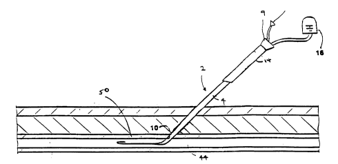
Referring to Figs. 1-lb, a device 2 according to
the invention is shown for treating an access channel to
the femoral artery after a catheterization (e.g.
10 angioplasty) or similar procedure. The device 2 includes
a catheter body 4 with a side port 10 to an inner lumen
12. A long hemostatic plug 14 can be slid axially (arrow
30) along the catheter body. A depth indicating system
22 of marks can indicate the axial location of the distal
15 end 26 of the plug relative to the port 10 when the
distal end of the plug and the port are inside an access
channel and out of view. When the port 10 is within an
access channel, the tissue of the ~h~nnel wall seals
against the port, preventing blood flow through the lumen
20 12. When the port 10 is within the blood vessel, blood
(which is at body pressures typically in the range of
120-200 mm Hg) flows through the port 10 into the channel
12. By detecting the flow of blood in the lumen 12 while
moving the catheter body axially, the side port 10 can be
25 axially located at a depth adjacent the vessel wall.
Then, using the depth indicating system 22, the plug 14
is slid over the catheter body to position the distal end
26 of the plug so that it is adjacent the port 10 (Fig.
lb) and, hence, axially adjacent the vessel wall, but
30 does not extend beyond the vessel wall into the vessel
lumen. The catheter body is removed from the channel,
leaving the plug 14 properly positioned in the channel.
The body 4, formed of a flexible, kink-resistant
material such as nylon, has an overall length L1, about
3S 25 cm, which is substantially longer than the depth of an
W095/05206 PCT~S94109300
,~ 216q304
- 15 -
A~c~cc ch~n~el to the femoral artery regardless of a
patient's weight, age, and anatomical variations. The
catheter body 4 has a proximal portion 3, an alignment
portion 6, and a flexible distal guiding portion 8. The
5 proximal portion 3 is relatively stiff, of substantially
constant outer diameter (e.g. 8 French), and remains
outside of the body. The alignment portion 6 includes
depth indicating system 22 and port lO and is also of
substantially constant outer diameter (e.g. 8 French).
lO The diameter of the alignment portion is similar to,
e.g., slightly greater than, the width of the access
c-hAnnel, but is not so much greater to cause tearing or
excessive stretching of the tissue forming the wall of
the channel. The diameter is sufficient to prevent blood
15 from leaking around the device and into the XurLo~ g
tissue. The proximal portion of the alignment portion,
corresponding to the region near an end mark 23 of
locating system 22, remains outside the access channel
above the skin in use.
The relatively long flexible distal portion 8, of
length, L2, about 5 cm, tapers to an outer diameter of
about 4.5 French or approximately one half the diameter
of the alignment portion of the device. Most of the
guiding portion 8 is disposed in the vessel in use. The
25 taper makes the distal portion more flexible than
proximal and alignment portions so that the distal
portion will easily deflect when engaging a vessel wall
to allow smooth axial motion when locating the side port
adjacent the vessel wall. (In embodiments, the portion 8
(and distal portions of the alignment portion) may be
made of a more flexible material than the stiffer
pushable proximal portions proximal thereof.) The stiff,
- pushable, proximal portions and the taper in the distal
portion facilitate dilation on entry into the ch~nnel.
W095l05206 PCT~S94/09300
~ 69 304 - 16 -
The catheter body 4 has an internal guidewire
lumen 11 (diameter about .040 inch), which extends from
the proximal end to a distal end opening 15, allowing
delivery of the catheter over a guidewire (about 0.038
5 inch). The guidewire lumen 11 communicates with a lumen
5 in a ~o~ne~tor 9 to a guidewire control device 17, such
as a Touhy-Borst valve.
The port 10, with a diameter, d1, about 2 mm or
less, e.g. about 1 mm, is positioned, L3, about 5 cm,
10 from the distal end of the body 4. The port 10 is a
notch-cut in the catheter body that c~ icates with
flow lumen 12. The flow lumen 12 is smile shaped to
provide a large cross section (about 1.9 mm2 for an 8F
outer diameter body) without substantially sacrificing
15 the strength of the catheter and its resistance to
kinking. (Typically, the flow lumen cross-sectional area
is about one-third the cross-sectional area of the
catheter.) The flow lumen 12 has a sealed distal end so
that it does not extend distally of the port 10. The
20 flow lumen 12 extends proximally through the catheter
body 4 to a lumen 7 in connector 9 that connects the flow
lumen 12 with tubing 13, leading to an indicator 16. The
indicator 16 is a clear plastic tube that provides a
visual indication when port 10 is within the vessel,
25 since blood flows through the port 10, lumen 12, tubing
13, and into the indicator 16. The indicator also
provides a visual indication when the port 10 is located
in the access channel, since blood will cease flowing in
the indicator 16. (The indicator may be coupled to the
30 tubing by a luer lock hub for easy aspiration of the
lumen if desired.) Other indicator arrangements are
possible. For example, the tubing 13 or polymer of at
least portions of the body 4 may be clear, so blood flow
can be seen.
woss/0s206 21693 PcTns~4/~93no
The plug 14 includes a hemostatic material in
~nnt-l ~ form so that it can be slid axially (arrow 30)
along the catheter body 4. The plug 14 includes distal
end 26 which is bevelled to ease entry into the body and
5 to match, roughly, the angle at which the access channel
penetrates a vessel wall. (Alternatively, the distal end
of the plug is tapered which also can aid entry into the
chAnn~l.) The outer diameter of the plug 14 is selected
based on the width of the access ch~nnPl. For example,
10 for an access ch~nnPl previously occupied by a 9.5 French
(outer diameter) access catheter (introducer), the plug
14, is between about 11-14 French (outer diameter). The
inner diameter of the plug substantially corresponds to
the outer ~;~mcter (8F) of the constant diameter portions
15 of the catheter body 4.
The plug is of selected length, L4, about 8 cm,
which is longer than the expected depth of the femoral
access ch~n~el so that a portion of the plug will extend
beyond the skin when the distal end 26 of the plug is
20 positioned adjacent the vessel wall. (The plug and, as
mentioned, the catheter, can be made of sufficient length
80 the device can be used on all patients without regard
to weight, age, etc.) In this manner, the plug can be
easily slid distally over the catheter into the access
25 site during insertion by manually grasping the exposed
portion. Further, once the plug is positioned within the
access channel, the exposed portion of the plug can be
grasped and pulled to adjust its depth or remove it from
the channel, if desired, even after the catheter body has
30 been removed.
The plug, including portions that remain outside
the body, and the alignment portion 6 of the catheter
- body 4 are constructed to allow accurate positioning of
the distal end 26 of the plug once the side port 10 has
35 been located adjacent the vessel wall. The alignment
WO 95/05206 ~ PCT/US94/09300
3~ 4
-- 18 --
portion 6 of the catheter body includes depth indicating
system 22 that indicates the distance to the side port
lO. The system 22 includes an end mark 23, which marks
the distance, L5, about 8 cm, from the side port lO,
5 corresponding to the axial length, L4, of the plug 14.
Referring particularly to the non-sectional side view
portion of Fig. lb, when the proximal end 24 of the plug
14 is aligned with the end mark 23, the distal end 26 of
the plug 14 is positioned ad~acent the side port lO, with
10 the side port positioned adjacent the vessel wall, the
distal end 26 of the plug can be located adjacent the
vessel wall without measuring the actual depth of the
ch~nl~Pl .
The depth indicating system 22 also includes a
15 series of additional marks 25 running distally that are
known dist~rlce~ from the side port 10. The marks 25,
which may be numbered to indicate actual distance
(numbers not shown), can be used to measure the actual
depth of the access channel by noting the mark adjacent
20 the surface of the skin when the port is located adjacent
the vessel wall. In addition, the plug also includes
graduated marks 31 that indicate the distance from the
distal end 26 of the plug 14. The marks 31 can be used
to accurately position the plug 14 in the access channel
25 by aligning with the skin surface the mark on the plug
that corresponds to the depth of the access channel that
was measured using the marks 25 on the catheter. In this
arrangement, the plug 14 can be slid over the catheter
body 4 and accurately positioned without the need to
30 accural;ely maintain the axial position of the plug or
catheter in the access channel. The marks 31 on the plug
are particularly useful when the plug is formed of an
opaque material that obscures marks on the catheter once
the plug is slid distally. The plug 14 also includes an
35 axially oriented alignment mark 27 and catheter body 4
-
W095/05206 ~ PCT~S94/093~0
2169304
-- 19 --
includes a corresponding mark 29. By aligning these
marks, the rotational orientation of the bevel on the
distal end 26 of the plug 14 can be selected. Mark 29
also is indicative of the rotational orientation of side
5 port 10. The marks on the catheter and plug can be made
by application of ink, laser radiation, etc.
Referring particularly to Fig. la, the plug 14 is
preferably formed of a biodegradable material so that it
need not be removed surgically after the access ch~nnel
lO has healed. The plug includes an inner layer 19 of soft
bovine collagen (about 0.3-0.5 mm thick) and an outer
layer 18 of a stiffer material. The soft collagen, of a
type formed as a freeze-dried dispersion, rapidly absorbs
blood cells and facilitates the body's natural healing
15 process by providing a surface for fibrin and clot
formation. The hemostatic material swells to fill the
internal lumen and block off the access site after the
catheter body is removed. The stiffer material may be,
for example, plastic, gelatin, or, particularly, a
20 stiffer, nonporous collagenous material (for example, 0.3
- 0.5 mm thick), of the type formed by dipping into a
collagen solution. The stiffer material supports the
softer, spongy inner material and provides a firm
gripping surface so that the plug can be easily slid
25 axially along the catheter body 4. The stiffer outer
layer 18 is kept thin so as not to inhibit movement or
cause discomfort in the patient. In the case of a stiff
collagenous outer layer, collagen may be selected which
softens quickly, e.g. in about 15 seconds after exposure
30 to tissue. This material also swells slightly and
- presses against the inner wall of the channel, which
helps anchor the plug, although anchoring is primarily
- achieved by fibrin that bridges across the plug and
adjacent the vessel wall. Both the inner 19 and outer
35 layer 18 degrade within the body. A thin coating (not
W095/05206 ~ PCT~S94/09300
~693~4 ~
- 20 -
shown) of gelatin may be placed at the interface between
the two layers to provide adhesion. A lubricant, for
example a hydrogel or silicone, may be placed on the
catheter body and, likewise, on the exterior surface of
5 the outer layer 18 to reduce friction when sliding the
plug into the body. The plug may have mec-h~n;cal or
pharmaceutical properties selected for a particular
application and may contain materials other than
collagen. Plugs of the types described herein are
10 available from Integra, p~ horough~ New Jersey.
Hemostatic plugs are also described in U.S. Patent
Application Serial No. 787,S18 by J.R. Haaga, filed
November 4, 1991, and U.S. Patent No. 4,838,280. The
entire contents of both of these cases are hereby
15 incorporated by reference.
Use
Referring to Figs. 2-2i, an access channel may be
treated using a device as described in Fig. 1, as
follows. An access channel is formed by making an
20 incision with a thin needle that punctures the tissue,
then widening the puncture using dilators. The c-h~nnel,
therefore, is characterized as a rip or tear of the
tissue. The walls of the incision rebound to fill the
incision opening unless a device such as a catheter is
25 provided in the incision to push the walls outwardly.
Referring particularly to Fig. 2, in a typical operation
where access is needed to the femoral artery 44, an
il.LLod~cer catheter 46 (e.g. 2-3.5 mm in diameter) is
positioned in an access channel 48 through tissue,
30 including skin 40 (usually about 0.25 inch thick),
underlying fascia 42 (usually about 1-2 inch thick) and
the wall 50 (usually about 1 mm thick) of the artery
(about 6-lO mm lumen diameter). During the operation,
the access catheter 46 is used to introduce diagnostic or
35 therapeutic catheters, e.g. angioplasty balloon
W095/05206 ~ PCT~S94/09300
21 69304
- 21 -
catheters. A valve 52 can be opened to deliver these
medical devices. Before the operation, anticoagulants
may be delivered through the access catheter 46 to
inhibit clot formation in the artery. After the
5 operation, the access catheter is left in the body for a
few hours until the anticoagulant has been taken up
systemically.
Referring particularly to Fig. 2a, to treat the
access ch~n~el after the operation, a guidewire 54 (0.038
lO inch) is passed through the access catheter 46 and into
artery 44. The guidewire may be 80 cm in length,
generally longer than the delivery device by about 40 cm,
and includes a J-tip to avoid puncturing the vessel wall.
Referring to Fig. 2b, the access catheter is then
lS removed from the access channel 48, leaving the guidewire
54 in place. The tissue that makes up the wall of the
access channel which is an incision or puncture, fills in
around the guidewire once the access catheter has been
removed. (Although, for clarity, the access channel is
20 shown in Fig. 2b as an open lumen.) Yet, significant
bleeding can occur through the channel if manual
comprecsion (arrow 58), by, for example, the physician's
hand 60 is not applied, since blood under pressure can
open the channel.
Referring to Fig. 2c, the device 2 is positioned
over the guidewire 54, with the plug 14 initially over
the proximal portion rem~i ni ~g outside the body. The
alignment portion 6 is primarily located within the body,
with at least a portion of the depth indicating system 22
30 visible above the surface 33 of skin 40. As illustrated,
the device 2 is initially positioned such that the port
10 is within the artery 44. Although the physician
cannot, of course, see the distal end of the catheter,
its location within the artery is indicated by blood flow
35 in the indicator 16, which was delivered through the port
W095/05206 ~ PCT~S94/09300
.
%~ ~93~ 4 - 22 -
10 and through the flow lumen 12. The catheter may also
be rotated while in the blood vessel, to assure that the
side port is not pressed against snd occluded by the
wall, giving a false indication that the port is in the -
5 ~sc~s channel.
Referring to Fig. 2d, the catheter device 2 is
moved proximally (arrow 63) until the port 10 is located
within the access channel. The wall of the access
~h~n~el seals against the port and prevents the flow of
10 blood through it. This condition is visually indicated
by the cessation of blood flow in the indicator 16.
Referring to Fig. 2e, the device 2 is iteratively
moved axially (arrow 64) to accurately locate the side
port 10 at a depth adjacent the wall 50 of the artery 44,
15 by observing the flow of blood and lack thereof, in the
indicator 16. While not necessary in all cases, the
device body may also be rotated about its axis to
rotationally orient the catheter so the distal end of the
plug can in turn be rotationally oriented to conform to
20 the shape of the opening in the vessel wall. For
example, when the access rh~nnel is at an angle with
respect to the vessel wall and a plug with a beveled
distal end is used to match the angular shape of the
opening at the vessel wall, it is desirable to first
25 orient the side port, as shown, so that any rotation of
the catheter about its axis will position the side port
deeper within the body. This rotational orientation may
be confirmed by rotating the catheter and obser~ing blood
flow. If the port is properly oriented, as rotation
30 begins, blood flow will increase until the body is
rotated to a position 180 from the initial position,
since at that point the port is positioned at the
greatest depth and becomes m~i m~ lly exposed to the flow
in the vessel. As rotation continues beyond the 180
35 point from the initial position, there is a gradual
wo9slo52o6 2 1 ~ 9f~0 ~ PCT~S94/09300
~'
- 23 -
decrease in flow. With the catheter in this rotational
orientation, the plug can be oriented (by alignment of
marks 29 and 27) so the beveled shape of the distal end
of the plug conforms with the vessel wall so the distal
5 end of the plug is flush with the vessel wall. Rotation
of the catheter body can also determine irregularities of
the shape opening in the vessel wall, such as torn or
stretched portions, that can be taken into ac~o~llL by the
physician in positioning hemostatic material. The
lO physician may also shape the distal end of the plug to
conform to the irregularities.
Referring to Fig. 2f, with the device 2 positioned
such that the side port 10 is located adjacent the vessel
wall 50, the hemostatic plug is slid axially into the
15 access channel and the proximal end 24 of the plug is
aligned with the end mark 23 of depth indicating system
22. In this position, the distal end 26 of the plug 14
is located adjacent the side port 10, which is, as
mentioned, adjacent the side wall 50. The plug may be
20 accurately positioned such that the distal end is not
substantially proximal of the superior surface of the
vessel wall.
As noted above, the plug 14 includes graduated
marks 31 and the catheter depth indicating system 22
25 includes graduated marks 25 in addition to the end mark
23. When the port 10 is located adjacent the vessel
wall, the number of graduated marks on the catheter above
the skin (or the depth reading of the particular mark
adjacent the skin) is noted by the physician to measure
30 the depth of the access c-hAnnel. While advancing the
plug 14, the axial position of catheter body 4 need not
be main~Aine~. ~he plug 14 may be properly positioned
with its distal end adjacent the vessel wall by noting
the marks 31 on the plug so that the depth of the plug in
35 the chA~el corresponds to the depth of the channel
W095/05206 ~ ~ PCT~S94/09300
Z~ 6~3~4 ~
- 24 -
previously measured by the use of the marks 25. If the
plug is formed of a transparent material, no additional
marks may be provided on the plug 14, since the marks 25
can be easily viewed through the plug to confirm that the
5 catheter is at the proper depth, with the port lO,
adjacent the vessel wall.
Referring to Fig. 2g, after accurately positioning
the distal end of the plug 14 adjacent the vessel wall,
the catheter 4 is removed from the access ch~nnel by
lO drawing it axially distally (arrow 65), while maintaining
manual compression (arrow 58, hand 60). The guidewire
may be removed before or after the catheter. An
advantage of the system is that, by maintAin;ng the
guidewire in the body throughout most of the operation,
15 the device can be easily removed and replaced if it
becomes desirable. The additional marks 31 on the plug
can be used to confirm that the plug is at the proper
depth, even after the catheter has been removed.
Referring to Fig. 2h, with the catheter 4
20 completely removed, a proximal portion 32 of the plug 14
still extends out of the access ch~nnel. As mentioned,
the overall length of the plug 14 is selected to be
greater than the length of the access ch~nnel~ which may
vary depending on the age, weight, etc. of the patient.
25 The portion 32 of the plug ext~n~ing beyond the skin
provides for accurate manual positioning of the plug in
the chAnnel, as discussed above, and also provides a
æafety feature once the plug has been located and the
catheter removed. Should it be the case that the distal
30 end of the plug has been improperly positioned, for
example, such that it extends into the artery 44, the
plug can be removed from the access channel without
surgery, by pulling proximally on the exposed portion 32.
Typically, using a two layer plug as discussed above, the
35 protective plug can be removed up to 3 hours after
W095/05206 2 1 ~ 93 o 4 PCT~S94'09300
;
- 25 -
implantation, a time after which the portions of the
protective plug within the body degrade beyond the point
which they can be removed as a unit by pulling axially on
the exposed portion 32. The effective removal time can
5 be varied by using different types of materials in the
plug.
Referring to Fig. 2i, after the waiting period to
ensure that there are no complications, the portion 32 of
the protective plug ext n~ing beyond the skin is cut off
10 with forceps. The portions of the protective plug
remaining in the channel degrade over time and need not
be removed.
Other Embodiments
Referring to Figs. 3-3a, a delivery device 78 is
15 illustrated which uses a side port vessel wall locating
system, but does not require sliding a hemostatic plug
over the catheter body during positioning. The devices
allow a one step delivery of a hemostatic material at a
desired location in the access channel. The embodiment
20 includes a catheter 88 with a side port 98 to an inner
lumen 112, a series of marks 92 indicating depth from the
side port, a short annular plug 94 of hemostatic material
positioned adjacent the side port, and a protective
sheath 90. In Fig. 3, the device is configured for entry
25 into the body and positioning using the side port, with
the sheath 90 positioned over the plug 94. After the
side port is positioned ad;acent the vessel wall, the
catheter is advanced axially distally the known distance,
L6, from the port to the distal end of the plug, using
30 the marks 92, to accurately position the plug adjacent
the wall of the vessel. The sheath is retracted to
expose the plug to the access channel. The sheath may be
- retracted so its distal end is adjacent the proximal end
of the plug. The catheter can be withdrawn, with the
W095/05206 ~ ~ PCT~Ss4/Os3nO
~ G93~4
- 26 -
sheath preventing axial distal movement of the plug. The
sheath can be removed thereafter.
Catheter 88 is a two lumen polymer extrusion of a
material such as nylon that exhibits good flexibility,
5 kink resistance, antithrombogenicity, maneuverability and
workability (in fabrication). Referring particularly to
Fig. 3a, catheter 88, of outer diameter 8 French (may be
specific to the arteriotomy size), has guidewire lumen
110 and smile shaped lumen 112. Guidewire lumen 110 is
10 typically 0.038 inch in diameter or greater. The lumen
112 has an almost semi-circular shape with a diameter
that may be close to twice that of the lumen 110, e.g.,
0.070 inch. The cross-sectional area is about 0.0019
inch2 (1.236 mm2) (about one-third the cross-sectional
15 area of the delivery device) for the lumen 112 and 0.0011
inch2 (0.710 mm2) for the guidewire lumen 110. The sizes
of these lumen are selected based upon the diameter of
catheter 88, maximizing blood flow, and allowing easy
maneuverability over a guidewire. The catheter 88 has
20 two proximal hubs, 80 and 82, attached to the lumens.
Each hub has a luer lock fitting and an extruded polymer
tube, 5-8 cm long. The hubs 80 and 82 are joined to the
catheter 88 at a connector 114. The connector is
injection molded or glued to the catheter to ensure a
25 tight seal. Hub 80 is connected to the lumen 110 and is
sized 0.038 inch to accept the guidewire. Hub 82 is
co~ected to the side port lumen 112 and allows blood
flow through the side port to reach the blood flow
indicator 108. Indicator 108 is a clear or translucent
30 sealed chamber made of a material such as polycarbonate.
It serves to allow visual confirmation of blood flow
while avoiding blood exposure or loss.
Catheter 88 is typically 25 cm long from the
bottom of a handle stop 84 to the tip of distal guiding
35 portion 100. Distal portion 100 is tapered to allow easy
wo 95/052n6 2 1 ~ ~ 3 0 PCT~S94/09300
- 27 -
entry to the puncture access site. Portion lO0 may be
made of a softer material than the rest of the catheter
to reduce the likelihood of tissue or vessel injury upon
insertion. Typically, the side port 98 is about 5 cm
5 from the distal tip of catheter 88 and is located 20 cm
from the proximal end of catheter 88. The side port 98
is a triangular skive over lumen 112 with a base of about
1 mm and a height of 1-2 mm. The size of side port 98 is
selected to allow sufficient blood flow while not
lO allowing for erroneous readings of flow by being too
sensitive when outside the body vessel or duct.
Catheter 88 further includes depth marks 92
located from the side port 98, proximally. These marks
92 are fabricated in a manner such as ink imprintation or
15 laser burning and are located, e.g., at every 0.5 cm
proximal from the side port 98 up to 10 cm proximal to
the side port 98. (Finer graduations may be used to more
accurately measure depth.) Marks 92 are preferably
located around the circumference of the catheter 88 and
20 may be numbered and/or indicated with a variety of colors
to help associate color with depth.
Just below the connector 114 is handle stop 84
that prevents removal of the protective sheath 9O from
the catheter 88 without first removal of the catheter.
25 The h~n~l e stop 84 stops syringe handles 86 which are
connected to the sheath 9O. The curved shape of the
h~l es 84 indicates their use by pulling rather than
pllching protective sheath 9O. (This indication may also
be given by providing ringlets.)
The h~n~l e 86 controls axial positioning of
protective sheath 9O. Protective sheath 9O is made of a
clear, flexible polymer material such as polyethylene.
The clear material allows visual observation of the marks
92 on the catheter 88. (If a clear material is not used
35 for the sheath, depth marks can be provided on the
W O 95/05206 PCT~US94/09300
~ ~93~4
- 28 -
outside of the sheath.) Protective sheath 90 is
typically thin, .010 inch, and made of an extruded tube
which is 4-6 French (1-2 mm) larger in outer diameter
than catheter 88 outer diameter. The thin sheath 90 is
5 flexible, kink resistant and readily collapsible under
manual pressure. The inner diameter of protective sheath
90, in accordance with the thickness, is 2-4 French (0.7 -
1.3 mm) greater than the outer diameter of catheter 88.
Protective sheath 90 is typically 17 cm long and, in the
10 position for entry into the body, the tip 96 is proximalto the side port 98. The distal tip 96 of protective
sheath 90 tapers to the outer diameter of catheter 88.
The tip 96 seals against the catheter to prevent body
fluid from reaching the plug during tissue entry. The
15 tip 96 opens up over the plug 94 during protective sheath
pullback. There is a gap of about 2 cm between the
handle 86 of the protective sheath and the handle stop 84
of the catheter. This distance between the handle 86 and
the handle stop 84 is dependent upon the length of
(hemostatic) plug 94. In full retraction, the distal end
of the tip seats just proximal of the plug. The tip 96
reseals onto catheter proximally to plug 94 to support
the plug 94 to inhibit it from being pulled proximally
when the catheter is withdrawn from within the protective
25 sheath 90. After the catheter is withdrawn, the tip 96
seals the sheath opening to prevent blood flow while the
hemostatic material promotes clot formation. The sheath
can thereafter be removed.
Referring to Figs. 4-4b, the tip 96 may be
30 fabricated of a compliant material, such as silicone
tubing. The elasticity of the tubing causes the tip 96
to form inwardly around the catheter body 88, forming a
seal to prevent body fluids from reaching the plug 94
(Fig. 4). When the sheath is withdrawn proximally over
35 the plug, the tip 96 is stretched elastically over the
W095/05206 PCT~S94/09300
~ 2 1 6 9304
- 29 -
plug and, distal of the plug, rebounds elastically to
form a seal over stop 116 (discllcc~ below), preventing
any blood flow through the sheath and providing a stop
that prevents the plug from moving any substantial
5 distance proximally when the catheter is removed (Fig.
4a). When the catheter 88 is removed from the sheath,
the tip 96 closes the end of the sheath, preventing flow
of body fluids (Fig. 4b).
Referring to Figs. 5-5b, in another embodiment,
10 the tip 96 may be formed by providing a slice 99
longitll~; nA 11y in the end of the sheath and providing an
elastic material 101 such as silicon tubing to bias the
end of the sheath closed. The tip 97 seals against the
catheter 88 when present (Fig. 5), seals over the stop
15 116 (discussed below) when retracted (Fig. Sa), and
closes the distal end of the sheath with the catheter
removed (Fig. 5b).
Annular collagen plug 94 is located over catheter
88 and, during positioning in the access c-hAn~el, held in
20 a state of compression by protective sheath 90. Plug 94
is typically a cylinder having an inner diameter equal to
that of catheter 88 and an outer diameter in a compressed
state equal to the inner diameter of protective sheath
so. Plug 94 is fabricated of a single layer of a
25 compressed matrix of hemostatic collagen, so while its
mounted dimensions were as described, its unloaded
diameter may be much larger. The unloaded size of plug
94 may be on the order of 30 French (lo mm) outer
diameter. The internal diameter is formed by forcing the
30 catheter 88 through the collagen matrix 94 when loading.
The axial length of the plug is known and does not vary
significantly after removal of the sheath or on exposure
to blood. Plug 94 is preferably short, about 2 cm long,
but may be of a different length generally equal to or
35 shorter than the insertion site. Accordingly, the plug
WO95/05206 pcT~ss4los3no
~ 6q3G4
- 30 -
94 typicalIy weighs about 30-50 mg but may change with
volume or hemostatic requirements. The distance from the
distal end 95 of the plug to the distal part 93 of the
port 98 is a known distance, L6, about 1 cm.
A clear heat shrunk polymer tube is used to form
stop 116. The stop is positioned on the catheter just
proximal of collagen plug 94. Typically, stop 116 is
located about 3 cm proximal the side port 98. This tube
is made of a material such as teflon. The stop, formed
10 by the ledge created by the end of the tube, stops plug
94 from distal motion when protective sheath 90 is
initially withdrawn. The tube and hence the stop 116
typically has a thickness of 0.010 inch. The inner
diameter of the tube is equal to the outer diameter of
15 catheter 88, as the tube is securely fixed. An adhesive
may be used to fix the tube upon catheter 88. The tube
is typically about 1-2 cm long. A longer tube may be
used to either aid fixation or to increase the strength
of catheter 88. In other embodiments, a stopping
20 mec-h~n;sm in the form of a bump, hook, balloon or the
like can be used to prevent proximal axial motion of the
plug during sheath retraction. The stopping ~ch~n;cm
may be activated from the proximal end of the device to
~O~L ~de from an otherwise uniform tubular catheter body.
Referring to Figs. 6-6d, the device 78 may be used
as follows. The wound or access channel 118 is seen in
Fig. 6 where skin 102 lies over subcutaneous tissue 104
(fat, fascia, muscle, etc.) which lies over blood vessel
106. Blood vessel 106 has a wall 120. Upon removal of a
30 CardiOVaSC~ r catheter or another medical device from
the ~h~nnel~ a guidewire 105 (0.038 inch, 80 cm) is fed
into the puncture to act as a guide for the device. When
first inserted, side port 98 is located far within blood
vessel 106. Blood pressure causes blood to flow through
35 the side port 98, lumen 112, and into indicator 108.
Wo95/05206 PCT~S94/09300
21 69304
- 31 -
This initial depth of insertion assures that the side
port extends into the blood vessel, as indicated by a
blood flow response (no blood flow would mean that the
side port has not entered the vessel). The device may be
5 rotated to assure that the side port 98 is not blocked-
off by the interior wall of the vessel lumen. As
illustrated, the sheath 90 extends axially distally over
the plug 94.
Referring to Fig. 6a, the device 78 is positioned
10 so that the side port is adjacent the vessel wall. The
device 78, with plug 94, is gradually pulled proximally
out of blood vessel 106. As withdrawal occurs, side port
98 eventually will be located at the level of the vessel
wall 120. once all of side port 98 is external to vessel
15 106 and at the level of vessel wall 120, blood flow
through the port will cease. This condition is indicated
by indicator 108. The cessation of blood flow is evident
at any level in the access site 118 external to vessel
106. It is therefore preferable to iteratively move the
20 device back and forth into and out of the vessel 106 to
assure the user that the exact level of vessel wall 120
has been reached without being excessively external to
vessel 106.
Referring to Fig. 6b, the plug is positioned
25 accurately adjacent the vessel wall and protective sheath
90 is withdrawn from over plug 94. Once the exact
position has been reached where the side port 98 is
located just superior to vessel wall 120, the measurement
from skin level 102 to the vessel wall 120 is noted by
30 observation of depth marks 92. As mentioned, plug 94 is
located, L6, 1 cm proximal to side port 98 (to provide
room for tapered tip 96 to close and seal over the plug
94). For exact placement of the plug after positioning
the side port, the device and plug 94 are then advanced
35 distally 1 cm forward. The advancement to a depth of 1
W095/05206 PCT~S94/09300
L~ ~9304
cm is noted using marks 92 which are external to the
body. The indicator 108 will once again indicate blood
flow. With the plug correctly positioned, protective
sheath 90 is withdrawn by pulling back on handles 86
5 until handle stop 84 is reached. Distally, tapered tip
96 will stretch open and around collagen plug 94. Plug
stop 116 will prevent collagen plug 94 from moving
proximally as protective sheath 90 is withdrawn. Once
the length of plug 94 is transversed by tapered tip 96,
10 the tip will then seal back around plug stop 116. The
tip 97 of the sheath prevents the plug from moving as the
catheter is withdrawn, as illustrated above in Figs. 4a
and 5a.
When exposed to blood, plug 94 will swell and
15 begin to expand and seat itself inside the channel.
Hemostasis begins as the plug 94 is exposed. The plug 94
works both mech~n;cally and hemostatically. Mech~nically
it will wedge itself in position within tissue tract 118.
Hemostatically, plug 94 works on an ionic level to
20 attract blood platelets and decrease the hemostatis time.
The plug may have mech~n;cal or pharmaceutical properties
selected for a particular application and may contain
materials other than collagen.
Referring to Fig. 6c, the catheter 88 of the
25 delivery device and the guidewire are ~uickly withdrawn
from within protective sheath 90. After the catheter is
withdrawn, tapered tip 96 closes down upon itself to
prevent any blood flow through the lumen. As mentioned,
protective sheath 90 also serves to help hold plug 94 in
30 place as catheter 88 is withdrawn. Tapered tip 96, being
of a smaller outer diameter than plug 94, will not allow
plug 94 to move within access tract 118. Further, in
cases where the sheath includes graduated marks, the mark
adjacent the skin level is noted once the sheath is
35 retracted. Should the plug move proximally during
W095/05206 Zl ~ 9 ~ o 4 PCT~S94/09300
.
- 33 -
removal of the catheter, the plug can be accurately
repositioned by pushing the sheath distally to the proper
depth.
Referring to Fig. 6d, manual compression is
5 applied. ~ntlA 1 compression will be applied over the
~Cce~C site 118 and plug 94 for a period of 5 min. This
period may not be nPcP~ry in non-vascular applications
or with various materials or pharmaceutic adaptations of
the plug 94. Plug 94 being biodegradable will eventually
10 be dissolved or remodeled by local cells.
Compression may initially be applied before
removal of the protective sheath 90 to allow time for
plug 94 to seat hemostatically. Blood pressure may be
strong enough to loosen the plug. Protective sheath 90
15 is flexible enough to collapse under manual compression.
Furthermore, protective sheath 90 is antithrombogenic
enough not to promote clotting on its own. Within 1 to 2
min, protective sheath 9O may be withdrawn to allow the
AcrP~cc site 118 to close upon itself. Protective sheath
20 9O may also be withdrawn immediately following removal of
catheter 88 if the plug 94 appears secure.
Referring to Fig. 7, an embodiment providing
automated sequential retraction of the sheath 9O and
catheter 88 is illustrated. The device includes a
25 housing 130 that serves as a handle and contains a drive
me~h~ni~m 132, shown in the loaded position. The sheath
9O extends through a first spring 134 and is attached to
a first collar 136. The catheter 88 extends through the
sheath 9O, through a second spring 138, and is attached
30 to a æPcon~ collar 140. The catheter may include a
further extension 142 to the end of the device body to
provide access to the catheter flow and guidewire lumens.
The first spring is held in the co~ essed state by a
rocker arm 144, which can pivot about pin 145, and the
35 ~econ~ spring held in the compressed state by the rocker
WO 95/05206 PCT/US94/09300
2~ 693~4
arm 146, which can pivot about pin 147. Rocker arm 145
includes a firing button 148.
In operation, the physician finds the proper
location for depositing the plug 94 using the port 98 and
5 indicator 108 in the manner discussed above. To deposit
the plug, the firing button 148 is actuated, causing the
rocker arm 144 to pivot and release the collar 136, which
causes the sheath to be retracted. The sheath is
retracted a distance, L8, preselected by the travel, L8',
10 of collar 136, which positions the distal tip 97 of the
sheath just proximal of the plug 94. Collar 136, near
the end of its travel actuates rocker arm 146, which
pivots to release the collar 140, causing the catheter to
be retracted. The catheter is retracted a distance, Lg,
15 preselected by the travel, Lg', of collar 140, which
positions the distal tip 148 of the catheter proximal of
the plug 94 and within the sheath 90. The device is then
removed from the access rhAn~el leaving the plug 94 at
the proper position. Construction details and
20 additional features for the drive mechAn;c~ including
components for arming the device and a safety m~ch~nism,
can be adapted from the drive mechAnisms for biopsy
needle devices disclosed in U.S.S.N. 07/583,080, filed
September 14, 1990 and U.S. 4,958,625. The entire
25 contents of both of these cases is hereby incorporated by
reference.
Still further embodiments are possible. In the
embodiment of Fig. 1, the plug is inserted into the
access chAnnel following the location of the vessel wall
30 using the delivery device. In the embodiment of Fig. 3,
the sheath and plug are inserted simultaneously with the
insertion of the delivery device and during location. In
the former, the plug is left in place to biodegrade. In
the latter a sheath is removed and the plug left in place
35 to biodegrade. Referring to Fig. 8, in another
W095/05206 pcT~ss4los3on
2 1 693a4
- 35 -
embodiment, the sheath and plug are inserted following
the location of the vessel wall and the sheath is then
removed leaving the plug to degrade. In this case, the
device 120 consists of a ruled side port delivery
5 catheter 122 with a stop 124 formed by a tube heat shrunk
or otherwise fixed to the catheter. The tube extends a
distance, L7, (2 cm, for example) proximal to the side
port creating a stop ledge 125. A collagen plug 126 (2
cm in length for example) lines the inside of a clear
(such as polyethylene, etc.) plastic sheath 128 at the
distal portion of the tube. The clear plastic sheath is
typically about 10 cm long and 13F in diameter (sized per
the medical procedure previously performed). The plug
126 is therefore initially located between the clear
15 plastic sheath 128 and the tube 124 and has a compressed
inner diameter equal to the outer diameter of the tube
124 (about 8.3F, for example) and a compressed outer
diameter equal to the inner diameter of the clear sheath
128, (ahout 12F, for example). The sheath 128 may be
20 beveled or tapered at the distal tip to aid during entry
into the body tissues.
The plug 126 is deployed in the following manner.
The plastic sheath 128 and plug 126 are initially
positioned on the proximal end of the delivery device as
25 shown. Following location of the artery wall using a
side port 10, the sheath 128 is used to advance the
collagen sponge forward. The sheath may have small
ridges on the inside wall to engage and carry the
collagen forward. The plug 126 fits between the sheath
30 and the tube 124 during advancement. Once the proximal
end of the plug 126 had passed beyond the ledge 125, the
collagen ~p~n~ sufficiently (either through relaxation
or fluid absorption) to prevent axial movement in a
reverse (proximal) direction. The sheath 128 is then
35 withdrawn and the plug, stopped by the ledge 125, stays
Wo9S/05206 PCT~Ss4/093n0
~ ~93~4 36 -
in its deployed position. The outer sheath is withdrawn
from the body, followed by removal of the catheter 122
and the application of pressure at the site, if
necessary.
Many other embodiments are possible. For example,
other emho~;ments can be m~ch~ni ~ed to allow automatic,
trigger-type actuation of a sheath, the catheter, and
even distal extension of the plug a known distance from
the side port. For embodiments relying on blood flow
10 indication, the flow through the side port can be
collected to sample fluid before the hemostatic material
is delivered. The side port can be used to deliver a
radiopaque dye to ensure positioning or to indicate lumen
patency after protective sheath deployment. The device
15 may also include radiopaque markings for initial
positioning or confirmation. A side port catheter not
carrying hemostatic material can be used to measure
channel depth for other reasons or hemostatic material
may be deposited subsequently by other means.
Still other embodiments are possible. For
example, the shape of the side port can be selected to
provide a desired flow pattern as it crosses the vessel
wall. The port may be triangular, arranged with the
widest part (base) of the triangle perpendicular to the
25 catheter axis and toward the distal end so that an abrupt
cessation or onset of blood flow occurs when the base
p~cs~s the vessel wall. Multiple side ports and flow
lumen arrangements can be used. Two side ports may be
positioned at different axial locations on the catheter,
30 each communicating with a separate flow lumen. The
presence of flow in one lumen and the absence of flow in
the other lumen is indicative that the vessel wall is
located between the two side ports. Other detectors that
indicate flow or pressure might be used, such as pressure
35 transducers or doppler effect detectors.
W095/05206 PCT~S94/09300
~ 2t 6~304
- 37 -
In other embodiments, rather than a side port
through the wall of the flow lumen, the port aCc~ccos the
flow lumen on the axis of the flow lumen. In one such
embodiment, the port is proximal of the distal end of the
5 catheter body a known distance, sufficient that the
tissue on the walls of the access channel seal against
the portions of the catheter distal of the side port to
stop blood flow into the rh~n~el and port. The alignment
marks and plug are arranged so the plug can be positioned
10 to extend this distance beyond the port to an axial
position adjacent the vessel wall. This emhoA;~Pnt may
be constructed by attaching a piece of straight length
tubing to a single (guidewire) lumen catheter and
disposing an annular plug over the catheter and tube. In
15 another embodiment of this type, the catheter may be
configured to create the access channel prior to the
operation. In this case, the distal end of the catheter
is similar to a hollow needle, i.e. it is made of a hard
material, such as metal, and sharpened to a point for
20 puncturing the skin. A port is located at the distal end
of the point and accesses a flow lumen through the
device. When the point first punctures the vessel wall,
blood flows through the port and the depth of the chAnnel
can be ~etermined using markings on the side of the
25 device. After the operation, the device can be
repositioned in the r-h~nnel to deliver a plug using depth
markings on the plug as discussed, for example, with
respect to Fig. 1, to properly position the plug adjacent
to vessel wall according to the depth previously
30 measured.
Systems can be dimensioned and configured for use
in various body vessels through which there is fluid
flow, particularly fluid flow under pressure, such as
other vessels in the vascular system, e.g., veins, or
35 other nonvascular vessels, e.g., the bile duct, hepatic
W095/05206 PCT~S94/09300
~ 6~3~4
- 38 -
duct, lymph duct, renal duct, cerebral spinal fluid
n~ t.
Other types of vessel wall locators that do not
indicate fluid flow may also bé used. A notch in the
5 side wall of an otherwise constant diameter of the
catheter body, not co~Pcted to a flow lumen, may be used
to locate a vessel wall or other tissue interface by
creating a vibration in the catheter body when the notch
passed across the interface, e.g. edge of the vessel
10 wall. An hourglass shaped catheter wall can be employed
to indicate the location of the vessel wall by noting the
resistance of the catheter to axial motion, with the
location of the side wall being indicated by a decrease
and an increase in resistance as the catheter is moved
15 axially. An expandable foam material can be used on the
catheter, that swells upon exposure to fluid in the
vessel. The expanded material would create resistance as
it is pulled across the vessel wall. The expansion of
the material is not so great as to prevent the catheter
20 from being withdrawn into the access ch~nnel. Other
detectors, e.g. chemical or biodetectors, such as a
detector of salts can be used. Detectors mentioned above
that do not rely on fluid flow can be used to position
material relative to the interface of different tissues.
25 For example, the locators that detect the difference in
texture or elasticity of different tissues by, e.g.
transmitting a vibration, to detect the interface of
tissues. Various systems are possible that detect a
location in a channel, including systems not located on
30 the wall of the member, may use the depth measurement
schemes as described above.
In still further embodiments, rather than an
Annlll~r plug positioned over a catheter body, the
collagen is held in a hollow plastic sheath which is
35 inserted, positioned, and removed, leaving the plug in
095/0s206 2 1 6 ~ o 4 PCT~S~ 93~^
- 39 -
the body. The collagen does not move outward with the
sheath due to a positioner or stopping member. This
positioner could be a plunger, small hook, or balloon,
e.g., located behind the collagen, that holds the
5 collagen axially in place during sheath removal. The
positioner is removed thereafter. The catheter may have
three parallel lumens including a guidewire lumen, a flow
lumen to a side port, and a lumen containing the plug.
An advantage of this system is a lower profile upon
lO insertion.
In further embodiments, a slidable securing ring
or a securing clamp positioner is provided which can be
selectively fixed to a plug or a protective sheath at a
desired distance from the distal end. The ring or clamp
l5 has a diameter significantly larger than that of the body
puncture, thus preventing any portion of the sheath
proximal of the ring or clamp from being advanced into
tissue. During device positioning, the ring prevents
advancement of the sheath beyond the depth that was
20 indicated by the delivery device. In cases (e.g., Fig.
8) where a separate, non-biodegradable sheath is removed
following deployment of the healing material, the clamp
or ring can be provided on the sheath and the depth at
which the sheath is stopped allows exact remote placement
25 of the degradable, healing substance. The ring also
stabilizes the device by preventing any further
advancement once the site has been reached and the site
is being manipulated, i.e., as the device is removed or
manual compression is applied. The ring or clamp could
30 also serve as a handle making it easier to push or pull
the sheath. The ring may be secured in a manner such as
twisting, similar to a Touhy-Borst locking mechAn;cm. A
clamp could have a spring mech~nicm~ deformable polymer
or a locking mechAni~m to secure it in place and allow
35 subsequent removal. Furthermore, the ring or clamp may
WO9S/05206 PCT~S94/09300
~ 93~4
have a ruler associated with it, extending proximally to
aid an accurate placement of the clamp on the plug or
sheath. The ring or clamp may be removed following
satisfactory positioning of the body healing substance.
Referring to Fig. 9, a clamp-positioner 150 is
shown for positioning a plug that also has the additional
function of removing the excess plug material after
positioning. The clamp 150 includes a relatively wide
hAn~le portion 152 and clamping arms 154. The clamping
10 arms 154 can be controlled (arrows 156) to adjust the
clamping pressure by a gear mech~n;sm 158 in the h~n~l e
portion 152. (Alternately, the arms may be spring biased
inward or constructed of a deformable material.) The
clamping arms further include blade r^mhPrs 160 for
15 cutting the plug material.
Referring to Figs. 10-lOc, in use, the device may
be positioned on the plug at the axial location from the
distal end corresponding to the depth of the access
r-h~nnel (particularly, Figs. 10 and lOa). This depth is
20 determined using a side port catheter, as discussed
above, for example with respect to Fig. 1. The clamp is
tightened so the blades 160 firmly grip or cut into the
plug. The catheter may be removed or remain in the body.
The plug 14 is then positioned (arrow 162) such that the
25 handle portion of the plug is adjacent the skin 164
(Fig. lOb). Finally, the clamp is rotated (arrow 166) so
the blades cut through the plug, to remove the excess
portion that extends above the skin when the plug is
properly positioned (Figs. lOb and lOc).
Referring to Fig. 11, in another embodiment, a
positioner 170 includes a clamping portion 172, that can
be fixed to the plug 14, and an adjustable leg assembly
174. The clamping portion 172 includes an adjustment
screw 176 to tighten clamp arms 178 around the plug to
35 fix the axial position of the positioner with respect to
W095/0S206 pcT~ss4los3nn
21 6930~
- 41 -
the plug. The clamping arms are joined to the leg
assembly at a pivot point so the leg assembly can be
pivoted (arrow 179) away from the surface of the body 180
during initial positioning. The leg portion 174 includes
5 an upper housing 182 which axially receives a lower
adjustable leg 184 (arrow 185). The leg 184 can be fixed
at a desired ext~n~ length with a screw assembly 186.
The leg 184 includes a series of graduated marks 187 and
extends to a foot 188 which is shaped to allow the
10 catheter body 4 and plug 14 to pass into an access
channel 48.
In use, the positioner 180 is clamped to proximal
end of the plug 14 as shown, but with the leg assembly
pivoted away from the body while the depth of the access
15 channel is determined. Once determined, the leg
assembly is adjusted so that the leg 184 extends to a
length that will position the distal end of the plug
adjacent the vessel wall when the foot rests on the body
surface 180. The proper extension length is selected by
20 observation of the alignment marks 187 on the adjustable
foot portion 184, which indicate depth in correspondence
with marks on the catheter body that measure the channel
depth. The foot and the plug are then slid axially
toward the body until the foot 188 rests on the body
25 surface 180, thus positioning the distal end of the plug
at the proper depth. The catheter body and guidewire can
be removed from the chAnnel, while the positioner
stabilizes the plug against axial motion. Finally, the
excess portion of the plug exten~;ng outside the body can
30 be removed and the clamp removed from the excess portion
for reuse. The clamp positioner can also be used to
steady the device during positioning of the plug by
resting the foot 188 on the surface 180 and allowing the
leg 184 to freely extend and retract as the plug is
W095/05206 PCT~S94/09300
~ 693G4 ~
- 42 -
positioned without using the marks 187 on the foot
positioner 184.
In further embodiments, rather than providing
marks on the plug, the insertion depth can be verified by
5 a me~-h~nical gauge or a slidable ruler attached to the
device. The gauge or ruler is slid into contact with the
skin of the patient and indicates the length of a plug
that is desired to extend beyond the surface of the body.
The ruler is preferably clear or its profile less than a
10 complete cylinder (e.g. half-cylinder) to allow
visualization of the plug that it is measuring. In the
latter embodiment, the substance can be seen under the
ruler in the areas not covered by the ruler.
For example, in one embodiment, a me~-h~n;cal gauge
15 is used in conjunction with a marking device to indicate
the length of the collagen plug desired to extend beyond
the surface of the body for providing the distal end of
the plug adjacent to, but not beyond, the blood vessel
wall.
Referring to Fig. 12, hemostatic plug 14 has a
predetermined length L1o, (e.g., 6 cm), longer than the
expected depth of the femoral access channel, so that the
plug extends beyond the surface of the body when the
distal end of the plug is adjacent the vessel wall. A
25 measurement gauge 200 has a length L11 (e.g., 8 cm) equal
to the distance between a point along the catheter body
where the plug is initially loaded and a point 204 along
the catheter body. Point 204 is located a distance equal
to the length of plug 14 from sideport 10 (e.g., 6 cm).
30 In this embodiment, an end surface 202 of connector 9 of
the bifurcation strain relief provides a convenient and
easily repeatable initial plug position, although, in
other ~mhoA; ments, a reference mark can be scribed or
otherwise marked along the length of the catheter. In
35 other words, measurement gauge 200 extends from end
W09S/05206 2 1 6 9 3 o ~ PCT~S94/09300
- 43 -
surface 202 (where plug 14 is initially loaded) to the
position on the catheter body (point 204) where the
proximal end of plug 14 should be aligned when the plug
is properly positioned.
The use of measurement gauge 200 in positioning
plug 14 is described in Figs. 13a-13d as follows.
Referring to 13a, hemostatic plug 14 is slid over
catheter body 4 with its proximal end abutting end
surface 202 of ro~nector 9. Referring to Fig. 13b, after
10 device 2 has been properly positioned so that port 10 is
accurately positioned at a depth adjacent wall 50 of
artery 44 (see Figs. 2c-2e), measurement gauge 200 is
placed adjacent, and as parallel as possible, to catheter
body 4 and plug 14 with distal end 206 of the gauge
15 resting, without force, on the skin. In this position,
proximal end 208 of gauge 200 is aligned with a position
along the length of plug 14 representing the level at
which the plug will extend from the body after proper
positioning. A line 210 is drawn on the outer surface of
20 plug 14 using a marker 212 (e.g., a skin marker for
marking incision sites) to record the level. Referring
to Fig. 13c, plug 14 is slid axially into the incision
chAnn~l until line 210 is aligned with the surface of
skin. In this position, the distal end of plug 14 is
25 located adjacent side port 10 as well as side wall 50.
As shown in Fig. 13d, device 2 can then be removed from
the incision channel by drawing it axially distally ,
while main~in;~g manual compression, as described above
in conjunction with Fig. 2g.
Referring to Fig. 14, in another embodiment, a
slidable ruler 214 has a series of marks 216 etched or
otherwise disposed on the outer surface of the ruler.
Numeric indications are provided with marks 216, which
are spaced in 0.5 cm increments. A 1-2 cm section at the
35 proximal end 218 of ruler 214 serves as a handle for the
W095/05206 PCT~S94/09300
~1 69304
- 44 -
ruler and is unmarked. Distal end 220 of ruler 214 is
generally beveled to provide a more accurate reading
along the delivery device and plug 14 which are generally
at an acute angle with respect to the surface of the
5 skin. Catheter body 4 includes a reference mark 222
positioned a distance equal to the length of plug 14 from
sideport lO (e.g., 6 cm). Although not generally
n~cDscAry, an additional reference mark 223 may be
provided to facilitate locating sideport lO.
Referring to Fig. 15a, after proper positioning of
device 2 within the incision channel so that sideport lO
is adjacent blood vessel wall 50, ruler 214 is placed
adjacent catheter body 4 with its distal end contacting
the outer skin surface of the body. Using marks 216, the
15 distance from the outer skin surface to reference mark
222 is noted. Referring to Fig. 15b, with ruler 214
positioned with respect to the outer surface of the body
and along catheter body 4, plug 14 is slid axially into
the incision channel until the proximal end of plug 14 is
20 aligned with the noted marking of ruler 214. Use of
ruler 214 maintains the reliability of the initial
catheter placement in the event that device 2 is
accidentally repositioned during the advancement of plug
14 along catheter body 4. In this position, the distal
25 end of plug 14 is located adjacent side port lO as well
as side wall 50. As shown in Fig. 15c, after device 2 is
removed from the incision channel, ruler 214 is once
again adjacent the outer surface of plug 14 to ensure
that the desired amount of the proximal end of the plug,
30 as noted on ruler 214, extends beyond the surface of the
skin.
In other embodiments, ruler 214 may not use series
of marks 216, but is deformable through bending or
folding to indicate the distance from the outer surface
35 of the body to reference mark 222 on catheter body 4. In
WO 95/05206 PCT/US94/09300
~ 21 693~4
- 45 -
a particular embodiment, ruler 214 may be fabricated to
be easily ripped along its length at a position
commensurate with the level of reference mark 222. In
another embodiment, a marker may be used to provide a
5 mark along the length of ruler 214 indicating the proper
distance from the outer surface of the body to reference
mark 222.
The use of a slidable securing ring or securing
clamp positioner, as described above, may be used in
10 conjunction with measurement gauge 200 or ruler 214 to
physically restrict insertion of plug 14 beyond the
correct length. In preferred embodiments, the gauge or
ruler, itself, is used to clamp the plug. In this
approach, the initial measurement of the desired height
15 of the plug ex~n~;ng from the body surface is made, as
described above in conjunction with Figs. 13b and 15a.
This initial measurement is then physically transferred
to the plug by securing the gauge or ruler around or to
the plug at the measured location on the plug. The gauge
20 or ruler, in this approach, serves as a pusher as a well
as a stop in the advancement of the plug. The clamping
or securing device may have any of a variety of forms,
including hose clamps, o-rings, spring clamps, ring
clamps with set screws, or even tape. Alternatively, the
25 clamp may be a C-clip, a snap or a clothes pin type
clamp.
Still other embodiments are within the following
claims.
What is claimed is: