Note: Descriptions are shown in the official language in which they were submitted.
WO 95/06487 PCT/US94/12128
~
INTRAVASCULAR MEDICAL DEVICE
Description
Technical Field
This invention relates generally to medical devices and, in
= particular, to an intravascular medical device treated with a
thrombolytic agent and or an antithrombogenic agent.
Background of the Invention
When medical devices such as catheters, wire guides,
cannulae, stents, and the like are introduced into the vascular
system of a patient and manipulated through the vessels thereof, the
blood vessel wall is commonly disturbed or injured. Thrombus often
forms at the injured site, and the blood vessel can experience
obstruction or closure. Should the medical device remain within the
vessel for an extended period of time, thrombus often forms on the
device as well. Both blood platelets and blood coagulation factors
play key roles in thrombus formation. Platelets adhere to foreign
objects in the blood or to injured vessel walls and then aggregate
to form platelet plugs. The coagulation factors interact in a
cascade of reactions that result in the cQnversion of soluble
fibrinogen into insoluble fibrin threads. The platelet aggregates
serve as anchors or attachment sites for fibrin, and the fibrin
threads form a mesh which entraps blood cells and more platelets.
The platelets also secrete procoagulant factors which further
promote fibrin formation. This positive feedback cascade continues
resulting in the formation of a network of platelets, fibrin and
entrapped blood cells which constitute a thrombus. As a result of
thrombus formation, the patient risks complications such as heart
attack, pulmonary embolism, and stroke.
Attempts to control thrombus formation and reduce thrombotic
vascular occlusion have traditionally involved the use of
systemically administered antithrombogenic agents. These include
both the anticoagulants, which inhibit the conversion of soluble
fibrinogen into insoluble fibrin, and the antiplatelet agents, which
-1-
WO 95/06487 2169639 PCT/g7S94/12128
inhibit platelet activity including adhesion, aggregation and the
secretion of procoagulant factors.
Surface treatments involving antithrombogenic agents like
heparin, thrombolytic agents like urokinase, or combinations thereof
are not new: Surface immobilized heparin was first reported in the
early 1960's, and surface immobilized urokinase or urokinase-heparin
preparations were reported in the early 1970's. However, the =
methods reported for immobilizing urokinase have involved either
covalent or ionic binding of urokinase rather than the simple
dispersion of urokinase throughout a carrier polymer matrix.
Covalent binding of urokinase, or any drug, chemically changes the
drug, often reducing or destroying its beneficial pharmacologic
activity. Ionic binding requires the use of binding agents, such as
the quaternary ammonium surfactants or the use of polymers
containing charged functional groups. These binding agents or
charged polymers are often toxic or cause local inflammatory
reactions.
One medical device such as an intravascular stent provides
a useful adjunct to percutaneous transluminal catheter angioplasty
(PTCA), particularly in the case of acute or threatened vessel
closure after an angioplasty procedure. A problem with the use of
intravascular stents is that stent implantation requires aggressive
and precise antiplatelet and anticoagulation therapy typically via
systemic intravascular infusion. Still, the incidence of thrombosis
complications remains significant. Furthermore, a side effect of
this systemic antiplatelet and anticoagulation therapy is increased
blood loss at the percutaneous entry site where the stent is
introduced into the vascular system. As a result, the incidence of
bleeding complications remains significant.
Summary of the Invention
The foregoing problems are solved and a technical advance is
achieved in an illustrative intravascular medical device having a
structure shaped and sized for introduction into the vascular system
of a patient. To advantageously minimize, if not eliminate,
inflammation, the structure has biologically inert properties and =
includes a thrombolytic agent and/or an antithrombogenic agent. In
-2-
WO 95/06487 PCTIUS94/12128
~ ~D~~.
one embodiment, the structure includes a biologically inert
material. In another embodiment, the structure includes a base
material and a coating of a biologically inert material.
Alternatively, the base material can include the biologically inert
material. Iin another aspect of applicant's invention where the base
material is not completely biologically inert, the base material
= advantageously includes an antiinflammatory agent or the
antiinflammatory agent can be included in a coating material which
is applied over the base material to minimize, if not eliminate,
inflammation of vascular tissue.
- The structure of the intravascular medical device also
includes a thrombolytic agent and/or an antithrombogenic agent. The
antithrombogenic agent advantageously minimizes thrombus formation.
The inclusion of a thrombolytic agent advantageously causes the
breakdown of existing macroscopic thrombi and/or prevents the
formation of macroscopic thrombi by causing the breakdown of
microscopic thrombi as they form. Preferred thrombolytic agents
include streptokinase, urokinase, and tissue plasminogen activators
(t-PA). However, any thrombolytic agent can be incorporated into
the structure of the device to reduce stent thrombosis.
The thrombolytic agent is advantageously included in the
structure of the device without ionic or covalent bonding to the
other materials of the structure. This eliminates toxic reactions
caused by ionic bonding agents and also eliminates the reduction of
thrombolytic activity caused by covalent bending. The thrombolytic
agent can be included in the base material of the structure or
homogeneously included with a coating material.
To advantageously minimize inflammatory reactions due to
materials of the structure that are not completely biologically
inert, an antiinflammatory agent is included in the structure of the
device. The antiinflammatory agent can be of the nonsteroidal type
such as salicylates, propionic acid derivatives, and others. The
antiinflammatory agent can also be of the steroidal type such as
cortisone, dexamethasone, betamethasone, prednisone, and others.
When the steroidal type of antiinflammatory agent is used, further
benefits from antiproliferative effects can be achieved, which help
-3-
WO 95/06487 co PCT/US94/12128
in the reduction of restenosis. The preferred steroidal
antiinflammatory agent is dexamethasone.
The inclusion of the antithrombogenic agent in the structure
of the device advantageously reduces thrombosis while eliminating
the side effects associated with systemic administration. The
antithrombogenic agent includes an anticoagulant and/or an
antiplatelet agent. The anticoagulants include, for example, a
heparin, hirudin, hirulog, agatroban, tick anticoagulant peptide,
antistasin, and a variety of other natural and synthetic inhibitors
of the coagulation factors. The antiplatelet agent includes, for
example, aspirin, dipyridamole, ticlopidine, sulfinpyrazone,
prostaglandins, von Willebrand factor antagonists, glycoprotein
Iib/IIIa antagonists, and others. The base material comprises
one or more of a metal, stainless steel, tantalum, nitinol, gold,
platinum, inconel, iridium, carbon, plastics, polymers, or a
biologically inert material. The structure can include a
biologically inert material or a biologically inert material can be
advantageously included in the base material of the structure. The
biologically inert material includes one or more of a cellulose,
cellulose compounds, cellulose-based polymers, cellulose esters,
cellulose ethers, cellulose acetate, cellulose nitrate,
polyurethanes, silicones, ethylene vinyl acetate copolymers,
polymethylemethacrylates, polyhydroxyethyl methacrylates,
polyethylene terephthalates, polytetrafluoroethylenes, polyether.
urethanes, polyethylene oxides, nylons, polyesters, polyamides,
polyimides, polyvinyl chlorides, polyvinyl acetates, polyolefins,
polystyrene, polypropylenes, polycaprolactones, epoxies, parylenes,
hydrogels, polyvinylpyrrolidone, polyvinyl alcohols, polyethylene
glycols, polyacrylamides, polyglycolyic acids, polylactic acids,
proteins, collagen, albumin, lipids, phospholipids, and
phosphatidylcholine.
The structure of the medical device can advantageously_
include one or more of the aforementioned base materials along with
a coating of one or more of the biologically inert materials with =
the thrombolytic agent and/or the antithrombogenic agent
homogeneously included in the coating and/or material.
-4-
WO 95/06487 PCTIUS94/12128
When the antithrombogenic and thrombolytic agents are
applied to the surface of a base material, a primer coating of
biologically inert material is applied to the surface of the medical
device structure for adhering at least one of the thrombolytic and
antithrombogenic agents.
In an alternative embodiment of the present invention, the
= intravascular medical device can include a structure of which the
base material and the thrombolytic agent are combined together. The
.antithrombogenic material is then applied or added to further
enhance the ability of the device to minimize and/or dissolve the
formation of thrombus thereon.
The method of treating a medica-1 device with a thrombolytic
agent comprises providing a base material for the medical device
along with the thrombolytic agent. The base material is treated
with the thrombolytic agent to advantageously dissolve the thrombus
on the surface of the medical device. The base material is
advantageously dipped into a solution of the thrombolytic agent and
then removed to allow the thrombolytic agent to dry thereon. The
steps of dipping and drying the base material and the thrombolytic
agent is repeated to form a desired concentration of thrombolytic
agent on the base material. The method further includes providing
a polymer or a biologically derived material and mixing the
thrombolytic agent with the polymer or biologically derived material
and applying the mixture to the base material.
Brief Description of the Drawing
FIG. 1 depicts a partial cross sectional view of an
intravascular medical device of the present invention with a
thrombolytic coating on the structure of the device;
FIG. 2 depicts the medical device of FIG. 1 with a coating
of an antithrombogenic agent on the base material of the device;
FIG. 3 depicts the medical device of FIG. 1 with a first
antithrombogenic agent coating formed on the base material and a
= second thrombolytic agent coating formed thereon;
FIG. 4 depicts the medical device of FIG. 3 with a primer
coating first applied to the base material for adhering the
-5-
WO 95/06487 - , PCT/US94/12128
216'963$ is
antithrombogenic and thrombolytic agent coatings to the base
material;
FIG. 5 depicts the medical device 10 of FIG. 3 with a primer
coating and three separate layers each of the antithrombogenic and
thrombolytic agent coatings applied thereto;
FIG. 6 depicts the base material of a medical device which
has been placed in human blood;
FIG. 7 depicts the base material of a medical device treated
with a thrombolytic agent and then placed in human blood; and
FIG. 8 depicts the medical device 10 of FIG. 1 with a
homogeneous coating of a thrombolytic agent, an antithrombogenic
agent, and a biologically inert material.
Detailed Description
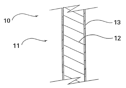
FIG. 1 depicts a partial cross-sectional view of an
intravascular medical device 10 such as a stent, catheter, wire
guide, cannula, and the like having a structure 11 shaped and sized
for introduction into the vascular system of a patient. The
structure of a stent typically includes a formed wire such as the
commercially available Gianturco-Roubin FLEX Stent from Cook
Incorporated, Bloomington, Indiana, for percutaneous introduction to
a failed angioplasty site. The<structure of a catheter, wire guide,
cannula, and the like are also well-known and commercially available
also from Cook Incorporated as well as other medical device
manufacturers. These intravascular medical devices are commonly
inserted into the vasculature of a patient using well-known
percutaneous surgical procedures. To advantageously minimize the
formation or removal of thrombus on the medical device, the
structure includes a thrombolytic agent 13 and a base material 12
treated with the thrombolytic agent. In FIG. 1, thrombolytic agent
13 is depicted as a coating on base material 12.
Base material 12 of the intravascular medical device
includes any one.of a number of different commercially available
biocompatible materials such as a metal, a plastic, a polymer, a =
biologically inert material, or a biologically derived material
suitable for the formation of the structure. The structure of the
intravascular medical device preferably includes a biologically
-6-
WO 95/06487 2.169638 PCTIUS94/12128
~
inert material so as to minimize, if not eliminate, an inflammatory
reaction of the vascular tissue of which the device is positioned
thereat. The metal comprises, amongst others, at least one from a
group consisting of titanium, stainless steel, tantalum, nitinol,
gold, platinum, inconel, and iridium, which are all commercially
available metals or alloys used in the fabrication of medical
devices. All of these metals are well-known to be biocompatibl'e
materials. The biologically inert material includes one or more of
a cellulose, cellulose compounds, cellulose-based polymers,
cellulose esters, cellulose ethers, cellulose acetate, cellulose
nitrate, polyurethanes, silicones, ethylene vinyl acetate
copolymers, polymethylemethacrylates, polyhydroxyethyl
methacrylates, polyethylene terephthalates,
polytetrafluoroethylenes, polyether urethanes, polyethylene oxides,
nylons, polyesters, polyamides, polyimides, polyvinyl chlorides,
polyvinyl acetates, polyolefins, polystyrene, polypropylenes,
polycaprolactones, epoxies, parylenes, hydrogels,
polyvinylpyrrolidone, polyvinyl alcohols, polyethylene glycols,
polyacrylamides, polyglycolyic acids, polylactic acids, proteins,
collagen, albumin, lipids, phospholipids, and phosphatidylcholine.
The biologically inert material can also be included in or can
constitute the entire base material or form a portion thereof. The
polymer comprises at least one from a group consisting of well-known
cellulose acetate, cellulose nitrate, silicone, polyethylene
teraphthalate, polyurethane, polyamide, polyester, polyorthoester,
and a polyanhydride. The polymer can also include one of the
aforementioned biologically inert materials. Biologically derived
material includes, by way of example, p'roteins, collagen, and
lipids. More broadly, the thrombolytic agent includes a plasminogen
activator which stimulates or augments the blood fibrinolytic system
which breaks down thrombi by breaking down insoluble fibrin into
soluble fibrin degradation products. The thrombolytic agent can
both cause the breakdown or lysis of existing macroscopic thrombi
and prevents the formation of macroscopic thrombi by causing the
lysis of microscopic thrombi as they form.
Thrombolytic agent coating 13 comprises at least one from a
group consisting of well-known and commercially available urokinase,
-7-
.~.~.
WO 95/06487 1x6?9 `+ 3 O p PCTlUS94112128
93
streptokinase, and tissue plasminogen activators (t-PA) These
thrombolytic agents are well-known and typically administered
systemically to dissolve, break up, or disperse thrombus.
Depicted in FIG. 2 is medical device 10 of FIG. 1 with a
second coating 13 of an antithrombogenic agent on base material 12.
This antithrombogenic agent includes an anticoagulant and/or an
antiplatelet agent for inhibiting the formation of thrombus on the
medical device. The anticoagulant agent typically includes heparin,
hirudin, hirulog, agatroban, tick anticoagulant peptide, and
antistasin. The antiplatelet agent typically includes aspirin,
dipyridamole, ticlopidine, sulfinpyrazone, prostaglandins, von
Willebrand factor antagonists, and glycoprotein Iib/IIIa
antagonists.
FIG. 3 depicts the medical device 10 of FIG. 1 with the
antithrombogenic agent coating 14 formed on base material 12 first
and thrombolytic agent coating 13 formed on top of coating 14.
FIG. 4 depicts medical device 10 of FIG. 3 wherein a primer
coating 15 has been first applied to base material 12 for adhering
the coatings of the antithrombogenic agent 14 and thrombolytic agent
13 to the base material. This primer material coating includes, for
example, well-known and commercially available cellulose esther,
cellulose nitrate, polyurethane, or a combination thereof. The
primer can also include any of the aforementioned biologically inert
materials.
FIG. 8 depicts medical device 10 of FIG. 1 wherein a
homogeneous coating 22 of a thrombolytic agent 13, antithrombogenic
agent 14, and a biologically inert material 15 has been applied to
base material 12. The biologically inert material of the
homogeneous coating is not ionically or covalently bonded to the
thrombolytic agent. This homogeneous coating does not affect the
strength or effectiveness of the thrombolytic agent. Furthermore,
the biologically inert material minimizes, if not eliminates,
inflammation of surrounding vascular tissue. An antiinflammatory agent 23 is
also included in the homogeneous mixture to minimize the
effects of any material of base material 12. Antiinflammatory agent
23 can also be included in the structure of base material 12. The
antiinflammatory agent can include a steroidal or a nonsteroidal
-8-
WO 95/06487 PCT/US94/12128
agent. The steroidal agent includes one or more of a cortisone,
dexamethasone, betamethasone, and prednisone, dexamethasone being
the preferred antiinflammatory agent. The nonsteroidal
antiinflammatory agent includes one or more of the salicylates and
propionic acid derivatives.
FIG. 5 depicts medical device 10 of FIG. 3 with primer
coating 15 with three separate layers of antithrombogenic agent 14
and three separate layers of thrombolytic agent 13 applied
thereover.
Although medical device 10 has been illustrated as having
separate coatings of a thrombolytic agent 13 and antithrombogenic
agent 14 applied thereto, it is to be understood and contemplated
that medical device 10 can be formed by mixing the antithrombogenic
agent, thrombolytic agent, and the base material together to form
the basic structure of the device. A primer can also be applied to
this mixture for facilitating the bonding of the two agents to the
base material. Alternatively, the intravascular medical device of
the present invention can also be a structure including any one or
more of the aforementioned thrombolytic agents and a base material
treated with the thrombolytic agent. It is also contemplated that
the base material can also include carbon such as associated with
pacemaker leads. The base material and thrombolytic agent can be
formed together and then extruded or formed to form the
intravascular medical device as desired. The antithrombogenic agent
can be applied in the form of a coating or, alternatively, also =
included in the mixture as previously discussed.
The method of treating a device with a thrombolytic agent
comprises the steps of providing a base material for the medical
device along with providing a thrombolytic agent and treating the
base material with the thrombolytic agent as will be described in
more detail hereafter. The step of treating the base material
includes dipping the base material such as stainless steel into a
solution of the thrombolytic agent such as urokinase. The base
material is removed from the solution and the thrombolytic agent
coating allowed to dry. The steps of dipping and drying the
thrombolytic agent on the base material is then repeated as many
times as desired. The method of treating a medical device with a
-9-
WO 95/06487 2169638 PCT/IJS94/12128
=
thrombolytic agent also includes providing at least one of a group
consisting of a polymer, biologically inert material, or
biologically derived material as previously described. The
thrombolytic agent and the polymer, biologically inert material, or
biologically derived material are mixed and then applied to the base
material.
Depicted in FIG. 6 is base material 17 such as stainless
steel of a medical device which has been placed in human blood. Red
blood cells 18, crenated red blood cells 19, platelet aggregates 20,
single platelets 21, and a large number of fibrin threads 22 have
formed on the untreated base material when placed in, for example,
human blood. FIG. 7 depicts base material 17 treated with a
thrombolytic agent as described herein. Only a small number of red
blood cells 18 and fibrin threads 22 appear to have formed on the
treated base material. Both of these figures illustrate samples of
stainless steel treated with urokinase and magnified 1,500 times.
A description of the materials and method used with in-vitro
thrombus deposition on three Gianturco-Roubin FLEX Stents from Cook
Incorporated will now be described. In-vitro thrombus deposition on
three Gianturco-Roubin coronary FLEX stents was examined. The
stents were 20-25 mm in length and designed to expand to 2.5-3.5 mm
in diameter. One stent was made from 0.006" diameter stainless
steel wire, a second from 0.006" diameter tantalum wire, and a third
from coated 0.006" diameter stainless steel wire. The third stent
was coated with a layer of primer (35066C, STS Biopolymers, Inc.,
Rush, New York) followed by 3 layers of heparin in a cellulose
polymer (Medicoat Heparin type 35066A, also commercially available
from STS Biopolymers Inc.). After deployment, the third=stent was
further coated with urokinase (AbbokinaseTM, urokinase for injection,
50,000 I.U./ml, commercially available from Abbot Laboratories, as
follows. The stent was dipped in the urokinase solution for
approximately 5 minutes, dried in room air for approximately 30
minutes, dipped in urokinase solution for approximately 1 minute,
dried in room air for approximatelv 30 minutes, dipped in urokinase
solution for 5-10 seconds and dried in room air for 30 minutes
before further handling. The uncoated stents were also deployed
-10-
WO 95/06487 2t69638 PCTIUS94/12128
~
., =. . = -
before use in the thrombus deposition experiment which will now be
described.
Each stent was suspended from the cap of a 6 ml test tube
for incubation in blood. Eighteen ml of human venous blood was
' S collected in a series of three 6 ml vacutainer tubes, each
containing 0.06 ml of heparinized normal saline (100 U of
heparin/ml). The blood, containing 1 U of heparin per ml, was then
carefully poured into the incubation tubes and the caps suspending
the stents were placed on these tubes. The tubes were positioned on
an inclined turntable rotating at approximately 20 rpm in a 37
degree C oven. The tubes were positioned so that the stent remained
totally immersed in blood for the entire incubation period which
lasted one hour. The tubes were positioned so as to rotate in a
well-known manner. After the one hour incubation in blood, each
stent was gently rinsed (2 dips of approximately 1-3 seconds
duration each) in 37 degrees C phosphate buffered saline and then
fixed in 3 percent glutaraldehyde in Milloniz's phosphate buffered
saline for at least 30 minutes before further processing. After
standard preparation (post-fixation in osmium, dehydration, critical
point drying and gold sputter coating) the stents were examined by
scanning electron microscopy.
The surfaces of the uncoated stainless steel and tantalum
stents were completely covered with a dense fibrin mesh containing
platelets and red blood cells. For each of these stents, there was
some variability in this covering from region to region. However,
there were no striking differences between the stainless steel and
tantalum stents and the coverage was visually estimated to be near
100 percent.
The surface of the H-UK coated stent (the third stent)
appeared strikingly different. The vast majority (visually
estimated at 90-95 percent) of the surface had only'a few adherent
red blood cells and a rare adherent platelet. There was also some
, variability in this covering and a visually estimated 5 to 10
percent of the surface had a slightly.y denser layer of adherent red
blood cells with a few platelets and an occasional fibrin thread.
The most striking difference between the coated and uncoated
stents was the fibrin deposition. Nearly 100 percent of the surface
-11-
WO 95/06487 PCT/[JS94/12128
s38
21~9
of the uncoated stents appeared covered with fibrin in contrast to
a visually estimated fibrin coverage of only 1-2 percent for the
coated stent.
Heparin is a mucopolysaccharide anticoagulant typically
obtained from porcine intestinal mucosa or bovine lung. Heparin
acts as a thrombin inhibitor by greatly enhancing the effects of the
blood's endogenous antithrombin III. Thrombin, a potent enzyme in
the coagulation cascade, is key in catalyzing the formation of
fibrin. Therefore, by inhibiting thrombin, heparin inhibits the
formation of fibrin thrombi. However, heparin's inhibition of
thrombin and fibrin formation is not 100 percent as evidenced by the
fibrin deposition on uncoated stents in heparinized blood.
Furthermore, heparin does not have fibrinolytic activity.
Urokinase is a plasminogen actuating enzyme typically
obtained from human kidney cell cultures. Urokinase catalyzes the
conversion of plasminogen into the fibrinolytic plasmin which breaks
down fibrin thrombi.
It is highly probable that both the heparin and urokinase on
the coated stent contributed to the dramatic reduction in fibrin
deposition on this stent. It has not been determined which of these
agents may have had the greater effect. Moreover, it has not been
determined whether the effects were localized near the surface of
the stent or whether the delivery of heparin or urokinase may have
caused anticoagulant and/or fibrinolytic effects respectively on the
entire 6 ml of blood in which the stent was incubated.
In another series of experiments performed by the inventors,
coated stents were implanted in the external iliac arteries of
rabbits for periods of up to six months. Although no inflammatory
reactions were observed in response to the cellulosic polymers used,
the quaternary ammonium binding agent benzalkonium chloride was
associated with an intense inflammatory reaction when included in
the stent coating. However, when the potent antiinflammatory
steroid dexamethasone was also included in the coating, the
inflammatory reaction was suppressed. Similar results were reported
(Lincoff, et al., "Local Delivery of Dexamethasone by an Eluting
Stent Attenuates the Adverse Response to Biodegradable Polymer in
the Porcine Coronary Artery", Circulation, Vol 88, No 4, Part 2,
-12-
WO 95/06487 ~+ +~ 1UC{j638 PCT/US94/12128
J
p. 1-655, October 1993) when stents coated with poly-l-lactic acid
(PLLA) or PLLA with dexamethasone added (DEX-PLLA) were implanted in
porcine coronary arteries. Severe inflammation was observed in
response to the PLLA coated stents. However, the inflammation was
substantially less in arteries implanted with DEX-PLLA coated
stents. Also, from a study of polylactic acid (PLA) microspheres
delivered into the rabbit carotid artery wall (Dev, et al.,
"Microspheres for Drug Delivery to the Arterial Wall: A Study of
Kinetics, Toxicity and Effects of Corticosteroid Loaded
Microspheres", JACC, p. 19A, February, 1994), it was reported that
arteries infused with unloaded microspheres showed inflammation
where arteries infused with dexamethasone loaded microspheres did
not.
It is to be understood that the above-described thrombolytic
treated intravascular medical device is merely an illustrative
embodiment of the principles of this invention and that other
thrombolytic treated intravascular medical devices may be devised by
those skilled in the art without departing from the spirit and -scope
of this invention.
-13-