Note: Descriptions are shown in the official language in which they were submitted.
WO 95/0578(1 21 ~ 9 g r~ 3 PCT/US94/09443
- 1 -
MODIFICATIONS OF VISUAL ACUITY BY THERMAL MEANS
BACHGROUND OF THE INVENTION
1. FIELD OF THE INVENTION
The present invention relates to a thermokeratoplasty probe that is
placed into direct contact with the outer surface of the cornea.
2. DESCRIPTION OF RELATED ART
Techniques for correcting vision have included reshaping the
cornea of the eye. For example, myopic conditions can be corrected by
cutting a number of small incisions in the corneal membrane. The
incisions allow the corneal membrane to relax and increase the radius of
the cornea. The incisions are typically created with either a laser or a
precision knife. The procedure for creating incisions to correct myopic
defects is commonly referred to as radial keratotomy and is well known
in the art.
Present radial keratotomy techniques generally make incisions that
penetrate approximately 95% of the cornea. Penetrating the cornea to
such a depth increases the risk of puncturing the decemets membrane
and the endothelium layer, and creating permanent damage to the eye.
Additionally, light entering the cornea at the incision sight is refracted by
the incision scar and produces a glaring effect in the visual field. The
glare effect of the scar produces impaired night vision for the patient. It
WO 95/05780 1~.9 9 ~~ PCT/US94/09443
-2-
would be desirable to have a procedure for correcting myopia that does
not require a 95% penetration of the cornea.
The techniques of radial keratotomy are only effective in correcting
myopia. Radial keratotomy cannot be used to correct an eye condition
such as hyperopia. Additionally, keratotomy has limited use in reducing
or correcting an astigmatism. The cornea of a patient with hyperopia is
relatively flat (large spherical radius). A flat cornea creates a lens system
which does not correctly focus the viewed image onto the retina of the
eye. Hyperopia can be corrected by reshaping the eye to decrease the
spherical radius of the cornea. It has been found that hyperopia can be
corrected by heating and denaturing local regions of the cornea. The
denatured tissue contracts and changes the shape of the cornea and
corrects the optical characteristics of the eye. The procedure of heating
the corneal membrane to correct a patient's vision is commonly referred
to as thermokeratoplasty.
U.S. Patent No. 4,461,294 issued to Baron; U.S. Patent No.
4,976,709 issued to Sand and PCT Publication WO 90/12618, all disclose
thermokeratoplastic techniques which utilize a laser to heat the cornea.
The energy of the laser generates localized heat within the corneal
stroma through photonic absorption. The heated areas of the stroma
then shrink to change the shape of the eye.
Although effective in reshaping the eye, the laser based systems of
the Baron, Sand and PCT references are relatively expensive to produce,
have a non-uniform thermal conduction profile, are not self limiting, are
susceptible to providing too much heat to the eye, may induce
astigmatism and produce excessive adjacent tissue damage, and require
long term stabilization of the eye. Expensive laser systems increase the
cost of the procedure and are economically impractical to gain
w0 95/05780 ~ ~ ~ ~ ~ j PCT/US94l09443
-3-
widespread market acceptance and use. Additionally, laser
thermokeratoplastic techniques non-uniformly shrink the stroma without
shrinking the Bowmans layer. Shrinking the stroma without a
corresponding shrinkage of the Bowmans layer, creates a mechanical
strain in the cornea. The mechanical strain may produce an undesirable
reshaping of the cornea and probable regression of the visual acuity
correction as the corneal lesion heals. Laser techniques may also
perforate Bowmans layer and leave a leucoma within the visual field of the
eye.
U.S. Patent Nos. 4,326,529 and 4,381,007 issued to Doss et al,
disclose electrodes that are used to heat large areas of the cornea to
correct for myopia. The electrode is located within a housing that spaces
the tip of the electrode from the surface of the eye. An isotropic saline
solution is irrigated through the electrode and aspirated through a
channel formed between the outer surface of the electrode and the inner
surface of the sleeve. The saline solution provides an electrically
conductive medium between the electrode and the corneal membrane.
The current from the electrode heats the outer layers of the cornea.
Heating the outer eye tissue causes the cornea to shrink into a new radial
shape. The saline solution also functions as a coolant which cools the
outer epithelium layer.
The saline solution of the Doss device spreads the current of the
electrode over a relatively large area of the cornea. Consequently,
thermokeratoplasty techniques using the Doss device are limited to
reshaped corneas with relatively large and undesirable denatured areas
within the visual axis of the eye. The electrode device of the Doss system
is also relatively complex and cumbersome to use.
i . .
WO 95/05780 PCT/US94/09443
_4
"A Technique for the Selective Heating of Corneal Stroma" Doss et
al., Contact & Intraoccular Lens Medical Jrl., Vol. 6, No. 1, pp. 13-17,
Jan-Mar., 1980, discusses a procedure wherein the circulating saline
electrode (CSE) of the Doss patent was used to heat a pig cornea. The
electrode provided 30 volts r.m.s. of power for 4 seconds. The results
showed that the stroma was heated to 70°C and the Bowman's membrane
was heated 45°C, a temperature below the 50-55°C required to
shrink
the cornea without regression.
"The Need For Prompt Prospective Investigation" McDonnell,
Refractive & Corneal Surgery, Vol. 5, Jan./Feb., 1989 discusses the
merits of corneal reshaping by thermokeratoplasty techniques. The
article discusses a procedure wherein a stromal collagen was heated by
radio frequency waves to correct for a keratoconus condition. As the
article reports, the patient had an initial profound flattening of the eye
followed by significant regression within weeks of the procedure.
"Regression of Effect Following Radial Thermokeratoplasty in
Humans" Feldman et al., Refractive and Corneal Surgery, Vol. 5,
Sept./Oct., 1989, discusses another thermokeratoplasty technique for
correcting hyperopia. Feldman inserted a probe into four different
locations of the cornea. The probe was heated to 600°C and was inserted
into the cornea for 0.3 seconds. Like the procedure discussed in the
McDonnell article, the Feldman technique initially reduced hyperopia,
but the patients had a significant regression within 9 months of the
procedure. To date, there has been no published findings of a
thermokeratoplasty technique that will predictably reshape and correct
the vision of a cornea without a significant regression of the corneal
correction.
WO 95/05780 ~~ pCT/US94/09443
c~
-5-
It would therefore be desirable to provide a thermokeratoplasty
technique which can predictably reshape and correct the vision of an eye
without a significant regression of the visual acuity correction.
Electrodes are subject to contamination, when RF electrical
current is used for thermokeratoplasty. For example, an electrolized
layer or protein film may form on the surface of the electrodes. Such a
filin may vary the impedance of the electrodes and affect the
performance of the instrument. Varying instrument performance may
create inconsistent results. Therefore it would be desirable to provide a
thermokeratoplastic probe that would have to be replaced by a new
device after a predetermined number of uses.
CA 02169943 2001-O1-08
WO 95105780 - PCTIUS9al09:1a3
-6-
SUMMARY OF THE INVENTION
The present invention is a thermokeratoplasty system and method
for locally heating and reshaping a cornea in a manner that produces a
minimal regression of the corneal correction. The system includes a
probe that is coupled to a power source which can provide a
predeterTnined power, frequency and time duration that creates a
thermal profile within the cornea which extends from the epithelium
into the corneal stroma. The electrical return of the probe is a lid
speculum which maintains the eye lids in an open position. The probe is
placed into contact with the cornea and energy is transferred from the
power source to the eye, through the lid speculum and back to the power
source. The energy from the power supply is focused by a probe tip that
locally heats and denatures the cornea, and causes a subsequent
shrinkage of corneal tissue. A pattern of denatured areas can be created
around the cornea to correct the vision of the eye. It has been found that
power no greater than 1.2 watts, for a duration no greater than 1.0
seconds, will sufficiently induce corneal shrinkage without any significant
regression of the visual acuity correction of the eye. The probe may have
an electronic circuit which prevents usage of the probe after a
predetermined number of procedures.
Accordingly, in one of its broad aspects, the present
invention resides in providing a thermokeratoplastic system
for reshaping a cornea, comprising: a power source which
contains a radio frequency generator that generates an
alternating current; a first electrode that is coupled to
CA 02169943 2001-O1-08
-6a-
said radio frequency generator and which transfers the
alternating current from said radio frequency generator to
the cornea which is in contact with said first electrode; a
lid speculum that is coupled to said power source and the
cornea to create a return path so that the alternating
current flows from said first electrode, through the cornea
and back into said lid speculum.
Also, in another of its broad aspects, the present
invention resides in providing a thermokeratoplastic probe
that is coupled to a source of power, comprising: an
electrode coupled to the source of power; a fuse coupled to
said electrode, said fuse preventing power from being
supplied to said electrode when said fuse is blown; and, an
electrical circuit that provides a fuse current to blow
said fuse after power is supplied to said electrode.
In another of its broad aspects, the present invention
resides in providing a thermokeratoplastic probe system for
reshaping a cornea, comprising: a handle; a tip that
extends from said handle, said tip having a sharp point
that can be inserted into the cornea; a power supply
connected to said tip, said power supply provides a pulse
of current at a power no greater than 1.2 watts and for a
time duration no greater than 1.0 seconds, such that the
current flows into the cornea through said inserted tip to
denature the cornea.
WO 95/05780 PCT/US94/09443
_7_
BRIEF DESCRIPTION OF THE DRAWINGS
The objects and advantages of the present invention will become
more readily apparent to those ordinarily skilled in the art after
reviewing the following detailed description and accompanying drawings,
wherein:
Figure 1 is a perspective view of a thermokeratoplastic electrode
system of the present invention;
Figure 1 a is a graph showing a waveform that is provided to the
probe of the system;
Figure lb is a graph showing the amount of typical vision correction
regression over time;
Figure 1 c is a representation of a nominal thermal profile within
the cornea produced by the electrode system of the present invention;
Figure 2 is a top view of an electrode probe of the system;
Figure 3 is a side view of the probe in Fig. 2;
Figure 4 is an enlarged view of the probe tip;
Figure 5 is a side view showing the probe being used to treat an
area of the corneal membrane;
Figure 6 is a top view showing a pattern of denatured areas of the
cornea;
Figure 7 is a perspective view of an alternate embodiment of the
probe;
Figures 8a-b show a method for performing a procedure of the
present invention;
Figure 9 shows a pattern of incisions and denatured areas to
correct for a myopic condition;
WO 95/05780 , ~ PCT/US94/09443
_ g _
Figure 10 shows another pattern of incisions and denatured areas
to correct for hyperopic conditions;
Figure 11 shows a preferred embodiment of the present invention;
Figure 11 a is an enlarged view of the tip of Figure 11;
Figure 12 is a perspective view of a probe with the return electrode
as a lid speculum that maintains the eye lid in an open position;
Figure 13 is a side view of an alternate probe tip embodiment;
Figure 14 is a side view of an alternate probe tip embodiment;
Figure 15 is a side view of an alternate probe tip embodiment;
Figure 16 is a side view of an alternate probe tip embodiment;
Figure 17 is a side view of an alternate probe tip embodiment;
Figure 18 is a side view of an alternate probe embodiment;
Figure 19 is a schematic of a circuit which limits the
use of a probe
beyond a
predetermined
useful
life.
WO 95/05780 PCT/US94/09443
_g_
DETAILED DESCRIPTION OF THE INVENTION
Referring to the drawings more particularly by reference numbers,
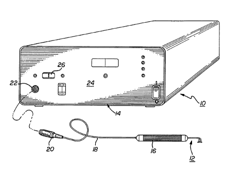
Figure 1 shows a thermokeratoplastic electrode system 10 of the present
invention. The system 10 includes an electrode probe 12 coupled to a
power supply unit 14. The power supply unit 14 contains a power supply
which can deliver power to the probe 12. The probe 12 has a hand piece
16 and wires 18 that couple the probe electrodes to a connector 20 that
plugs into a mating receptacle 22 located on the front panel 24 of the
power unit. The hand piece 16 may be constructed from a non-
conductive material and is approximately 0.5 inches in diameter and 5
inches long.
The power supply 14 provides a predetermined amount of energy,
through a controlled application of power for a predetermined time
duration. The power supply 14 may have manual controls that allow the
user to select treatment parameters such as the power and time duration.
The power supply 14 can also be constructed to provide an automated
operation. The supply 14 may have monitors and feedback systems for
measuring tissue impedance, tissue temperature and other parameters,
and adjust the output power of the supply to accomplish the desired
results. The unit may also have a display that indicates the number of
remaining uses available for the probe 12.
In the preferred embodiment, the power supply provides a
constant current source and voltage limiting to prevent arcing. To
protect the patient from overvoltage or overpower, the power unit 14
may have an upper voltage limit and/or upper power limit which
terminates power to the probe when the output voltage or power of the
unit exceeds a predetermined value. The power unit 14 may also contain
WO 95/05780 PCT/US94/09443
- 10 -
monitor and alarm circuits which monitor the resistance or impedance of
the load and provide an alarm when the resistance/impedance value
exceeds and/or falls below predefined limits. The alarm may provide
either an audio and/or visual indication to the user that the
resistance/impedance value has exceeded the outer predefined limits.
Additionally, the unit may contain a ground fault indicator, and/or a tissue
temperature monitor. The front panel of the power unit typically
contains meters and displays that provide an indication of the power,
frequency, etc., of the power delivered to the probe.
The power unit 14 may deliver a power output in a frequency range
of 5 KHz- 50 MHz. In the preferred embodiment, power is provided to
the probe at a frequency in the range of 500 KHz. The unit 14 is
designed so that the power supplied to the probe 12 does not exceed 1.2
watts (V~. The time duration of each application of power to a particular
corneal location is typically between 0.1-1.0 seconds. The unit 14 is
preferably set to deliver approximately .75 W of power for 0.75 seconds.
Figure 1 a shows a typical voltage waveform that is applied by the unit 14.
Each pulse of energy delivered by the unit 14 is a highly damped signal,
typically having a crest factor (peak voltage/RMS voltage) greater than
10:1. Each power dissipation is provided at a repetitive rate. The
repetitive rate may range between 4-12 KHz and is preferably set at 8
KHz.
The system has a switch which controls the application of power to
the probe 12. The power unit 14 also contains a timer circuit which
allows power to be supplied to the probe 12 for a precise predetermined
time interval. The timer may be a Dose timer or other similar
conventional circuitry which terminates power to the probe after a
predetermined time interval. The unit may also allow the user to apply
WO 95/05780 PCT/US94/09443
- 11
power until the switch is released. As one embodiment, the power
supply may be a unit sold by Birtcher Medical Co. under the trademark
HYFRECATOR PLUS, Model 7-797 which is modified to have voltage,
waveform, time durations and power limits to comply with the above
cited specifications. .
The power unit 14 may have a control member 26 to allow the user
to select between a "uni-polar" or a "bi-polar" operation. The power
supply 14 may be constructed to provide a single range of numerical
settings, whereupon the appropriate output power, time duration and
repetition rate are determined by the hardware and software of the unit.
The front panel of the power unit may also have control members (not
shown) that allow the surgeon to vary the power, frequency, timer
interval, etc. of the unit. The return electrode (not shown) for a uni-polar
probe may be coupled to the power unit through a connector located on
the unit. The return electrode is preferably a cylindrical bar that is held
by the patient, or an eye fixation electrode.
It has been found that at higher diopters, effective results can be
obtained by providing two different applications at the same location.
Listed below in Table I are the power settings (peak power) and time
duration settings for different d'iopter corrections (-d), wherein the
locations (Loc) are the number of denatured areas in the cornea and
dots/Loc is the number of power applications per location.
WO 95/05780 PCT/US94/09443
...
- 12-
TABLE I
-d DOTS/LO LOC PWR (W) TIME(SE
C C)
1.5 1 8 0.66 .75
2.5 2 8 0.66 .75
3.5 2 8 0.83 .75
4.5 2 16 0.66 .75
6.0 2 16 0.83 .75
Using the parameters listed in Table I, the procedure of the
present invention was performed on 36 different patients suffering from
some degree of hyperopia. A pattern of 8-16 denatured areas were
created in the non-vision area of the eye. Patients who needed higher
diopter corrections were treated with high applications of power. Figure
lb shows the amount of regression in the vision correction of the eye.
The eyes were initially overcorrected to compensate for the known
regression in the procedure. As shown in Fig. lb, the regression became
stabilized after approximately 60 days and completely stabilized after 180
days. The error in overcorrection was within +/- 0.5 diopters.
Figure 1 c shows nominal thermal profiles produced by the
application of power to the cornea. As known to those skilled in the art,
the cornea includes an epithelium layer, a Bowmans membrane, a stroma,
a Descemets membrane and a endothelium layer. Without limiting the
scope of the patent, the applicant provides the following discussion on
the possible effects of the present method on the cornea of the eye.
WO 95/05780 PCT/US94/09443
~~~~ w
- 13-
When power is first applied to the cornea the current flows through the
center of the tissue immediately adjacent to the probe tip. The
application of power causes an internal ohmic heating of the cornea and a
dehydration of the tissue. The dehydration of the tissue rapidly increases
the impedance of the local heated area, wherein the current flows in an
outward manner indicated by the arrows in Fig. 1 c. The cycle of
dehydration and outward current flow continues until the resistance from
the tip to the outer rim of the corneal surface, and the full thermal
profile, is significantly high to prevent further current flow of a
magnitude to further cause denaturing of the corneal tissue. The direct
contact of the probe with the cornea along the specific power/time
settings of the power source creates a thermal profile that denatures both
the Bowman's membrane and the stroma. The denaturing of both the
Bowman's membrane and the stroma in a circular pattern creates a
linked belt type contracted annular ring. This annular ring will create a
steepening of the cornea and sharpen the focus of the images on the
retina. To control and minimize the denatured area, the surface of the
eye is kept dry by applying either a dry swab to the cornea or blowing dry
air or nitrogen across the surface of the eye.
The design of the power source and the high electrical resistance
of the denatured area provides a self limit on the amount of penetration
and area of denaturing of the cornea. Once denatured, the cornea
provides a high impedance to any subsequent application of power so that
a relatively low amount of current flows through the denatured area. It
has been found that the present procedure has a self limited denatured
profile of approximately no greater than 75% of the depth of the stroma.
This prevents the surgeon from denaturing the eye down to the
decemets membrane and endothelium layer of the cornea.
WO 95/05780 PCT/US94/09443
~~,6°~
- 14-
Fig. 1 c shows nominal thermal profiles for diopter corrections of
-1. 5 d, -2.5-3.5 d and -4.0-6.0 d, respectively. In accordance with Table
I, a-1.5 diopter correction creates a denatured diameter of approximately
1 mm and a stroma penetration of approximately 30%. A -2.5-3.5 d
correction creates a denatured diameter of approximately 1.13 mm and a
stroma penetration of approximately 50%. A -4.0-6.0 d correction
creates a denature diameter of approximately 1.25 mm and a stroma
penetration of approximately 75%.
Figures 2-5 show an embodiment of the probe 12. The probe 12
has a first electrode 30 and a second electrode 32. Although two
electrodes are described and shown, it is to be understood that the probe
may have either both electrodes (bipolar) or just the first electrode
(unipolar). If a unipolar probe is used, a return electrode (indifferent
electrode) is typically attached to, or held by, the patient to provide a
"return" path for the current of the electrode.
Both electrodes 30 and 32 extend from the hand piece 16 which
contains a pair of internal insulated conductors 34 that are contact with
the proximal end of the electrodes. The first electrode 30 has a tip 36
which extends from a first spring member 38 that is cantilevered from
the hand piece 16. The electrode 30 is preferably constructed from a
phosphor-bronze or stainless steel, wire or tube, that is 0.2-1.5 mm in
diameter. The spring portion 38 of the first electrode 30 is preferably
50 millimeters (mm) long. In one embodiment, the tip 36 has an
included angle of between 15-60°, 30° nominal, and a nose radius
of
approximately 50 microns. A majority of the electrode 30 is covered
with an insulating material to prevent arcing, and to protect non-target
tissue, the user and the patient. The relatively light spring force of the
WO 95!05780 PCT/US94/09443
- 15
probe provides a sufficient electrode pressure without penetrating the
cornea.
The second electrode 32 includes a disk portion 40 which extends
from a second spring member 42 that is also cantilevered from the hand
piece 16. The disk portion 40 is spaced a predetermined distance from
first electrode 30 and has an aperture 44 that is concentric with the tip
36. In the preferred embodiment, the disk portion 40 has an outer
diameter of 5.5 mm and an aperture diameter of 3.0 mm. The disk 40
further has a concave bottom surface 46 that generally conforms to the
shape of the cornea or sclera.
In one embodiment, the bottom surface 46 has a spherical radius of
approximately 12.75 mm and a griping surface to assist in the fixation of
the eye. The second electrode 32 provides a return path for the current
from the first electrode 30. To insure proper grounding of the cornea,
the surface area of the disk 40 is typically 20-500 times larger than the
contact area of the tip 36. In the preferred embodiment, the second
spring member 42 is constructed to have a spring constant that is less
than one-half the stiffness of the first spring member 38, so that the
second electrode 32 will have a greater deflection per unit force than the
first electrode 30. As shown in Fig. 3, the tip 36 and disk 40 are typically
located at angles a' and a" which may range between 30°-180°,
with the
preferred embodiment being 45°. As shown in Fig. 5, the probe 12 is
pressed against the cornea to allow the second electrode 32 to deflect
relative to the first electrode 30. The second electrode 32 is deflected
until the tip 36 is in contact with the cornea.
For surgeons who prefer "two handed" procedures, the probe could
be constructed as two pieces, one piece being the first electrode, and the
other piece being the second electrode which also stabilizes the eye
WO 95/05780 PCT/US94109443
- 16
against corneal movement. Although the probe has been described and
shown denaturing a cornea, it is to be understood that the probes and
methods of the present invention can be used to denature other tissues
to correct for wrinkles, incontinence, etc. For example, the probe could
be used to shrink a sphincter to correct for incontinence. The technique
would be basically the same with small closely spaced dots forming a
tightening line, belt or cylinder.
Figure 6 shows a pattern of denatured areas 50 that have been
found to correct hyperopic conditions. A circle of 8 or 16 denatured
areas 50 are created about the center of the cornea, outside the visual
axis portion 52 of the eye. The visual axis has a nominal diameter of
approximately 5 millimeters. It has been found that 16 denatured areas
provide the most corneal shrinkage and less post-op astigmatism effects
from the procedure. The circle of denatured areas typically have a
diameter between 6-8 mm, with a preferred diameter of approximately 7
mm. If the first circle does not correct the eye deficiency, the same
pattern may be repeated, or another pattern of 8 denatured areas may be
created within a circle having a diameter of approximately 6.0-6.5 mm
either in line or overlapping. It has been found that overcorrected
hyperopic conditions may be reversed up to 80% by applying a steroid,
such as cortisone, to the denatured areas within 4 days of post-op and
continued for 2 weeks after the procedure. The procedure of the
present invention can then be repeated after a 30 day waiting period.
The exact diameter of the pattern may vary from patient to patient,
it being understood that the denatured spots should preferably be formed
in the non-visionary portion 52 of the eye. Although a circular pattern is
shown, it is to be understood that the denatured areas may be located in
any location and in any pattern. In addition to correcting for hyperopia,
WO 95/05780 PCT/US94/09443
- 17-
the present invention may be used to correct astigmatic conditions. For
correcting astigmatic conditions, the denatured areas are typically
created at the end of the astigmatic flat axis. The present invention may
also be used to correct radial keratotomy procedures that have
overcorrected for a myopic condition.
The probe and power settings have been found to create denatured
areas that do not reach the Decemets membrane. It has been found that
denatured areas of the Bowmans layer in the field of vision may disturb
the patients field of vision, particularly at night. The present invention
leaves a scar that is almost imperceptible by slit lamp examination 6
months after the procedure. It has been found that the denatured areas
generated by the present invention do not produce the star effect caused
by the refraction of light through the slits created in a corrective
procedure such as radial keratotomy.
Figure 7 shows an alternate embodiment of a probe 60 which has a
plurality of first electrodes 62 coupled to a cage 64. The cage 64
includes a first ring 66 separated from a second ring 68 by a number of
spacers 70. The cage 64 can be connected to a handle (not shown)
which allows the surgeon to more easily utilize the probe 60.
The first electrodes 62 extend through apertures 72 in the rings
66 and 68. The electrodes 62 can move relative to the cage 64 in the
directions indicated by the arrows. The probe 60 has a plurality springs
74 located between the rings and seated on washers 76 mounted to the
electrodes 62. The springs 74 bias the electrodes 62 into the positions
shown in Fig. 7. In the preferred embodiment, the probe 60 includes 8
electrodes arranged in a circular pattern having a 7.0 millimeter
diameter.
WO 95/05780 ~ PCT/US94/09443
- 18 -
In operation, the probe 60 is pressed onto the cornea so that the
electrodes 62 move relative to the cage 64. The spring constant of the
springs 74 is relatively low so that there is a minimal counterforce on the
tissue. A current is supplied to the electrodes 62 through wires 78
attached thereto. The probe 60 is preferably used as a uni-polar device,
wherein the current flows through the tissue and into a return electrode
attached to or held by the patient.
Figure 8a and 8b show a preferred method of correcting for
hyperopic conditions using the electrode system of the present
invention. As shown in procedural block 100 refractive readings are
initially taken of both eyes with, and then without, cycloplasia. In
procedure block 102, the interoccular pressure and cornea thickness at
the center of the eye are taken with a tonometer and pacymeter,
respectively. If the interoccular pressure is 20 mm Hg or greater, for
LO.P. reduction, 1 drop of a .5% solution marketed under the trademark
"Betagan" is applied to the cornea twice a day for 2-3 months and then
initial test are repeated. A topography reading of the eye is then taken to
determine the shape of the cornea in procedural block 104.
Approximately 30 minutes before the application of the electrode,
the patient is given a mild tranquilizer such as 5 mg of valium, and the
surgeon administers drops, such as the drops marketed under the
trademark "Madryacil", to dilate the pupil and freeze accommodation, in
block 106. Immediately before the procedure, 2 drops of a topical
cocaine commonly known as "Proparacaine" is administered to the eyes
in block 108. In block 110 an in line microscope light is directed to the
cornea for marking purposes. Then the lighting may be directed in a
lateral direction across the cornea. Laterally lighting the eye has been
WD 95/05780 PCT/US94/09443
- 19-
found to provide good visualization without irritating or photobleaching
the retina.
In procedural block 112, the surgeon marks 8 or 16 spots on the
cornea, wherein the pattern has a preferred diameter of approximately 7
mm. The surgeon sets the power and duration setting of the power unit
to the proper setting. In block 114, the surgeon then places the tip at
one of the spot markings and depresses the foot switch of the system, so
that power is supplied to the probe and transferred into the cornea. This
process is repeated at all of the spot markings. The epithelium of the
denatured areas are then removed with a spatula in block 116. If a
diopter correction of -2.5-3.5 d, or -4.0-6.0 d is required the tip is again
placed in contact with the spots and power is applied to the cornea to
generate a deeper thermal profile in the stroma. The procedure is then
checked with an autorefractor.
The eyes are covered with a patch or dark glasses, and the patient
is given medication, in block 118. The patient preferably takes an
antibiotic such as a drug marketed under the trademark "Tobrex" every 2
hours for 48 hours, and then 3 times a day for 5 days. The patient also
preferably takes an oral analgesic, such as a drug marketed under the
trademark "Dolac", 10 mg every 8 hours for 48 hours and a drug
marketed under the trademark "Globaset" every 8 hours for 48 hours. If
the patient has been overcorrected, the procedure can be reversed by
waiting 3-4 days after the procedure and then administering to the eyes
1 drop of a steroid such as cortisone, 3 times a day for 1-2 weeks.
Figure 9 shows a pattern of denatured areas 130 combined with a
pattern of incisions 132 that can correct myopic conditions. The
incisions can be made with a knife or laser in accordance with
conventional radial keratotomy procedures. The incisions are made from
WO 95/05780 PCT/US94/09443
'~1 _20_
a 3.5 mm diameter to within 1 mm of the limbus at a depth of
approximately 85% of the cornea. Denatured areas are then created
between the incisions 132 using the procedure described above. The
power unit is preferably set at 0.75 W of power and a time duration of
0.75 seconds. The slow heating of the cornea is important for
minimizing regression, and as such 0.75 seconds has been found to be a
preferable time duration to account for the patients fixation ability and
the surgeons reaction time. The denatured areas pull the incisions to
assist in the reshaping of the cornea. This procedure has been found to
be effective for diopter corrections up to + 10.0 d. Penetrating the cornea
only 85% instead of conventional keratotomy incisions of 95% reduces
the risk of puncturing the decemets membrane and the endothelium
layer. This is to be distinguished from conventional radial keratotomy
procedures which cannot typically correct for more than 3.5 diopters.
The denatured pattern shown in Fig. 6 has been shown to correct
up to 7.0 diopters. As shown in Figure 10, a circumferential pattern of
incisions 134 may be created in addition to a pattern of denatured areas
136, to increase the correction up to 10.0 diopters. The incisions will
weaken the eye and allow a more pronounced reshaping of the eye. The
pattern of incisions may be created at either a 6 mm diameter or a 8 mm
diameter. The incisions typically penetrate no greater than 75% of the
cornea. The contractive forces of the denatured areas may create gaps in
the incisions. It may be preferable to fill the gaps with collagen or other
suitable material.
Figure 11 shows an alternate embodiment of a probe which has a
single electrode 140. The electrode 140 has a tip 142 which is
preferably 0.009 inches in diameter. The tip extends from a spring beam
144 that is bent so that the surgeon can place the tip onto the cornea
WO 95/05780 PCT/US94/09443
-21 -
over nose and brow without impairing the surgeon's vision. The spring
beam 144 is preferably insulated and is 0.2-1.5 mm in diameter. The
spring beam 144 extends from a base 146 that is inserted into the hand
piece. The base 146 is preferably constructed from stainless steel and is
0.030-0.125 inches in diameter, with a preferred diameter of 0.060-
0.095 inches.
As shown in Figure 11 a, the end of the tip 142 is preferably flat and
has a textured surface 148. The textured surface 148 slightly grips the
cornea so that the tip does not move away from the marking when power
is applied to the eye.
As shown in Figure 12, the probe 200 has a return electrode lid
speculum 202 that maintains the eye lid in an open position. The
speculum 202 has a pair of cups 204 located at the end of wire 206. The
cups 204 are placed under an eye lid and maintain the position of the lid
during the procedure. Extending from the lid speculum 202 is a wire
208 that is typically plugged into the unit 14 "return" connector. It has
been found that the procedure of the present invention will produce
more consistent results when the probe 200 uses the lid speculum 202
as the return electrode. The impedance path between the probe 200 and
the lid speculum 202 is relatively consistent because of the relatively
short distance between the lid speculum 202 and the probe 200, and the
wet interface between the cornea and the lid speculum 202.
Figures 13-15 show alternate probe tip embodiments. The tips
have steps that increase the current density at the corneal interface. The
tips are preferably constructed from a stainless steel that is formed to the
shapes shown. The tip 220 shown in Fig. 13 has a cylindrical step 222
that extends from a base 224. The step 222 terminates to a point,
although it is to be understood that the end of the step 222 may have a
WO 95/05780 PCT/US94/09443
_22_
flat surface. In the preferred embodiment, the base 224 has a diameter
of 350 microns (um), and the step 222 has a diameter of 190 microns
and a length of 210 microns.
The tip 230 shown in Fig. 14, has a first step 232 extending from a
base portion 234 and a second step 236 extending from the first step
232. The end of the second step 236 may be textured to improve the
contact between the probe and the cornea. In the preferred
embodiment, the first step 232 has a diameter of 263 microns and a
length of 425 microns, the second step 236 has a diameter of 160
microns and a length of 150 microns . The tip 240 shown in Fig. 15, has
a first step 242 that extends from a base portion 244 and a second
tapered step 246 that extends from the first step 242. In the preferred
embodiment, the first step 242 has a diameter of 290 microns and a
length of 950 microns. The second step 246 has a diameter of 150
microns, a length of 94 microns and a radius of 70 microns.
Figures 16 and 17 show alternate probe tip embodiments which
have an outer electrode concentric with an inner electrode. The
electrodes are coupled to the unit so that the electrodes can provide
current to the cornea either simultaneously or sequentially. By way of
example, it may be desirable to initially apply power to the cornea with
the inner electrode and then apply power with the outer electrode, or
apply power with both electrode and then apply power with only the
outer electrode. Assuming the same current value, the inner electrode
will apply power with a greater current density that the outer electrode.
The dual electrode probes allow the surgeon to create different thermal
profiles, by varying the current densities, waveforms, etc. of the
electrodes.
WO 95/05780 ~~ PCT/US94/09443
-23-
The probe 250 shown in Fig. 16 has an inner electrode 252 that is
concentric with an intermediate layer of insulative material 254 and an
outer conductive layer 256. In the preferred embodiment, the inner
electrode 252 may have a diameter of 125 microns and extend from the
outer layers a length of 150 microns. The outer layer 256 may have
diameter of 350 microns. The inner electrode 252 may be capable of
being retracted into the insulative layer 254 so that the inner electrode
252 is flush with the outer electrode 256, or may be adjusted between
flush and full extension, either manually or under servo control.
Fig. 17 shows another alternate embodiment, wherein the probe
260 has an additional outer sleeve 262. The sleeve 262 has an internal
passage 264 that supplies a fluid. The fluid may be a gas that stabilizes
the current path to the cornea or a relatively high impedance solution
(such as distilled water) which provides a coolant for the eye.
Figure 18 shows an economical detachable probe 270 embodiment.
The probe tip 270 has a conductive wire 272 that is located within a
plastic outer housing 274. The probe tip 270 has a flexible section 276
that extends from a body 278, preferably at a 45° angle. The tip 280
extends from the flexible section 276, preferably at a 90° angle.
Extending from the opposite end of the handle 278 is a male connector
282. The connector 282 may have a conductive sleeve 284 that is
inserted into the socket 286 of a female probe connector 288. The end
of the wire 272 may be pressed between the inner surface of the sleeve
284 and the outer surface of the male connector 282 to provide an
electrical interconnect between the tip end 280 and the female probe
connector 288. The sleeve 284 may have a detent 290 to secure the
probe tip 270 to the probe connector 288. The probe tip end 280 may
WO 95/05780 PCT/US94/09443
-24-
have distal shape configurations similar to the tips shown in Figs. 11, 13,
14, 15, 16, or 17.
Figure 19 shows a circuit 300 that will prevent the use of the probe
tip beyond a predetermined useful life. The circuit 300 has a plurality of
fuses 302 that are blown each time the probe is used for a procedure.
The probe is rendered inoperative when all of the fuses 302 are blown.
The circuit 200 typically has 10-30 fuses 302, so that the probe can only
be used 10-30 times. The circuit 300 (not shown) is preferably located
on a printed circuit board (not shown) mounted to the probe. The fuses
302 may be covered with a flash inhibitor such as silica sand to prevent
fuse alloy splatter/spray when the fuses are blown.
In the preferred embodiment, the fuses 302 are connected to
drivers 304 that are coupled to a plurality of serial to parallel shift
registers 306. The clock pin (CLK) pins and input pin D of the first shift
register are connected to the unit 14. The unit 14 initially provides an
input to the first shift register and then shifts the input through the
registers 306 by providing a series of pulses on the clock pin CLK. An
active output of a register 306 will enable the corresponding driver 304
and select the corresponding fuse 302. The unit 14 may clock the input
through the shift registers 306 in accordance with an algorithm
contained in hardware or software of the unit, wherein each clock signal
corresponds to the end of a procedure. By way of example, a clock signal
may be generated, and a fuse blown, upon the occurrence of four shots
that have a power greater than 0.16 W and a duration greater than 0.25
seconds.
The circuit 300 may have a separate sample unit 308 that is
coupled to the unit 14 and the fuses 302. The sample unit 308 may have
an optical coupler 310 which isolates the unit 14 from power surges, etc.
WO 95105780 PCT/US94/09443
_ _
or may be any voltage or current threshold/comparator circuitry known
in the art. The sample unit 308 may have a relay 312 that closes a switch
when the fuses 302 are to be sampled. The sample circuit 308 samples
the fuses 302 to determine how many fuses 302 are not blown. The
number of remaining fuses 302, which correlate to the amount of
procedures that can be performed with that particular probe, may be
provided by a display on the unit 14. By way of example, after sampling
the fuses, the unit 14 may display the number 6 providing an indication
that 6 more procedures can be performed with the probe. A 0 on the
display may provide an indication that the probe must be replaced.
To sample the fuses 302, the unit 14 sets relay 312 to "sample" and
clocks an input through the registers 306. If the fuse 302 is not blown
when the corresponding driver 304 is enabled by the output of the
register, the optical coupler 310 will be enabled. If the fuse 302 is blown
the optical coupler 310 will not be enabled. The process of enabling a
driver 304 and monitoring the output of optical coupler 310 is repeated
for each fuse 302. The unit 14 counts the number of viable fuse links
remaining to determine the remaining useful lifes of the probe.
While certain exemplary embodiments have been described and
shown in the accompanying drawings, it is to be understood that such
embodiments are merely illustrative of and not restrictive on the broad
invention, and that this invention not be limited to the specific
constructions and arrangements shown and described, since various
other modifications may occur to those ordinarily skilled in the art.