Note: Descriptions are shown in the official language in which they were submitted.
WO 95/066~0 2 i 7 û 7 7 7 - PCT/US94/10261
EP~LIGRIN, AN ~ ;LIAL LIGAND FOR INTEGRINS
1. Field of the Invention
The invention relates generally to epithelial cell receptors and ligands which
are useful for adhering epithelial cells to a substratum.
2. Background ofthe Invention
The invention is predicated upon a basic underst~n~ing of epithelial cells and
tissues studied. Such epithelia, which cover free surfaces and line body cavities and
ducts, have been studied microscopically for at least three centuries. Recently the
biochemi~try and molecular biology of epithelial cells and tissues have been
extensively investig~ted However, the seemingly simple question of how the cells in
epithelial tissues are driven to become speçi~li7~d has r~.m~ined unanswered. The
present invention provides reagents that allow us for the first time to unravel the inter-
and intracellular signals that direct epithelial cell di~erel"iation. More fim(l~m~nt~lly,
the subject reagents permit one to finally decipher what has been a tangled web of
suspected interactions involving a wide variety of cell types, some of them non-epithelial, in order to understand and modulate at a molecular level how the cells are
driven to di~el ellliate to fulfill specialized functions in the body. Per~inentbackground information concerning these heretofore disparate systems follows.
2. 1 Abbreviations:
By way of introduction, the following abbreviations are used in this disclosure:9 BPA, Bullous Pemphigoid Antigen; CD3, cellular determinant #3, a Iymphocyte
surface antigen marker; CP, cicatrical pemphigoid, an autoimm~me dermatological
disease; EBA, epidermolysis bullosa acquisita, an autoimmlme dermatological disease;
WO 95106660 PCT/US9~/10261
2iL7 0 77 7 - 2 -
ECM, extrac~ r matrix; FAs, focal adhesions; HD-BSA, heat denatured bovine
serum albumin; HFK(s), human foreskin keratinocyte(s); HFK-ECM, human foreskin
keratinocyte-extr~cç~ r matrix; kd, kilodaltons of molecular mass as determined by
SDS-PAGE; MAbs, monoclonal antibodies; Mr, molecular radius by SDS-PAGE,
S applu,~;...~ting molecular mass; SACs, stable anchoring contacts; and SDS-PAGE,
sodium dodecyl sulfate-polyacrylamide gel electrophoresis.
E200, E170, E145, E135, E100, and E36 refer to the constituent and
associated glycoplo~ehls of the subject epithelial ligand complex epiligrin, having
apparent molecular weights of 200+20kd, 170_20kd, 145+20 kd, 135~15 kd,
100_10 kd, and 36+5 kd, respectively.
Ep-1, 1-1, and 8-6 refer to the disclosed cDNA clones deposited under ATCC
accession numbers 75540, 75539, and 75538, respectively.
Throughout the specification, the notation "(#)" is used to refer to the
doc-lm~nt~ listed in the appended Citations section.
2.2 Epithelial Cells:
The invention is pre-lic~ted upon a basic underst~n~lin~ of epithelial cells andtissues studied. Such epith~ which cover free surfaces and line body cavities and
ducts, have been studied microscopically for at least three centuries. Recently the
bio.~h~mi.ctry and molecular biology of epithelial cells and tissues have been
20 extensively investig~ted However, the seemingly simple question of how the cells in
epithelial tissues are driven to become speci~li7ed has r~m~inçd unanswered. Thepresent invention provides reagents that allow us for the first time to unravel the inter-
and intrac~ll--l~r signals that direct epith~ l cell di~~ lLiation. More filn(l~ment~lly,
the subject reagents permit one to finally decipher what has been a tangled web of
25 suspected interactions involving a wide variety of cell types, some of them non-
epithelial, in order to understand and modulate at a molecular level how these cells are
driven to di~elt;"liate to fulfill their spe~i~li7ed functions in the body. Pertinent
background information concerning these heretofore disparate systems follows.
The significance of epithelial tissues as a protective barrier is readily appalellt
30 in the body as the lining of body cavities, blood vessels, digestive tract"~,~,."~,y
glands, urogenital, endocrine, reticuloendothelial systems, respiratory surfaces,
placenta, and surrounding the nerves and brain. The epithelia also forms the basis for
the epidermis, cornea, and conjunctiva.
2.3 Epithelial tissues are rather unique in their ability for continuous regulated
35 self-renewal, and in their ability to polarize and control cellular division and the
subsequent di~l~llLiation of the tl~llghter cells. In attempting to explain how
WO 95/06660 2 ~ 7 ~ 7 7 7 PCT/US94/10261
3
epithelial cells may decide how and when to differentiate, it has been suggested that
perhaps gradients of growth factors or interactions with extr~cell~ r matrix (ECM)
may inflllçnce the cells. However, the biochemical mech~ni~m~ remain largely
unknown.
2.4 The epithelial b~ment membrane is a common histological feature of
columnar, stratified, and squamous epithelia. Another prominent feature is a
proliferative basal (stem) cell layer resting on a ba~Pment membrane. When viewed
through the light microscope, an epithelial b~çmçnt membrane may include lucent
and dense regions termed, respectively, the Lamina lucida and Lamina densa,-which
are sandwiched between an overlying cellular stroma (stroma), made up of basal stem
cells and fibroblasts, and an underlying collagenous matrix. B~cPment membranes ar
thin but continuous sheets that separate epithPlinm from stroma and surround nerves,
muscle fibers, smooth muscle cells, and fat cells (1-4). The molecular composition of
the b~PmPnt membrane varies with speci~li7ed cellular functions and with the
develop,.,t;"~al stages, shape, structure, and architect~lre of di~erenL epithelia (5). In
the simplest model, ~aePm~nt ~ nes contain at least type IV collagen (1, ~-8),
laminin(7-8), çnt~ctin(g), and heparin sulfate proteoglycans(10-11). When co-
electrophoresed in SDS-PAGE (12) under red~lcing conditions, purified EHS tumor
laminin was reported to have appa e,ll molecular sizes of 400 kd and 200 kd, ent~ctin
was 158kd, and nidogen was lOOkd (Klçinm~netal., Biochemistry 25:312-318
(1986)).
2.5 The human skin. for example, is an epithelial tissue composed of the
epiderrnis and the dermis. The dermis is relatively acpll~ r and composed of secreted
cell products, e.g., collagens and heparin-sulfate- and chondroitin-sulfate-
proteoglycans. In contrast, the epidermis is essenti~lly cellular, co"~ il-g a layer of
cells resting on a basement .I-e...b.~.le, termed the basal (stem) cells that are covered
by a layer of cornified cells, termed the stratum corneum. Central questions in skin
biology have been, (1) how do the cells in the basal layer commit to become cornified,
and (2) how do cells decide which ~ughtpr cells will become cornified, and which30 will rernain in the basal layer to provide the germinal basis for future generations of
cells? Histological Px~min~tion provides little insight. The viable inner malpi~hian
layers of the skin, from which the cornified cells arise, are composed of the basal cell
layer, the stratum spinosum and the stratum granulosum. The cell types in these areas
include at least keratinocytes, melanocytes, Merkel cells, Langerhans cells, and35 migratory immune cells. Cell division in the basal (stem) cell layer forms the basis for
WO 95/066C0 . . PCT/US94/10261
2~70771 -4- ~
the continuous self-renewal of the skin, and it is thought that decisions on the fate of
the cl~l-ghter cells are made in this layer.
2.6 Two types of daughter cells appear to be created by cell division in the
basal (stem) cell layers of the skin. The first tl~lghter cell, which will continue to
5 divide; and the second cl~llghter cell, which will di~erellliate and ultimately become
cornified. Distinctive cellular features that may define stages in the diLrerell~iation of
the second cl~llghter cell include at least the acquisition first, of a fl~tt~ned cell shape
with cytoplasmic keratohyalin granules, ivolucrin, and cytokeratin fil~m~nt.s
(characteristics of cells in the stratum spinosum); second, of greater amounts of
10 cytoplasmic keratin and a submembranous envelope formed of proteins cross-linked
by epidermal tr~n~gll-t~min~e (characteristics of cells in the stratum granulosum); and
third, the acquisition of di~tin~ hin~ features associated with cornified anuclear cells
such as extensively cross-linked dense submembranous envelopes (i.e., characteristics
of cells in the stratum corneum). The molecular mer.h~ "l.~ determining "first
15 d~lghter" and "second ~lghter" status, as well as the mPçh~ni~m~ which control
epidermal cell di~renLiation into cornified anuclear cells, are largely unknown, but
these mech~niem~ appear to be coor~lin~ted; i.e., cells enter and leave the m~lpi~hi~n
layer at appro~illlaLely the same rate; they appear to be polarized, i.e., from the basal
(stem) cell layer to the apical cornified layers; and they appear to be self-re~ ting~
20 i.e., processes by which the cornifie~l layers are renewed can e~e~;Li~ely compensate
for variation in the rate of m~çh~nical sloughing of cells from the surface in di~eltnL
parts of the body. The molec~ r processes by which this remarkable coordination of
cells is achieved in skin or other epithelial tissues are largely unknown, at present.
2.7 The ~tt~çhment of proliferatin~ basal (stem) cells to the b~sement
25 membrane occurs at limited points of cellular contact. Contact of epithelial cells, in
general, with the b~Pnn~nt membrane has been thought to have potential functional
significance for m~ ;";l-g cellular polarization necessary for asymmetric cell
division, e.g., to give rise to the ~ictinctively di~elenL types of ~ ght~r cells, as well
as for s~t~ining the continuous morphogenetic process through which progeny of
30 stem cells dirrel~llLiate into cornified epithelial cells in skin or into Schwann cells and
cells of the spinous strata surrounding nerves. However, there has been (and is
currently) a lack of detailed knowledge regarding the cellular biology and mo!ecular
bioch~mi~try involved in these post~ ted polarization and morphogenetic processes.
Thus, the meçh~ni~m~ controlling proliferation of stem cells and commitment of the
35 ~llghter cells to dirrelt;ll~iation are largely unknown.
WO 95/06660 21 7 0 7 7 7 PCT/US94110261
,,,
2.8 The ultrastructure of the att~chment points where basal cells are in
contact with the basement membrane exhibits characteristic features that are
id~ntifi~le in applopliately fixed and stained tissues (and cultures). The
ultrastructural features have been termed hemide~mosomes (14-16), focal
adhesions (17, 18), and h~mide~mosome-like stable anchoring contacts (SACs) (19).
Focal adhesions and SAC/h~mide.~mosomes are structurally and functionally distinct
adhesion structures (19, 20). Focal adhesions have been observed in motile cells in
association with actin-co~ g stress fibers (20, 21), while SACs appear to be
tin~ hed as a structural component of stationary cells which only form in vitro
after cells stop migrating. The function of SACs and focal adhesions is currently not
clear, either with respect to their possible role in motility or to other possible roles in
the cell biology of the epidermis. However, it has been observed that the laminadensa may be connected to stroma through anchoring fibrils (22), such as those
observed in cells which appear to be linked to h~midesmosomes (23-25).
SAC/h~midç~mosome structures have also been observed to be associated with
cytoplasmic interrnet1i~te fil~m~nt~ (26, 27) which have a Bullous pemphigoid antigen
(BPA) id~ntifi~ble by indirect immlmoflllorescence.
2.9 Studies of basal cell interactions with ba~ment membranes have been
complicated by lack of suitable in vitro model systems as well as by changes occurring
in the structure, shape, and composition of b~çmP.nt membranes during development
and acquisition of speçi~li7ed cellular functions (5). There has been a near total lack
of in vitro models by which basal (stem) cells might be studied. Keratinocytes are one
in vitro epitheli~l model system. These cells are not basal (stem) cells, but they do
represent a major cellular con.ctitll~nt of epidermis. Human keratinocytes have been
isolated and cultured from stratified or squamous epithelia in vi~ro under controlled
con-litione either using fibroblast feeder layers and conditioned medium(28-30);mediurn col-lSt;";l~g at least epidermal growth factor (31); keratinocyte growthmetlillm (KGM) co,~ at least hydrocortisone, low-calcium, insulin, and insulin-
like growth factor-l (32, 33) serum-free (34, 35) or supplemented MCDB 153 basalnutrient m~dillm (36). One recent study has suggested that 85-90% of keratinocyte
clones, derived from growing and cloning normal human skin keratinocytes, may bederived from the basal (stem) cell layer and 10-15% from the suprabasal layers of the
epiderrnis (36). The pre~u~ Li~e "suprabasal" keratinocytes expressed markers ofterminal di~elellLiation (i.e., ivolucrin) but still possessed the ability to synthe~i7e
DNA. These fin~ling~ s~ gested to the investigators that some "suprabasal"
keratinocytes may exist in an altered state of "non-terminal" di~lellliation wherein
WO 95/06660 PCT/US9~/10261
6
217077~ - -
they are still capable of cell division (36). Others have termed possibly related strains
of keratinocytes "nondi~el~"li~ting keratinocytes" (37). Ivolucrin is one marker for
keratinocyte differentiation in vitro. It is a cytosolic protein of human keratinocytes
with a reported appalellL Mr of 140 kd on SDS-PAGE (38); the gene has recently
5 been reportedly cloned (39) and its re~ tion studied in cells in vitro (40). Ivolucrin
is useful as a marker for an early stage in the terminal differentiation of keratinocytes
since it is synth~si7e~1 shortly after keratinocytes leave the basal (stem) cell layel at a
time when cellular enlargement has begun, but before onset of envelope cross-
linking (41, 42). Ivolucrin has been reported to have undergone a relatively rapid
evolution with the possibility of 3 alleles in monkeys (43, 44). Cytoke~ins are a
second useful marker for keratinocyte differentiation in vitro. There are at least five
cytokeratins which may be expressed by keratinocytes in vitro using Western
imm--noblot analysis and commercially available monoclonal antibodies AEl and
AE3: these include cytokeratins No. 5 (58 kd), No. 6 (56 kd); No. 14/15 (50 kd); No. 16 (48 kd); and No. 17 (46 kd) (45).
Keratinocyte di~lenLiation can be incluced in vitro, at least to the extent thatthe cells change morphology into cells resembling cornified epithelia. This process
can be indllced in tissue culture with calcium or with ionophores (46, 47). When such
keratinocyte difI~rell~iation is intl~lced in tissue culture, epidermal tran~ -t~min~e
20 can become activated in the cells with coincidçnt development of a cross-linked
submembranous protein envelope. During cross-linking, cytosolic ivolucrin becomes
associated with the submembranous protein envelope as do two other proteins which
are reportedly found in keratinocytes but not in fibroblasts. These two proteins have
reported a~palell~ molecular sizes on SDS-PAGE of 210 kd and 195 kd (48). In an
25 in vifro reconctitutecl system it was s~ ested that addition of ivolucrin promoted
cross-linking of proteins (49). Thus, while keratinocytes are useful as an in vi~ro
model for some molecular processes involved in epithelial di~le-lLiation, they are not
basal (stem) cells and are clearly ~lictin~liched from them with at least ivolucrin as a
marker. In addition, the past studies of keratinocytes has not approached at a
30 molecular level the possible interactions which may occur between receptors in basal
(stem) cells and ligands in the b~cçment membrane.
2.10 T i~nds which mediate the bindin~ of basal (stem) cells to the epithelial
b~cçment membrane are largely unknown. The known b~cçmçnt membrane
components in the lamina lucida layer of the epithelium include at least l~minin,
35 nidogen, and heparin sulfate proteoglycan, and in the lamina densa they include
types IV and VII collagen (5, 50, 51). The possible cellular receptors which may bind
wo 95/06660 2 1 7 ~ 7 71 PCT/US94/10261
to these ligands include at least the integrin adhesion receptors (for reviews
see 52-55).
2.11 Integrins are a family of receptor ~lycoproteins with two noncovalently
associated polypeptide chains of di~~ .L molecular sizes (the larger termed the a
chain and the smaller the b), forming a structure termed a heterodimer. The
respective chains have amino acid sequence homology, and the integrins serve a
similar function at least as receptors for cellular adhesion to extr~cçll~ r matrix
glycopro~eills. Six a chains and at least four b chains have recently been i~entifie-l,
giving at least 24 di~elen~ theoretical heterodimers which could act as receptors for
cellular adhesion. An alignment of the a6 chain amino acid sequence with the a3
chain reportedly showed approx,lllalely 37% identity (#56). The molecular eventsand meçh~nieme governing control of the biosynthesis and assembly of the di~renl a
and b chains in di~lelll cells and tissues are largely unknown, as is the possible
existence of several of the theoretical integrin structures. In T-lymphocyte;" as
opposed to epithelial cells, the activation of cells with interleukin-2 is correlated with
induction of eA~JI ession of the a3,~1 integrin on the cell surface (#57).
2.12 Biological functions of inte~rins in tissues and cells include (1) the
possible medi~tiQn of the ~tt~chm~nt of T- and B-lymphocytes and platelets to
b~ePmP.nt membrane via integrins a3~1, a2,~l and a6~4 and (2) a possible role inhemostasis and homeostasis for these integrins, the latter by contributing to the
m~int~n~nce ofthe structure ofthe inte~lm~.nt and epithelia (#s 19-21; 57, 58).
2.13 Possible associations between integrins and laminin ligands include
reports that the a2,~ 1 integrin is a collagen receptor in human fibrosarcoma
cells (59, 60) with affinity for laminin in some cells (61, 62). T ~minin is a disulfide-
bondec7 glycoprotein complex composed of three distinct polypeptide chains. T ~minin
was first isolated from mouse Engelbreth-Holm-Swarm (Elts) tumor (#7), and the
subunits were originally deei~n~ted as follows: A(400 KDa), B1 (220 KDa), and
B2 (210 KDa). However, in light of many recent reports describing multiple isoforms
of l~minin, the original subunits of EHS laminin (laminin-1) have now been deeign~ted
as al (400 KDa), ,~1 (220 KDa), and ~1 (210 KDa) (#125).
,~ a6,~4 integrin has also been suggested as a laminin receptor in human colon
carcinoma cells (63), but it reportedly does not bind to the E8 domain of l~minin a
ligand domain of laminin that interacts with a6,Bl integrin (64). a3,~l is reportedly
one of the most widely expressed integrins in tissues and in cultured epithelial and
non-epithelial cells. It also has been suggested as a possible nonspecific laminin
receptor in cells (57, 65). The reports of an association of a3,~l with laminin either
wo 95/06660 PcT/uss~/l0261
2~7~77 -8- )
have not determined the apparent binding affini.ty of the interaction or have
determined the association by assays which permit only a relational comparison, i.e.,
relatively strong or weak. T stminin is reportedly a poor ligand for adhesion ofcultured human foreskin keratinocytes (20, 21). In tissue culture, antibody reactive
5 with a3,~1 reportedly substslntizllly inhibited adhesion of human foreskin keratinocytes
to ~K-extrac~ -lzlr matrix. In contrast, antibody reactive with a6B4 had only a
minor effect, but when both antibodies were added together, adhesion of HFK to
H~K-ECM was reportedly completely inhibited (20) but no ligand was identified.
Thus, it is not apparent whether the interactions of a3~l, and a6,~4 with laminin are
10 physiologically meslnin~fill whether laminin is a ligand, or what the physiologically
meslningfill ligands for these integrins may be in skin.
2.14 The distribution of the ~4~ _3Bl and _2BI inte~rins in tissues is
varied. The a6,~4 form of integrin is limited primarily in epithelial and Schwann cells
surrounding my~linslted nerves (64) and is down-re~llslte~l in di~~ islted spinal
cells (20, 21, 66). SAC/hemide~mosome structures have also been observed to be
associated with Bullous pemphigoid antigen (27, 67). In contrast, the a3Bl and a2,Bl
integrins are widely expressed in tissue and particularly evident in proliferating
epithelial cells (20, 21) and in transformed cells and activated Iymphoblastoid cells.
At the ultrastructural level, the a6~4 a3Bl and a2Bl integrins have been vi~lslli7ed by
association with focal adhesions (rather than SACs) and actin-cot~ g stress fibers
in motile cells (20, 21). In addition, a3Bl (68) and possibly a2Bl have been
implicated in cell-cell adhesion because they have been observed to relocate from
areas of cell-substrate contact to areas of cell-cell contact in cells, and because
afflibodies to the ,Bl integrin polypeptide inhibit cell-cell contact in cells
in vifro (20, 21, 69). In general, a6B4, a3Bl, and a2Bl appear only in the
prolirt;~ ,g basal cell layer; a6~4 appears to be restricted to regions of the stem cell
basal plasma membrane (58, 20, 21); a3~1 appears on basal lateral and apical regions
of the stem cell plasma membrane; and a2~1 appears primarily on the apical and
lateral regions of the stem cell plasma membrane (59, 20, 21). Thus, while integrins
a3Bl and a6B4 have been recognized as glycoplotehls involved in cell-substrate
contact in vi~ro, the available information has created a tangled web which does not
permit a determination of which interactions may be physiologically me~nin~fill
in vivo.
2.15 Integrins are reported to play a possible role in Iymphocyte activation. Ithas been reported recently that in T-lymphocytes, the interaction of cells contslining
a3,~1 integrin with collagen and a second signal such as initi~ted by binding of
WO 95/06660 21 7 0 7 7 7 PCT/US9~/10261
_ g _
antibody to CD3 to the cell surface integrin may trigger cellular activation. Whether
such effects may also be triggered by integrins in non-lymphoid cells is not known, at
present. It has been reported that a complex substrate composed of a gel formed
from purified l~mininJ type IV collagen, heparin sulfate proteoglycan, entactin and
S nidoge:n inr~lced clustering of melanocytes (13), formation of tubular structures by
Sertoli cells (70), in vivo growth of neurons (71), and in vitro growth of Schwann
cells and liver cells (72). However, it is not known whether the complex substrate
intl~lced these effects, or whether the complex substrate favored the growth of a few
cells which already possessed these features. Epithelial cells grown with this complex
substrate, in general, were reported to assume a much greater polarity than on plastic,
collagen, l~minin or fibronectin, but again the molecular basis for this reported
change is at present unclear.
2.16 ~ n~nt transformation of normal human keratinocytes in vitro has
been reported to impair their ability to di~~ Liate, stratify, and form cornified
epithelia in vitro, and these properties were correlated with inability of the cells to
syntheei7e ivolucrin and re-eA~.ession of fetal cytokeratins (73). Studies of ivolucrin
tissue clistribution in cases of skin and lung carcinoma have also suggested that basal
cell car~inom~.e may be negative for ivolucrin with low tr~negll7t~min~ee activity while
squamous cell carcinomas may be positive for ivolucrin (74-77). In these respects the
transformed keratinocytes and basal cell carcinomas seemed to resemble basal (stem)
cells. However, the validity of such inte~le~alions based on ivolucrin has been
brought into doubt by the finding that ivolucrin is universally present in both benign
acne and keratotic lesions as well as in m~lign~nt lesions in skin (78) or cervical
tissues ~(79).
2.17 The mech~nieme by which cancer cells of epitheli~l ori~in arise are
lar~ely unknown~ and since these m~.h~ ,.s are unknown it is difficult to structure
treatments to restore normal growth control to m~lign~nt cells. Recently it has been
reported that m~lign~n~ human cells may be intlucec~ to assume a non-m~ n~nt
phenotype in vitro by fusion with diploid human keratinocytes. The non-m~ n~nt
phenotype in the fused cells was reportedly correlated with the continued ~ -c;s~ion
of ivolucrin as a marker of keratinocyte terminal dif~le..Liation, i.e., cells which
reportedly lost the ability to produce ivolucrin during in vitro culture also reacquired
the ability to grow progressively in animals (80).
2.18 Mer.h~ni.eme involved in psoriasis and autoimmune dermatological
3~ diseases are also largely unknown. However, it is reported that epidermal tissues may
show decreased tr~n~glllt~min~ee activity and premature appearance of ivolucrin in
WO 95/06660 PCT/US9~/10261
217~777 -lo- ~
the basal cell layer (81), s~lg~estin~ a possible premature terminal di~erl-Liation of
basal (stem) cells in this disease, but not suggesting any meçh~ni.cmc by which this
condition may be caused or corrected. Similarly, bullous pemphigoid (BP), cicatrical
pemphigoid (CP), and epidemolysis bullosa acquisita (EBA?, are autoimmunc
S dermatological rlice~ces where autoantibodies have been reported (in some patients)
that bind to antigens in pathological and normal b~cemrnt membranes. In BP and CP,
using immllnoelectron microscopy, immllnoreact~ntc have been reported to be in skin,
and associated with the lamina lucida (82-86) while, in contrast, EPA
immllnoreact~ntc reportedly are localized just below the Lamina densa (87).
~lltoantibodies present in some BP patients' sera also reportedly bound ~ntip;çnc in the
Lamina lucida (88, 89) and in EBA they reportedly bound to antigens in the Lamina
densa (87, 90). The appalenl molecular sizes on SDS-PAGE reported for the BP
antigens were 220 kd and 240 kd (91) and the EBA were 290 kd and 145 kd (90).
Using suction blisters and split skin techniques to separate the basal layer from the
b~c~ment membrane, BP ~ntigrne (BPA) were reportedly identified in the "roofs" of
the blisters and split skin (i.e., associated with the cells and not with the b~cemrnt
l,lt;",b,~le) while CP ~nti~nc were reportedly located in the "floors" (i.e., associated
with the b~crmçnt melllbl~lle) (92, 93). BPA has been associated by immlln~electron
microscopy with ultrastructural elçmrntc resembling SACs (26, 27). These
ultrastructural studies have made the association between the presence of
immllnore~ct~nts in the Lamina lucida and the antibodies that are present (in some
patients) to the ~nti~ne presumed to be present on the basal surfaces of the basal
(stem) cells in BP, but it is not clear at present what significance these fin~ling~ may
have for underst~n-ling these autoimmllne dice~ces. Similarly, it is not clear at present
how these fintling~c may relate to the cell biology and bioçhrmi~try of normal epithelia.
2.19 Epili~rin is a recently çl~1rid~tecl epithelial b~cement membrane
component that mediates cell adhesion via inte~rins a3~1 and a6~,~ (113). Epiligrin, a
complex of several glycoproteins, is located in the lamina lucida of the basement
membrane where the complex comes in direct contact with the overlying epithelialcells. A major conctit~lçnt of this complex is a 170 kd protein (E170) that is encoded
by the LamA3 gene (#135). E170 is the a3 chain of epiligrin, not to be confused with
the a3 chain of integ~in (#135).
Epiligrin and nicein, a similar glycoprotein complex, have been shown to be
absent from the basement membrane of patients with the gravis form of junctionalepidermolysis bullosa (#114, 121, 126, 136). Junctional epidermolysis bullosa is a
blistering disorder of the skin that is characterized by a separation of basal cells from
WO 95/066~0 2 1 7 0 7 7 7 PCT/US94110261
- 11 -
the ba~m.?nt membrane due to a decreased number of hemi(lesmosomes (#126-129).
These data establish that epiligrin interactions with integrin a6~4 in hemiclesmosomes
are important for anchorage of basal cells to the basement membrane. Furthermore, it
was shown that epidermotrophic T-lymphocytes can interact with epiligrin via integrin
5 a3,~1 and this interaction may medi~te T cell infiltration of the epidermis during
pathogenic cutaneous infl~mm~tion (#123). Taken together, these results indicated
that epiligrin interactions with both integrin a3,Bl and a6,B4 are physiologically
polLallt. Similar observations were made in studies by Weit7:m~n et al. (1993),
Niessen et al. (1994), and Rousselle and Aumailley (1994) (#s 130-132).
Epiligrin is the major adhesion ligand present in epidermal basement
melllbr~les and it has been shown to medi~te basal cell adhesion via integrins a3,~l in
focal adhesions and a6,~4 in hemide~mQsome adhesion structures (113, 133, 134).
3. Sumrnary of the Invention
The present invention is prerlic~ted upon the discovery that adhesion of
15 epitheli~l and Iymphoid cells is modified by epiligrin, an epitheli~l ligand complex
composed of ~ llficle linked gl~coplo~eins of 200+20 kd, 170+20 kd, 145+20 kd,
135+15 kd, and 100110 kd, with an associated intr~cell~ r 36+15 kd glycoprotein.Epiligrin, as well as constituent epithelial ligand glycopro~eills and peptides disclosed
herein, are ligands for the a3,~1 and oc6,B4 integrins. Epiligrin and its constituents are
20 usefill for modifying adhesion of epithelial and Iymphoid cells. Certain embodiments
of the invention thereby provide reagents and methods for restoring normal growth in
epithelial cells, e.g., in auto;.. ~.. e disease and carcinoma. Other embodimentc
provide reagents and methods for inhibiting the binding of activated Iymphoid cells to
epithelial cells through the integrins, e.g., for controlling infl~mm~tion in epithelial
tissues.
The present claims are directed to nucleic acids that encode E170 epithelial
ligand glycoplo~eins, and particularly to such nucleic acids capable of hybridizing
under stringent conditions to one or more of the disclosed "Ep-l" (ATCC
No. 75540), "1-1" (ATCC No. 75539; deposited under the strain de~ign~tion
"NAS-3 1-1"), and "8-6" (ATCC No. 75538; deposited under the strain de.~ign~tion"NAS-3 8-6") nucleotide sequences. The gene encoding E170 is called LamA3, and
is located at human chromosome 18qll.2. The subject nucleic acids are useful in
- expression systems fQr production of E170 epithelial ligand glycoproteins, and they
also find use as diagnostic and therapeutic agents in identifying and treating patients
with the gravis form of junctional epidermolysis bullosa and other epithelial diseases.
wo gs/06660 Pcr/uss~/l026l
2~70777 12- ,
The claims are directed also to antibodies directed against proteins, as well as to the
epiligrin complex itself and the proteins encoded by the claimed nucleic acids.
4. Brief Description of the Drawings
FIGURE l shows glycoproteins extracted as the epithelial ligand glycoplotein
5 complex (epiligrin) from extracellular matrix.
FIGURE 2 depicts glycoproteins in the epiligrin complex which are not related
to known b~çment membrane components.
FIGURE 3 shows that the purified HFK-ECM does not contain fibronectin or
1~minin
FIGURES 4A-4K illustrate the SACs "ring structures" in which epiligrin is
deposited in ECM.
FIGURES SA-5K depict the co-localization of a6~4 (GoH3, FIGURE 5B),
the subject epithelial ligand integrins (PlEl, FIGllRES 5A, 5C and 5E), BPA
(FIGI~RE 5D), and a3,~1 (PlF2, FIGURE SF) in the SACs contained in the purified
l S HFK-ECM.
FIGURES 6A-6F demonstrate the att~chm~nt of human foreskin fibroblasts
to HFK-ECM (FIGI~RES 6D-6F) but not other ECM components.
FIGURE 7 illustrates a specific test cell assay for epiligrin in which specific
adherence of cells to epiligrin is mo~ tecl
FIGURES 8A-8P illustrate the loc~1i7~tinn of epiligrin in epith~ basement
membranes of skin, tonsil, and lung, and shows epiligrin distribution in sweat glands,
Iymphoid follicle germinal centers, and in submucosal glands.
FIGURES 9A-9F illustrate the ultrastructural localization of epiligrin in
epith~ m
FIGURE lOA s~hem~tic~11y depicts the strategy used to clone the El70
component of epiligrin .
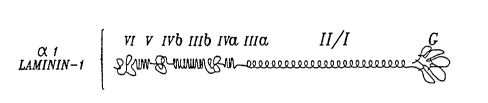
FIGURES lOB-lOC depict a comparison of the domains of al laminin-l and
a3 epiligrin, as well as the overlapping cDNA clones (FIGURE lOD) used in
Example 15 to compile the nucleotide sequence encodingEl70 epithelial ligand
glycoprotein, i.e., as shown in FIGURES lOF, l lA-l lC, AND l5A-lSF.
FIGURE lOF shows the nucleotide sequence of El70 epithelial ligand
glycoplolein cDNA clone "l-l" from position l to position 664 (SEQ ID No. l).
(The sequence of clone l-l was edited to remove a common cloning artefact, the
first l 50bp of the Ep-l cDNA consisting of a cloned fragment of a rRNA.)
f~t~ H~D SHEET (RULE 913
WO 95/06660 2 1 7 0 7 7 7 PCTIUS94/10261
- 12/1 -
FIGURES 1 lA-l lC show 1994 bp (SEQ ID No. 2) of the nucleotide
sequence of E170 epithelial ligand glycol)ro~ein compiled from cloned cDNAs as
depicted in FIGURE lOA. PCR primers used in cDNA cloning were:
RtCll~lE~ SHEET (R~JLE 91)
wo 9s/06660 PcT/Usg~/10261
2170777 -13- 1~
MR-12 corresponding to (nucleotides 183-198 in FIGI~RE 1 lA), MR-l l (nucleotides
340-357 in FIGURE llA), MR-8 (nucleotides 640-657 in FIGURE llA), MR-6
(nucleotides 700-719 in FIGURE llA), MR-7 (nucleotides 992-1012 in
FIGURE llB), MR-5 complement to (nucleotides 1055-1073 in FIGURE 11B),
MR-4 (nucleotides 1277-1296 in FIGURE 1 lB) and~MR-3 (nucleotides 1709-1723 in
FIGURE llC)). The positions at which di~lenL cDNA clones begin are position 1,
where"l-l" begins, and positionl216, where EP-l begins. CloneEP-l ends at
position 1742. FIGURE llA shows the sequence from position 1 to position720.
FIGURE 1 lB shows the sequence from position 721 to position 1500. FIGURE 1 lC
shows the sequence from position 1501 to position 1994.
FIGURES llD-llE show srhem~ically the positions of restriction
endonuclease sites within the E170 nucleotide sequence shown in FIGURES llA-
llC.
FIGURE 12A shows the relative positions of the primers within the EP-l
E170 sequence.
FIGURE 12B shows the nucleotide sequences of 8 primers useful in PCR
methods for isolating nucleic acids encoding E170 epi~h~ l ligand gl~coprotein, as
described in F~mple 15.
FIGURE 13 depicts sçl~e~ lly the steps in a representative PCR assay
method for isolating nucleic acids encoding E170 epithelial ligand ~lycoplotein.FIGURE 14 depicts sch~m~tir,~lly the steps in a representative 5' RACE
system for PCR cloning of cDNAs encoding E170 epithelial ligand glycoprotein, asdescribed in Example 15.
FIGURES 15A-15F depict the nucleotide sequence compiled from sequenring
cDNA clones corresponding to the a3EpA transcript.
FIGURES 16A-16B show experiments demonstrating that the clone Ep-l
expresses a fusion protein that corresponds to at least a portion of the 170 kDasubunit of epiligrin.
FIGURES 17A-17B show a Northern blot analysis of a3Ep mRNA and
illustrates that two distinct transcripts are detect~ble.
FIGURES 18A-18C illustrate the sequence variability in domain IIIa, near the
amino-terminal portion of the protein encoded by a3Ep.
FIGURES l9A-19R show the amino acid sequence encoded by a3EpA
FIGURES 20A-20H illustrate the localization of epiligrin mRNA and protein
in 48 hour human wounds, using in situ hybridization with probes derived from a3Ep
Rt~ SHEET (RULE 91)
WO 95/06660 21 7 ~ 7 7 ~ PCT/US9~V10261
- 13/1 -
FIGURES 20A,20C, AND 20E show the wound site, or compound to normal
skin shown in FIGURES 20B, 20D, and 20F.
FIGURES 20A-20B are labeled with epiligrin anti-sense probe,
FIGURES 20C-20D with epiligrin sense probe, and FIGI~RES 20E-20F with Keratin
5 anti-sense probe.
FIGURES 20g and 20H show wound sites labeled with anti-epiligrin and anti-
alpha3 antibodies, respectively.
FIGURE 21 shows the localization of the human LamA3 gene to
chromosome 18ql 1.2.
FIGURES 22A-22B are graphical representations depicting that integrins
a6,~4 and a3~l mediate anchorage and motility, respectively, on epiligrin via distinct
signal p~lllW~ly:i, as described in Example 18.
5. Detailed Description of the Preferred Embodiment
The scientific literature contains several examples wherein the discovery of
15 ubiquitous extracell~ r ploteins (e.g., laminin and fibronectin) led to the subsequent
RECTIFIED SHEET (RULE 91)
WO 95/06660 PCTIUS9~110261
.
- 14-
2~777
icl~.ntific~tion and purification of the cellular receptors binding these ligands. In
contrast, in the present case the inventors recognized that existing background art
which identified laminin as a putative ligand for binding two of these cellular
receptors, a3,~ 1 and a6~4 integrins, probably described physicochemiç~lly minor5 binding interactions. The inventors had previously observed that the a3,~l and a6~4
integrins were co-distributed in epithelial tissues incl~ltling human skin; however, the
~ignific~nce of this observation was not readily appalt;llL and the distribution could
have been due to both cellular receptors binding to laminin in the tissues. However, in
fli~tinction to the te?~chinp;e in the literature, they reasoned that (a) laminin was not the
10 ligand, and (b) that the two cellular receptors co-distributed because they shared some
other (new) extrac.~ r matrix complex as a ligand. With this recognition of the
problem, they sought to identify the novel ligand. Because it was not possible to
investigate this problem in tissue sections of human biopsy material, they recognized
that it would be necç~, y to select the proper cell type for study in vitro. Since they
15 had previously observed that the two forms of integrin were present together on cells
in regions of human skin that contained keratinocytes, fibroblasts, and other
speri~li7ed epithelial cells, they focused on those regions as a possible source of cells
for in vitro study. In considering among these possible di~lellL cell types the
inventors recognized that human fetal keratinocytes (HFKs) expressed both the a3~l
20 and a6,~4 integrins. Although keratinocytes were recognized by the literature to be a
di~l~ ted form of epithelial cell, both with respect to their microscopic appealailce
and their biosynthetic activities, the inventors reasoned that cultures of these cells
might syntheci7e and secrete the novel extracell~ r matrix ligand, and might be
suitable for in vitro study. (In fact, as the detailed description of the invention
25 (appearing below) shows, if they had chosen to study fibroblasts or continuous
epithelial cell lines, they would not have s~lcceeded in identifying the ligand which is
an embodiment of this invention.) Armed with this recognition of the problem and its
possible solution, the inventors succeeded in identifying the novel ligand for the a3~l
and a6,B4 integrins. Surprisingly, the cellular receptors and ligands identified and
30 isolated from keratinocytes in tissue culture were the same as those utilized by the
basal (stem) cells in epithelial tissue.
In the research described below, the molecular me~h~ni.~m.c by which epithelial
cells establish contact with the b~mem membrane are elllçi~l~ted, the cell receptor
and its extracell~ r basement membrane ligand are identified and subst~nti~lly
35 purified, and the m~çh~ni~m~ are unraveled through which growth and differentiation
are controlled in the epith~lillm The novel ligand which is a subject of the invention
WO 95t06660 2 ~ 7 0 ~ 7 7 PCT/US94/10261
- 15 -
is termed "epiligrin." Research from other laboratories has identified GB3
antigen/nicein (Verrando et al., 1987, 1988) and kalinin (Rouselle et al., 1991;Marinkovichetal., 1993) which are probably identical to epiligrin. Epiligrin is a
covale:ntly linked glycoproLein complex that mediates epithelial cell att~çhment to the
5 b~cPmPnt membrane through a3,~l integrin acting as a cellular receptor. In this
application, the terms "epiligrin" and "epithelial ligand glyco~ teins" are usedhl~e~ e~bly to refer to the same glycoprotein complex. Individual protein
components of epiligrin are so...el;...es referred to as "epithelial ligand glycoproteins."
Epiligrin is present in the lamina lucida of basement membrane and is associated with
those cell lllellll.l~e ultrastructural features previously termed focal adhesions. These
focal adhesions are located on the basal surface of the cells in areas of contact with
the b~cP.m~nt membrane substratum, and they are also involved in cell motility.
Epiligrin also interacts with the a6~4 integrin, a cellular receptor that is present in
ultrastluctural membrane features previously termed hemidesmosomes and stable
adhesion complexes. The invention provides an underst~ntling for the first time, of
how these two di~len~ ultrastructural features (frozen in time by fixation for electron
microscopy) can function in a living cell to merli~te adhesion, control of cell gowth,
and det~ on of the fate of d~llghter cells derived from cell division in the basal
layer of the epith~ m
Burgeson et al. (#125) have proposed nom~n~l~tl-re that categorizes epiligrin
as a kind of "l~minin " Accol~lh1gly, the inventors have denoted the gene encoding
E170 as "LamA3." However, following this nomen~l~t--re can convey the misleadinghllplession that E170 is dçmonctrably similar to the a3 chains of laminin 5, 6, and 7.
First, there is no published sequence available for the Burgeson scheme's a3 chain of
laminin-5, laminin-6, or laminin-7 which would provide any way of comparing those
sequences with the a3EpA and a3EpB sequence. Second, the new nomençl~t~-re is
based on the unproven assumption that a3 chains of laminin-5, laminin-6, and
laminin 7 are intlictin~lich~hle. In contrast, the Applicants' data clearly show that
there are two distinct a3Ep transcripts, suggesting that there is heterogeneity in the
a3 polypeptide chains (135). In addition, the a3 chain described for nicein (#137)
collLaills a significantly smaller G domain and is reported to have a difrele.lL structural
olg~ ;on than the Applicants observed for the a3EpAor a3EpB transcripts of
epiligrin. This suggestc the possibile existence of a third a3Ep transcript (#137), but
no nucleotide seqeunces for nicein are known, and nicein may encoded by a locus
distinct from LamA3.
WO 95/06660 PCT/US9-1/10261
21~0~7~ - 16_ ~
The a3,~l, integrin which binds epiligrin is one of the most widely expressed ofall integrins in tissue, but its physiological ligand has not been idenfified until now.
Novel test cell assays, extracçll~ r matrix compositions, and immlmochemical
reagents were created which allowed identification for the first time of epiligrin, in
5 basement membranes as the physiologically significant ligand for plasma membrane-
based a3,~l and a6,B4 integrins. Only a few epithelial cells in culture (e.g.,
keratinocytes) express significant q~ntities of epiligrin, and this glycoprotein complex
is of a large molecular size and has poor solubility in aqueous solutions. Epiligrin's
size and poor solubility have undoubtedly contributed to the lack of previous
10 recognition of this ligand in binding to integrins.
The role of the a6,~4 integrin as a receptor for the epithelia ligand(i.e., in
stable adhesion complexes) is less h~,plessi~/e than the adhesion mediated by the a
integrin but is potentially more significant. The fintling~ described below in(lic~te that
a6,~4 integrin is involved in cellular adhesion to b~çm~nt membranes, and it may also
15 localize the focal adhesions in a pattern which encircles the regions of the stable
anchoring contracts. This process of encircl~m~nt as well as the localization of a6~4
integrin in cell-cell adhesion sites, deterrnines the fate of the d~--ghtçr cells formed by
division in the basal (stem) cell layer of the epithP.li-.m
Migration of epithelial cells is an important aspect of at least wound he~ling,
20 ;.ln~..",~l;on, and tumor met~t~ei~ Focal adhesions co"l~;"ii-g a3~l integrin are
involved in cell movement, and the stable anchoring contacts co.~ g a6,~4 integrin
are involved in stopping cell movement. Epiligrin binds to both a3,~l and a6~4
integrins This inventive recognition, pursuant to the present disclosure, allows one
skilled in the art to identify specific binding partners to epiligrin (as disclosed in
25 greater detail, below), and provides for the first time compositions which can modify
movement and adhesion of cells of epithelial origin. The invention also provides, for
the first time, an underst~ntling at the molecular level of how polarized self-re~ ted
growth and di~ele"liation are achieved in epitheli~l tissues through the binding of the
tr~n~memhrane integrins in the plasma membrane to extrac~ r epiligrin, the
30 epithelial ligand complex, and possibly through intrac.?ll~ r ~ign~ling accomplished by
the 36+15 kd epiligrin glycop~tein. These events occur at discrete plasma membrane
sites in the stable anchoring contacts, and a second cytoplasrnic polypeptide was also
discovered to be a recognized SACs protein termed Bullous pemphigoid antigen.
Armed with the new inrol",aLion and underst~n~ling provided in the present
35 disclosure, one skilled in the art is able to recognize how m~ n~nt carcinoma cells
may arise through loss of the control mech~ni~m~ provided by epiligrin, and how it is
W095/06660 21 7 0 7 7 ~ PCT/US94110261
- 17-
possible to consider reestablishing these normal control mech~nicm.~ in carcinoma ceils
by using the underst~ncling provided by the invention to select for epiligrin derivatives
and other pharm~ceutical agents that induce the cells to correct their defect inepitheli~l ligand regulation.
S These conc~ ioni are based on the following fintling~ and on reintell,leLaLion
of previous reports in light of the new insights gained from the present invention are:
(i) Purified epiligrin was shown to induce cell adhesion and localization of the a3~1
integrin in focal adhesions better than l~minin, fibronectin, or collagen. Further, cell
adhesion to epiligrin was specifically inhibited with monoclonal antibodies to-a3,~l
integrin. (ii) Epiligrin was the major component of the extrac~ r matrix
synthç~i7ed by human rules~ill keratinocytes. In cultures of stationary keratinocytes,
epiligrin was deposited and co-distributed with the tr~n~m~mbrane a6,~4 integrin and
with cytoplasmic bullous pemphigoid ~nti~ens which are recognized components of
htomide,smosome like stable adhesion complexes. All three of these components in the
stable adhesion complexes were resistant to sequential extraction with detergent,
2 M Urea/l M NaCI, and 8 M Urea. In contrast, the ,Bl-co"~ l;ng integrins in thefocal a~lhe~io~ were not stable to this extraction. The a3,~1 integrin-coll~ g focal
adhesions were observed to form rings around the periphery of the a6,B4 integrinco,~l~;nillg stable anchoring contacts. (iii) In tissue, epiligrin localized in most
epithelial b~mt?nt lllc;l~lbl~nes, but not in the basement membranes of muscle, or
endothelium. At the ultrastructural level, epiligrin localized to the lamina lucida csf the
epidermaVderrnal basement membrane of skin. Con~i~tçntly, epiligrin localized with
the a3,~l, integrin in the basal plasma membrane, as well as with the a6~4 integrin-
collt~ g hçmidçcmosomes of basal (stem) cells. These data indicate that epiligrin is
the ligand for which a3~l and ~6~4 act as recep~ol~.
The subject epiligrin derived from HFK is an epithelial ligand glycopl~ein
complex that in~ des at least three major covalently linked di~lllfide-bonded
glycoproteins having appalellL molecular sizes of 170 kd, 145 kd, and 135 kd. A
glycoprotein of 36 kd is also associated with the epiligrin complex. Other observed
components of epiligrin include a 100 kd protein that is antigenically related to the
145kd protein, and a 200kd protein that is antigenically related to the i70kd
protein. The individual epithelial ligand glycoproteins are visible following reduction
and SDS-PAGE (under re~lcin~ conditions). These con.~tituçnt glycoproteins are at
times referred to herein by reference to their apparent molecular weight on
SDS-PAGE, i.e., E200, E170, E145, E135, E100, and E36, respectively. The 145 kd
protein in some in~t~nces is referred to as E145/100. The subject epithelial ligand
WO 9S/06660 PCT/US91/10261
2~ 77 -18-
complex has the ability to bind to a6~4 and a3~1 integrins and thereby modify
cellular adhesion to a substratum (#s 113, 133, 134).
Skilled artisans will recognize a variety of epithelial cells from which the
subject epiligrin and its con~tit~lPnt glycoproteins may be purified, or subst~nti~lly
S pure nucleic acids prepared. The following direction is provided with regard to
rep,esel,L~Li~e sources of conctitllent epiligrin glycoproteins. E36 may be found in the
culture supe",a~anl (CS) and extracellular matrix of H~K cells (~K-ECM); E100
acc~-m-ll~tes in CS; E170 is usually not found in CS but may be found in a
Triton X-100 extract of HFK-ECM as well as in the insoluble HFK-ECM fraction
after the Triton X-100 extraction; and E200, E140, and E130 are usually not found in
CS or the Triton X-100 soluble fraction of HFK-ECM, but only in the Triton X-100insoluble fraction of HFK-ECM. A variety of biochemical and immunochemical
methods may be utilized to purify the subject epiligrin glycoproteins, e.g., afflnity
chrc).l.alography in buffers co~ ;";"g Triton X-100 and/or mixtures of ionic,
15 nonionic, or zwitterionic detergents. Tre~tm~nt with proteases (e.g., trypsin) may be
useful for prepa ~lion of soluble epithelial ligand glycopeptides, some of which, while
failing to medi~te cellular adhesion to a surface may still retain the ability to block
cellular adherence to epithelial ligand coated surf~.P~s. The following
immlmoçhPmic~l direction is provided with regard to the ~ntig~nic rel~tetlnes~ of the
subject epiligrin glycopro~i.. s: E200 appears to be antigenically related to E170;
E145 appears antigenically related to E100; and E135 does not appear to be related to
other glycoproteins in the epithelial ligand complex. The inventors currently believe
that E170 may be derived from E200 by proteolytic degradation (and/or processing)
and, in a similar manner, E100 may be derived from E145.
Epiligrin antigens are associated with the basal surfaces of basal (stem) cells in
epithelia at limited points of cellular contact with ba~çment membranes.
Embodim~nt~ of the invention also relate to the isolation of epiligrin glycoprotein
complexes for modifying adhesion of cells to substrata and for achieving polarized and
self-reg~ tecl growth and di~len~iation in cells of epithelial origin. Other
embo~imPnt~ relate to antibodies to the epiligrin glycoprotein complex for modifying
cellular adhesion to substrata and for identifying epiligrin-like antigens in biological
fluids, as well as epiligrin antigens for identifying antibodies in patient samples. Still
other embodiments relate to nucleotide sequences of E170 epithelial ligand
glycoprotein useful as specific probes for measuring the presence of epithelial ligand
mRNA in a tissue, as well as the level of expression in different cells in the tissue.
Embodiments of the invention provide compositions and test methods for identifying
WO 95106660 21 7 n 7 7 7 PCT/US94/102GI
- 19-
tli.~e~ed epithelial cells, and for tli~tin~ hing at least between the epithelial
abnormalities in such autoimmllne dermatological diseases as bullous pemphigoid,cicatriical pemphigoid, and epidemolysis bullosa acquisita.
The subject test methods and compositions are useful for determining the level
S of expression of epiligrin in a tissue. Expression of epiligrin is a hallmark of a
regenerating epithelial tissue (see Example 15). The level of ~ s~ion of E170
epiliglin glycoprotein was fourid to provide a tool useful for ~li.ctin~ hing between
regenerating epitheli~l tissues (where expression was high) and non-regenerativeepithelial tissues or m~lign~nt tissues (where ~A~lession was low). Diagnostic
histopathology is frequently complicated because it is not easy to tli.~tin~ h tissue
repair (e.g., resulting from traumatic injury or infection) from an abnormality that
might be a neoplastic or preneoplastic event. E~ it-g the levels of E170 epitht li~l
ligand ~ ,ression (e.g., using imml-nt~-histochemical techniques, in sifu hybridization
with an oligonucleotide or cDNA probes, or PCR of isolated tissue mRNA), is useful
for rli~tin~ hing between repair and m~lign~nt (or pr~m~lign~nt) changes in epithelial
tissues.
The invention provides nucleic acids capable of hybridizing under stringent
conditions to at least one nucleotide sequence selected from the group con~ ting of
the nucleotide sequence shown in FIGURES 1 lA-l lC, the cDNA clone Ep-l (ATCC
No. 75540) shown in FIGURE lOF, the cDNA clone 1-1 (ATCC No. 75539), and the
cDNA clone 8-6 (ATCC No. 75538), or the nucleotide sequences shown in
FIGURES 15A-lSF. The subject nucleic acids are preferably capable of encoding anE170 epithelial ligand glycoprotein. A partial nucleotide sequence of nucleic acid
encoding E170 epithelial ligand glycoprotein is provided in FIGURES llA-llC
compiled from the cDNAs shown in FIGURE lOD as sc.h~m~tically depicted in
FIGURE lOA. The entire nucleotide region encoding E170 is depicted in
FIGURES lSA-lSF, and corresponds to the sequence of a3EpA, one of the two
distinct a3Ep transcripts discovered by the Applicants. FIGURES lSA-lSF consist of
a composite sequence derived from the several overlapping clones shown in
FIGURE lOD.
Although only a single (+) strand of the cDNA is shown in
FIGURES lOF, 1 lA-l lC, and 1 SA-l SF, those skilled in the art will recognize that the
complementary (-) strands are thereby disclosed as well. According to the convention
used herein to describe PCR primers, the "(-) strand" is complementary to E170
mRNA.
RE~ S~ RULE 9
WO 95/06660 PCT/US94/10261
2~7~77~ -20- ~
By nucleic acid molecule is meant DNA, RNA, and/or synthetic nucleotide
sequences such as oligonucleotides that are the same as, homologous with, or
complementary to, at least one helical turn (about 10 to 15 nucleotides) of the
illustrated E170 epithelial ligand glycoprotein nucleotide sequence. At least two
5 alternative forms of E170 transcripts are disclosed herein in ~K cells, one mRNA of
about 5 kb and another of about 6 kb. Both mRNA species are identifi~ble to those
skilled in the art in RNA from HFK by standard Northern blotting methods (e.g.,
using radiolabeled Ep-1 as a probe as illustrated in Example 15). The invention
relates to at least four classes of E170 encoding nucleotide sequences, 1) alternative
10 splicing transcript sequences, 2) sequences resulting from genetic polymorphism of
E170, 3) sequences resllltin~ from translocation of E170 in tumorigenesis and genetic
~ e~e~, and 4)sequences of E170 family members having greater than75%
homology with E170 over a conserved region of at least 30 nucleotides. In all cases
the latter four classes of E170 nucleotide sequences are i~ntifi~ble as hybridizing
under stringent conditions with an E170 nucleotide sequence of FIGU~ES 1 lA-1 lC,
e.g., cDNA clone 1-1, Ep-1, or 8-6. While the several clones depicted in
FIGURE 10D encompass the entire nucleotide sequence encoding an E170 epithelial
ligand glSlcopl~)tein, skilled artisans will recogr~ize that additional cDNA clones may
be obtained using nucleotide sequences contained within the subject cDNAs as probes
and primers for obtaining additional cDNA clones. An illustrative example of a PCR
cloning method for obtaining additional cDNA clones through PCR cloning is
provided in Example 15, below. PCR primers are additionally provided in
FIGURE 12B and Table 1, below, and the steps of an illustrative PCR method are
outlined in FIGURE 13.
TABLE 1
Primers for PCR Primer-extended Sequencing
Primer 4 (SEQUENCE ID NO: 9): 5' AGCACGAAGGTCACTGAGTT 3'
Primer 5 (SEQUENCE ID NO: 10): 5' AAGTCACCTGAAGGCACG 3'
Primer6 (SEQUENCEIDNO: 11): 5' TGGACGTGCGACTTGACCAG 3'
Primer 13 (SEQUENCE ID NO: 12): 5' AACTCGCTTGCAGTTGAC 3'
Primer 14 (SEQUENCE IDNO: 13): 5' GATGGCTGTGGATCTTTG 3'
Primer 15 (SEQUENCEIDNO: 14): 5' TCCACAGCAAGTGCTATG 3'
Primer 16 (SEQUENCE IDNO: 15): 5' ATGACAGTGCTGTCTGGAC 3'
Primer 17 (SEQUENCEIDNO: 16): 5' TCTCCGAGATGGTCTTCATG 3'
Primer 18 (SEQUENCE IDNO: 17): 5' TTATCTGCATCAGTCAGAGC 3'
Primer20 (SEQUENCE IDNO: 18): 5' TGACCAGTGAGCTGTACATC 3'
Primer29 (SEQUENCE ID NO: 19): 5' AGAGACCATTCGATTCAGAT 3'
Primer 30 (SEQUENCE ID NO: 20): 5' AGCTTCTGAGAAATAGCAAA 3'
F~ lE~ SHEET (RULE 91)
wo 95/06660 2 ~ 7 0 7 7 7 PCT/US9 ~/10261
- 21 -
The PCR method in FIGURE 13 was used successfully to isolate mRNA encoding
E170 from normal epidermal tissue as well as from cells of patients with
Epidermolysis bullosa. Primers MR-4 and MR-7 and primers MR-5 and MR-7
(FIGURES 12A-12B) and the primers shown in Table 1 have also been used for PCR
5 amplification and isolation of genomic DNA from normal and patient samples. The
latter isolated gemonic DNA contained both intron and exon sequences. The introncoded for a junctional amino acid sequence between an EGF-like region and a helix
region in E170, and the exon was recognized by the presence of non-coding sequence
and stop codons.
The subject nucleic acid capable of hybridizing under stringent conditions to a
nucleotide sequence in FIGI~RES llA-llC and FIGURE 15, (e.g., cDNA clones
"Ep-l", 3-1-1, 5-4-2, 3-8-6, 5-4-1, 3-8-2, or 8-6-1), find a variety of in vitro and
in vivo uses. For instance, in a plefelled embodiment the nucleic acids are useful (as
illustrated in Example 15) in expression systems that produce E170 epithelial ligand
glycoprotein. The t;~essed epiligrin glycoproteins, in turn, find a variety of uses:
e.g., as adhesive agents for cells; as antigens for production of antibodies; and, as
antigens useful in detection of patient ~llto~ntibodies such as those described in the
serum of patients with acquired subepidermal blistering diseases (Domologe-
~lllt~çh et al., citation #114, incorporated herein by reference).
In another example, the subject nucleotide sequences of the subject nucleic
acids are useful for constructing ~nti~n~e oligonucleotides (as illustrated in
Example 15, below). The ~nti~en~e oligonucleotides have nucleotide sequences
capable of hybridizing under stringent conditions with the subject nucleic acids and
are complement~ry with a nucleotide sequence encoding an E170 epithelial ligand
~,lycoploLein. The subject anti~en~e nucleotides have been used sllcces~fillly for
in situ hybridization, as shown in FIGURE 21. The subject anti~en~e nucleotides may
be further characterized by their ability to transiently inhibit t;~ression of an epiligrin
gene in a cell, e.g., by transiently binding and inhibiting translation of an rnRNA
encoding an epiligrin con~tit~lent F.pitheli~l cells whose expression of epiligrin was
transiently blocked by ~nti~n~e oligonucleotides did not adhere as strongly to
CM in vitro, and they became more rounded in appearance and form
multicellular aggregates in suspension. The cells in the aggregates were observed to
be di~e~ tin~ Thus, it is considered most likely that one or more regulatory
fee~lbaçk mech~ni~rn~ exist in epithelial cells through which the binding of epiligrin to
its a3~l receptor tr~n~d~lces a signal through a second messenger pathway that stops
cellular proliferation and induces dirrelentiation. It is thought highly likely that
RE(;Il~ltl) SHEET ~RUEE 91)
WO 9S/06660 PCT/US9~/10261
2~707~ -22_ ~
abnorrnalities in the latter signal tr~n~dl~ction pathway will exist in certain epithelial
cells because of defects in expression levels of epiligrin, or abnormalities in one or
more epiligrin glycoproteins or in the epithelial a3,Bl integrin. The affected cells may
exhibit a phenotype of either uncontrolled growth or premature di~el enLiation.
Antisense nucleic acids (e.g., oligonucleotides) may thus be useful for ind~lçing
epithelial difrelellLiation in rli~e~sed cells that are exhibiting uncontrolled growth
resl]lting from a failure to properly regulate epiligrin expression. In an additional use
for the subject ~nti~n~e nucleic acids, ~ t;s~ion of E170 epiligrin glycoprotein was
increased in rapidly dividing cells in the migratory tongue of epithçlillm in wound sites
(Exarnple 16). Exuberant (uncontrolled) wound healing is a frequent condition inscarring and keloid formation, and poor quality wound healing is also a problem
encountered in large wound sites, e.g., in burn patients and in diabetic and paraplegic
patients with dicubitous ulcers. In the latter ulcers a thin tongue of migratingepith~ m may forrn across a wound site, but the cells frequently fail to properly
initiate terminal di~t;rel.Liation. ~nti~Pn~e nucleic acids may be useful therapeutically
for intl~1c.ing epith~ l di~lellLiation in ulcers, and for lesLolii~g normal di~erell~iation
to prevent keloid formation and scaring. conditions. The subject ~nti~en~e nucleic
acids may be introduced into a host cell by transfection (e.g., of an oligonucleotide) or
by tr~n~ ction of a nucleic acid encoding an ~nti~çn~e nucleic acid (e.g., usingretroviral vectors ). The subject ~nti~en~e nucleic acids are all characterized by their
ability to hybridize under ~lingelll conditions with a (+) or a (-) strand of a nucleic
acid encoding an E170 epith~ l ligand glycoprotein, e.g., as represented in
FIGURE lOF, FIGURES 1 lA-1 lC, and FIGllRE 15.
Methods are disclosed in Example 16, below, for up-re~ ting ~x~ules~ion of
epiligrin through the addition of TGFa or TGF,~ to epithelial cells. These methods
may be useful for increasing cA,ules~ion of epiligrin in patients suffering dimini.~hed
synthetic capacity, e.g., in patients with a variety of blistering disorders and idiopathic
urticarias (hives). Skilled practitioners will note that an effective dosage of TGFa or
TGF,~ may be determined in screening assays (i.e., in vitro and in vivo in animal
models) where the dosage in contact with the epithelial cells is esc~l~ted in a stepwise
manner until synthesis of an epiligrin glycoploteil- is increased (i.e., as measured by
mRNA or protein). Also, a variety of systernic and topical methods for application
may be tested by t;~ )g the levels of expression of an epiligrin glycoprotein in the
treated cells before and after the tre~tm~nt.
The subject nucleic acids also find use in gene therapy for in(l~lcing over-
t;x~uression of epiligrin in ~i~e~eed cells, and for gene replacement therapy in genetic
F~E(;~ ED SHEET (RULE 91)
wo 95/~C6~ 21 7 0 7 7 ~ PCT/US9~/10261
disease. For example, junctional epidermolysis bullosa gravis can be a lethal genetic
disease of infants that is associated with failure to normally express epiligrin. ~er;e
L~ rer may be accomplished using vectors (e.g., a retroviral vector) cor,l~ g a
construct that has in serial array: a promoter, a subject nucleic acid that encodes one
or more epithelial ligand glycoproteins, e.g., E170, and a polyA tail. In genetic
repl~c~m~nt therapy for treating lethal junctional epidermolysis bullosa gravis,con~ e CAI lession of epiligrin in vivo may result in establi~hment of epithelillm-
basement me~ e integrity in rli~e~ed epidermal tissues as well as in the lung,
urogenital tract, ga~Lro;"le~ l tract, and other sites of epiligrin cA~uression
(representatively illustrated in the Examples, below).
These and other aspects of the invention are described below.
5.1 Definition of Terms:
The following terms used herein are intended to have the meanings set forth
below:
"Epithelial ligand glycol)loteill" means a con.ctituçnt glycoprotein of the
epithelial ligand complex epiligrin.
"Subst~nti~lly-pure" means of a purity sufficient that more than 70% of the
polypeptides in the l~lep~Lion can be determined by SDS-PAGE and protein st~ining
to be the composition so specified.
"Covalently linked" means polypeptides chemically bonded to one another, as
through for example (but not limited to) di.~ulficle-bonds, thiol-ester bonds, ester
bonds, amide bonds, or the like.
"Capable of binding" means physical interaction between two materials, such
as between a specific binding partner and a ligand, where the interaction is sufficiently
strong to permit measurement of a ~h~mic~l association (or dissociation) constant
(i.e., Ka or Kd).
l'Capable of hybridizing under stringent conditions" means ~nnealing of a
nucleic acid molecule to at least a region of the disclosed E170 epithelial ligand
glycoprotein nucleic acid sequence (whether as cDNA, cRNA, mRNA, or genomic
DNA), or to its complçment~ry strand under standard conditions, e.g., high
temperature and/or low salt content, which tend to disfavor hybridization of
noncompl~m~nt~ry nucleotide sequences. A suitable protocol is described in
~ni~ti~, T., et al. (#118 which is hereby incorporated by reference), at
pages 387-389), wherein following the hybridization step filters are washed in
0.1X SSC, 68C for 2 hours. Other protocols for achieving stringent hybridization
are well-known to those skilled in the art, and can be selected from those presented in
WO 95/06660 PCT/US9~/10261
2~7~777 -24-
Maniatis (#118). Such hybridizing molecules may be related to the disclosed
sequence by deletion, point mutation, base substitute, fr~meshift alternative ORFs,
mRNA splicing or processing, or post-transcriptional modification (e.g., methylation
and the like).
"Substratum" means an insoluble material upon which cells may be deposited
by gravity.
"Non-adhesive substratum" means a substratum to which fewer than 20% of
the cells will bind in24 hours at 37C and from which 80% of the cells can be
removed by washing with ..,e~li-.,.. e.g., such a substratum is provided by
10 microbiological grade poly~lylene plastic petri dishes.
"Epithelial cells" means, in this disclosure, the cells origin~tin~ through mitosis
in epith~ l tissues which cover the free surfaces of the body and line the body
cavities and ducts, as well as cells of epithelial origin such as m~lign~nt carcinoma
cells. Further examples of epithelial cells as they are commercially available are
15 provided in Table 2, below, as listed in the "Catalogue of Cell Lines and Hybridomas",
6th Edition, 1988, the American Type Culture Collection, Rockville, Maryland.
"Modulate" means to effect an increase or decrease of a specified parameter to
a measurable extent.
"Adhesion assay" means an assay con(3~1cted with test cells, such as HT1080 in
20 Example 6 below, to measure adhesion of cells to a protein-coated "non-adhesive"
substratum under defined test conditions of tissue culture.
"Di~,t;"~iation" means a staged process, e.g., in development, through which
a cell progressively acquires tli~tin~ h~bly new phenotypic attributes.
"Confluent cell culture" means a culture in which more than 85% of the cells
25 are observed microscopically to be in physical contact with their neighboring cell.
"R~ci~t~nt to digestion" means that no substantial change in physical
properties is observed following incubation of the polypeptide with an enzyme for a
substantial period of time.
"Co-migrate" means substantially the same electrophoretic migration when
30 two polypeptides are either run together in the same lane of an SDS-PAGE gel, or
when they are run side-by-side in adjacent lanes.
"Molecular size" means the a~a,~ molecular radius of the polypeptide as
observed under denaturing conditions in SDS-PAGE, and as recorded in kilodaltons(kd+) of mass as determined by comparison with other polypeptides of known
35 molecular mass.
WO 95/06660 21 7 ~ ~ 7 7 PCT/US94/10261
- 25 -
Table 2
Examples of Commercially-Available Human Epithelial Cells
Tissue Name/ATCC No. Description
Endometrium RL95-2/CRL1671 Adenosquamous carcinoma
Skin WM-1 15/CRL1675 epitheloid melanoma
WS-I/CRL1502 fetal skin
Pancreas AsPC-I/CRL1682 adenocarcinoma
PANC-I/CRL1469 epitheloid carcinoma -
Stomach AGS/CRL1739 adenocarcinoma
Bladder UM-UC-3/CRL1749 bladder carcinoma
HT-1 197/CRL1473 bladder carcinoma
Colon CCD841CoN/CRL1790 fetal epithelial-like
NCI-H548/CCL249 adenoca~ .ol.. a
Tongue SCC-9/CRL1629 squamous cell carcinoma
Kidney ACHN/CRL1611 adenocarcinoma
Cervix C-4I/CRL1595 cal~iino.lla
CaSki/CRL1550 epiderrnoid carcinoma
Ovary PA-1/CRL1572 teratocalcinollla
Epidermis A-431/CRL1555 epidermoid carcinoma
Breast ZR-75-1/CRL1500 .,.~.. ~.y carcinoma
MCF-7/HTB22 adenocarcinoma
Pharynx Detroit 562/CCL138 carcinoma
Adrenal cortex SW-13/CCL105 adenocarcinoma
Lung WI-38/CCL75 fetal diploid
5.2 Keratinocyte Extrac~ r Matrix and Immunoprecipitation of Epiligrin:
The Major Glycoprotein Complex in Adhesive HFK-ECM
For this study, the extr~c~ r matrix synthesi7ecl and secreted by HFKs shall
30 be referred to as E~K-ECM and that synthesized and secreted by HFFs as HFF-ECM.
Endogenous HFK-ECM is that which is intrac~ r or plasma membrane associated.
HFK-ECM secreted into the conditioned culture merlillm during the time course of an
- assay, or that which can be purified from culture dishes or glass cover slips (after the
removal and/or extraction of the HFKs, as by the three-step extraction procedure35 detailed below), is lerell~;d to as exogenous HFK-ECM.
WO 9_/OG~0 PCT/US94/10261
2170777 -26-
To identify a physiologically ~ignific~nt ligand for a3~l and/or a6,~4 integrinsin epith~ l cells, we first examined the composition of the ECM produced by HFK.Radiolabeled HFK-ECM and HFK were prepared by incubating HFK in culture dishes
for 15 hours in KGM co,.l~inil-p; 35S-methionine, 3H-glucosamine, or 35so4-2, and
1 mg/ml HD-BSA (Sigma) as a carrier protein. Radiolabeled HFKs were sequentiallyextracted in a sequential three-step extraction procedure, as described pre~,iously
(Wayner and Carter, 1987): (1) with 1% (w/v) Triton X-100 (Sigma; to solubilize
membranes and cytoplasmic conctit~l~nt~) and 2 mM N-ethylmaleimide (Sigma, to
prevent intramolecular cross-linking); (2)with a solution cor,l~in;llg 2MUrea and
1 M NaCl (to remove nuclear and cytoskeletal components); and (3) with 8 M Urea
(to solubilize residual cellular components). All extraction buffers contained 1 mM
phenylmethyl sulfonyl fluoride (PMSF; Sigma Chemical Co., St. Louis, MO) and a
protease inhibitor and 2 mM N-ethylmaleimide (Sigma) as an inhibitor of
intramolecular cross-linking. The con~titllent radiolabeled glycoproteins were
separated by SDS-PAGE (12) and vi~ li7ed by fluorography. The results of the
sequential extraction procedure are presented in FIGURE 1 where lanes 1-3 show the
~,ly~ioplu~eins extracted in the steps 1, 2, and 3 (above), respectively.
To .olr~mine the nature of the 8 M urea-insoluble HFK-ECM glycoproteins
re,ll~ ing on culture dishes after the extraction step 3, 0.5% (w/v) SDS was added to
the culture dishes, and glycoploteins were physically dissociated by meçh~nic~l
scraping with a rubber policeman. The glycoproteins obtained in this manner did not
enter an 8% SDS-PAGE gel (FIGURE 1, lane 5) unless they were reduced on
SDS-PAGE under red~lçing conditions (i.e., with 2-mercaptoethanol; 2ME) and
vi~ li7ed by fluorography. They consisted ec~enti~lly of at least five major
glycoproteins vi~u~li7ed by protein st~ining with Coomassie brilliant blue (FIGURE 1;
lane 8) or following biosynthetic~lly radiolabeled with 35S-methionine (FIGURE 1,
lane 4), or 3H-glucosamine (FIGURE 1, lane 9); these glycop~oteins having appalen~
Mr of 200kDa, 170 kDa, 145 kDa, 135 kDa, and 36kDa (FIGURE 1, lane9).
(Migration of molecular mass standards are indicated in the left margin of FIGURE 1
(i.e., 180, 116, 84, 58, 49, and 37 kd).) The HFK-ECM glycoproteins detected with
protein stain showed slightly decreased amounts of the 200 kd glycoprotein
(FIGURE 1, lane 8). The five major glyeoplo~eins were desi~n~ted E200, E170,
E145, E135, and E36, based on relative molecular mass under red~lcing conditions on
8% SDS-PAGE. The E170 band was incon~i~tently resolved into two bands
(FIGURE 1, lane9). Under non-red~lr.ing conditions the five glycoproteins did not
enter the polyacrylamide gel (FIGURE 1, lane 5), intlic~ting that they were subunits of
wo 95/06660 2 1 7 0 ~ 7 7 PCT/US94/10261
- 27 -
one or more high molecular mass complexes, cross-linked by intermolecular ~ 1fide
bonds. This mass (or masses) is known as epiligrin. Although the glycoproteh
subunits were not labeled with 35S04-2, three additional sulfate-labeled components,
probably glycosaminoglycan or proteoglycan, were also present in the exogenous
5 HFK-ECM (FIGURE 1, lane l0, marked with *). In control experiments, metabolic
labeling for di~elellt times did not detect any precursor product relationship among
the five glycopl~tein subunit~ of the complex. However, antigenic similarities suggest
that E200 is a precursor to El70. Comparison of the molecular masses of the fiveglycoprotein subunits in the complex to known b~çment melllbl~ne components
l0 failed to detect any obvious relationships. To evaluate further any possible
relationship b~ween the exogenous HFK-ECM glycoplo~eins and the collagens, non-
reduced and reduced (2-nlc;lca~loethanol; Sigma) 3~S-methionine biosyntheticallyradiolabeled HFK-ECM was treated at 37C for 18 hours with 100 units/ml
coll~g~n~ce (Advanced Biofactures, Form III) under con(lition~ which degrade
15 collagen standards, as described previously (98). The collagenase-digested
radiolabeled HFK-ECM was extracted using the same three-step extraction procedure
described above, and the glycopl oleins were separated using SDS-PAGE and
vi~u~1i7ed by fluorography. None of the five major glycoploleill components in
HSK-ECM was ~ligested with collagenase either when non-reduced (FIGVRE l,
20 lanes 6 and 7) or reduced prior to digestion, indicating that they were not collagens.
In addition, the HFE~-ECM ~,lycoproteills (FIGURE 2, HFK-ECM) did not co-
migrate on 8% SDS-PAGE with purified protein standards of EHS sarcoma laminin
(FIGU~E 2, EH5-LN; LN A; LN Bl and B2), fibronectin (FN); when vi~u~li7ed by
st~ining for protein with Coomassie blue (FIGURE 2, PROTEIN), çnt~tin~ or
25 ten~çin~ but El70 did co-migrate with pepsinized human placental laminin
(FIGURE 2, conlpale HFK-ECM to H LN). In contrast (and as expected), proteins
in con-litioned medil-m from HFK cells (HFK CS, FIGURE 2) contained a multiplicity
of proteins, some of which co-migrated with the protein standards (FIGU~E 2,
HFK CS). To further evaluate any possible relationship between fibronectin (or
30 laminin) and the components in exogenous HFK-ECM, three types of experiments
were con-lucted. First, the glycoproteins in exogenous HFK-ECM were separated onSDS-PAGE, blotted onto nitrocellulose as described previously (98) and tested for
their imm1moblot reactivity with rabbit antibodies directed toward laminin (Anti-LN;
FIGURlE 2) or fibronectin (Anti-FN; FIGI~RE 2). Anti-FN bound to antigens in
35 HFK-conditioned metli11m (HFK CS) and in purified fibronectin (FN) but not inhuman pl~cçnt~l laminin (H LN), sarcoma EHS laminin (EHS LN) or HFK-ECM;
7 PCT/US9~/10261
- 28 -
anti-LN bound to antigens in HFK-CS, H LN, and EHS-LN but not in FN or
HFK-ECM (FIGURE 2). In summary, imml-nnblotting of HFK-ECM with anti-
laminin or anti-fibronectin (FIGURE 2, Panel "Anti-LN and Anti-FN") failed to detect
any relationship between these known extrac~ r matrix glycopro~ehls and t};e
5 glycoproteins in HFK-ECM. Second, polyvalent antibodies to laminin or fibronectin
were also used to prepare an immllnople~ e of exogenous HFK-ECM.
Tmmllnol)reci~ ion with antibodies against l~minin fibronectin, t~n~cr.in, ~nt~ctin, or
Bullous pemphigoid antigen (BPA) failed to detect any immllnological cross-reaction
among those known BM proteins and the five major glycoprotein subunits of the
10 HFK-ECM. Third, HFK-ECM was scraped from the substratum and used to
immlmi7e rabbits to induce antiserum. The res--lt~nt antisera did not react withlaminin or fibronectin, but did react with E170, E145, and E135 in the HFK-ECM.
In order to further characterize the HFK-ECM, we prepared monoclonal
antibodies (MAbs) against the HFK-ECM glycoprotein complex using HFKs as an
15 immlmogen.
5.3 Bindin~ Pal lllt;l ~ as Exemplified by Monoclonal Antibody to HFK-ECM
Binding pallllel~ as exemplified by MAbs to HFK-ECM were produced by the
methods of Oi and ~e~ ,~nhel~, (99) and Taggart and Samloff (100) as described (59).
Spleen cells from RBF/DN mice immlmi7ed with cultured HFKs were fused with
20 NS-l/FOX-NY myeloma cells. Viable heterokaryons were selected in RPMI 1640
me-lillm supplem~nted with adçnine/aminopterine/thymidine.
Hybridomas PlEl (ATCC No. HB10681) and PlH8 (ATCC
No. HB10682)producing antibody specifically directed to HFK-ECM were selected
using immlmofluorescence microscopy and HFK-ECM or H~F-ECM on glass cover
25 slips. We selected MAbs PlE1 and PlH8 that reacted with HFK-ECM but not
HFF-ECM produced by the dermal fibroblasts. PlEl and PlH8 were cloned by
limiting dilution. PlEl and PlH8 (with rabbit anti-mouse IgG) immlmoprecipitatedfive relatively minor di~ulfide-bonded subunits from the conditioned culture medillm
of 35S-methionine-labeled HFKs. The results presented in FIGURE 2 show examples
30 of three (1, 2, 3) such immlmoprecipi~a~ion experiments con~ cted with PlEl in
which HFK cells were metabolically radiolabeled with 35S-methionine, as described
above, and antigens in the conditioned medillm (FIGURE 2; CS) and HFK-ECM
(FIGURE2; ECM) were immlmoprecipitated. The five subunits of the PlE1
antigen(s) co-migrated with the five major glycoprotein subunits of the exogenous
3~ HFK-ECM (FIGURE 2, compare ECM in the far left lane of the figure with PlE1,
experiment 1). The E170 subunit labeled as "E180" in FIGURE 2) in the precipitated
WO 95/06660 21 7 0 7 ~ ~ PCT/US94/10261
- 29 -
PlEl antigen was increased relative to the other subunits in all three experiments,
suggesting thatE170 contained the epitope recognized by the PlEl MAb. In a
similar manner, the E36 component of glycoprotein complex was increased in the
imml-noprecipitate prepared in this manner between radiolabeled HFK-CS and PlH8,5 suggesting that E36 may contain the antigenic epitope for PlH8. The possibility that
the gl~coprotein complex recognized by PlEl may contain ~ntigen~ previously
i~çntified was once more evaluated, this time ~Itili7:ing immunoprecipitation
techniques. Preclearing of H~K conditioned culture medium (FIGURE 2; CS) by
immunople-;i,uiL~Lion with polyvalent anti-fibronectin (FIGURE 2, experiment 2-; FN),
10 polyvalent anti-~nt~c.tin, or mouse monoclonal anti-tenascin prior to plecipila~ion with
PlE1 had no effect on subsequent precipitation of PlEl ~ntigPn.~ How~ver,
preclearing (i.e., imm--nopre~ g) with polyvalent anti-laminin (FIGURE 2;
experiment 3, LN) removed E200 and 50% of the other subunits from the PlEl
p~ci~ilale. Since none of the glycoproteins (i.e., E200, E170, E145, E135, or E36)
15 reactecl with anti-laminin antibodies by Western imm--noblotting after SDS-PAGE
(FIGURE 3), we conclude that: (a) E200 in the complex is not l~minin; and
(b) glycoplotei5a complex may be associated with laminin so that it forms a complex
that can be p,~cipil~Led with the anti-laminin antibody. This type of interaction of
laminin has not been reported previously, and the composition of the ~lecipiL&Le20 differs significantly from previously reported complexes of laminin interacting with
other glyco~rotei,ls.
In ~ n~y, the PlEl and PlH8 ~ntig~n~ correspond in molecular sizes to the
glycoproteins in exogenous HFK-ECM, which consists of at least five subunits: E200,
E170, ]E145, E135, and E36, which are vi~ li7ed on SDS-PAGE after reduction, and25 that are distinct from any previously identified adhesion ligand(s) present in basement
Inelllbl~nes or extrac~ll--l~r matrix. The complex recognized by PlEl and Pl~I8 .s
the major component of exogenous ~K-ECM and a,so a minor component in E~K-
conditioned culture medi--m Based on the unique characteristics of this complex, and
in order to simplify the following discussion, we shall henceforth refer to the
30 covalently linked glycol)lo~ein complex as "epithelial ligand" or "epiligrin."
5.4 Epiligrin Distribution in Motile and Non-Motile HFKs
The o5~ n of epiligrin deposited in E~K-ECM was e~mined by
immunof,uorescence microscopy, using MAb PlEl and PlH8 and antibodies directed
toward other extrac~ r matrix adhesive ligands. It was found that PlH8 stained
35 on,y cells which were permeabilized to allow st~ining of cytoplasmic proteins,
WO 95/06660 ; PCT/US9~/10261
- 30 -
2~7~777
indicating that the PlH8 antigen was a cytoplasmic con.~titllçnt of cells expressing
a3,~l integrin. Subsequent studies, detailed below, utilized only the PlEl MAb.
HFKs were grown for24 hours on glass cover slips coated with either
fibronectin (FIGURES 4A-4B) or BSA (FIGURES 4C-4K), as described above (see
"Cellular Adhesion to Extracell~ r Matrix Adhesive Ligand-Coated Substrates").
Glass cover slips and cells were then incubated with mouse or rat MAbs or rabbitpolyclonal primary antibodies diluted in 1% heat HD-BSA overnight as previously
described (21). The cover slips were washed with PBS; in~llbate~ with dilutions of
affinity-purified, species-specific, FITC-conjugated goat anti-mouse/rat IgG or
rhodamine-conjugated goat anti-rabbit IgG secondary antibodies (respectively) for 1
hour, washed with PBS, and fixed with 2% formaldehyde prior to
immllnofluorescence microscopy. The olgani~a~ion of epiligrin was dependent on the
ligand to which the HFKs were ~tt~çhed. When HFKs attached to fibronectin
(FIGURE 4A), collagen, or l~minin, the cells migrated over the ligand surface leaving
trails of epiligrin.
To investigate whether the a3- and a6-co"~ g integrins (i.e., a3,~l, and
a6,B4) were associated with epiligrin on the cell surface, tests were con~ cted
~im~11t~neously to visualize the integrin and its putative ligand on HFKs using a double
immllnofluorescence technique. Epiligrin (PlEl, FIGI~RES 4A, 4C, 4F, and 4I) waslocalized relative to a6 (FIGURES 4B and 4 D; GoH3), BPA (FIGURE 4G) and
a3,~l, (FIGllRE 4J; PlF2). SACs and FAs were identified in each field by
hlLelrelellce reflection microscopy (FIGURES 4E, 4H, and 4K). Arrows in panels I,
J, K identify a3,~l in FAs in relation to epithelial ligand in SACs. HFE~s were
incllb~te~3 with: (1) mouse MAb anti-a6 (GoH3); followed by (2) incubation with
rhodamine-conjugated goat-antimouse IgG and IgM; after which the cells were fixed
and reacted with (3) biotinylated with mouse PlEl-MAb; followed by (4) fluorescein
Avidin. In migrating HFK cells, a6,B4 was expressed on the apical surface of the cells
and at the trailing edge. Small quantities of a6,~4 co-distributed with the epiligrin
antigen in the trails of these cells (FIGURE 4B).
As described (20), when HFKs are grown on BSA-coated surfaces, the cells
migrate less and form h~mi~esmosome-like stable anchoring contacts ~SACs) on their
basal surface. In the present study, by immllnofluorescence microscopy, all the SACs
on the basal surface of the stationary HFK cells contained a6,B4 and most contained
BPA. As seen in FIGURE 4C, HFKs grown on BSA deposited epiligrin antigen on
the substrata in "ring-like structures" characteristic of SACs. The distribution of
epiligrin antigen (PlEl; FIGURE 4A, 4C, 4F) in relation to a6 (GoH3;
~t~ FIEl) SHEET (RULE 91)
WO 95/06660 217 0 7 7 7 PCT/US94/10261
- 31 -
FIG~RES 4B and 4D), and BPA (FIGURE 4G) was most strikingly similar in the
"ring structures." To further di.~tin~ h SACs from focal adhesions, interferencereflection microscopy (IRM) was performed basically as described (Izzard and
Lochner, 1976) and was used to identify focal adhesions (FAs) in the same field as the
5 two color immllnofluorescence which identified the SACs. FAs were also localized
by the antibody exclusion technique (101). Epiligrin (identified by PlE1) in
a6~4/BPA-SACs corresponded to contact sites with the adhesion surface as
deternlined by interference reflection microscopy (FIGURES 4E and 4H). The co-
locali7:~tion and similar stabilities indicated that the deposits of epiligrin were at
10 adhesion sites linked to a6,~4/BPA-SACs.
As an additional test for the loc~li7~tion of epiligrin in SACs, since we had
previously observed that a6,~4 and BPA in SACs are extraordinarily stable and are
resistant to sequential extraction with 1% Triton X-100 detergent and
2Murea/rMNaCl, while the ~l-co"~ g integrins in focal adhesions are soluble
15 under these extraction conditions (20), we therefore next ~mined the distribution of
epiligrin, a6,~4, BPA, and a3,~l in HFKs SACs extracted in this manner. HFKs were
grown for 24 hours on s~ ces coated with BSA, then extracted with 1%
Triton X-100 detergent followed by 2 M urea col.~ 1 M NaCl. The adherent
cell residue co..~ SACs was stained with anti-epiligrin (PlE1, FIGURES 5A,
5C, and 5E), anti-a6(,~4) (GoH3, FIGURE 5B), anti-BPA (FIGURE 5D), and anti-a3
(FIGURE 5F; PlF2). Co~ ly, we observed that epiligrin antigen was present
in sequentially extracted HFK SACs (PlEl; FIGI~RES 5A, 5C, and 5E). In these
studies a6, a3, ,Bl, epiligrin, or BPA antigen was vi~ li7ed using a double
immlmofluoresc~nce technique. Epiligrin co-distributed with the a6 (GoH3;
FIGURE 5B), and BPA (FIGURE 5D) in the extracted HSK-ECM. The co-
distribution of epiligrin with a6~4 and BPA in both non-extracted and sequentially
extracted HFKs (Triton X-100, 2 M urea/lM NaCI, above) in~lic~ted a stable linkage
between the cytoplasmic intermçfli~te fil~ment BPA, the intrinsic membrane a6,~4integrin and the extrac~lll-l~r epiligrin glycoprotein complex ligand, as major
con~tit~-ent~ of h~.midesmosome-like SACs.
5.5 The Epithelial Ligand Complex Epili~rin is a Ligand f or a a3~l-FAs
We have previously reported that a3,Bl in HFK-ECM is localized into FAs in
- pluxillliLy to, but excluded from, a6~4/BPA-SACs (20, 21). H~Ks that deposited
epithelial ligand in SACS, usually localized a3~l to FAs at the periphery of SACs as
detecte(l by interference reflection microscopy (FIGURES 4I-4K). Epiligrin was not
dçtect~hle by immunofluorescence in the a3,Bl-FAs probably due to physical
REl;IIFlEU SHEET (Rl~LE 91)
WO 95/06660 PCT/US9~/10261
21~77~ -32-
exclusion of anti-a3~l antibodies from the adhesion sites (20, 21, lOl). In contrast to
the results obtained above with extraction of a6~4/BPA/epiligrin in SACs, the a3~l
integrin in FAs was readily solubilized with 1% Triton X-100 detergent
(FIGURES SE-5F), further tli~tin~ hing the ol~,ani~ion and stability of FAs fromSACs (20). We con~i~t~ntly observed a3,~l integrins in FAs encircling the a6,~4
integrins in the SACs s~lggesting to us a functional relationship and/or common ligand
for both integrins in the SAC and FA adhesion structures.
To further evaluate the role of a3,Bl in adhesion to epiligrin in isolation fromthe a6~4 intiegrin, we first sought to identify cells by imml-n~fiuorescence microscopy
that did not express a6~4 or epiligrin ~ntigPn, but did express a3,Bl. Nine dirrelenl cell
populations were Px~mine-l, in~ inp; HFFs; HT1080 fibrosarcoma; Tera-2
teratocarcinoma; T-47D m~mm~ry carcinoma cells; Ovcar-4 ovarian carcinoma; and
FEPE1 L-8, FE-A, T-47D and FE-H18L, four HSK cell lines resulting from
papillomavirus L,~llsrullllation. F.x;.l";,li~l;t n of the nine cell lines and primary HFKs
(for comparison) by immllnoflllorescence microscopy with PlE1 identified only one --
the plhllaly HFKs, that produced significant quantities of epiligrin antigen (thus,
further justifying our initial belief that HFK may produce an ECM that is unique from
other ECMs). In contrast to p.hllaly HFKs, human foreskin fibroblasts (HFFs),
HT-1080 fibrosalcollla, Tera-2 teratocarcinoma, and T-47D ~ ly tumor
carcinoma cells, while positive for a3,~1, were all negative for eA~"les~ion of epiligrin.
Ovcar-4, an ovarian carcinoma cell line, and FEPElL-8, FE-A, and FE-H18L, the
human papilloma virus-transformed-HFKs (94, 95), expressed epithelial ligand but at
low levels relative to HFKs. Expression of a6~4 was also investi~ted with
immllnofluorescence microscopy. Of the nine cell lines and primary HFKs cells, only
the HFKs, T-47D, FEPElL8, FE-A, and FE-H18L cells expressed a6,Bl. Thus, HFF
and HT1080 fibrosarcoma cells t;A~lessed a3~1, but not epithelial ligand or a6,~4 and
provided us a model system to study the interactions of a3,~l with the epithelial ligand
glycoprotein complex.
We previously observed that HFFs localized the a5,~l and a2,~l integrins in
focal adhesions when the cells were attached to fibronectin- and collagen-coatedsurfaces respectively (21), and this property was considered to be associated with
ligand-ind~lced receptor redistribution on the cell surface. The a3,~l integrin was not
previously ex~min~cl during the interaction of these cells with fibronectin or collagen.
To study a3,~1, integrin redistribution, HFFs were attached to surfaces coated with
fibronectin (FIGURE 6A), type I collagen (FIGURE 6B), laminin (FIGURE 6C), or
HFK-ECM (FIGURES 6D-6F) for 1 hour. The fixed and perrneabilized cells were
P~ECTIFIED SHEET (RULE 91)
WO 95/06660 21 7 0 7 7 ~ PCT/US94/10261
- 33 -
stained with anti-a3~l (PlF2, FIGURES 6A-6D). The panels D-F
(FIGURES 6D-6F) are all the same field. The field in FIGURE 6E was stained for
epiligrin (PlEl), and FAs were detected by interference reflection microscopy asshown in Panel F. Arrows in FIGURES 6D-6F indicate localization of a3,~1 in FAs
that contact the adhesion surface and exclude anti-epiligrinantibody. When HFFs
~tt~r.l~ed to fibronectin or collagen, they distributed a3~l, over the entire cell surface
with no localization of a3,~l in FAs (FIGI~RES 6A and 6B). When att~che(l to
surfaces coated with l~minin, the laminin ind~lced thin FAs that were weakly positive
for a3,Bl (FIGURE 6C). Thus, neither fibronectin, collagen, nor laminin appears to
10 consti~ute a major ligand capable of redistributing the a3,Bl integrin on the surface of
human foreskin fibroblasts in vitro. In contrast, HFFs that attached to HFK-ECM for
only 1 hour localized a3,Bl into FAs as determined by interference reflection
microscopy and exclusion of the PlE1 antibody (FIGURES 6D-6F). The a3,Bl co-
distributed with epiligrin in both FAs and the ring structures characteristic of SACs.
15 These results indicated that epiligrin, as the major component of HFK-ECM,
controlled the formation of a3~l-FAs better than any previously identified ECM
ligand.
5.6 Tmm~1nopurification of Epili~rin
Epiligrin was immllnopurified from conditioned culture medium This was
20 accomplished in a stepwise fashion, first, by affinity-purification of MAb PlE1 from
hybridoma culture me~illrn on Protein G-Sepharose (Pharmacia, Piscataway, NJ).
Second, the purified monoclonal antibody was covalently coupled to Affigel-lO (Bio-
Rad Laboratories, Richmond, CA; forming the PlE1-affinity-colurnn). Third,
conditioned culture m~rlillm from confiuent cultures of HFKs was passed over a
25 gelatin-Sepharose column (Pharmacia) to remove fibronectin. Fourth, the flow-through from the gelatin sepharose colurnn was then passed over the PlE1-affinity-
colurnn. Unbound protein was removed by washing with PBS; and then the bound
epithelial ligand antigen was eluted with 3 M KSCN and dialyzed against PBS. Theepiligrin purified on the PlEl affinity-column contained the complex of E200, E170,
30 E145, andE135 covalently linked subunits as well as theE36 and E100, although lower levels of E200 were present than in HFK-ECM.
5.7 Specificity of an Inte~rin a_~l-Mediated Cellular Adherence to Epili~rin
Soluble, purified epiligrin glycoprotein complex was coated on non-adhesive
polystyrene plastic surfaces to examine its ability to promote adhesion of HT1080
35 cells through the a3,~1 receptor. Inhibition of cell adhesion to various ligands was
performed as previously described (20, 21, 59). The specificity of the adhesion for
~tL~ IEL) SHEET ~RULE 91)
WO 95/06660 , ~ ~ PCT/US9~/10261
2170777 ~l
a3~ l was evaluated by testing for inhibition of adhesion with anti-a3~ 1, MAb (P lB5).
For colllpalison, epiligrin (EN; 1 Il/rnl), human plasma fibronectin (FN) 10 ~/ml),
type I collagen (CN; 10 ~ml) EHS laminin (LN; 10 ,u/ml), or BSA (5 mg/ml) were
coated on non-adhesive plastic surfaces (2 hours), washed, and blocked with heat5 denatured BSA for 1 hour. The cells were labeled with Na2 slCrO2 (New F.n~l~ndNuclear; 50 ,uCi/ml for 2-4 hours) and vere allowed to adhere (in the presence of the
following inhibitory antibodies) to the protein-coated surfaces in the presence of the
hybridoma sup~ aL~nls for 1.5 hours. The inhibitory antibodies indicated in
FIGURE 7 include: SP2 as a control, non-inhibitory antibody. P4CIO, inhibits cell
10 adhesion via all ,~l-co~ g integrins. GoH3, anti-a6(~l) laminin receptor in
HT1080. PlH5, anti-a2~l collagen receptor. PlD6, anti-aS,~l fibronectin receptor.
PlB5, anti-a3~l epiligrin receptor. The bars in FIGURE 7 represent the mean values
of three assays. Un~tt~chPd cells were removed by washing and the adherent cellsdissolved in SDS/NaOH and q~ ntit~ted in a gamma counter. The results presented
in FIGURE 7 show that purified epiligrin medi~ted adhesion of HT1080 cells to the
previously non-adhesive plastic surface, and the cellular adhesion to epiligrin-coated
plastic was blocked in a specific manner by the PlB5 MAb to the a3,~1 integrin.
More specifically, the data presented in FIGURE 7 show that HT1080 cells ~tt~ch~d
to epiligrin-, fibronectin-, and collagen-coated and non-adhesive plastic surfaces and
laminin (FIGURE 7). Antibodies against the aS,~l, (PlD6) fibronectin receptor,the
a2,Bl (PlH5) collagen receptor, and the a6~1 (GoH3) laminin receptor (64, 102)
specifically inhibited HTlD80 adhesion to their corresponding ligands but had noinhibitory effect on adhesion to epiligrin. Anti-~l MAb (P4C10) inhibited cell
adhesion to all ligands involving ,BI-co.l~ l;llg integrins.
The testing was next ç~cp~nlled to include cells other than HT1080. PlB5
(anti-a3,~l) was found to inhibit the adhesion of E~K and FE-H18L cells to epiligrin.
These results established a3,~l, as a plilllaly receptor for cell adhesion to epiligrin.
Further, the MAb inhibition results clearly fli~tin~ h cellular adhesion to laminin via
a6~l from epiligrin via a a3~l. These fintling~ will establish a test cell assay system
useful for identifying epiligrin produced by other epithelial cells.
5.8 Epili~rin Localization in Normal Epithelial B~ement Membrane
We compared the distributions of epiligrin to l~minin, a3,~l and ,~1 by
immlln~peroxidase st~ining of cryostat sections of normal skin, tonsil (below), and
lung (below) (FIGURES 8A-8P). The results show cryostat sections of the SKIN
(human neonatal roleskh~) (FIGURES 8A-8D) and SPLIT SKIN (skin from a patient
with junctional epidermolyses bullosis that spontaneously separates between the BM
Rtt;ll~lW SHEET (RULE 91)
WO 95106660 217 0 7 7 7 PCT/US94/10261
and basal cells) (FIGURES 8E-8H). TONSIL (FIGURES 8I-8L) and LUNG
(FIGURES 8M-8P) were stained with the indicated antibodies (ANTI-EPILIGRIN
LIGA~D, etc.). (The m~gnific~tion in FIGURES 8A-8R prior to photographic
enlargement was 160x.) (Arrows and letter abbreviations are used in
- 5 FIGI~RES 8A-8P to identify the following structures: BM= epidermal basement
l.lt;lllb.~le; S = epithelial sweat glands in derrnis; D duct of sweat gland in dermis;
V = venule; BC = basal cells of epiderrnis; C = BM of capsular epitheli--m;
LF = Iymphoid follicles (gerrninal centers); E = BM of ciliated epithelillm in bronchus;
M=bundles of smooth muscle cells ~djacçnt bronchus; and SMG=submucosal
glands.)
The distribution of receplol~, and ligands in tissue was determined by
imml-noperoxidase microscopy of the cryostat sections. Cryostat sections (,u) were
prel)aled from human tissues embedded in OCT me~ m after snap freezing in
iso~enl~lL/liquid nitrogen. All sections were fixed with 2% formaldehyde in 0.1 M
NaCacodylate pH 7.2 in 0.1 M sucrose for 20 mimltes and then permeabilized with
1% Triton X-100 for 15 mimltes The sections were incubated with primary
antibodies and peroxidase-conjugated secondary antibodies.
In skin (FIGURE 8A-8D), antibodies against epiligrin, l~minin, a3~1 and ~4
localized to the BM region sel)~lillg the epidermis and dermis and to epithelial sweat
glands (S) and ducts (D) in the dermis. Epiligrin antigen was not present in laminin-
positive endothelial BMs in venules (V) in the dermis.
5.9 Epiligrin Anti~en Localization in Diseased Epithelial B~eement Membrane
Split skin from a patient with junctional epidermolyses bullosa (JEB) was also
analyzed by immllnoperoxidase staining in FIGllRES 8R-8H for epiligrin antigen,
l~minin, a3,~l, and ,B4. JEB is an inherited disorder that results in disruption of basal
cell ~tt~hm~nt to the BM with a corresponding decrease in hemiclecmnsomes
(103, 104). In cryostat sections of skin from a patent with JEB (FIGURES 8E-8H),the basal epidermis spontaneously separated from the BM. Epiligrin antigen and
laminin both localized to the BM floor of the split skin indicating that epiligrin was a
component of the BM. In contrast, a3,~1 and ,~4 localized to the roof of the split in
the basal cells (BC).
5.10 Epili~rin Localization in Lymphoid Tissues
In tonsil (TONSIL, FIGI RES 8I-8L), PlE1 (ANTI-EPILIGRIN) localized to
lymphoid follicles (LF), or gerrninal centers as a fine fil~m~ntous network, possibly in
reticular fibers, and the b~em~nt membrane of the lymph node capsular
epith~ m (C). In contrast, laminin (ANTI-LAMININ (R5922), FIGURE 8I) was
RECTIFIED SHEET (RULE 91)
WO 95/06660 PCTIUS9~110261
217077~ -36-
weakly expressed in the Iymphoid follicles and capsular BM and strongly expressed in
the venous sinuses (V). Both a3,~l and ,~4 were expressed in basal cells associated
with the capsular BM. However, neither a3,~1 nor ,~4 was detect~ble as major
components of Iymphoid follicles.
5.11 Epiligrin Localization in Lclng Tissues
In lung (FIGIJRES 8M-8P), epiligrin and laminin localized to the BM of
ciliated epithçlil-m (E) and submucosal glands (SMG) in the bronchus. a3,~1 was
adjacent to epiligrin antigen in the basal plasma membranes of the ciliated epithelial
cells. In contrast, ,B4 was only sporadically expressed along the basal plasma
membrane (E). Both laminin and a3~1 were strongly expressed in bundles of smoothmuscle cells (M) and veins (V) while epiligrin was absent.
5.12 Epili~rin Localization in Other Tissues and Organs
Epiligrin was absent from BM of heart muscle, mesothelillm brain, and
glomerulus and tubules in kidney, while laminin was expressed. Epiligrin was present
in the BM sepa~ g the i"~e,~l;"~l epith~ m from the lamina propria. These results
identified epiligrin as a component of epithelial BMs particularly in organs of
endodermal and ectodermal (but not neural) origin. Epiligrin was not present in
muscle, neural, and endothelial BMs.
5.13 Epili~rin. a3,Bl. and ~,~ LocalizeattheCell-Ra~ementMembrane
Junction
The ultrastructural loc~li7~tinn of epiligrin, a a3,~l, and ~4 was determined byimmlmnelectron microscopy of human skin. Electron micrographs are presented in
FIGURE 9A-9F of immlmQperoxidase-stained human neonatal foreskin which was
stained with the following antibodies and achieving the following results:
(FIGURE 9A) control ~ g with SP2 culture (sul)el"ala"l is negative;
hemidesmosomes and desmosomes are detect~kle); (FIGURE9B) PlF2 (a3,~1 is
detectable on the apical, lateral, and basal membranes; staining was also observed in
desmosomes); (FIGURE 9C) and (FIGURE 9D) E3 1 (the ,~4 subunit is localized to
the basal surface and increased staining localized to hemicle~mosomes at the origin of
keratin fil~ment~); and (FIGURE 9E) and (FIGI~RE 9F) PlE1 (epiligrin is present in
the lamina lucida along the entire basal membrane but particularly a(lj~c.çnt
hemidçcmosomes). (Abbreviations used in FIGURES 9A-9F include: BM = basement
membrane; IC = intercellular contacts; BS = basal membrane surface;
KF = interme~i~te fil~ments; HD = h~midesmc)somes; D = desmosomes; and
C = collagen fil~ment~ )
RECTIFIED SHEET ~RULE 91)
WO 95106660 2 1 7 ~ 7 7 7 PCT/US9~110261
- 37 -
We reported previously that a6,B4-con~ining SACs in E~Ks are homologous
to hemidesmosomes in skin (20). Consistent with this suggestion and the report of
Stepp et al. (19), immunoelectron microscopy localized ,B4 to hemidesmosomes at
origins for intermediate fil~ments (FIGURES 9C-9D).
- 5 For immllnt~electron microscopy, tissue was fixed (30 mimltes in 3%
pa.~o,l,laldehyde/0.5% glutaraldehyde in PBS) prior to freezing and cryostat
sectioning (6,u), followed by imml]noperoxidase st~ining with PlF2 and PlE1.
Because MAb 3E1 required milder fixation conditions, tissue was cryostat sectioned,
then fixed (2% I)al~r~.llllaldehyde in PBS for 15 min~ltes) followed by peroxidase
st~ining and further fixation (2.0% paraformaldehyde and 2.5% glutaraldehyde in
0.1 M Ca-codylate buffer). All tissue sections were post-fixed for 1 hour in 1%
osmium tetroxide, alcohol dehydrated and infiltrated with Epon 812-tm resin
(Polysciences, Warrington, PA) as described (105). Thin sections were cut
(600-800 A). No uranyl acetate staining was performed.
Con~i~tçntly~ epiligrin localized in the lamina lucida of epidermal BM
(FIGURES 9E-9F). Epiligrin appeared to directly contact the entire basal surface of
the basal cell plasma lllembl~ne but was elevated in concentration aclj~cent b4-Co~ g hemi~lesmosomes a3,~1 localized along the basal membrane as well as the
lateral and apical membranes of basal cells (FIGI~RE 9A), consistent with the dual
roles for a3,~l, in adhesion to epithelial ligand glycoprotein complex and in cell-cell
adhesion (21). In many fields, we observed a3~l localization in desmosomes
indicating an association of a3,~l and epiligrin with desmosomes. Nonspecific
st~ining of controls (FIGURE 9A) was not detectable while hPmi~çsm~somes and
desmosomes were identifi~ble with anti-a3~l and PlE1.
The ~oregoi~g is exemplified by the representative examples that follow.
Specific protocols are described in the appended Materials and Methods section.
E~AMPLES
6. Example 1
Process for Preparin~ HFK-ECM and HFF-ECM
Human foreskin keratinocytes (HEiKs) were prepared as described by Boyce
and Ham (106) and ~ ed by serial passage in serum-free keratinocyte growth
medium (KGM; Clonetics, San Diego, Ca) co-.~ g insulin, 10 ng/ml epidermal
growth factor hydrocortisone, and 50 ,ug/ml bovine pituitary extract.
RECTIFIED SHEET ~RULE 91)
WO 95/06660 PCT/US94/10261
- 37/1 -
217~777
Keratinocytes have a distinctive composition of glycoproteins which is
extracted with SDS sample buffer (12) and is vi~ li7~d by SDS-PAGE (12): namely,(a) cytokeratins glycoproteins No. 5 (58 kd); No. 6 (56 kd); No. 14/15 (50 kd);
RECTIEIED SHEET (RULE 91~
WO 95/06660 2 ~ 7 0 7 7 7 PCT/US94/10261
- 38 -
No. 16 (48 kd); and, No. 17 (46 kd); (b) and the ivolucrin glycoprotein. Several or all
of these keratins are vi.c--~li7ed by protein staining, e.g., with Coomassie brilliant blue,
or alternatively by Western imml~noblotting (107) with monoclonal antibodies AEland AE3 (45). Ivolucrin is vicu~li7ed at an appal~,lL Mr of apploxi,llaLeiy 140 kd in
human keratinocytes by SDS-PAGE (107). Keratinocytes also have a distinctive
ultrastructure in tissue culture visible by electronmicroscopy where the cellular
colonies are 5 to 6 cells thick with characteristic keratin fil~mçntc, tonofil~m~ntc, and
numerous desmosomes.
Human foreskin fibroblasts (HFFs) were prepared by protease/coll~ n~e
digestion (Methods in Enzymology). For preparation of HFK-ECM or H~F-ECM of
HFK or HFF cells, respectively, were seeded in KGM into 7.5 cm di~meter tissue
culture plastic dishes, and the dishes were inc~lhated at 37C in a hl-mi~lifiedatmospl~ere consisting of 95% air/5% C02 preferably for 1-3 days. The HFK-ECM
or HFF-ECM was prepared by a three-step extraction procedure. First, the adherent
HFK and HFF cells and their membrane and cytoplasmic conctit~çntc were removed
by extraction with deLelg~lll in the continuous presence of protease inhibitors and
2 mM N-ethylm~ imi(le (to inhibit intramolecular cross-linking). Suitable d~Lel~t;llLs
and concentrations for this first step include for example 1% (v/v) Triton X-100-tm
anionic detergent (Sigma), Empigen BB-tm Zwitterionic detergent or 100 mM octyl
glucoside. Suitable protease inhibitors include diisopropyl fluorophosphate (DFP,
Sigma), bçn~mi~in~ polybrene, kallikrein inhibitor, or phenyl methyl sulfonyl
fluoride (PMSF; Sigma), which may be used individually or in conlbillation as
n~cess~ry to inhibit cellular protease activity (as evidenced by succes~fill plepal~ion
by the complete three-step extraction procedure of HFK-ECM capable of adhering
HFF or HT1080 cells in a manner inhibitable with antibodies to the a3,BI integrin; see
below; Example 6, "Functional Properties of Epithelial Ligand"). (These same
protease inhibitors were used at the same concentrations in the solutions used in the
second and third steps, below.) The second step, in the three-step extraction
procedure, involves solubilizing and removing nuclear and cytoskeletal components
with a solution co"~ g2 M urea (ammonium-free), 1 M NaCI, and protease
inhibitors; and the third step involves solubilization of any residual cellular
components with a solution of 8 M urea and protease inhibitors. The ECM rem~inin~
ch~l to the culture dish after the three-step extraction procedure of HFK culture
dishes, or HFF culture dishes, is referred to as HFK-ECM or HFF-ECM, respectively.
HFK-ECM can be solubilized into 0.5% w/v SDS by scraping the culture
dishes with a rubber policeman; its constituent glycoproteins consist e~.centi~lly of four
WO 95/06660 ~ ~ PCT/US9~110261
217~777 _39_
to five covalently linked r1~ fide-bonded glycoproLeins with an appa, en~ Mr
> 450 kd; i.e., they do not enter an 8% SDS-PAGE (12), traverse the upper 3.5%
st~cking gel, and stop at the 8% gel interface; and the four to five covalently linked
glycoproLeins can be separated by reduction and SDS-PAGE run under red~lcin~
conditions (12) into components with appa~ Mr of 200 kd, 170 kd, 145 kd, 135 kd
and 36 kd. In contrast, the matrix produced by cultured fibroblasts consists
~ssenti~lly of a non-covalently linked dissociable complex of type I collagen,
fibronectin, and heparin-cont~ininp: and chondroitin-sulfate-co~ )g proteoglycans
(108), most of which is extracted by the three-step sequential extraction procedure.
7. Example 2
Process for Ple,oaling Antibody Bindin~ Partners to Epili~r~n as
Exemplified by Rabbit Antisera and Monoclonal Antibody
Specific for Epiligrin Glycoprotein Complex
Useful antigens for producing monoclonal and polyclonal antibodies include
HFK cells, and also include HFK-ECM (prepared as described in Example 1), or
individual glycoproteins present in HFK-ECM which are physically separable by
techniques obvious to those skilled in the art incll1rling at least SDS-PAGE.
Various procedures well known in the art are useful for the production of
polyclonal antibodies to ~ntig~nic epitopes in HFK-ECM. Various host animals canbe immllni7ed by injection with HFE~-ECM proteins, fr~gm~nt~ thereof, or synthetic
peptides constructed to mimic the amino acid sequence in an HFK-ECM protein.
Adjuvants may be used to ~ugm~nt the immllne response and immllnogenicity of small
proteins, and peptides may be enh~nced by coupling them to larger "carrier"
molecules.
Monoclonal antibodies for therapeutic use may be human monoclonal
antibodies or chimeric human-mouse (or other species) of monoclonal antibodies.
Human monoclonals may be made by numerous techniques known in the art and the
subject of many reviews and detailed techniques m~ml~ls, e.g., Olsson et al., Methods
in Enzymology 92:3-16 (1982). Similarly, techniques are well known by which
chimeric antibody molecules may be prepared cont~ining a mouse V-region antigen
binding domain with human constant regions. Molecular cloning of antibodies may
also be used to construct recombinant DNA molecules which encode monoclonal
antibody, and chimeric monoclonal antibodies from dif~l ~;llL species, and thesemethods are also well known to those skilled in the art.
HFK cells for use as an immllnc~gen to produce monoclonal antibodies were
prepared as follows. First, HFK cells were seeded in plastic tissue culture dishes.
WO 95/06660 2 1 7 0 7 7 7 PCT/US9~/10261
- 40 -
After 7 days the cells were det~ched from the substrata and collected by
centrifugation. The cell pellet was resuspended in PBS and mixed with an equal
volume of Complete Freund's Adjuvant. 0.2 ml was injected into each of 2sc and 2im
sites in RBF/Dr mice. Third, the animals were boosted after 14 and 21 days.
HFK-ECM was collected from the surface of culture dishes by scraping with a
rubber policeman into 0.5% SDS and dialyzing against PBS to remove the SDS. ThisHFK-ECM aggregated protein suspension was mixed with an equal volume of
Complete Freund's Adjuvant and 100,ul ofthe adjuvant solution was injected at each
of 2sc and 2im sites in New Zealand white rabbits. The animals were boosted- at 14
and 21 days.
Tmmllne spleen cells from HFK-immllni7ed mice were prepared and fused with
NS-I/FOX-NY murine myeloma cells using polyethylene glycol as described
(59, 99, 100). (These and the following methods (below) are also useful for producing
monoclonal antibodies from mice immlmi7ed with HFK-ECM and individual
gl~cop~ eins present in HFK-ECM). Viable heterokaryons were selected in
RPMI 1640 supplemetlted with a~l~nin~,/aminopterin/thymidine (AAT; 100).
Heterokaryons producing antibodies (termed "hybridomas") specific for HFK-ECM
and not binding to HFF-ECM were selected. The clones cle~ign~ted PlEl and PlH8
were derived, as an example, by this method.
Other immlmochemic~l methods for selecting positive hybridomas producing
antibodies reacting with epithelial ligand glycoprotein complexes will be obvious to
those skilled in the art, in~ in~ at least selection by ELISA, RIA, and Western
blotting using purified epithelial ligand ~ntig~n~ (Example 3, below) and/or
HFK-ECM and HFF-ECM.
It will also be understood that antibodies other than monoclonal antibodies
may be produced (e.g., by immllni7.ing rabbits, goats, or other animals), and will be
equally useful.
The specific immlmochemical properties of monoclonal (or other)
antibodies specific for epiligrin antigens are detailed below in Example 5.
8. Example 3
Process for Preparing Epiligrin
Epiligrin (epithelial ligand complex) was substantially purified by meçh~nicallyscraping HFK-ECM (prepared in Example 1) into 0.5% SDS. When subject to
reduction of disulfide bonds, e.g., with DTT or2-mercaptoethanol, the epithelialligand complex prepared in this manner exhibited epithelial ligand glycoproteins with
W O 95/06660 - PCTrUS9~/10261
2170~7~ - 41 - ~
apparent molecular sizes of 200 kd (E200), 170 kd (El70), 145 kd (El45), 135 kd
(El35), and 36 kd (E36) when reduced and electrophoresed under reduçing conditions
on 8% SDS-PAGE with a 3.5% st~.king gel (12).
A mixture co..~ g predol.lh~ ly a covalently-linked epithelial ligand
5 gl~copl otein complex was substantially purified from the conditioned me~ lm of ~K
cells as outlined in FIGURE 3. This was accomplished in a stepwise fashion involving,
first, affinity purification of MAb PlE1 from hybridoma culture m~illm on Protein G-
sepharose using the methods recomm~nded by the m~mlf~ctllrer OEharmacia,
Piscatawy, NJ). Second, the PlE1 antibody was immobilized on a matrix (i.e., to
10 form a PlE1-affinity-column) by covalently coupling purified monoclonal antibody to
Affigel-10 according to the m~mlf~r,tllrer's instructions (Bio-Rad Laboratories,Richmond, CA). Third, conditioned merlillm from confluent cultures of HFK cells
was passed over a gelatin-sepharose column (Pharmacia) to remove fibronectin.
Fourth, the flow-through from the gelatin-sepharose column was passed over the
15 PlE1-affinity-column; unbound protein was removed by washing with PBS until the
wash had an OD280 of less than 0.001 units; the bound epithelial ligand gl~coplotein
complex was eluted with 3 M KSCN, and the fractions co..~ g the eluted protein
were pooled and dialyzed overnight at 4C against at least 10 volumes of PBS. The
subst~nti~lly-purified covalently-linked epithelial ligand glycoprolein complex purified
in this manner (i.e., from conditioned media) comprised predolllinallLly E170, E145,
E135, and E36, although low levels of E200 andE100 were also present by
SDS-PAGE.
It will be understood by those skilled in the art that MAb PlE1 is purified
using affinity cllrollla~ography on other chlc,rllaLographic resins co~ g
compositions binding murine Ig (i.e., protein A-sepharose, or anti-IgG or protein
M-sepharose); or alternatively, by specific binding of MAb PlE1 to epithelial ligand
complex or epithelial ligand glycoprotein covalently bound to a matrix (e.g.,
Affigel~l, Bio-Rad or CNBr-sepharose, Pharmacia).
It is also obvious to those skilled in the art that the relatively large molecular
size of the epithelial ligand glycoprotein complex (c~lc. ll~ted to at least greater than
450 kd to 650 kd, ~c~uming equimolar amounts of each epithelial ligand
gl~;oplo~eill), and insolubility of HFK-ECM glycoproteins in aqueous solutions will
be used to advantage in purification schemes designed to separate these complexes
from numerous smaller soluble cellular components. Purification of epithelial ligand
glycoprotein complex from HFK-ECM or HGK-conditioned merlillm by conventional
column chlomalography was not possible due to the relatively poor solubility of the
WO 95/06660 2 1 ~ ~ ~ 7 7 PCT/US94/10261
- 42 -
complex and its large molecular weight. It is also obvious that reduction and
alkylation of the ~ fide bonds will be useful for producing epithelial ligand
glycopro~eins, where intramolecular cross-linking (leading to formation of aggregates)
is inhibited by 2 mM n-ethyl m~ imide and similar ç~mic.~l agents. However, it was
5 also discovered that reduction and alkylation leads to inactivation of the epiligrin
adherence-promoting activity and it is appa,e"~ that preserving the secondary
structure of epithelial ligand glycoproteins is important to preserving their functional
activity.
The epiligrin (E170) protein was substantially purified from either HFK-ECM
10 or the affinity-purified epithelial ligand gly~iopro~ein complex by SDS-PAGE. It is
understood by those skilled in the art that epithelial ligand for integrins is also purified
by other conventional means from conditioned media of other epithelial cells under
other conditions of growth.
The physical, imml-nochP.mic~l, and functional properties of the epithelial
15 ligand glycoplotein complex epili~rin is det~iled in Examples 4, 5, and 6, respectively,
below.
9. Example 4
Physical Properties of HFK-ECM. and Epithelial Li~and Glycop[olein Complex
~IFKs were seeded into 7.5 mm plastic tissue culture dishes, as described in
20 Example 1, and inc~lbated in KGM metli~-m (suppl~m~nted as described in Example 1)
and co~ ;.,;"g 35S-methionine or altern~tively3H-glucosamine. HFK-ECM was
prepared according to Example 1, which incl~ldes sequential extraction with 1%
Triton ~-100, 2 M urea/l M NaCl, and then 8 M urea. These conditions are known
by those skilled in the art to frequently be s--ffi~içnt to dissociate non-covalently
25 associated proteins, and also, to be sufficient to denature other proteins. HFK-ECM
is also stable to extraction with 6 M gl-~ni~line hydrochloride and 4 M sodium
trichloroacet~te. Thus, it may be said that the epiligrincomplex is relatively stable to
denaturing conditions, and relatively resistant to extraction from the plastic tissue
culture substrata (although it has been observed that some low levels of epithelial
30 ligand glycoprotein complex are extracted with 8 M urea).
~- 35S-methionine biosynthetically-radiolabeled HFK-ECM glycoproteins
(above) and non-radiolabeled HFK-ECM glycoproteins were solubilized into 0.5%
SDS (w/v) (sodium dodecyl sulfate; Bio-Rad) by mechanical scraping of the tissueculture dishes with a rubber policeman, and the glyco~luteins solubilized in this
35 manner were then subjected to SDS-PAGE essenti~lly according to T aemmli (12)
WO 95/06660 ~ PCT/US9~/10261
2~7a7~7 -43
op. ci~ on 8% gels under non-red~l~.ing and red~1~ing conditions using a 3.5% st~cking
gel. Non-radiolabeled glycoproteins were vi.s~li7ed by staining with Coomassie
Brilliant Blue (Bio-Rad) and the radiolabeled glycoproteins were vi~ li7ed by
fluorography although other methods known to those skilled in the art for detecting
biosynthetically radiolabeled glycoproteins are equally useful. Under reducing
conditions five glycoproteins were vi.cll~li7~d in epilig~in complex with appa~
molecular sizes in the 8% gel of 200 kd (E200), 170 kd (E170), 145 kd (E145),
135 kd (E135) and 36 kd (E36), (FIGURE 1, lanes 4 and 6, 35S-methionine
biosynthetically radiolabeled); lane 9 (3H glucosamine-radiolabeled); and lane 8 (non-
radiolabeled). The five glycoproteins appeared to be present in about equal amounts
in the biosynthetically radiolabeled HFK-ECM samples (FIGURE 1, lanes 4 and 9).
An appl-oxilllale combined Mr for the non-reduced epithelial ligand glycoproteincomplex was calculated to be at least 450 kd-650 kd (exclllrling any contribution of
proteoglycan to the molecular weight, see below). The non-radiolabeled sample ofHFK-ECM appeared to contain lesser amounts of the E200 precursor (relative to
E170, E145, or E135) than that present in biosynthetically-radiolabeled samples. This
may be because there are di~el~nces in the amounts of E200 in the epithelial ligand
~,lycol)roteill complexes at di~t;lel-L stages in HFK cell growth (e.g., subconfluent vs.
confluent). It is also possible that (1) recently synthe~i7ed E200 may be covalently
associated with the epithelial ligand glycol)lotein complex and that (2) with time,
reduction of the ~ lllficle bonds (e.g., with glutathione or ~i~lllfide-exchange) may
lead to liberation of processed peptides from the complex, leaving behind a 170 KDa
protein. Under nonre~lucin~ conditions the epithelial ligand complex in HFK-ECM
was vi~ li7ed as a high molecular weight complex which did not enter the 8% gel.(FIGURE 1, lane 5).
The five glycoproteins in the epithelial ligand complex were not
biosynthetically radiolabeled with 35So4-2 (FIGURE 1, lane 10) indicating that they
are not s~llf~ted proteoglycans. However, independent of the epithelial ligand
complex the HFK-ECM (biosynthetically radiolabeled for 15 hours with 50 IlCi/ml of
35so4-2 in KGM cont~ining 1 mg/ml HD-BSA as a carrier protein) contained three
high-Mr sl-lf~tecl proteoglycans that were associated with the HFK-ECM (FIGI~RE 1,
lane 10) and thus with the epithelial ligand complex in the ~FK-ECM (FIGURE 1,
lane 9). The first s~ lf~ted proteoglycan did not enter the 3.5% st~cking gel and the
second barely entered the st~c~ing gel but did not enter the 8% running gel.
RECTIFIED SHEET (RUEE 91)
WO 95/06660 2 1 7 0 7 7 7 PCT/US94/10261
- 44 -
10. Example 5
Tmmunochernical Properties of Epithelial Ligand Anti~en. Epithelial Li~and Complex~
~, and Monoclonal Antibody to Epili~rin
Monoclonal antibodies PlE1 and PlH8 selected (above, Example 2) for
S specific binding to HFK-ECM and not to ~F-ECM, e.g., PlE1, bound to
35S-rnethionine radiolabeled epiligrin complex in HFK-conditioned media (prepared
as in Example 3, above) and the immlmoprecipitate formed by adding a second
antibody (i.e., rabbit or goat anti-murine IgG and IgM H and L chain sera) with
carrier proteins (e.g., diluted murine sera co~ ;";"g murine IgG and IgM) contained
E200, E170, E145, E135 and E36. (FIGURE 2, lanes marked "PlE1").
PlEl did not bind to any of the epithelial ligand glycoplo~eil~s when they had
been reduced and subjected to SDS-PAGE under red-lcing conditions, suggesting that
the PlE1 antigenic epitope may be conroll,lalional and denatured in these tre~tm~nt~.
The glycoploteins immllnoprecipitated by PlE1 include relatively greater
amounts of E170 than E135, E145, or E200. These finrlinge indicate: a) that E170may contain the PlEl-reactive antigenic epitope in the epiligrin complex; and b) that
E170 may also exist in HFK-conditioned media as a glycoplo~ein independent of the
epiligrin complex.
PlH8, while binding to ~K-ECM and soluble epiligrin complex in HFK-
conditioned media, did not bind to the endogenous epiligrin complex in HFK cellsunless the plasma membrane was permeabilized to allow entry of antibodies into the
cytoplasm. PlH8 immlmoprecipitated E36 from HFK-conditioned media in amounts
relatively greater than E200, E170, E145 or E135 indicating. a) that E36 co,lLaills the
PlH8 antigenic epitope of the epiligrin glycoprotein complex; and b) that E36 is also
present in HFK-conditioned media as a glycoproteill independent of the epithelial
ligand complex.
Verification that monoclonal (or polyclonal) antibody is directed to epiligrin
complex is obtained by 35S-methionine or 3H-glucosamine biosynthetically-
radiolabeling HFK cells, collecting the conditioned merlillm, and using the antibody in
question to form an immlmop~e~ iLa~e with the biosynthetically radiolabeled antigens
in the conditioned medillm Antigens in the epiligrin complex exhibit characteristic
molecular weights of 170+20 kd, 145+20 kd, 135+20 kd, 36_15 kd (with variable
amounts of E200+20 kd) under reclllcing conditions on 8% SDS-PAGE and when run
- according to T.7~mmli (12).
Additional verification that monoclonal (or polyclonal) antibody is directed to
epiligrin can be obtained by t;~",;~ g the pattern of staining of cells in epithelial
RECTIFIED SHEET (RUlE 91)
Wo gs/06660 ~ ~ - - PCr/US94/10261
.
~7~777 45
tissues by imml-noperoxidase staining with cryostat sections of tissues (prepared as
described above, see "Epithelial Ligand Localization in Normal Epithelial BM") (see
"Epithelial Ligand, a3,~l, and ~4 localization at the cell-BM Junction"). Antibodies to
epiligrin stain basement membrane materials in normal skin, tonsil, and lung essent~ y
S as described (see "Epiligrin Localization in Normal Epithelial BM"; "EpiligrinLocalization in Lymphoid Tissues"; "Epiligrin Localization in Lung Tissues"; above).
Antibodies to epiligrin do not stain BM materials of heart muscle, mesothelillm brain,
or glomerulus and tubules in kidney (see "Epiligrin Localization in Other Tissues and
Organs").
When HFK cells are grown on BSA-coated surfaces the epiligrin complex is
contained within characteristic "ring structures" in the ECM that are vi~n~li7ed by
immllnnfluorescent st~ining with PlEl (see, "Epiligrin Distribution in Motile and
Non-motile HFKs"). An example of these characteristic "ring structures" formed by
epithelial ligand glycoprotein complex in HFK-ECM is provided in FIGURES 4A, 4C,and 4F. Confirmation that "ring structures" produced by other cells of epithelial
origin (i.e., Epith-ECM; i.e., other than HFK) contain epiligrin will be possible using
monoclonal antibodies to epiligrin, such as PlEl, and as a pler~lled embodiment,using these anti-epiligrin antibodies in a double antibody imm-lnofluoresc~nce
microscopic technique with antibodies binding a6,~4 integrin (e.g., FIGURES 4B
and 4D (GoH3)) or BPA (e.g., FIGURE 4G). In the latter case, the simlllt~neous
presence of epiligrin ~ntig~n~ and a6,B4 or BPA in a "ring structure" will confirm that
the ECM structure coll~ahls the epiligrin complex. Additional verification that the
"ring structures" identified in this manner contain epiligrin will be obtained by
extracting the epithelial-ECM sequentially with 1% Triton X-100, 2 M urea/lM NaCI,
and then 8 M urea as described in Example 1, above, and in connection with
FIGURES SA-5F. The "ring structures" co"l~;";l-g epiligrin complex will be stable
under these conditions, in a manner similar to those in HFE~-ECM
(FIGURES SA-SE).
11 . Example 6
Functional Properties of Epili~rin Complex: Cell Adhesion Assays
Test cells which express functional a3~1 integrin will bind to ligand in the
epiligrin complex, and if epiligrin is coated onto the surface of a normally non-
adhesive substratum (epithelial ligand-coated substratum) the interaction between the
epiligrin and the a3~l integrin is sufflcient to modulate adhesion of the test cells, in
this case, by increasing their adhesion to the epiligrin-coated substratum. The
adhesion of the test cells to the substratum will be optimal after24 hours of
RECTIFIED SHEET (RULE 91)
WO 95/066G0 2 1 7 ~ 7 7 ~ PCT/US94/10261
- 46 -
inc~lb~tion at 37C to allow washing to remove non-adherent cells, and the relative
number of adherent cells will be determined by microscopically counting the adherent
test cells or by radiolabeling the test cells prior to incubation in the assay, such as with
NaslCrO4 or other suitable label known to those skilled in the art. The plt;rt;l-ed
embo-liments relate to test cells which do not express the a6,~4 receptor or epiligrin in
amounts sufficient to be detecte~l by immllnofl~lorescence microscopy, and HFF and
HT1080 fibrosarcollla cells are examples of such test cells. Polystyrene plastic petri
dishes (i.e., bacteriological grade petri dishes as opposed to tissue culture plastic
dishes) are examples of a non-adhesive substratum which will be useful for coating
10 with epiligrin. The epiligrin coating on the non-adhesive substrate may be applied by
any of a variety of means known to those skilled in the art (e.g., by so~king spraying,
dipping, etc.) using a concentration of protein sufficient to accomplish the desired
result of test cell adhesion to the epiligrin-coated ~,lbsLl~ m. Epiligrin useful for
coating the non-adhesive substratum will be, as an example, the epithelial ligand
15 glycopr~lein complex purified in Example 3, above, from HFK-conditioned me~illm,
although other cellular sources, plt;p~ e purification methods, and subst~nti~lly
equivalent epith~ l ligand compositions will also be useful. To verify that the
adherence of the test cells to the epiligrin-coated substratum involves a specific
binding interaction between a3,~1 integrin and the epiligrin, it will be obvious to one
20 skilled in the art that controls will be re4ui.ed incl~l~ling at least non-coated non-
adhesive sul,sLl~a int~lb~tecl for the same period of time with the test cells. In
addition, it will be useful to test the specificity of the adherence of test cells to the
epiligrin-coated ~ubsLl~ m ~Itili7:ing reagents specifically binding to either the
receptor (e.g., monoclonal antibodies, such as PlB5 to the a3~l integrin, or MAb to
25 the a3 or .~1 chains; or peptide portions of the epiligrin complex which willcompetitively or non-competitively inhibit specific binding of the receptor), oralternatively, to the ligand (e.g., monoclonal antibodies to epiligrin which will inhibit
adhesion of test cells in a specific manner or peptide portions of a3,~l integrin
polypeptide chains which will inhibit the adhesion of test cells in a specific manner).
As an additional test for epithelial ligand complexes, the ability of such ligands
to co-distribute on epithelial cells with a3,~l, a6,~4, or BPA antigens will be useful.
Examples of these embo~lim~nte of the invention can be found above (see "Epiligrin
Distribution in Motile and Non Motile HFKs" and "Epiligrin is a Ligand for a3,~l-
FAs").
WO 95/06660 PCT/US9~/10261
2~77~' _47_ ~
12. Example 7
Epiligrin Adherence of Lymphoid Test Cells
Human peripheral blood Iymphocytes lack cellular a3~l integrin but acquire
this receptor when they are activated in tissue culture with Interleu-k-in-2 and5 Interleukin-3 (59). Such tissue cultures contain a class of nonspecific "killer"
lymphocyte commonly referred to as LAK cells (Iymphokine-activated killer cells).
LAK have been tested, previously by others, for their potential therapeutic anti-tumor
effects in cancer patients by intravenous fusion. One observed property of LAK cells
infused in this manner was a capacity to localize nonspecifically in epithelial tissues.
Activated Iymphoid cells t;AI~lesshlg a a3,~l receptors bind in a specific manner
to epiligrin-coated non-adhesive substrata in the cell adhesion assay described in
Exarnple 6, above, or Example I 1, below. The binding of activated lymphoid cells to
epiligrin-coated substrata is inhibited in a specific manner by reagents specifically
binding to either the a3,~1, receptor or to epiligrin, such as that described inExample 6, above and Example I I, below. One of ordinary skill in the art is able to
use the test cell assay described here with activated lymphoid cells to screen for
reagents (e.g., peptides mimi~ing the receptor binding domain in epithP1i~1 ligand, or
alternatively, integrin peptides mimicking the receptor domain which specifically
inhibit binding of activated lymphoid cells to epiligrin). These reagents are also useful
for specifically inhibiting the binding of activated Iymphoid cells at sites of chronic and
acute infl~mm~tinn, for example, but not limited to, autoimm~ne dermatological
e~ce~ rhe~lm~toid arthritis, graft-versus-host disease and transplant rejection sites.
Emborlim~nt~ of the invention relate to activated Iymphoid cells which include but are
not limited to LAK cells; interleukin, cytokine, and specific antigen-activated T- and
B-lyrnphocytes; and activated mononuclear phagocytes (e.g., but not limited to,
tre~tment with LPS), and/or antigen-activated mast cells (e.g., but not limited to,
tre~tm~nt with allergens).
13 . Example 8
Wound Healin~ Compositions
Wound strength depends on cellular and molecular factors which include
granulation tissue deposition, deposition of extr~cç~ r matrix, and re-
epitheli~li7~tion. Re-epith~ 1i7~tion depends upon migration of epithelial cells from
the periphery of the wound site in a migratory tongue into the wound site. This
migratory process is encouraged and promoted by epiligrin complex, epiligrin
component glycoproteins, and portions thereof; particularly a6,B4 and a3,~l receptor
binding portions of epiligrin glycoplu~eins. Agents which stimlll~te increased
WO 95/066G0 2 ~ 7 ~ 7 7 7 PCT/US9~/10261
- 48 -
expression a6,~4 and a3~l integrins on cells also promote cellular migration, which is
advantageous in wound healing.
The epiligrin compositions and receptor binding portions disclosed herein
promote the formation of SACS, and the proliferation of basal (stem) cells in
5 epithelial tissues by cytokines, since they act as "second signals" to potentiate the
action of cytokines. The binding of a6,~4 and a3,B 1 receptors to the epiligrin complex
serves as a n~lr,le~tion site for the formation of SACs, and stim..l~te~ synthesis of
epiligrin glycoproteins which l~ltim~tely results in a migratory cell becoming
st~tinn~ry. l hus, migratory behavior also promoted by agents which down-regulate
epiligrin gly~ioproLeill synthesis, or hll~lrele with formation of the epiligrin complex.
These agents and compositions are within the scope of the embo~iment~ of the
invention, and the methods and processes of the invention provide examples of how
these agents may be iclentifie(l
The premature terminal di~elwlLiation of basal (stem) cells in wound sites
slows the process of wound healing and contributes to wounds having lesser tensile
~Llt;ngLh than wounds in which terminal di~ele,lLiation of epithelial cells can be slowed
or completely retarded to allow proliferation of basal (stem) cells. Also, epiligrin
complex provides a natural b~çnlent membrane material for basal (stem) cells andepith~ l tissue explants which favors terminal di~,e-,Liation of the epithelial cells
into cornplex structures such as sweat glands and hair follicles, this process is not
currently possible with existing wound healing compositions. Epiligrin is also useful
for screening reagent compositions in vitro that promote wound healing and epithelial
cell growth in vivo, for example, but not limited to, cytokines and growth factors,
epithelial ligand peptides, and a3,~1 receptor binding partners, such as described in
Example 6, above, and in Examples 10 and 11, below.
14. Example 9
Role for Epiligrin in Polarized Asy,nmetric Cell Division and Growth
and Dirre,~nLiation of Cells of Epithelial Origin
Asymmetric cell division of epithelial basal cells is characterized by retention30 of one proliferative daughter cell at the BM (the first ~ ghtP.r cell) and dissociation
of the other di~ele,~ ting ~llght~r (second ~l~llghter cell) from the basal layer with
movement into the upper layers (i.e., the Malpighian layer of the skin and spinous
strata in nervous tissue) (109). In general, a3~1 and epiligrin are associated with
proliferating basal cells (57, 110, 111), and epiligrin synthesis ceases as cells leave the
35 basal layer. Cellular control of asymmetric cell division is manifest throughcytoplasmic polarization created by asymmetrical localization of adhesion sites ~e.g.,
WO 95/06660 PCT/US9~/10261
217~777
SACs and FAs) on the cell plasma membrane of the basal stem cells. Such asymmetry
involves cytoplasmic glycoproteins associated with SACs and FAs and their
interactions with epiligrin and the a3,Bl and a6,~4 integrins in these structures, e.g.,
cytoplasmic actin and the 36+15 kd epithelial ligand associated glycoprotein. These
5 (and other) cytoplasmic glycoprotein components of SACs and FAs bind to
cytoplasmic cy-toskeletal elçm~onts and create the cytoplasmic polarization sufficient to
create a first tlA~lghter cell which is di.~tinctly di~len~ in cytoplasmic olg~ Qn
from the second ~Aughter cell. a6,~4 and a3,~ 1 interact with epiligrin in the
proliferative first ~lA--ght~r at the basal surface of the cell (i.e., in association with
10 BM). a3,(~l relocates to pro~illlal sites of intercellular cell-cell adhesion in the basal
cell which creates an asymmetrical force upon the difrere~.l;Ating second ~lAalghtçr cell.
The lack of physical binding of the second clA.-ghter cell to the ba~mçnt membrane,
the down-regulation of epiligrin synthesis, and increased cell-cell adhesion in the
upper Malpighian or spinous layers creates a physical force to draw the di~re~.l;Ating
1~ second clA.~Ighter away from the proliferative first rlA~lghter l?e~ ion of these
polarized adhesion functions fAr.ilitAtçs separation of the ~A~lght~r cells and the
res--ltAnt polarized/asymmetric cell division in epithelial tissues. Epiligrin functions to
l,lAi~ the proliferative functions of the basal (stem) cells through dual roles in
anchoring the cell to the substratum and promoting (as a second signal) the activities
20 of cytokines. Lack of epiligrin functions to create the class of "second ~lA.~lghter cells"
col",..;lle(l to di~el~ iation. Thus, the epiligrin complex, epithelial ligand
~,lycopro~eil~s, and portions thereof (particularly ligand portions which interact with
the a3~l and the a6~4 integrins) are useful for promoting basal (stem) cell growth in
epithelial cells and modulation of epiligrin synthesis promotes differentiation of
25 epithelial cells such as cancer cells, cells in autoimm-lne disease states, and cells in
psoriasis. Thus, the di~,c;"Lialion of these cells reverses the processes by which the
cells cause disease. In addition, a3~l receptor specific binding partners, such as (for
example) antibody to the receptor, promotes aggregation of cells lacking epiligrin, and
this also is useful for mimicking the effects of epiligrin in in~ çing proliferation and
30 di~l~"liation of epithelial cells.
15. Example 10
Use of Epiligrin to Identify the Receptor Binding Portions of
the a_,~_ and a~ Integrins ~.
An Ali~nment of the adjusted a6 amino acid sequence (as reported by
35 Tamura, 56) with the a3 amino acid sequence as they appear in G~n~RAnk results in
3 7% identity, a value greater than with any other integrin chain. Amino acid sequence
-
2~7~771
WO 9~ G~ PCT/IJS94/10261
- 50 -
of a protein that is related to the functional properties of the protein are frequently
evolutionarily conserved. The present finding that epiligrin complex binds to both
a3,~1 and a6,~4 provides for the first time a relationship between these two integrins
and a fi~nction~l basis for regions of conserved amino acid sequence(s). Knowing thht
S there is a 37% sequence identity between a3 and oc6, and that both a3~l and a6,B4
bind to epiligrin, one can select and employ conserved peptide sequence(s) to
modulate binding of epiligrin to the a3~4 and a6~4 integrins as follows. "T.imited
sequence portions" of the (a3 and a6 chains, i.e., peptides which are at least 30%
homologous or id~ntic~l over at least 3 to 30 amino acids, are identified by direct
colllpalison of the aligned amino acid sequences. From these template "limited
sequence portions" in a3 and a6 chains, homologous peptides are constructed ("a3 or
a6 test peptides") which are composed of amino acid sequences that are at least 30%
homologous and/or id~ntic~l to the amino acid sequence in the "limited sequence
portions " Such "limited sequence portions" and/or "a3 and a6 test peptides" are then
assayed pursuant to this disclosure to de;~e~ ine and select "reagent peptides" which
will function as inhibitors ("a3 and a6 inhibitory test peptides"), antagonists, and
agonists of the natural binding of a3~l and a6~4 integrins to the epiligrin.
In a ,ep,ese"~aLi~re example, cellular adhesion assays such as those described
in Fx~rrlrle 6, above, are used to dt;le",.,ne and select which of the "limited sequence
portions" and "a3 and a6, inhibitory test peptides" inhibit cellular adhesion by at
least20% to epiligrin-coated ~ul~s~"lla at a physiologically significant molar
conce~ ion (i.e., to determine the "a3 or a6 inhibitory peptides"). Test cells are
allowed to adhere to epiligrin-coated substrata in tissue culture metlillm within 2 to 24
hours, and preferably 2 to 18 hours, and this adherence of the test cells is inhibited by
at least 20% when the "a3 or a6 inhibitory test peptide" is added to the tissue culture
medium at a physiologically si nific.~nt molar conce"~ ion, i.e., of 10-5 M to
10-10 M and preferably 10-6 M to 1o-lo M M. A plurality of assays with a plurality of
test cells and "a3 or a6 inhibi~o~y test peptides" are run to identify the "a3 or a6
inhibitory peptide(s)" which inhibit test cell adherence in the assay by at least 20% at a
30 physiologically significant molar conce,lL,~Lion.
The selected inhibitors, antagonists, and agonists may be engineered to, e.g.,
improve their stability, half-life in blood or binding affinity for ligands. Such
improvements are made either by substitution of one or more amino acid(s) in the it "
a3 or a6 inhibitor peptide" for a "natural amino acid" (the "natural arnino acid" is that
35 amino acid that is present more than 40% of the time at that particular position when
the a3 and a6 chains from at least 5 di~erel~L animal species are properly aligned).
WO 95/06660 PCT/US9~/10261
217~7~7 51- ~
Improvements are also made by biochemical or chemical modification of the "a3 ora6 inhibitor peptide" to create at least one synthetic amino acid, or a plurality of
synthetic amino acids, in the amino acid sequence of the "a3 or a6 inhibitor peptide"
to produce "a3 and a6 inhibitor analogues." Alternatively, by col~ll,laLionally
5 modeling the "a3 and a6 inhibitor peptides" and their interaction with epiligrin, it is
possible to construct an "a3 or a6 inhibitor mimetic" which mimics the "a3 or a6inhibitor peptide" interaction with epiligrin by filling at least 50% of the three-
(lim~n~ional fluid space occupied by the "a3 or a6 inhibitor peptide" in tissue culture
mer~ m at physiological pH and ionic sl,~ng~ll when the "a3 or a6 inhibitor peptide"
10 is interacting with the epiligrin CO111P1CA~
16. Example 11
Use of a_ and a6 Inhibitor Peptides: a_ and a6 Inhibitor Analogues: and a3 and a6
Inhibitor Mimetics for Inhibiting the Binding of Lymphoid Test Cells
to Epithelial R~cement Me,n~,ane Compositions
Certain of the compositions described in Example 10 are useful for inhibiting
the binding of activated T Iymphocytes through the integrins to ba~m~nt .,.t;.nb.~le
compositions having at least epiligrin. Using the test cell assay disclosed in
Example 7 (above), it is possible to assay a plurality of the inhibitor, ~-~agolfist, and
agonist compositions disclosed in Exarnple 10 to determine which compositions
20 inhibit by at least20% the adherence of Iymphoid test cells at a phvsiologically
...e~l~in~fi-l concentration, e.g., between 10-5 M to 10-1 M (i.e., "Iymphoid inhibitory
compositions"). The "Iymphoid inhibitory compositions" are useful for preventingand slowing the acc~m~ tion of activated Iymphoid cells (as defined in Example 7) at
sites of chronic or acute i..lli."...."l;on, for example (but not limited to) in graft vs.
25 host disease, transplant rejection, autoimmllne dermatological and rhe -m~tic di~e~eec,
such as rhe~-m~tQid and nonrhe lm~toid arthritis, Bullous pemphigoid, CP, and EBA.
17. Example 12
Use of Epiligrin to Identify Autoantibodies in Patient Sera
Epiligrin complex and its con~tit~lent antigens (as described in F.~mples 1-3
30 and as, for example, ,orepared according to Example 3) are useful for identifying
autoantibody in patients with autoimm~ne disease, for example (but not limited to)
greater than 50% of the p~tiente with cicatrical pemphigoid and less than 20% of the
patients with BP or EBA, they are useful for ~iistin~ hing CP from BP and EBA.
Tmmllnochemical diagnostic assay formats, for example, which are useful
35 include at least enzyme-linked imml-no~ orbent assays (ELISA), radioimml-noa~s~ys
(RIA), fluorescence immlmoa~ys (FIA), Western immlmoblot assays, time-resolved
wo g~o-c~o 2 1 7 0 7 7 ~ PCT/US94/10261
- 52 -
fluorescence assays (TRF), particle ~ tin~tion assays (e.g., latex, red cell, etc.). In
these assays the bound antibody (or antigen) is separated from free antibody (orantigen) by physical means involving, for example, the use of a solid-phase adsorption
of antibody (or antigen) in tubes, microtiter plates, and on polymeric membranes,
5 dipsticks, and beads (e.g., m~gnetic beads or polystyrene or nylon-66-tm beads); or,
alternatively, bound antibody (or antigen) is separated from free antibody (or antigen)
througl1 the use of a washing step wl~el ei., the assays are run in steps involving at least
binding of antibody (or antigen), separation of bound antibody (or antigen) from the
free antibody (or antigen) with washing, and assaying for the amount (or presence) of
10 bound antibody (or antigen).
18. Example 13
Epiligrin Complex for Promoting the Growth of Epithelial Basal (Stem)
Cells in Biopsy Materials Use for Transplantation
Fpiligrin-cornplex-coated subsl.~L~Im is optimal for promoting the growth of
15 epithtq.li~l basal (stem) cells and for preventing the diLrelellliation of this population of
cells into other cell types, such as keratinocytes in the skin. For example, plilllaly
cultures of epitheli~l cells, e.g., from biopsy s~mrle~, are grown on epiligrin-coated
substrata in KGM metlillm (e.g., keratinocyte growth ...e~ co~ g growth
factors such as PDGF, EGF, FGF, and insulin, and serum, such as 1-10% fetal bovine
20 serum), and the basal (stem) cells in the s~mrles continue to exhibit mitotic activity.
To confirm the mitotic activity of basal (stem) cells in these cultures, and to
tin~ h from the mitotic activity of other co~ ting cell types (e.g., fibroblastsin skin biopsy samples) it is possible to observe the basal (stem) cells microscopically
after fixing, st~ining and embedding the cell layer for autoradiography, e.g., using a
25 photographic emulsion. Basal (stem) cells cultured on epiligrin-coated substrata
retain their mitotic activity for a period of time which is longer than that of biopsy
samples grown on control substrata, i.e., E~D-BSA; 7 to 14 days is a convenient
length of time for ~ses~ g this activity (although it is also possible to visually assess
the cultures on a daily basis by inverted microscopy, and those skilled in the art are
30 readily able to determine the optimal time for dete---lini-.g mitotic activity in an
individual experiment). The mitotic activity is microscopically visible in the
autoradiograph as increased numbers of silver grains over the nucleus of the basal
- (stem) cells. In any cases where doubt may exist as to the nature of the cells in the
mitosis assay, it is possible to combine autoradiographic analysis with the uses of
35 imml-nochemic~l or other di~.e..Liation markers and in these cases, the basal (stem)
cells lack the di~e.e..~iation marker and are thus di.ctin~iched
WO 95/06660 PCT/US94/10261
~7 ~ r~ r~ 53 -
The ability of epiligrin-complex-coated substrata to support mitosis and
growth of epith~ l basal (stem) cells creates, for the first time, the opportunity to
establish tissue cultures of contin--Qusly prolirel~Ling and di~el~ ting epithelia that
mimic normal biological processes and provides a superior source of epith~ l cell
5 sheets for tr~ncpl~nt~tion. Such cell sheets are obtained from any epithelial tissue; for
example, in the case of skin they are useful for skin transplants; in the case of
epithelial cells in the bone marrow and Iymphoid tissues they are useful in bonelllallOW transplantation methods; and, in the case of ga~llu;.~esl;.~l ulcers, the cell
sheets are useful for repopulating d~.n--~ed areas of ulcerated tissues througb non-
10 invasive tr~ncpl~nt~tion procedures, such as through the use of a ç~theter.
19. Example 14
Therapeutic Compositions
The subject epiligrin complex, and epiligrin glycoproteins and peptidecompositions may be ~ ed to a human patient or other m~mm~ n host in
15 need of treatm~nt by a variety of convention~l routes of ~minictration~ inr.llltling
orally, parenterally, intravenously, i lll~peliloneally, intradermally, subc~lt~neously or
intr~m--cc~ rly. Compositions may also be a-lminict~red transdermally (as in a lipid-
soluble vehide for a timed-release skin patch), or by nasal or oral in.ctill~tion into the
lungs (as with a nebllli7~r). In general, these compounds will be arlminictçred at
20 dosages between 1 and 250 mg per kg body weight of the subject to be treated per
day. However, some variation in dosage will necec.c~rily occur depending on the
condition of the patient being treated, and the physician will, in any event, determine
the apl)lopliate dose for the individual patient.
Pharm~ce~ltic~lly acceptable salts can be readily prepared from sorbinil and
25 sorbinil analogs by conventional methods. Thus, such salts may be prepared byllcaling the sorbinil or sorbinil analog with an aqueous solution of the desiredpharm~ce -tic~lly acceptable metallic hydroxide or other metallic base and evaporating
the res lting solution to dlyness, plerel~bly under reduced pressure. Alternatively, a
lower alkanoic solution of the sorbinil or sorbinil analog may be mixed with an
30 alkoxide of the desired metal, and the solution subsequently evaporated to dryness.
The pharrn~ce~ltic~lly acceptable hydroxxides, bases, and alkoxidçs include those with
cations that form metal salts with the acidic compounds of sorbinil and its analogs and
that are nontoxic at the dosages ~tlminictered to a patient in need of treatmentSuitable cations for this purpose include, but are not limited to, pot~cci-~m, sodium,
35 ammonium, c~lcil-m and m~gnPcillm
WO 95/06660 2 ~ 7 0 7 7 7 PCT/US94110261
- 54 -
The compounds may be a~lmini~tered alone or in co",binalion witl1
pharm~ceutic~lly acceptable carriers, in either single or multiple doses. Suitable
pharmitceutical carriers include inert solid diluents or fillers, sterile aqueous solutions
and various nontoxic organic solvents. The pharm~ce Iflcal compositions formed by
5 co"lbinil,g the sorbinil or sorbinil analog with the pharm;t~eutically acceptable carrier
are then readily admini~tPred in a variety of dosage forms, such as tablets, powders,
1O7çnge~, syrups, injectable solutions and the like. These pharm~ce~lticitl
compositions can, if desired, contain additional ingredients such as flavorings, binders,
excipieilts, and the like. Thus, for purposes of oral atlmini~tration, tablets cor~ g
various ~.yciltient~ such as sodium citrate, calcium carbonate, and calcium phosphate
may be employed along with various fli~integrants such as starch, and preferablypotato or tapioca starch, alginic acid, and certain complex ~ilicat~, together with
binding agents such as polyvinylpyrrolidine, sucrose, gelatin, and acacia. Additionally,
lubricating agents, such as m~g~ ;"", stearate, sodium luaryl sulfate, and talc are
often useful for tableting purposes. Solid compositions of a similar type may also be
employed as fillers in salt and hard-filled gelatin capsules; prerellt;d materials ~o. this
purpose include lactose or milk sugar and high molecular weight polyethylene glycols.
When aqueous suspensions of elixirs are desired for oral atlmini~tration~ the çsst~.nti~tl
active ingredient therein may be combined with various sv~re~le~-il-g or flavoring
agents, colored matter or dyes, and, if desired, emulsifying or suspending agents,
together with ~ e~t~ such as water, ethanol, propylene glycol, glycerin, and
colllbillalions thereof. For parel~ l atlmini~tration, solutions of the sorbinil or
sorbinil analog in sesame or peanut oil or in aqueous propylene glycol may be
employed, as well as sterile aqueous solutions of the co"t;*)onding water soluble
pharmitce~ltically acceptable metal salts previously described. Such an aqueous
solution should be suitably buffered if Iteces~try and the liquid diluent first rendered
isotonic with sufficient saline or glucose. These particular aqueous solutions are
especially suitable for intravenous, intraml~cul~r, subcutaneous and intraperitoneal
injection purposes. In this colmec~ion, the sterile aqueous media employed are all
readily obtainable by standard techniques well known to those skilled in the art.
Additionally, it is also possible to a~mini~tçr the aforesaid compounds topically ~,ia an
applu~liate solution suitable for the present purposes at hand.
- l['he subject compounds, when form~lated as described above, will typically be
packaged with printed instructions specifying their use as anti-cancer or anti-
infl~mm~tory compounds, e.g., for reestablishing normal growth control in carcinoma
cells, or for inhibiting adhesion of activated Iymphoid cells to epith~ m, respectively.
WO 9~/06660 PCT/US94/10261
2170~77 55 ~
20. Materials and Methods
20.1 Cells and Cell Culture
Normal newborn human ro~ kill keratinocytes (HFKs) were prepared as
described by Boyce and Ham (1985) and ~ ;t~ ;ne~l in serum-free Keratinocyte
S Growth Medium (KGM; Clorletics~ San Diego, CA) co"l~;"il-g insulin, epidermal
growth factor (10 ng/ml), hydrocortisone, and bovine pituitary extract. The FE-A,
FEPElL-8 and FE-H18L cell lines are HFKs that have been L~;1n~re~;led with
tran~r.,l.ni.lg genes E6 and E7 from human papilloma virus 16 and 18 (94; 95).
Primary cultures of human foreskin fibroblasts (HFFs) were prepared by
coll~gr.n~ee digestion of neonatal r~Jleskins (e.g., Methods in Enzymology). HT1080
human fibrosa,col"a cells were obtained from the American Type Culture Collection
(Rockville, MD). Tera-2 cells, human embryonal carcinoma, were obtained from
Dr. Bruce Fenderson (Bio",ti"lb,~,le Tn~titute, Seattle, WA). OVCAR-4 cells (human
ovarian carcinoma and T-47D cells (human ",~"",~,y tumor) were obtained as giftsfrom Dr. Arnoud Solmellbe,g (Central Lab. of Nethrrl~n~e Red Cross, Amsterdam
Holland). Peroxidase-, fluoresceill-, and rhodamine-conjugated (goat) anti-mouse and
anti-rat IgG and IgM (H and L chains) or peroxidase and rhodamine-conjugated
(goat) anti-rabbit IgG and IgM (H and L chains) were obtained from Tago, Inc.
(Burlin~m~, CA).
20.2 Antibodies and TmmlmQrhemic~l ~e~rnt~
Peroxidase-, fluo,c;scein-, and rhodamine-conjugated (goat) anti-mouse and
anti-rat IgG and IgM (H and L chains) or peroxidase and rhodamine-conjugated
(goat) anti-rabbit IgG and IgM (H and L chains) were obtained from Tago, Inc.
(Bull;~ .lle, CA). Fluolescei,l-cQnjug~tecl avidin was from Vector Labs
(Burling~me, CA). N-hydroxysllcrinimi~lQ-Biotin was from CalBiochem (La Jolla,
CA).
MAbs to the integrins a3,~1 (PlB5, PlF2), a2~1 (PlH5), aS~l (PlD6), and
~Bl (P4ClO) have been described (20, 21, 57, 59, 96). PlH5 and PlD6 inhibit
fibroblast, keratinocyte, and platelet adhesion to collagen-coated and fibronectin-
coated substrates, respectively (21, 57, 59, 97). MAb P4C10 reacts with all ~1-
col-l~;"ing integrins and inhibits cell adhesion to l~minin, collagen, and fibronectin
(20, 21). SP2 is a control-conditioned culture me~illm from the SP2 mouse
melanoma. Monoclonal anti-trn~cr.in, F9A5, was prepared in this lab (Maxwell andCarter, unpublished results). Monoclonal anti-a6 (GoH3) was from Dr. Arnoud
Somle~ g (Amsterdam, Holland) and inhibits platelet adhesion to laminin via a6~l(Sonnenberg et al., 1988) and carcinoma adhesion to laminin via a6~4 (63). Rabbit
WO 9S/06660 2 ~ 7 ~ 7 7 7 PCT/US94/10261
- 56 -
anti-laminin (R5922) and anti-fibronectin were prepared as previously described (59
and 98, respectively). Mouse MAb 3El against integrin b4 was a gift from Dr. EvaEngvall (La Jolla Cancer Res. Ctr., La Jolla, CA). Rabbit polyclonal antiserum
against the carboxy tPrmiml~ of the Bullous pemphigoid antigen (BPA; R1086) was a
gift from Dr. John R. Stanley (Dermatology Branch of the National Tn.~tit~1tes of
Health, Bethes~l~, MD). Rabbit polyclonal anti-çnt~ctin was from Upstate
Biotechnology, Inc. (Lake Placid, NY).
20.3 Extracellular Matrix Adhesive T ig~nrl,s
Mouse laminin (derived from EHS sarcoma, grown in mice) was purchased
from Collaborative Research Inc. (Bedford, MA) or prepared in this lab. Plasma
fil,ronec~ and collagen type I were prepal-ed as described (Wayner et al., 1988).
F.nt7~ctin was from Upstate Biotechnology, Inc. (Lake Placid, NY). Tenascin and
pepsini~;ed human placental laminin were from Telios (San Diego, CA).
20.4 Cellular Adhesion to Extr~cç~ r Matrix Adhesive Li~and-Coated
Sub~ les
For imml-nofluorescence and hlLt;lrerence reflection microscopy HFK-ECM
was prepa,t;d by growing HFKs (or E~F) for three days in KGM on acid-washed
glass cover slips (25 mm ~ mp~tpr) The adherent cells were removed by a three-step
sequential extraction procedure: first, with 1% v/v Triton x-100 del~ ;llL (Sigma) in
lOmM sodium phosphate, buffered, pH7.4, 0.14M saline (PBS); second, with
2 M urea/l M NaCl; and, third, with 8 M urea. The HFK-ECM was rli~ested with
DNase I for 30 mim!tçs in 1% w/v HD-BSA (Sigma)/PBS. The reslllting cover slips
were washed with PBS, and blocked with HD-BSA (i.e., to avoid non~ecirlc bindingof test antibody to the glass).
To test adhesion of cells to purified extMcPlllll~r matrix ligands, acid-washed
glass cover slips (25 mm rli~met~r) were derivatized with dimethyldichlorosilane(Pierce, Rockford, IL); then coated with purified ligands (1 to 10 "N" symbol
protein/ml); and finally blocked with 1% w/v HD-BSA in PBS as previously described
(20, 21). Cells were adhered to the cover slips in KGM metlillm for periods of 1 hour
30 to 3 days.
21. Example 15
Complete nucleotide sequence. transcripts~ and polypeptide
co"esl)onding to E170 epiligrin glycoprotein
As disclosed above, epiligrin is a major component of epithelial ba~PmPnt
35 melllbl~nes that m~di~tP~ basal cell adhesion via integrin a3~l. In an effort to further
characterize epiligrin glycoploteins, a cDNA ~_A~Iession library was screened using a
WO 95106660 PCT/US9~/10261
2i7~77~ 57-
polyclonal rabbit antibody prepared against the extr~c~ r matrix of human foreskin
keratinocytes, i.e., HFK-ECM. A lambda gtll ~ ..t;s~ion library generated from
human keratinocytes was screened with an afflnity purified polyclonal antibody
prepared against HFK-ECM using a method previously described (124). A 600 base
pair cDNA clone, referred to as Ep-I (deposited at the American Type Culture
Collection, Rockville, MD and ~c~igned ATCC No. 75540) was isolated, plaque
purified, and used for additional studies.
Ep-1 was shown by two independent imml~nological methods to express a
polypeptide immllnologically in~ tin~ h~hle from the 170 kDa component of
epilif~rin, herein referred to as E170. First, Ep-1 cDNA was expressed as a fusion
protein, and the fusion protein was shown to serve as a specific antigen in plepa,~lion
of immlmosorbents that affinity purified anti-epiligrin antibodies from a polyclonal
anti-HFK-ECM serum. For these expe~ L~ the Ep-1 fusion protein was
immobilized on nitrocellulose and then incubated with anti-HFK-ECM polyclonal
~l~iselulll. The nitrocellulose blot was washed and the bound antibody eluted using
0.1 M glycine-HCl, pH 3Ø The eluate was neutralized using 1 M Tris-HCl, pH 7.4,
and dialyzed against 0. lM phosphate buffer, pH 7.4 0.14M saline (PBS). Finally, the
Ep-1-expressed fusion-protein-adsorbed antibodies were tested for immllnoreactivity
against epiligrin, in this case purified by affinity chl(,nlaLography on monoclonal PlE1
(as described in the Examples above). The adsorbed EP-1 fusion protein-reactive
antibodies bound specific~lly to E170 in epiligrin after electrophoresis in SDS-PAGE
(under re~lçing conditions) and following blotting of the polypeptide onto
nitrocellulose (i.e., in a Western blot).
Second, polyclonal imml-ne serum was raised to the Ep-1 fusion protein and
shown to react specifically in immllnoblot analysis with the E170 epiligrin
glycoprotein. For these experiments Ep-1 cDNA was cloned into pGEX-lN (Amrad
Corporation, Australia), which expresses a portion of the glutathione S-transferase
gene. The fusion protein encoded by the chimeric construct was purified using
glutathione agarose beads and then SDS-PAGE. An SDS-PAGE gel band co.~ g
the purified fusion protein was used to immllni7e a rabbit, and the res~ ing anti-serum
was tested for immlmoreactivity to purified epiligrin by Western blot analysis against
the Ep-1 fusion protein, HFK-ECM (under reci~lring conditions), and PlE1-purified
epiligrin. Anti-GST-Ep-1 fusion protein serum bound specifically to E170 in these
Western blot analyses.
The experiments depicted in FIGURES 16A-16B illustrate the t:A~lhllc;
showing that Ep-1 fusion protein eAI~lessed in pGEX-rN can purify antibodies that
REl;l1FlEL) SHEET ~RUL~ 91)
WO 95/066G0 21 7 0 7 77 PCT/US94/10261
_ 5g _
recognize the 170-kDa subunit of epiligrin. The left panel (PROTEIN) shows a
Coomassie Blue stain of purified epiligrin (PlE1 Ag) under reduding (+2-ME) and
nonre~ cing conditions (-2-ME). The subunits of epiligrin are de~ign~ted E170,
E150, E135, and E100, based on molecular size in kilodaltons. "A" identifies high
molecular mass disulfide-bonded aggl egales of epiligrin. The right panel
(IMMUNOBLOT) shows that the fusion protein expressed by the clone (Ep-1) can
immunopurify antibodies from anti-HFK ECM polyclonal antiserum that react
spedifically with the 170-kDa subunit of epiligrin (PlE1 Ag). A second band of
slightly lower molecular weight is detect,.hle and may corespond to a degradation
product or a second related polypeptide. The negative control antibody (CONTROL)showed no reactivity to epiligrin. B, Ep-1 fusion protein was used to generate arabbit polyclonal antiserum. The Ep- 1 immune serum reacts specifically with the 170-
kDa subunit of epiligrin on an immllnoblot co~ ;";l~g purified epiligrin (PIE1 Ag) and
~K-con~itioned culture media (Cond. Media). A nonspecific band was detectecl by
the ple;~."~lne serum in the lane co"l~ g conditioned media. It should be noted
that this band does not comigrate with the second band of slightly lower molecular
weight tnat is detected by the Ep-1 immllnf~. serum. The negative control (Pre-
Immune Serum) showed no reactivity to purified epiligrin (PlE1 Ag).
Heterogeneity of lr~r~s~l i~ts hybridizing to Ep-l probe. Northern blot
analysis was con~lcted using RNAs from human foreskin keratinocytes, fibroblasts,
and Llhll.ÇJlmed keratinocytes with32P-DNA filll length Ep-l cDNA used as the
probe. The results of these experiments showed (as expected) no expression of Ep-l
mRNA in fibroblasts.
Interestingly, in ~K cells Ep-1 transcripts were present as a doublet of bands
at about 5kb and about 6kb, intlic~tinp; two mRNA species for E170, and the
possibility of alternatively spliced forms of E170. To further ~l~lcid~te these two
forms of transcript, the cDNA probe Ep-1 was used to examine factors that affectw~les~;on of epiligrin mRNA in cultured cells (FIGURES 17A-17B). Total cellular
RNA was isolated by the ~l,.ni~ine isothiocyanate/ultracentrifugation method
(Kingston et al., 1991), run on a 1% denaturing agarose gel, and transferred to
nitrocellulose (Brown, 1993). DNA was radiolabeled with 32p by random priming,
hybridized overnight at 42C in50% rolllld-llide solution (# 118, Maniatis), andwashed at 54C in 0.1 x SSC, 0.1% SDS.
This rnRNA analysis revealed two dirrerent-sized a3Ep transcripts
(approximately 5 and 6 Kb in size) to be present in HFK RNA (FIGURE 17A,
lane 1). Lanes 2 and 3 indicate respectively that both a3Ep transcripts were present at
RECrIFIED SHEET (RULE 91)
WO 95/06660 ~ PcT/Usg~/10261
217~77~ ~
lower levels in RNA isolated from FEP18-11 and FEPlL-8, two lines of HFK that
have been immortalized with human papilloma virus (FIGURE 17A) (Kaur and
McDougall, 1988; Kaur et al, 1989). These papilloma virus transformed cells lines
display altered adhesive capabilities (ref. to Carter et al, 1990b; Kaur and Carter,
5 1992) which may be due in part to the down regulation of epiligrin mRNA that has
been observed by the inventors. HFF cells, which do not express epiligrin, were
incl~lded in lane 4 as a control (FIGURE 17A). Positive hybridization to tubulinmRNA in~licAtes that comparable amounts of RNA were loaded in FIGURE 17A,
lanes 1-4.
These observations were consistent with the results of multiple sequence
nmP.nt~ which had inc~ ted that there are two distinct a3 transcripts that display
variability within domain IIIa (see FIGURES 18A-18B). First, the cDNA clone 5-4-1
was independently isolated and shown to encode an amino-terminal domain which isdi~lellL from that presented in FIGURE 19. This second distinct Ll~l1s~ , referred
15 to as a3EpB IllAil~ homology to al laminin throughout domain III1 and into
domain IV (FIGURE 18B). Second, PCR was performed in order to determine if
both the a3 transcripts are expressed. Briefly, primer 11, which corresponds to a
region of nondivergent sequence in cDNA clones 3-1-1, 5-4-1, and 5-4-1, was used to
syntheci~e cDNA from mRNA using reverse lla-1scli~se. Next, primers 11 and 14,
20 as well as primers 11 and 16, were used to amplify distinct PCR products. A control
RNA sample was in~ de(l to ensure that the PCR products were generated from
reverse-transcribed cDNA and not genomic DNA. DNA seql~nçing of the PCR
products generated from reverse-transcribed rnRNA confirmed that both the a3
message forrns are present in HFK mRNA. Since the cDNA clones 5-4-1, 5-4-2, and
25 3-1-1 contain sequences which are absolutely identical throughout the 3' end of each
clone (FIGURE 18C), the most reasonable explanation is that they represent two
di~el en~ products of the same gene. Third, sequencing of genomic clones
corresponding to the LamA3 gene contained sequences for both the a3EpA and the
a3EpB transcripts. It should be noted that the sequence provided in FIGURE 18B
30 was ded~lced from seq~l~n~.ing of the cDNA clone 5-4-1 and does not correspond to
the 5' end of the a3EpB ~ sclip~. A 5' RACE experiment was performed, and the
size of the anchored PCR product revealed that the 5' end of the a3EpB transcript
extends approximately 2.0 kb beyond the sequence presented in FIGURE 18B.
Prelimin~ry seq~lçn~.ing of the RACE product revealed that the a3EpB transcript
35 m~int~ine similarity to al laminin and encodes a second EGF-like region upstream of
domain IV. Despite the sequence of the a3EpB transcript being incomplete, these
RECTI~IE~ SHEET (RUEE 91)
WO 95/06660 2 1 7 0 7 7 7 PCT/US9~/10261
- 60 -
data show clearly that the differential expression of the two a3Ep Ll~ns~ would
produce variability in domain IIIa, leading to the presence of two a3 epiligrin chains
with dirrerellL amino-terminal sequences.
The Applicants also examined the effect of di~elenLiation in HFK cells on the
- 5 level of a3Ep transcripts. FIGURE 17B shows that when proliferating HFK cells are
in~ ced to di~erell~iate by adding calcuim (1.3 rnM Ca++, lane 2) or by increased cell
density (lane 3), a3Ep ~ures~ion is conco""";til"lly down re~ te~ In contrast,
RNA isolated from subconfiuent cultures expressed significantly higher levels of a3Ep
mRNA (FIGURE 17B, lane 1). Under these conditions, a third transcript wa
detect~ble that appeared to be migrating behind the 7.5 Kb molecular weight marker
(lane 1). Therefore, while the 6 Kb transcript appears to be the most abundant, there
are clearly additional a3Ep transcripts present in HFK RNA. As shown below, the
Applicants have succeeded in isolating cDNA clones corresponding to at least twodistinct a3EpA transcripts.
These fin(linge indicate that e,-~lession of E170 epiligrin glycoprotein (as
measured with antibodies to E170 and probes for E170 mRNA) is an early marker for
cl ""~ "~ent of cells to di~elel~Li~le into a cells of the epithelial lineage. In norrnal
epithelial dirrele-lliation E170 is down-re~ ted so that mature basal epithelial stem
cells (and their progeny) express only low levels of E170 mRNA. Basal stem cells,
however, retain the capacity to express E170 following wounding (as described inExample 16, below), and E170 eA~"ession is increased in the epithelial "tongue" of
cells that are migrating into wound sites. Thus, E170 is a useful marker for ~ese~eeing
damage to, and regeneration of, epithelial tissues.
F~z~ci~7fion of nucleotide sequences encodingE170. As disclosed above, the
first cDNA isolated that encodes a E170 epiligrin glycoprotein (Ep-l), was obtained
by expression screening with polyclonal antibody. Since the experiments described
above (e.g., FIGURES 16A-16B) provided convincing evidence that Ep-l encodes a
portion of the 170 kDa subunit of epiligrin, the inventors utilized Ep-l as a probe to
identify additional cDNA clones by screening cDNA libraries. Additional overlapping
cDNA clones were isolated also by using the 5' RACE system. Both of these
methods are rlieclleeed below, and are schern~tically depicted in FIGURE lOA.
Analysis of the overlapping cDNA clones revealed two distinct transcripts that
- share some degree of sequence and structural homology with the al chain of laminin
(see F][GURES lOB-lOC). To m~int~in coneieten~e with the laminin nomencl~t--re
proposed by Burgeson et al., the gene for epiligrin A chain has been deeip:n~te(l as
LamA3 and the two distinct transcripts as a3EpA and a3EpB.
RECTIFIED SHEET (RUEE 91)
WO 9~l06660 - PCT/US9~/10261
2~7~777 -61-
As compared with a3EpA, a3EpB mRNA encodes a larger amino-terminal
domain and contains additional EGF repeats and sequences corresponding to
domain IV of al laminin (FIGURE 18B). FIGURES 18A and 18B, compare the
partial amino-terminal sequences of proteins encoded by the a3EpA and a3EpB
cDNAs. FIGURE 18C depicts the common protein domain of a3EpA and a3EpB,
which is encoded by cDNA clones 3-1-1, 5-4-1, and 5-4-2 (FIGI~RE lOF). Nucleic
acid seqllçnçing of PCR products showed that ~K cells contain two distinct a3
transcripts in which either sequences A and C are continuous or sequences B and C
are continuous.
Sequence Encoding El 70:
(1) 5' RACE system (GIBCO BRL, Gathersburg, MD). Primer MR-3
(FIGURES 12A-12B) was used to prepare cDNA from total cellular RNA using
reverse transcl;pLase. The cDNA clones that were generated were then tailed withdCTP using terminal transferase. Next, PCR was performed using an anchor primer
(i.e., a primer hybridizing to the polyC tail) and primer MR-4 (FIGURES 12A-12B).
The res-llting PCR product (also referred to herein as the "race-product") was shown
by seq~en~ing and hybridization in Northern and Southern blots to overlap with the
original Ep-1 cDNA clone. The steps involved in this RACE method are depicted inFIGURE 14.
(2) Library screening with nucleic acid probes. Primers MR-4 and MR-8
(FIGURES 12A-12B) were used to synth~ei7e a 650bp fragment that overlaps with
the Ep-1 cDNA. This 650bp fragment was then labeled with 32p and used to rescreen
the cDNA library (FIGURE l OA). Over 100 potential positive clones hybridized on a
primary screening. Since this number of positive clones seemed too high, it was
~llmed that the amplified sequence was also hybridizing with a common sequence in
several gene products. To impart specificity to the screening method the same filters
were rescreened using 32P-radiolabeled Ep-l as a probe. Four positive clones were
identified in this screening and were isolated for further testing: cDNA clones
"l-l"(about 2kb in size), "2-3"(about l.lkb), "8-2"(about lkb), and "8-6" (about1.6kb). Clones 1-1 and 2-3 contain cDNA overlapping with each other and with
cDNA in clone Ep-1, as well as with the 650bp PCR product (above).
By nucleotide sequçncing, Ep-1 was shown to contain 600bp encoding a helix
region of the E170 polypeptide. A cloning artifact was also identified in the first
150bp of the clone sequence, namely, ribosomal RNA nucleotide sequence unrelatedto E170. (The compiled nucleotide sequence in FIGURES llA-llC and 15A-15F
were edited to remove the unrelated rRNA sequence.)
RECTIFIED SHEET (RULE 91
Wl:) 95/06660 21 7 0 7 7 7 PCT/US9~/10261
- 62 -
The overlapping cDNAs used to compile the E170 nucleotide sequence
(FIGI~RES 15A-lSF) are shown in FIGURE 10I). Sequence analysis revealed that
clone Ep-l encodes a helix region of E170 glycoprotein; clone "1-1 " an EGF-likeregion of E170 (FIGURE 10F); and, clone "8-6" encodes a potentially globular region
5of E170. The complete E170 nucleotide and protein sequences are shown in
FIGURES 15A-lSF and the composite nucleotide sequence of E170 as contained in
1-1, the Race-product (above), and EP-1 is shown in FIGURES 1 lA-1 lC.
Characterization of the E170 transcript and the protein it encodes. The
composite sequence provided in FIGURES 15A-lSF depicts the entire -a3EpA
10transcript. The adenine residue presented as nucleotide 1 (FIGURE 15A) is based on
sequence analysis of a 5' RACE product that showed this residue to correspond to the
5' end of the a3EpA transcript.
The open reading frame shown in FIGURES 15A-15F corresponds to the a
3EpA transcript and encodes a probable signal peptide followed by domains IIIa, II/I,
15and G. FIGURES 15A-lSF depict the nucleotide sequence and FIGURES 19A-19R
the polypeptide sequence. The initiator methionine (amino acid 1) was de~ign~tecl as
such because it is followed by a sequence that likely corresponds to the signal peptide
(FIGURE 19A), and it is flanked by a nucleotide sequence that is consistent with the
con~PnC -s sequence reported for translation initiation of ~ e mRNAs (#137).
20The proposed signal peptide is followed by a short protein domain and then by a
cysteine-rich domain comprised of multiple EGF repeats with similarity to those found
in domain IIIa of the laminin al chain (ref. Sasaki et al., 1988). Domain IIIa
(FIGU~ES 19B-19C, residues 46-201) ofthe a3EpA translation product contains two
complete EGF repeats that show conserved spacing between cysteine residues 1, 2,25and 8 and between cysteine residues 5, 6, and 7. This conserved spacing is strictly
ecl in the EGF repeats that have been identified in all three laminin chains
(Sasaki et al., 1987, 1988; Sasaki and Yamada, 1987). The last EGF repeat of the a
3EpA chain diverges from the al chain of laminin and is a partial repeat cont~ining
4 cysteine residues that conform with the arr~ngem~ont described by Sasaki et al.,
30(1988). The sequence identity in domain IIIa between the human a3 and al (Nissinen
et al., 1991) lamirlin chains was found to be 46%, whereas a colllpalison of the human
a3 and a2 merosin chains (Ehrig et al., 1990; Vuolteenaho et al., 1994) revealed a
slightly higher sequence identity (48%). Regions of high sequence homology across
domain III were also observed upon comparison of the a3 chain of epiligrin with a
35Drosophila a chain homologue (ref., Garrison et al., 1991). In particular, a
di~lclllially expressed region encoding the first one-half EGF repeat of domain IIIa
RECTIFIED SHEET (RULE 91)
_
WO 95/06660 . . - PCTNS9~/10261
2~77~ -63-
in the a3EpB transcript (FIGURE 18B) showed high conservation across the
Drosophila and vertebrate a chain homologues. The sequence identity between the a
3 and al laminin chains is only 22% in domains I and II; however, the structuralsimilarity is m~int~ined
FIGURES 19A-19R diagram several features of the translated amino acid
sequence of a3EpA. The proposed signal peptide (S.P.) is identified in FIGURE l9A.
Structural protein domains and the 3'-untr~n~l~ted sequence (3' UTR) are shown in
FIGI~RES 19B-C, 19D-H, 19I-J, 19K-L, 19M-N, 190-P, and 19Q-R. Cysteine
residues are shown as "Cys". The first cysteine residue of the EGF-repeats are
marked with a black triangle and the first two cysteine residues in domains I/II are
marked with a black diamond. Potential glycosylation sites are boxed, and consensus
sequences for adhesion recognition sites are shaded.
The a3 chain of epiligrin contains a series of heptad repeats
(FIGURES 19D-H, residues 202-793) which are characteristic of proteins that forman a-helical coiled coil structure (ref., Cohen and Parry, 1986; Beck et al., 1990).
The presence of an a-helical domain in the a3 chain of epiligrin suggests that it is
capable of interacting with laminin chains to form a heterodimeric coiled structure
(Hunter, 1990). The two cysteine residues which are found at the beginning of
domain II (residues 202 and 205) could stabilize epiligrin heterotrimers by forrning
intermolecular ~ nlfide bonds as was proposed previously for EGS laminin (Sasakiet al., 1988). These cysteine residues are also present in the ~3 chain of laminin-5
(Blk; Gerecke et al., 1994) and y2 chain (B2t; Kallunki et al., 1992) s~1~estin~ that
they may be important for stabilizing the isoforms of laminin that are found in skin.
The a3 chain of epiligrin contains two potential cell adhesion recognition
sequences, RGD (FIGURE 19G, residues 658-660) and LDV (FIGURE 19D,
residues313-314), within the a-helical domain. These sequences were shown to
metli~te cell adhesion to fibronectin through integrin a5~1 (Wayner et al., 1989; Guan
and Hunes, 1989; Komoriya et al., 1991), respectively. A sequence SKVAV
(FIGURE 19H, residues 765-769) was also identified at the carboxyl-terminal end of
the a-helical domain which is homologous in sequence and position to a regulatory
sequence in the laminin al chain (SIKVAV) that merii~ted cell growth (Kubota et al.,
1992) and met~ct~ces (K~nçmoto et al., 1990).
Amino acids 794 through 1713 (FIGURE 19I), comprise the G domain of the
a3EpA ~ sclii)l. In EHS sarcoma l~minin, the G domain has shown to have an
internal repeating structure composed of five tandem repeats with appl~,~hllately
180 amino acids per repeat (Sasaki et al., 1988). Data compiled from a multiple
RECTIFIED SHEET (RULE 91)
WO 9 ,/OG6G0 217 0 7 7 ~ PCT/US9~tlO261
- 64 -
sequence ~lignm~nt (Schuler et al., 1991) indicated that the a3 chain of epiligrin
contains five subdomains which are individually related to the Drosophila and
vertebrate a chains. A Dotplot analysis (Maizel and Lenk, 1981; Wilbur, 1983)
- comparing the a3 chain to itself and to other achains revealed that the a3
5 subdomains (G1-G5) are more closely related across di~rell~ a chains than they are
to one another. This is consistent with a report by Garrison (1991) which compared
the sequence of the G domain form Drosophila with other laminin a chains. A
BLAST protein search (Altschul et al., 1990) revealed that the G4 and G5
subdornains of epiligrin are also related to the heparin sulfate proteoglycan core
10 protein (Noonan et al., 1991) and the neurexin family of proteins (Ushkaryov et al.,
1992).
Although multiple sequence alignments (Schuler et al., 1991; Altschul et al.,
1990) identified five Gsubdomains in a3 epiligrin (FIGURES 19I-R,
subdomainsGl-G5) that are related to Gsubdomains described by Sasaki et al.
(1988), there is evidence of increased sequence divergence in the ot3 chain. A
co"l~alison of the a3 Gdomain to either the al (Ni~enen et al., 1991) or a2
(merosin; Ehrig et al., 1990) G domains revealed less than 25% identity. In contrast,
when ~he al and a2 chains are compared with one another, they share 41.8%
sequence identity across the G domain (VuoltePn~ho et al., 1994). This suggests that
a3 chain of epiligrin may lep~es~nl a new category of functionally distinct a chain
homologues.
The structure of E170 glycopl~tein de~lced from the nucleotide sequence of
FIGU~E 15 has overall similarity to the structure found in laminin A chain, but (as
shown in the Examples, above) antibodies to laminin fail to bind to epiligrin, PlEl
fails to bind to l~minin, polyclonal antibodies raised to epiligrin fail to bind to l~minin,
and 32P-radiolabeled full length Ep-l used as probe in Northern blots fails to detect
any species of mRNA other than 5kb and 6kb transcripts that are applol)liate in size
for E170 glycoprotein.
As disclosed in the Examples, E170 polypeptide, antibodies to E170, and
E170 mRNA are markers useful in identifying abnormal patterns of epiligrin
expression and thus monitoring basal cell diIrele.lliation in the epithelium. For
instance, epiligrin is not expressed in basal cell carcinoma and skin diseases such as
junctional epidermolysis bullosa gravis (as disclosed in the Examples, above; and also
independentlyconfirmed, 114).
Moreover, the amino acid sequences shown in FIGURES 19A-R provide one
skilled in the art with the information needed to create synthetic polypeptides whose
REt~llFItL~ SHEET (RULE 91)
WO 95/06660 PCTIUS9~/10261
217~77 -65-
properties resemble those of epiligrin E170. Synthetic polypeptides of the invention
comprise at least 5 amino acids, corresponding to at least 15 consecutive nucleic acids
from the squence of FIGURE 15. Methods are well known to those skilled in the art
for synthç~i7in~ polypeptides having a predetermined amino acid sequence.
Epiligrin expression in healing wounds. To further exarnine changes in the
patterns of ~A~"ession of epiligrin in regenerating tissues, expression of Ep-l mRNA
was investig~ted in wound sites using in sit~l hybridization-and immunoçhe.miç~lst~ining. For the in situ work, Ep-1 (600 bp) was cloned in both orientations into a
Bluescript ~x~les~ion vector (Stratagene, La Jolla, CA) so that sense and anti-sense
cRNA probes could be syntheci7ed using the T7 promoter. cRNA probes were
labeled with digoxigenin-11-UTP (Boehringer M~nnhtoim Biochemicals, St.
Louis, MO) and then cleaved by alkaline hydrolysis to an average size of
100-150 nucleotides. Probes were incubated with tissue slides cont~inin~
formaldehyde-fixed cryostat sections of punch biopsies from wounded epidermis
(inr~ ing ~jaç~nt non-wounded epidermis), and processed for immlmohistochemical
st~ining Bound probes were vi~n~li7ed using affinity purified anti-digoxigenin
antibody coupled to alkaline phosphatase and a colorimetric assay was used to
localize bound probe within the tissue. (Ep-1 sense probes were used as a control and
they showed no signal above background on the in~iç~ted tissues.)
The highest observed steady state of a3Ep mRNA observed by the inventors
has been in subconfluent keratinocytes that are still actively migrating. Taking this
observation one step further, the inventors used the above-described methods to
examine the ~A~.,ession of this mRNA in human skin harvested 55 h after the
introduction of a wound. The wounding of skin results in the transition of
keratinocytes from a sedçnt~ry to a migratory state (Grinnell, 1992). As shown in
FIGURE 20A, the anti~n~e cRNA probe localized a3Ep mRNA to keratinocytes in
the wound site, but not to keratinocytes fl~nking the wound site (FIGURE 20B). The
sense probe (FIGI~RES 20C-20D) was negative as expected. An anti~çn~e probe for
keratin 14 (FIGURES 20E-20F) showed that keratin 14 mRNA is ~etect~ble in the
basal cells of both wounded and normal epidermis. FIGURES 20E and 20H illustratethe loç~li7~tinn of epiligrin and integrin a3~l in the wound site. Tmmllnoperoxidase
st~ining using anti-epiligrin (rnAb PlE1, FIGURE 20G) and anti-a3 antibodies (mAb
PlF2, FIGURE 20H) shows that epiligrin protein is present in the newly synthe~i7Pd
b~.~ement me",b,~ne (BM) (FIGURE 20H, arrows), and integrin a3,~l relocalizes
from the apical and lateral surfaces to the basal surface (FIGURE 20H, arrows). The
epiderrnis and dermis are marked as E and D, respectively.
Rt~ EL) SHEET (RIJLE 91)
W095/06660 217 a 7 7 7 PCT/US94/10261
- 66 -
The localization of the a3Ep transcript in the wound site is correlated with thereolg~ni7~l;on of integrin a3~1 from the apical and lateral to the basal surface of the
keratinocytes (FIGU~E 20H) in contact with the newly synthe~i7ed epiligrin present
in the b~.c~m~nt membrane (FIGURE 20G). The reol~ ion of integrin a3~1
S suggests that keratinocytes may utilize integrin a3,~ 1 in cell/substrate adhesion
me~i~ted by epiligrin for motility during wound repair. Thus, exlJ-ession of E170
epithelial ligand glycoprotein in epidermal tissues ~i~ting~ hes between regenerating
epithelial tissues (where expression is high) and non-regenerative epithelial tissues or
m~lign~nt tissues (where ~A".ession is low). Many situations exist in
10 histopathological e~c~",;,.~tion where it would be most helpful to be able to ~i~tin~li.ch
between a normal regene.~Li~e event in an epithelial tissue (e.g., resulting from
traumatic injury or infection) and an abnormality that might be a neoplastic or
preneoplastic event. E~i.lllil-il-g E170 epiligrin glycoprotein eApres~ion (e.g., using
immllnor~h~mic7~1 techniques or in sifu hybridization) provides a way in which normal
15 legenw~ e events can be di~tin~ hed from pr~m~lign~nt and m~lign~nt events.
22. Materials and Methods for Example 15
~2.1 DNA Sequ~ncin~ cDNA inserts were amplified using Taq Polymerase
(Perkin-Elmer Cetus), purified on an agarose gel, and directly sequenced using either
the Taq DyeDeoxyTM Terminator Cycle SequPnçin~ Kit (Applied Biosystems, Inc.,
20 Foster City, CA) or a modified version of Sanger et al. (120) with Sequenase (USB,
Cleveland, OH). Deduced amino acid sequence was co---pa-~d to the available databases through searches with both Blast/ncbi nlm nih gov. and Blitz/EMBL-
heidelberg.de using the homology matrix described by Henikoffand Henikoff(l 15).22.2 Northern Blot Analysis. Human foreskin keratinocytes, fibroblasts, and
25 transformed keratinocytes were prepared as previously described (113; 166-117).
Total cellular RNA from each of these cell types was isolated by the gll~ni~inP
isothyocyanate method, run on a 1% denaturing agarose gel, and transferred to
nitrocellulose (118). Ep-l cDNA (0.6kb) was radiolabeled with 32p by random
prirning, hybridized overnight at 42C in 50% forrnarnide solution (118), and washed
30 at 54C in 0. lX SSC/0. 1% SDS.
22.3 In Situ Hybridization. Ep-l (600 bp) was cloned in both orientations
into a Bluescript eA~.es~.on vector (Stratagene, La Jolla, CA) so that sense and anti-
sense cRNA probes could be synth~si7:ed using the T7 promoter. cRNA probes were
synthçci7e~ using T7 RNA polyrnerase and labeled with digoxigenin-l l-UTP
35 (Boehringer Mannheim Biochemicals, St. Louis, MO). Full length probes were then
~ECTIFIED SHEET (RULE 91)
WO 9S/06660 PCT/US9~/10261
217~77 -67- ~
cleaved by alkaline hydrolysis to an average size of 100-150 nucleotides. Probes were
incub~ted with slides cont~ining formaldehyde-fixed cryostat sections overnight at
50C in hybridization buffer (50% form~micle/5X SSC/lmg/ml yeast tRNA/100,ug/ml
heparin/lX Denhardts/0.1% Tween 20/0.1% CHAPS). Slides were treated for 30
minl-tes at 37C with 20 ug/ml of RNase A, washed twice at room temperature with2X SSC for 30 mimltç~ and washed twice at 50C in 0.1X SSC for 30 minlltes.
Next, slides were equilibrated in PBS and processed for imml-nohistochemical
st~ining An afflnity purified anti-digoxigenin antibody~coupled to alkaline
phosphatase (Boehringer M~nnheim Biochemicals) followed by a colorimetric assay
was used to localize bound probe within the tissue.
22.4 Tissue Preparation. Punch biopsies of wounded epidermis, including
adjacent non-wounded epidermis, was taken from a human volunteer at several
dirrerel.l time points (22, 55, and 79 hours) and then embedded directly in OCT.Cryostat sections (12 ~m) were placed on aminoalkylsilane-treated slides, fixed in 4%
p~follllaldehyde, treated with 0.25% acetic anhydride in 0.1 M triethanolamine,
rinsed in 2X SSC, and then dehydrated through a series of alcohol solutions (30%,
50%, 70%, 95%, 100%)
23. Example 16
Regulation of Epiligrin Expression: Effects of Cell Density Woundin~ Onco~enic
Tran~r~ ion and Growth Factors
Epidermal wound healing is characterized by migration of the regenerating
epith~ m in a migratory tongue across a bed of newly formed granulation tissue.
Using immlmochemical techniques and several different monoclonal antibodies
directed to epiligrin, it has been observed that epiligrin is deposited in the b~cçm~nt
membrane at the leading edge of the migratory tongue of epithelillm 24, 48, and
72 hours after wounding in normal human volunteers. In these wound sites epiligrin
and a3,~1 were localized to the basal surface of the cells in the migrating epithelial
tongue. In si~u hybridization with ~nfi~çn.ce cRNA probes specific for E170 identified
elevated E170 mRNA levels in the epithelial cells in the wound site (see, e.g.,
FIGURE 20A). These data suggest that a3,Bl and epiligrin are involved in wound
repair, perhaps in relation to deposition and assembly of epiligrin in the b~cem~nt
membrane upon which the epithelial cells must migrate. Further, when the migrating
epithelial tongue reestablished contact with the surrounding epidermis, the level of
epiligrin ~;A~lession observed imm--nocytochemically was decreased. Thus, epiligrin
~ ession may serve to limit migration of the epithelial tongue and prevent keloid
formation in the epidermis. In contrast to the finrlings with epiligrin, laminin and
RECTiFlED SHEET ~RULE 91)
WO 95/06660 ~ 1 7 0 7 7 7 PCT/US9~/10261
- 67/1 -
a6,B4 were reduced in the cells at the leading edge of the migratory epidermal tongue,
REC~IFIED SHEET (RULE 91~
WO 95106660 ~ PCT/US94/10261
- 68 -
217~777
consistent with a possible role of laminin in forming stable (i.e., non-migratory)
h~midecmosome structures for non-migrating cells.
Cell-cell contact down-reg~ tes express of certain cellular proteins in normal
cells, but not tumor cells, and is commonly studied by establishing cell cultures at
5 dirre~e,ll cell densities. A comparison was made of E170 expression in HFK cells
grown at low and high cell density (i.e., confluence) using Northern blot analysis to
min~o. the levels of expression (FIGURE 17B). E170 t;A~lession was dram~tic~lly
increased in cells grown at low density, and t;A~res~ion was decreased in confluent
cells. Further, detaçhm~nt of HFK cells from the substratum in vitro caused a
10 decrease in the level of epiligrin expression observed immlmohistoçh~miç~lly.Oncogenic tran~ro""aLion of HFK cells, i.e., using retroviral vectors encoding HPV
E6 and E7, resulted in decreased ,Ap-~s~ion of E170 (FIGURE 17A). Considering
these fin~ling~ in the light of epiligrin cA~ule~ion during wound healing (above) and
disease, it seems likely that lowering cell density at an epithelial site (i.e., by
15 wounding) may induce increased epiligrin t;A~les~ion, and det~hm~nt of the
epidermis from dermis (e.g., in blistering di~e~es) may inhibit ~Aplession of epiligrin.
In the latter case, decreasing cell density and re~lo~ing epith~ cell contact with a
material co..~ g epiligrin may prove useful for therapeutic intervention in epithelial
dysjunction ~ e~es such as acquired and genetic epidermal disorders, ulcerative
20 colitis, Crohns disease, and cholera. It should also be noted that decreased t;~lession
of epiligrin in transformed cells contrasts markedly with the observed ~A~les~ion in
wound sites, and that pathological met~t~ of m~lign~nt cells into tissue sites may
be tii~tin~ hed from normal regeneration by t;~ g the level of epiligrin
t;A~lession in the cells (e.g., using in si~u hybridization or immllnocytoçhemic~l
25 methods). In the latter case normal regenerating cells have high levels of epiligrin
expression and tumor cells have low levels.
The combined results (above) suggest that interaction of epiligrin ~h the
a3~1 receptor re~ tes migration and growth of epithelial cells. Additional
expelilllenls were cond~lcted in which the receptor-ligand interaction was disrupted
30 and the effects were observed. Tncub~tion of keratinocytes in vitro with anti-a3~l
was observed to inhibit cell adhesion to epiligrin, stop migration of the cells on the
tissue culture surface, and induce cell-cell adhesion that resulted in rounded balls of
aggregated cells. The di~erell~iative state of the cells in the keratinocyte aggregates
was ex~mined using immllnohistoçh~mic~l identification of involucrin, a marker for
35 epithelial cell di~l~;llLialion. Keratinocytes in the cellular aggregates exhibited
increased ivolucrin expression, and a subcellular morphology consistent with
WO 95/06660 2 ~ 7 0 7 7 7 PCT/US91/10261
- 69 -
di~l~;n~ialion. Thus, the results are consistent with the notion that inhibition of
epiligrin binding to a3,~l induces terminal di~erenliation in keratinocytes. Theobserved effects of epiligrin are contrasted with those of fibronectin, which interacts
with a5~1 receptors in cells to induce decreased (rather than increased) t;A~les~ion of
S involucrin. Because fibronectin is t;A~ressed in wounded epidermis, and no in normal
regenerating epiderrnis, it is most likely that a5~l-fibronectin interactions inhibit
di~e~ liation during wound repair, while the a3,~l-epiligrin interaction plays a role in
normal epithelial regeneration by preventing basal stem cells from di~èlç~ tin~ (i.e.,
while in co~ntact with epithelial ligand) and by stim~ tin~ terminal di~lellliation in
10 progeny cells (i.e., when released from contact with epiligrin).
The role of the a3~l-epiligrin interaction in regenerating epidermis was
studied further by testing the effects of derrnal growth factors, i.e., TGFa and TGF~,
on eA~-ession of epiligrin in vifro as measured by Northern blotting for E170 mRNA.
For these cA~elill,ents, HFK cells were established in cell culture and grown to15 approxilllalely 75% confiuency so that eAIllession was down-re~ ted and the cells
assumed a resting state. Next, to deterrnine whether dermal growth factors couldinduce ~A~ules~ion of epiligrin, 30ng/rnl of TGFa, or 20ng/ml of TGF,~, were added to
sep~te cultures and the cells were cultured for an additional 24 hrs. A control
culture was inr.l~-led in which the metlillrn was changed but no growth factors were
20 added. Finally, epithelial ligand cA~.Iession was measured by Northern blotting for
E170 mRNA. The results obtained showed about a 2-fold increase in epiligrin
expression with either TGFa or TGF,~ over the level of ~Aplession seen in the control
culture.
24. Example 17
Chromosomal Localization of the Human LamA3 Gene
Fluorescent in situ hybridization was pelrolllled using the Pl genomic clone
DMPC-HFF#1-1034F10 which contained coding sequences for the human LamA3
gene. The results, shown in FIGURE 21, localized the human LamA3 gene to
chromosome 18qll.2. The human laminin ~1 chain (LamAl) was also found on
30 chromosome 18 but mapped to 18pll.3 (Nagayoshi etal., 1989), a locus distinct from that of LamA3.
The probe for the chromosomal localization was prepared by nick-translation
with biotin label and was hybridized to normal human metaphase cells as described
previously (VanDevanter and Yirdaw, 1993). Hybridized probe was detected with
35 Texas red avidin and chromosomes were identified by fluorescence R-banding with
W O 95/06660 - PCTrUS9~/10261
- 70 -
217a~7~
chromomycin A3/distamycin A (VanDevanter et al., 1994). Texas red and
chromoymcin A3 signals were exited independently using a Nikon Optiphot-2
epifluorescence microscope and captured as 8-bit black and white digital TIFF images
using an intçn~ified CCD camera and a SAMBA 4000 image analysis system (Tm~ging
5 Products International, Chantilly, VA). Chromomycin A3 intensities were linearly
reduced to generate gray countersignals, digitally added to Texas red signals collected
from the same fields, and printed as black and white images on a Phaser IISDX dye
sublimation printer (Tecl,L-ollix, Beaverton, OR).
25. Example 18
Novel Assay for Epili~rin Function
25.1 Trypsin-treated HFK-ECM: "Epili~rin-ECMt"
As described above, difrere"Lial extraction with Triton X-100 and urea may be
used to prepare a tissue culture surface coated with substantially purified epiligrin
complex. The yield of epiligrin coated matrices is relatively low, and so studies were
15 con~ cted to determine whether clet~rhing HFK from ECM with trypsin (i.e., as cells
were p~ ged) left an epiligrin that might be suitable for use in adhesion assays. To
~ tin~ h epiligrin made in this manner from those of previous methods the
preparations are referred to herein as "epiligrin-ECMt". Epiligrin-ECMt was an
adhesive substrate in adhesion assay, and surprisingly, the substrate adhered cells
20 more rapidly (i.e., more than >90% of HFK were att~ched in less than 5 minlltes) and
tightly (i.e., not easily removed with strong shaking) than epiligrin substrates.
As a first step, the relative role of epiligrin in the ~tt~çhment was inv~.stig~ted
using anti-epiligrin monoclonal antibody and a3-transfected K562 cells (a3-K562~.
W~ et al. (1993) reported that expression of a3 integrin in K562 was sufficient
25 for adhesion to HFK-ECM di~elellLially extracted to prepare an epiligrin-coated
substratum. These fin~ing.c were confirmed and extended in the present studies.
When 5lCr-labeled a3-K562 were incubated with di~ren~ ECM ligands, the cell
plerel~llLially att~.hed and spread on the epiligrin-ECMt. EHS l~minin, collagentype I (COL; a2,Bl-me~ ted) and BSA failed to support adhesion of a3-K562. A
30 partially purified laminin plep~Lion from human placental basement membranes also
failed to induce adhesion. Adhesion of a3-K562 to epiligrin-ECMt was inhibited with
anti-a3,~1 (PlH8) and anti-~l (P4C10) monoclonal antibodies, COIlrll~ g the
involvement of both the ~l~nsre~iLed a3 and the .~l subunits in ~tt~chment (i.e., the a5
and ,B1 subunits are biosynthetic products of K562 cells, and adhesion of a3-K562
WO 95/06660 2 ~ 7 ~ 7 7 7 PCT/US9~/10261
- 71 -
cells to fibronectin was inhibited by anti-a5 confirming the existence of the a
receptor). Anti-as had no effect on adhesion to epiligrin-ECMt.
As a second step, the adhesion of HFK to epiligrin-ECMt was tested. While
the results with a3-K562 (above) followed a relatively predictable pattern, a
surprising observation was made with HFKs. Adhesion of HFKs to epiligrin-ECMt
was not inhibited with anti-a3 or anti-~l, (i.e., s~lg~esting the involvement of a
receptor other than a3,~l) but, spreading of HFK on epiligrin-ECMt was inhibitedwith both anti-a3 and anti-~l. These combined results suggested that a3~l medi~ted
spreading of HFK but some other receptor merli~ted att~chment.
25.2 Dual Roles of a3~1 and a6,B,~ in ~tt~chment and Spreadin~ of HFK.
As described above, adhesion of human roreskhl keratinocytes (HFK) to
epiligrin is me~ ted through c3,~1 in focal adhesions and a6~4 in h~midçcmosomes.
Studies were cond~lcted in which the relative roles of a3,Bl and a6~4 were determined
i) by investig~ting epiligrin-ECMt adhesion of a3 transfected K562, human
fibroblasts, and HT1080 cells, (all of which have only the ~c3,~1 receptor) and
COlllp&l;llg the results with those obtained with HFK (having both the a3~l and a6,~4
receptors); and, ii) by ev~ ting a6,~4 ,.,ech~ mc at 4C, (i.e., a condition that
inhibits a3,~l-dependent cell adhesion). The results obtained in these expelilllt;llls are
ed below.
For a3~l Illerli~ed adhesion, ~tt~hment and motility of a3 transfected K562,
human fibroblasts, arld HT1080 cells were blocked by monoclonal antibodies to a3,~l,
and epiligrin, or by culture at 4C; in the latter case, 4C inhibits energy metabolism
required for actin rearrangement in the cytoskeleton and test cells that express a3~1,
but not a6,~4 (e.g., HFF, HFM, and HT1080), adhere to epiligrin-ECMt and collagen
at 37C but not to any ligand at 4C. Adhesion to epiligrin via a3,~l indllced
transmembrane sign~ling through tyrosine-phosphorylation of a focal adhesion kinase
(FAK; 125kDa).
For a6~4 mediated adhesion, the ~tt~chmçnt of HFK occurred without
spreading at 4C, did not require energy metabolism and actin cytoskeletal
rearrangement, and was inhibited by anti-a6 alone~ Thus, a6,~4 was capable of
medi~t;ng ~tt~chment of HFK independent of a3,~l. Hemidesmosome adhesion
structures assembled on epiligrin at 4C contained a6, ~4 and Bullous pemphigoidantigen I (BPAI) as a Triton X-100-insoluble complex. a6~4 mediated adhesion of
HFK to epiligrin was di~tin~ hed from that metli~ted by ,~1 integrins in the following
manner: (1) it was more rapid; (2) it occurred at 4C; (3) it did not result in cell
spreading; (4) it did not induce phosphorylation of FAK; (5) it did not induce
WO 95/06660 PCT/US9~/10261
2~ 7~77~ -72- --
formation of Triton X-100 detergent-insoluble adhesion structures (i.e., the type
involved when both a3~1 and a6,~4 merli~te adhesion, below). At 4C HFK failed to
adhere to fibronectin and EHS l~minin, indicating that under these conditions the
assay is specific for a6~4 receptors, and that the a6,~4 receptors forrn stable adhesion
S contact only with epiligrin at 4C.
For a3,~l and a6~4 meAi~ted adhesion, HFK ~tt~r.hment and motility at 37C
required both a3~1 and a6,~4; monoclonal antiboaies to a3 blocked spreading but not
~tt~chmPnt; and, anti-a6 blocked neither spreading nor ~tt~çhment, but the
combination of anti-a3 and anti-a6 successfully blocked both spreading and
att~rhm~nt. In addition, both a3~l and a6~4 appeared to mediate their effects
through epiligrin, since anti-epiligrin monoclonal antibodies blocked adhesion through
both receptors.
Thus, epithelial cells, in contrast to many other cells that express only a3~1
receptors, control motility on epiligrin (i.e., through a3,~ l and L~ e
.cign~ling) and stable anchorage on epiligrin (i.e., through a6,~4) by di~,e.,lial
e,~,uression of these two integrin receptors in hemidesmosomes.
The foregohlg may be better understood in connection with
FIGURES 22A-22B. As shown in FIGURE 22A, suspended cells (1) that express a3
.~1 (incll-~ing a3 KS62 cells, melanocytes, HT1080 cells, and fibroblasts) attach and
spread on epiligrin via a3~l at 37C (3), but not at 4C (2) or in the presence of anti-
a3 or anti-epiligrin. ~tt~hm~nt and spreading involves formation of FAs and
phosphorylation of FAK (FAK ~ P-FAK). As shown in FIGURE 22B, suspended
cells (1) that express a3~1 and a6,~4 (in~lu-ling HFKs) utilize a3,~1 for motility
(~tt~chment, spreading and rnigration) at 37C (3) and a6,~4 for stable anchorage
without spreading at 4C (2) and 37C (3). Adhesion at either temperature is blocked
with anti-epiligrin monclonal antibodies. HFKs anchored at 4C via a6,~4 (2) will
spread via a3,~1 when warmed to 37C (3). This spreading is blocked with anti-a3~1
Anchorage via a6,~4 at 4C does not require phosphorylation of FAK while spreading
via a3,~l induces tyrosine phosphorylation of FAK (3).
25.3 The a6~,~ Receptor Participates in Adhesion and HDs in Migratory HFK
at 37C.
The effects of anti-a6 on the distribution of the a6, ,~4, BPAl, epiligrin, Talin
protein components of HDs and FAs was examined using immllnr~histochemical
techniques and interference reflection rnicroscopy to identify co-localization of the
proteins in FAs and HDs. Anti-a6-treated cells continued to adhere and spread, but
failed to form colonies and acquired an elongate morphology indicative of migratory
Rt~ IE~ SHEET (RULE 91)
WO 95/06660 21 7 ~ 7 7 7 PCTJUS94/10261
- 73 -
cells. Anti-oc6 did not disrupt localization of a3 or Talin in FAs, but did disrupt the
co-localization of a6, ,~4, and BPA1 (a cytoplasmic component of HDs) in FAs.
These results confirm that at 37C a6 associates with ~4 and BPAl in imm~tllre ~s
and contributes to adhesion of HFKs on epiligrin-ECMt.
25.4 Domain-Specific Anti-Epiligrin Antibodies.
- It was considered highly likely that the epiligrin ligand protein amino acid
sequence domain involved in integrin a3~l receptor binding to epiligrin-ECMt were
di~e.t;ll~ than those involved in a3~l or a6,~4 binding to non-trypsin-treated epiligrin.
To identify the additional functional domain in epiligrin, monoclonal antibodies were
selected that inhibited binding of cells to epiligrin-ECMt. For these experiments HFK
were grown on cellophane sheets, and the HFK-ECM deposited on the sheets by the
cells was used as an antigen for immllni7~tion of BALB/c mice. Hybridoma clones
were selected based on immunoreactivity in a solid phase ELISA with epiligrin (i.e.,
solid phase antigen). Three monoclonal antibodies from the resulting library of clones
were selected for ~dr~ition~l testing to for their ability to immllnoprecipitate35S-methionine biosynthetic~lly-radiolabeled HFK-ECM proteins (i.e., in the presence
of Staphlococcal Protein A). Anti-epiligrin monoclonal antibody PlEl was used as a
control. Immul.oplec.i~ ted proteins were evaluated on SDS-PAGE under re~l--cing(i.e., using 2-mercaptoeth~nol) or non-reducing conditions. The results showed that
monoclonal antibodies PlE1, C2-9, G3-3, and B4-6 specifically immllnop~ ipiL~te a
lfide-bonded high molecular mass complex from HFK-ECM (non-reduced) that
contained only E170, E135, and E100/145 (i.e., follGwing reduction). Monoclonal
antibod;es C2-9, G3-3, and B4-6 were not reactive with human or mouse l~minin,
tçn~cci~ thrombospondin, collagen types I, IV or VII, fibronectin, human placental
baeemellt membrane proteins, or BSA.
Next, monoclonal antibodies capable of inhibiting adhesion and spreading of
HFK Ol1 epiligrin-ECMt were identified using 51Cr-radiolabeled test cells, zn~ tbe
assays described in the Examples above in regard to PlE1. Adhesion of a3-
transfected K562 cells to epiligrin-ECMt (i.e., via the a3,Bl receptor) was inhibited to
about 43% (of the control level of cpm bound) by monoclonal antibody C2-9, but not
by D3-4, B4-6, G3-3 or PlE1. Control experiments demonstrated that C2-9 had no
inhibitoly effect on binding of the a3-K562 to collagen (i.e., through the aS,~lreceptor). Thus, monoclonal antibody C2-9 identifies an arnino acid sequence domain
present in epiligrin-ECMt, and epiligrin, that is a functional requisite for binding by at
least a3,~1 integrin receptors.
WO 95/06660 PCT/US94/10261
7 7 ~ - 74 -
25.5 Kinetics of Bindin~ of Epili~rin at 4C and 37C.
Kinetic binding assays were cond~lcted wherein the binding of a6~4 receptor
bearing test cells (i.e., HFK) to epiligrin-ECMt was evaluated at 4C and 37C. The
rate of adherence of HFK test cells, or HFF control cells, to epiligrin-ECMt was5 deterrnined using slCr-radiolabeled HFK and the test cell assays described above.
The results are presented in TABLE 3, below.
Table 3
4C Assay 37C Assay
Time (% Adherent Cells) (% Adherent Cells)
(minl1tes) HFF HFK HFF HFK
0 1-2 30 0 0
2 1-2 28 4 10
1-2 28 6 10
1-2 25 8 20
1-2 37 18 26
1-2 35 29 36
1-2 31 30 38
1-2 30 38 50
1-2 35 57 60
Adherence of HFK test cells to epiligrin-ECM:t was rapid, occurring to
",Ax;,.,~l values within 2 mimltes~ and adherence was specific for a6~4 receptor10 bearing test cells, i.e., a6,B4 receptor-negative HFF control cells did not adhere under
these conditions. Similar results were obtained with HT1080 control cells.
25.6 Composition of ECM Assembled by HFK at 4C and 37C.
Previous studies indicated that detergent extraction (i.e., Triton X-100, as
above) solubilizes a3,~1 but not a6, ,~4 or BPAI that are associated with SACs forrned
15 at 37C. Since HFKs ~tt~çhed but did not spread at 4C, a question arose as to the
composition of the ECM under these conditions. E~K were allowed to adhere
overnight at 4C to epiligrin-ECMt. The next day the cells were removed with
Triton X-100 and fluorescence microscopy was used to tox~mine the
detergent-insoluble cellular skeletons left by cells, with the following fin~ling~: namely,
20 a6, ,~4 and BPAI were co-localized within the residual detergent insoluble ECM
material. a3~l was not detected in the extracted ECM. These results suggest thatHFK adhesion to epiligrin at 4C is mediated via a6,~4 in HDs that results in the
formation of Triton-insoluble SACs. '~
WO 9S1066~0 2 i 7 0 7 7 ~ PCT/US9~/10261
- 75 -
25.7 A Specific Assay for Identification of Functional Epili~rin in Tissues.
The binding of cells co~ g a6,B4 receptors to epiligrin-ECMt at 4C,
descrilbed above, formed the functional basis for a novel microscopic epiligrin-specific
"rosetting" assay. In this assay cryostat sections of normal human skin were
S incubated at 4C with fluorescently-labeled a6,~4 receptor bearing HFK cells (i.e.,
HFK co~ il);n.~ cytoplasmic FITC). Functional epiligrin in tissues was i(lçntified by
binding of the labeled cells at 4C to only the basal stem cell b~m~nt membrane
layer in the human epidermis. In contrast, at 37C the labeled cells bound to so many
di~ cells types that no definitive structures could be identified. Thus, the 4C10 assay provides a simple and definitive test for identifying functional epiligrin ligand in
normal and diseased epithelial tissues incl~ltling e.g. epithelia in skin, gastric mucosa,
lung, organs, endocrine glands, and the like. Suitable a6,~4 receptor bearing test cells
that have been used in the assay include HFK and human papilloma virus transformed
immortalized HFKs (i.e., ElL-8; Kaur et al., 1992). As a negative control, (e.g., for
15 IIIA;IIIA;IIil~g a cons~ 4C telll~e~ re during the assay), it may be desirable to use
an a6,~4 receptor-negative control cell on a parallel cryostat section of tissue.
Suitable negative control cells that have been used include HFFs, melanocytes,
HT10~0 cells, and ~3-K562 cells. As a positive control, (e.g., for the overall health
of the tissue section and for the activity of the a6~4 receptor on the test cells),
20 a6,~4 l~cep~or bearing test cells were incubated with the cryostat tissue section at
37C (i.e., to allow both the a6~4 and a3~l recep~or).
Epidermal basal stem cells in patients with Junctional Epidermolysis Bullosus
(JEB) gravis are defective in epiligrin synthesis (Domloge-Hultsch et al.) and the
patients exhibit a lethal blistering disease. It is thought highly likely that patients who
25 synthesize functionally defective epiligrin (e.g., mutant epiligrin) will present clinically
in a wide variety of sePmingly unrelated skin t1i~e~ces~ which upon testing according
to the assay of the invention will share a cornmon underlying epiligrin-related
etiology. Skin biopsy samples collected from patients exhibiting less severe forms of
blistering and skin ~ e~es, e.g., non-lethal lEB, psoriasis, and the like, may be
30 processed as follows. First, pieces of the biopsy sample are quick frozen in
preservative and prepared for cryostat sectioning according to routine procedure,.
Second, the 4C epiligrin-rosetting assay (developed above with FITC-labeled HFKcells), is used to identify locations in the patient tissue where functional epiligrin, (i.e.,
capable of binding an a6~4 receptor positive test cell), is resident. It may be
35 convenient to use a tissue sample of similar type from a normal individual as a positive
control in the assay. Patients lacking a normal functional epiligrin are identified by
WO 95/06660 PCT/US9~/10261
21 ~77 -76-
qualitative and/or q~ e difference in the binding of the labeled a6,B4 receptor
positive test cells. For example, the biosynthesis of a functionally defective epiligrin in
a patient may be identified by fewer test cells binding, or a decreased rate of binding,
at histological locations that would normally bind large numbers of test cells (i.e., in a
5 normal tissue). Decreased numbers of test cells may be observed binding in theb~e~ment ll,elllbl~ e zone in a skin biopsy sample within a 5 rninute time interval. (In
a similar manner, patients having auto;,."""l-e anti-epiligrin antibodies may show
decreased binding of the test cells in tissues as a result of auto-antibodies blocking
epiligrin ligand binding sites for the a6,~4 receptors on the test cells.) Alternatively,
10 qualitative alterations in the distribution of epiligrin can be identifiçd according to the
assay, and these may result from defects in FA, HD, or SAC formation in the tissues
of a patient, e.g., an actin cytoskeletal defect (or related energv metabolism defect)
that pl ;;~en~s epiligrin from co-localizing with a6, ,~4, and BPAI in HDs.
WO 95/06660 2 1 7 0 7 7 7 PCT/US94/10261
- 77 -
26. Citations
1. Kef~lides, N.A., Int. Rev. Connect. Tissue Res. 6:63-104, 1973.
2. Vracko, R., Am. J. Pathol. 77:314-338, 1974.
3. Timpl, R. and Martin, G., IN: Immunoçhçmi~try of Collagen (Furthmayr, H.,
Ed., vol. 11, pp- 119-150, CRC Press, Boca Raton, FL) 1982.
4. Laurie, G.W. and C.P. Leblond, Histochem. Cytochem. 31:159-163, 1983.
5. Yurchenko, P. and J C. Schittny, FASEB J. 4:1577-1590, 1990.
6. Orkin, R.W., P. Gehron, E.B. McGoodwin, G.R. Martin, T. Valentine, and
J.R. Swarm, J. Exp. Med. 145:204-220, 1977.
7. Timpl, R., H. Rohde, P. Gehron Robey, S. Rennard, J.M. Roidat and G.R.
Martin, J. Biol. Chem. 254:9933-9937, 1979.
8. Chung, A.E., 1~. Juffe, J.L. ~reeman, J.P. Vergness, K.E. Broginski, and B.
Carlin, Cell 16:277-287, 1979.
9. Carlin, B., R. Jaffe, B. Bender, and A.E. Chung, J. Biol. Chem. 256:5209-
5214, 1981.
10. Kanwar, Y.S. and M.G. Farquhar, Proc. Natl. Acad Sci., USA, 76:4493-4497,
1979.
11. ~csçll J.R., W.C. Leyshon, S.R. Ledbetter, B. Tyree, S. Suzuki, K. Masoto,
K. Kimata, and H.K. Kleinman, J. Biol. Chem. 260:8098-8105, 1985.
12. T,~m mli, U.K., Nature 227:680-685, 1970.
13. Klçinm~n, H.K. et al., Bioçhçmi~try 25:312-318, 1986.
14. Staehlin, L.A., Int. Rev. Cytol. 39:191-283, 1974.
15. Jones, J.C.R., K.M. Yokoo, and R.D. Goldman, Cell Motil. and Cytoskeleton
6:560-569, 1986.
16. Shienvold, F.L. and D.E. Kelly, Cell Tissue Res. 172:289-307, 1976.
17. Griepp, E.B. and E.S. Robbins, Epithelium in Cell and Tissue Biology. Ed. L. Weiss), Urban & Swarzenburg, Inc., Baltimore, MD, 1988.
18. Burridge, K., K. Fath, T. Kelly, G. Nuckolls, and G. Turner, Ann. Rev. Cell
Biol., 4:487-525, 1988.
W095/06660 PCTrUS9~/10261
217~777 -78-
19. Stepp et al., 1990.
20. Carter, W.G., J. Cell Biol. 111:3141-3154,1990.
21. Carter, W.G., E.A. Wayner, T.S. Bouchard, and P. Kaur, 1990, J. Cell Biol., 110: 1387-1404.
22. Keene, D.R., L.Y. Sakai, G.P. Lunstrum, N.P. Morris, and R.E. Burgeson, J.
CellBiol. 104:611-620,1987.
23. Keene et al., 1988.
24. Sakai, L.Y., D.R. Keene, N.P. Morris, and R.E. Burgeson, J. Cell Biol.
103:1577-1586,1986.
25. Gipson et al., 1983.
26. Stanley, J.R. Clin. Invest. 94:617-623,1989.
27. Tanaka, T., N.J. Korman, H. ~himi~l, R.A.J. Eady, V. Klaus-Kovtun, K.
Cehrs, and J.R. Stanley, J. Invest. Dermatol. 94:617-623,1990.
28. Green, H. and J.G. Rheinwald, U.S. 4,016,036 (1977).
29. Green, H. et al., U.S. 4,304,866 (1981).
30. Green, H. et al., Proc. Nat. Acad. Sci., USA, 76:5665-5668,1979.
31. Rheinwald and H. Green, Nature 265:421-424; 1977.
32. K~m~lti T., M. Howard, and R.F. Brooks, Development 106:283-293,1989.
33 K~m~lti T., Z. McIvor, M. Howa d, and M.R. Green, Exp. Cell Res.
18S:453-463,1989.
34. Haake, A.R. and A.T. Lane, In Vitro Develop. Biol. 25:592-560,1989.
35. Pillai, S., D.D. Bikle, M. Hincenbergs, and P.M. Elias, J. Cell Physiol.
134(2):229-237,1988.
36. Wilke, M.S., M. Edens, and R.E. Scott, J. Natl. Cancer Inst. 80:1299-1304,
1988.
37. Adams, J.C. and F.M. Watt, J. Cell Biol. 107(5):1927-1938,1988.
38. Michel, S., R. Schmidt, S.M. Robinson, B. Shroot, and U. Reichert, J. Invest.
Dermatol. 88:301-305,1987.
WO 95/06660 2 ~ 7 0 7 7 ~ PCTrUS9~/10261
- 79 -
39. Eckert, R.L. and H. Green, Cell 46:583-589,1986.
40. Eckert, R.L. andE.A. Rorke, Environ. HealthPerspect. 80:109-116,1989.
41. Watt, F.M., J. Invest. Dermatol. 81(1 Suppl.):lOOs-103s,1983.
42. Murphy, G.F., T.C. Flynn, R.H. Rice, and G.S. Pinkus, J. Invest. Dermatol. 82:453-457,1984.
43. Simon, M. and H. Green, J. Invest. Derrnatol. 92:721-724,1989.
44. P~enL~u, N.L., R.L. Eckert, and R.H. Rice, Proc. Natl. Acad. Sci., USA
84:7571-7575,1987.
45. Hronis, T.S., M.L. Steinberg, V. Defendi, and T.T. Sun, Cancer Res.
44:5797-5804,1984.
46. Cline, P.R. and R.H. Rice, Canc. Res. 43:3203-3207,1983.
47. Watt, F.M., J. Invest. Dermatol. 81(1 Suppl.):lOOs-103s,1983.
48. Simon, M. and H. Green, Cell 36:827-834,1984.
49. Simon, M. and H. Green, Cell 40:677-683,1985.
50. Martin, G. and R. Timpl, Ann. Rev. Cell Biol. 3:57-85,1987.
51. Bec~ K., L. Hunter, and J. Engel, FASEB, 4:148-160,1990.
52. Hynes, R.O., Cell, 48:549-554,1987.
53. Rouslahti, E., Ann. Rev. Biochem. 57:375-413,1988.
54. Hemler, M.E., Tmmllnnl Today 9:109, 1988.
55. Buck, C.F. and A.F. Horwitz, Ann. Rev. Cell Biol. 3:179-205,1990.
56. Tamura, R.N., C. Rozzo, L. Starr, J. Chambers, L. Reichardt, H.M. Cooper,
and V. Quaranta, J. Cell. Biol. 111:1593-1604,1990.
57. Wayner, E.A., W.G. Carter, R.S. Piotrowicz, and T.J. Kunicki, J. Cell Biol.,
107:1881-1891,1988.
58. DeLuca, M., R.N. Tamura, S. Kajiji, S. Bondanza, P. Rossino, R. C~ncedcl~
P.C. Marchisio, and V. Quaranta, Proc. Natl. Acad. Sci. USA, 87:6888-6892,
1990.
59. Wayner, E.A. and W.G. Carter, J. Cell. Biol. 105:1873-1884,1987.
WO 95/06660 PCT/US9~/10261
217~7~ -80-
60. Santoro, 1986.
61. Elices, M.J. and M.E. Hemler, Proc. Natl. Acad. Sci. USA 86:9906-9910,
1989.
62. Langvino et al., 1989.
63. Lotz, M.M., C.A. Korzelius, and A.M. Mercurio, Cell Re~ tion 1:249-257,
1990.
64. Sonnenberg, A., C.J. Linders, P.W. Modderman, C.H. Damsky, M. Aumailley,
and R. Timpl, J. Cell Biol. 110:2145-2155, 1990.
65. Gehlsen, K.R., K. Dickerson, W.S. Argraves, E. Engvall, and E. Rouslahti, J.
Biol. Chem. 264:19034-19038, 1989.
66. Adams, J.C. and F.M. Watt, Cell 63:425-435, 1990.
67. Stanely, J.R., J. Clin. Invest. 83:1443-1448, 1989.
68. K~llfm~nn, R., D. Frosch, C. Westphal, L. Weber, and C. Klein, J. Cell Biol. 109:1807-1815, 1989.
69. Larjava, H., J. Pelton~n, S.K. Akiyama, H.R. Gralnick, J. Uitto, and K.M.
Yamada, J. Cell Biol. 110: , 1990.
70. Hadley et al., J. Cell. Biol. 101: 1511 - 1522, 1985.
71. Madison et al., , 1985.
72. Carey, D. and M. Todd, unpublished results and L. Reid, unpublished results
cited in ~leinm~n et al., #13, above.
73. Bernard, B.A., S.M. Robinson, A. Semat, and M. Carmon, Canc. Res.
45:1707-1716, 1985.
74. Said, J.M., A.F. Sassoon, I.P. Shint~k~l, and S. Banks-Schlegel, J. Invest.
Dermatol. 82:449-452, 1984.
75. Murphy, G.F., M.J. Warhol, and R.H. Rice, J. Invest. Dermatol. 82:453-457,
1984.
76. Levitt, M.L., A.F. Gazdar, H.K. Oie, H. Schulter, and S.M. Thacker, Cancer
Res. 50:120-128, 1990.
77. Peterson, L.L., J.G. Zettergren, and K.D. Wuepper, J. Invest. Dermatol.
81(1 Suppl.):45s-100s, 1983.
-
WO 95/06660 2 ~ 7 0 7 7 7 PCT/US94/10261
- 81 -
78. Kvedar, J.C., J. Fewkes, and H.P. Baden, Arch. Pathol. Lab. Med.
110:183-188, 1986.
79. Elsayed, A., R.M. Richart, and C.P. Crum, Gynecol. Oncol. 26:25-34, 1987.
80. Harris, H. and M.E. Bramwell, J. Cell Sci. 87:383-388, 1987.
81. Bernard, B.A., S.M. Robinson, S. V~n~ e, J.N. Mansbridge, and M.
Darmon, Br. J. Dermatol. 112:647-653, 1985.
82. Schaumberg-Lever, W.F., J. Invest. Dermatol. 64:47-49, 1979.
83. Holubar, K., K. Wolfe, E.H. Beutner, J. Invest. Dermatol. 64:220-225, 1975.
84. Honi~sm~nn, H., G. Stingl, K. Holubar, E. Wolff-Schreiner, K. Konrad, and
K. Wolff, J. Invest. Dermatol. 66:262, 1976.
85. Nieboer, C., D.M. Boorsma, and M.J. Woerdeman, Br. J. Hematol.
106:419-422, 1983.
86. Fine, J.D., G.R. Neises, and S.I. Katz, J. Invest. Dermatol. 82:39-43, 1984.
87. Yaoita, H., R.A. Bri~m~n, T.J. Lawley, T.T. Provost, and S.I. Katz, J.
Invest Dermatol. 76:288-292, 1981.
88. Stanley, J.R., P. Hawley Nelson, S.H. Yuspa, E.M. Sherach, and S.I. Katz,
Cell 24:897-903, 1981.
89. Nisengard, R.J., S. Jablonska, E.H. Beutner, S. Shu, T.P. Chorzelski, M.
Jarzabek, M. Blascyk and G. Rzesa, Oral Surgery 40:365-375, 1975.
90. Woodley, D.T., R.A. Bri~g~m~n, E.J. O'Keefe, A.O. Inman, L.L. Queen, and
W.R. Gammon, N. Engl. J. Med. 310:1007-1013, 1984.
91. Stanley, J.R., P. Hawley Nelson, S.H. Yuspa, E.M. Shevach, and S.I. Katz,
Cell24:897-903, 1981.
92. Fine, J.D., J. Invest. Dermatol. 82:39-43, 1984.
93. Fine, J.D., Collagen Rel. Res. 5:369-377, 1985.
94. Kaur, P. and J.K. McDougall, J. Virol. 62:1917-1924, 1988.
95. Kaur, P., J.K. McDougall, and R. Cone, J. Gen. Virol. 70: 1261-1266, 1989.
96. Wayner, E.A. et al., 1989.
97. Kunicki et al., 1988.
WO 95/06660 PCT/US9~/10261
~17~7~t 82-
98. Carter, W.G., J. Biol. Chem. 2S7:13805-13815, 1982.
99. Oi and L. Herzenberg, IN: Selected Methods in Cellular Immunolo~y (B.B.
Mishell and S.M. Shiigi, Eds.), W.H. Freeman & Co. Publishers, San
Francisco, CA, pp. 351-373, 1980.
100. Taggart and Samloff, Science 219: 1228-1230, 1983.
101. Neylakh, A.A., I.S. Tint, T.M. Svitkina, A.D. Bershadsky, and V.I. Gelfand, Exp. Cell. Res. 149:387-396, 1983.
102. Aun~ailley, M., R. Timpl, and A. Sonnenberg, Exp. Cell Res. 188:55-60, 1990.
103. Katz, S.I., J. Arner. Acad. Dermatol. 11:1025-1037, 1984.
104. Haber et al., 1985.
105. Mar, H. and T.N. Wight, IN: Colloidal Gold: Principles Methods. and
Applications, vol. 2, (Ed. M.A. Hayat), Acad. Press, Inc., N.Y., 1989.
106. Boyce, S.T. and R.G. Ham, J. Tiss. Cult. Meth. 9:83-93, 1985.
107. Towbin et al., Proc. Natl. Acad. Sci. USA 76:4350-4354, 1979.
108. Michel, S.R. et al., J. Invest. Dermatol. 88:301-305, 1987.
109. Wolpert, L., J. Cell Sci., Suppl. 10:1-9, 1988.
110. Pl~nt~f~ber, L.C. and R.O. Hynes, Cell 56:281-290, 1989.
111. Hernler, M.E., Tmmllnol. Today 9: 109, 1988.
112. Carter, W.G., Wayner, E.A., Bouchard, T.S., and Kaur, P. (1990) J. Cell Biol.
110: 1387-1404.
113. Carter, W.G., Ryan, M.C., and Gahr, P.J. (1991) Cell 65:599-610.
114. Domloge-Hultsch, N., Gammon, W.R., Grig~i~m~n, R.A., Gil, S.G., Carter,
W.G., and Yancey, K.B. (1992~ J. Clin. Invest. 90: 1628-1633.
115. Henikoff, S. and Henikoff, J.G. (1992) Proc. Natl. Acad. Sci. USA
89: 10915-10919.
116. Kaur, P. and McDougall, J.K. (1988) J. Virol. 62: 1917-1924.
117. Kaur, P., McDougall, J.K., and Cone, R. (1989) J. Gen. Virol. 70:1261-1266.
W O 95/06660 PCTrUS94/10261
- 83 - 2~70777
118 l\/l~ni~ti.c, T., Fritsch, E.F., and Sambrook, J. (1982) Molecular Cloning: A
Laboratory Manual; pp. 149-184, 382-387. Cold Spring Harbor Laboratory,
Cold Spring Harbor, New York.
119. Rousselle, P., Lunstrum, G.P., Keene, D.R., and Burgeson, R.E. (1991) J.
Cell Biol. 116:567-576.
120. Sanger, F., Nicklen, S., and Coulson, A. (1977) Proc. Natl. Acad. Sci. USA
74:5463-5467.
121. Verrando, P., Hsi, B-L., ~eh, C-J., Pisani, A., Serieys, N., and Ortonne, J-P.
(1987) Exp. Cell. Res. 170: 116-128.
122. Verrando, P., Blanchet-Bardon, C., Pisani, A., Thomas, L., Cambazard, F.,
Eady, R.A.J., Schofield, O., and Ortonne, J-P. (1991) Lab. Invest. 64:85-92.
123. Wayner, E.A., Gil, S.G., Murphy, G.F., Wilke, M.S., and Carter, W.G. (1993) J. Cell Biol. 121: 1141-1152.
124. Young, R.A. and Davis, R.W. (1983) Proc. Natl. Aca. Sci. USA
80:1194-1198.
125. Burgeson, R.E., Chiquet, M., De~ n R., Ekblom, P., Engel, J.,
Kleinman, H., Martin, G.R., ~ene~ 7.i, G., Paulsson, M., Sanea, J., Timpl,
R., Trygguason, U., Yamada, Y., and Yurchenco, P.D. (1994). Matrix
Biology 14:209-211, 1994.
126 Gil, S.G., Brown, T.A., Ryan, M.C., and Carter, W.G. J. Invest. Dermatol.,
1994, in press.
127. Fine, J.D., Bauer, E.A., Bri~gm~n, R.A., Carter, D.M., Eady, R.A.J., Esterly,
N.B., Holbrook, K.A., Hurwitz, S., Johnson, L., Lin, H., Pearson, R., and
Sybert, V.P. J. Am. Acad. Dermatol. 24:119-135, 1991.
128 Uitto, J., and Christiano, A.M. J. Clin. Invest. 90:687-692, 1992.
129. Weit7:m~n~ J.B., Pasqualin, R., Takada, Y., and Hemler, M.E. J. Biol. Chem. 268:8651-8657, 1993.
130. Niessen, E.H.M., ~llikm~n, I., and Sonnenber, A. Exp. Cell Res.
211:360-367, 1994.
131. Rouselle, P, and Aumailley, M., J. Cell Biol. 125:205-214, 1994.
132. Xia,
133. Ryan, M.C., .Tizard, R., VanDevanter, D.R., and Carter, W.G. J. Biol. Chem. 269 (in press).
WO 95/06660 PCTrUS9~/10261
217~777 -84-
134. Verrando, P., Pisani, A., and Ortonne, J.-P. Biochem. Biophys. Acta
942:45-56,1988.
135. Baudoin, C., Aberdam, D., Ortonne, J.-P., and Mt?ne~ i, G. J. Invest.
Dermatol. 102:549 (abstr.), 1994.
136. Altschul et al., J. Mol. Biol. 214:403-410,1990
137. Brown, T., in Current Protocols in Molecular Biologn, Vol. I, pp. 4.9.2-4.9.6,
1993 ~ ~;
138. Carter, W. et al., in The Integrins: The Biological Problems (Takada, Y., ed.),
1994
139. Cohen, C., and Parry, A.D., Trends Biochem. Sci. 11:245-248,1986.
140. Ehrig., K., et al., Proc. Natl. Acad. Sci. USA 87:3264-3268,1990.
141. Garrison, K., et al., J. Biol. Chem. 266:22899-22904,1992.
142. Gerecke, D.R. et al., J. Biol. Chem. 269: 11073-11080,1994.
143. Grinnell, F., J. Cell. Sci. 101:1-5,1992.
144. Guan, J.L., and Hynes, R.O., Cell 60:53-61,1989.
145. Hunter, I., et al., Eur. J. Biochem. 188:205-211,1990.
146. K~lhlnki P., et al., J. CellBiol. 119:679-693,1992.
147. Kanemoto, T., et al., Proc. Natl. Acad. Sci. USA 87:2279-2283,1990.
148. Kaur, P., and Carter, W.G., J. Cell Sci. 103:755,1992.
149. Komoriya, A., et al., J. Biol. Chem. 266:15075,1991.
150. Kozak, M., J. Cell. Biol. 115:887,1991.
151. Kubota, S., et al., J. Biol. Chem. 267:4285,1992.
152., Maizel, J.V., and Lenk, R.P., Proc. Natl. Acad. Sci. USA 78:7665,1981.
153. Marinkovich, M.P., et al., Lab. Invest. 69:295,1993
154. Nagayoshi, T., et al., Genomics 5:932,1989.
155. Ni~en~n M. et al., Biochem. J. 276:369,19gl.
WO 9S/06660 PCT/US94/10261
-85. 2~7~777
156. Noonan, et al., J. Biol. chem. 266:22939, 1991.
157. Rouselle, P., and Aumailley, M., J. Cell Biol. 125:205, 1994.
158. Sasaki, M., and Yamada, Y, J. Biol. Chem. 262:17111, 1987.
159. Sasaki, M., et al., Proc. Natl. Acad. Sci. USA 84:935, 1987.
160. Sasaki, M., et al., J. Biol. Chem. 263:16536, 1988.
161. VanDevanter, D.R., and Yirdaw, G., Genes Chromosomes & Cancer 6:190,
1992.
162. VanDevanter, D.R., et al., Proc. Natl. Acad.Sci.USA, 91:5858, 1994.
163. Vuolteen~ho~ R., et al., J. Cell Biol. 124:381, 1994.
164. Wayner, E.A., et al., J. Cell Biol. 109:1321, 1989.
165. Wilbur, Wj., and Lipman, D.J., Proc. Natl. Acad. Sci. USA 80:726, 1983.
W O 95/06660 PCTrUS9~/10261
2~7077~ -86- ~
SEQUENCE LISTING
(1) GENERAL INFORMATION:
(i) APPLICANT: Carter, William G.
Gil, Susanna A.
Ryan, Maureen C.
(ii) TITLE OF INVENTION: Epiligrin, an Epithelial Ligand for
Integrins
(iii) NUMBER OF SEQUENCES: 30
(iv) CORRESPONDENCE ADDRESS:
(A) ADDRESSEE: Christensen, O'Connor, Johnson, and Kindness
(B) STREET: 1420 Fifth Avenue
(C) CITY: Seattle
(D) STATE: WA
(E) COUNTRY: USA
(F) ZIP: 98101-8100
(v) COMPUTER READABLE FORM:
(A) MEDIUM TYPE: Floppy disk
(B) COMPUTER: IBM PC compatible
(C) OPERATING SYSTEM: PC-DOS/MS-DOS
(D) SOFTWARE: PatentIn Release #1.0, Version #1.25
(vi) CURRENT APPLICATION DATA:
(A) APPLICATION NUMBER: US na
(B) FILING DATE: 02-SEP-1994
(C) CLASSIFICATION:
(viii) ATTORNEY/AGENT INFORMATION:
(A) NAME: Shelton, Dennis K.
(B) REGISTRATION NUMBER: 26,997
(ix) TELECOMMUNICATION INFORMATION:
(A) TELEPHONE: (206) 682-8100
(B) TELEFAX: (206) 224-0779
(2) INFORMATION FOR SEQ ID NO:1:
(i) SEQUENCE CHARACTERISTICS:
(A) LENGTH: 24 base pairs
(B) TYPE: nucleic acid
(C) STRANDEDNESS: single
(D) TOPOLOGY: linear
(ii) MOLECULE TYPE: cDNA to mRNA
(A) PCR primer; see FIGURE 12B
(iii) HYPOTHETICAL: NO
(vi) ORIGINAL SOURCE:
(A) ORGANISM: Homo sapiens
(xi) SEQUENCE DESCRIPTION: SEQ ID NO:l:
TGGCATTGAG GATGTTCTCG TAGG 24
(2) INFORMATION FOR SEQ ID NO:2:
(i) SEQUENCE CHARACTERISTICS:
(A) LENGTH: 30 base pairs
(B) TYPE: nucleic acid
(C) STRANDEDNESS: single
(D) TOPOLOGY: linear
(ii) MOLECULE TYPE: cDNA to mRNA
(A) PCR primer; see FIGURE 12B
(iii) HYPOTHETICAL: NO
(vi) ORIGINAL SOURCE:
(A) ORGANISM: Homo sapiens
RECl IFIED SHEET (RULE 91)
W 095/06660 2 1 7 0 7 7 7 PCT~US94/10261
-87-
(xi) SEQUENCE DESCRIPTION: SEQ ID NO:2:
ACGTAAGCTT AGCACGAAGG TCACTGAGTT 30
(2) INFORMATION FOR SEQ ID NO:3:
(i) SEQUENCE CHARACTERISTICS:
~ (A) LENGTH: 28 base pairs
(B) TYPE: nucleic acid
(C) STRANDEDNESS: single
(D) TOPOLOGY: linear
(ii) MOLECULE TYPE: cDNA to mRNA
(A) PCR primeri see FIGURE 12B
(iii) HYPOTHETICAL: NO
(vi) ORIGINAL SOURCE:
(A) ORGANISM: Homo sapiens
(xi) SEQUENCE DESCRIPTION: SEQ ID NO:3:
ATATGTCGAC AAGTCACCTG AAGGCACG 28
(2) INFORMATION FOR SEQ ID NO:4:
(i) SEQUENCE CHARACTERISTICS:
(A) LENGTH: 20 base pairs
(B) TYPE: nucleic acid
(C) STRANDEDNESS: single
(D) TOPOLOGY: linear
(ii) MOLECULE TYPE: cDNA to mRNA
(A) PCR primer; see FIGURE 12B
tiii) HYPOTHETICAL: NO
(vi) ORIGINAL SOURCE:
tA) ORGANISM: Homo sapiens
(xi) SEQUENCE DESCRIPTION: SEQ ID NO:4:
TGGACGTGAG ACTTGACCAG 20
(2) INFORMATION FOR SEQ ID NO:5:
(i) SEQUENCE CHARACTERISTICS:
(A) LENGTH: 20 base pairs
(B) TYPE: nucleic acid
(C) STRANDEDNESS: single
(D) TOPOLOGY: linear
(ii) MOLECULE TYPE: cDNA to mRNA
(A) PCR primer; see FIGURE 12B
(iii) HYPOTHETICAL: NO
(vi) ORIGINAL SOURCE:
(A) ORGANISM: Homo sapiens
(xi) SEQUENCE DESCRIPTION: SEQ ID NO:5:
ATGTCATCCG GAATGTGCAC 20
(2) INFORMATION FOR SEQ ID NO:6:
(i) SEQUENCE CHARACTERISTICS:
(A) LENGTH: 18 base pairs
(B) TYPE: nucleic acid
(C) STRANDEDNESS: single
(D) TOPOLOGY: linear
(ii) MOLECULE TYPE: cDNA to mRNA
(A) PCR primer; see FIGURE 12B
(iii) HYPOln~ CAL: NO
(vi) ORIGINAL SOURCE:
(A) ORGANISM: Homo sapiens
hE~ ltl) SHEET (RUEE 91)
W 095/06660 i PCTrUS94/10261
2~70777 -88-
~xi) SEQUENCE DESCRIPTION: SEQ ID NO:6:
TACGACACCT GTGTGATG 18
t2) INFORMATION FOR SEQ ID NO:7:
(i) SEQUENCE CHARACTERISTICS:
(A) LENGTH: 18 base pairs
(B) TYPE: nucleic acid
(C) STRANDEDNESS: single
(D) TOPOLOGY: linear
(ii) MOLECULE TYPE: cDNA to mRNA
(A) PCR primer; see FIGURE 12B
(iii) HYPOTHETICAL: NO
(vi) ORIGINAL SOURCE:
(A) ORGANISM: Homo sapiens
(xi) SEQUENCE DESCRIPTION: SEQ ID NO:7:
ACGGCGTTGC CATAGTAG ~ 18
(2) INFORMATION FOR SEQ ID NO:8:
(i) SEQUENCE CHARACTERISTICS:
(A) LENGTH: 18 base pairs
(B) TYPE: nucleic acid
(C) STRANDEDNESS: single
(D) TOPOLOGY: linear
(ii) MOLECULE TYPE: cDNA to mRNA
(A) PCR primer; see FIGURE 12B
(iii) HYPOTHETICAL: NO
(vi) ORIGINAL SOURCE:
(A) ORGANISM: Homo sapiens
(xi) SEQUENCE DESCRIPTION: SEQ ID NO:8:
TAGGCCTGGA TACTATCG 18
(2) INFORMATION FOR SEQ ID NO:9:
(i) SEQUENCE CHARACTERISTICS:
(A) LENGTH: 20 base pairs
(B) TYPE: nucleic acid
(C) STRANDEDNESS: single
(D) TOPOLOGY: linear
(ii) MOLECULE TYPE: cDNA to mRNA
(A) PCR primer; see TABLE 1
(iii) HYPOTHETICAL: NO
(vi) ORIGINAL SOURCE:
(A) ORGANISM: Homo sapiens
(xi) SEQUENCE DESCRIPTION: SEQ ID NO:9:
AGCACGAAGG TCACTGAGTT 20
(2) INFORMATION FOR SEQ ID NO:10:
(i) SEQUENCE CHARACTERISTICS:
(A) LENGTH: 18 base pairs
(B) TYPE: nucleic acid
(C) STRANDEDNESS: single
(D) TOPOLOGY: linear
(ii) MOLECULE TYPE: cDNA to mRNA
(A) PCR primer; see TABLE 1
(iii) HYPOTHETICAL: NO
(vi) ORIGINAL SOURCE:
(A) ORGANISM: Homo sapiens
RE~ ltl) SHEET (RULE 91)
W O9~/0G660 -89- 21 7 o 7 7 7 PCTrUS9~/1026
(xi) SEQUENCE DESCRIPTION: SEQ ID NO:10:
AAGTCACCTG AAGGCACG 18
(2) INFORMATION FOR SEQ ID NO:11:
(i) SEQUENCE CHARACTERISTICS:
(A) LENGTH: 20 base pairs
(B) TYPE: nucleic acid
(C) STRANDEDNESS: single
(D) TOPOLOGY: linear
(ii) MOLECULE TYPE: cDNA to mRNA
~A) PCR primer; see TABLE 1
(iii) HYPOTHETICAL: NO
(vi) ORIGINAL SOURCE:
(A) ORGANISM: Homo sapiens
(xi) SEQUENCE DESCRIPTION: SEQ ID NO:11:
TGGACGTGCG ACTTGACCAG 20
(2) INFORMATION FOR SEQ ID NO:12:
(i) SEQUENCE CHARACTERISTICS:
(A) LENGTH: 18 base pairs
(B) TYPE: nucleic acid
(C) STRANDEDNESS: single
(D) TOPOLOGY: linear
(ii) MOLECULE TYPE: cDNA to mRNA
(A) PCR primer; see TABLE 1
(iii) HYPOTHETICAL: NO
(vi) ORIGINAL SOURCE:
(A) ORGANISM: Homo sapiens
(xi) SEQUENCE DESCRIPTION: SEQ ID NO:12:
AACTCGCTTG CAGTTGAC 18
(2) INFORMATION FOR SEQ ID NO:13:
(i) SEQUENCE CHARACTERISTICS:
(A) LENGTH: 18 base pairs
(B) TYPE: nucleic acid
(C) STRANDEDNESS: single
(D) TOPOLOGY: linear
(ii) MOLECULE TYPE: cDNA to mRNA
(A) PCR primer; see TABLE 1
(iii) HYPOTHETICAL: NO
(vi) ORIGINAL SOURCE:
(A) ORGANISM: Homo sapiens
(xi) SEQUENCE DESCRIPTION: SEQ ID NO:13:
GATGGCTGTG GATCTTTG 18
(2) INFORMATION FOR SEQ ID NO:14:
(i) SEQUENCE CHARACTERISTICS:
(A) LENGTH: 18 base pairs
(B) TYPE: nucleic acid
(C) STRANDEDNESS: single
(D) TOPOLOGY: linear
(ii) MOLECULE TYPE: cDNA to mRNA
(A) PCR primer; see TABLE 1
(iii) HYPOTHETICAL: NO
(vi) ORIGINAL SOURCE:
(A) ORGANISM: Homo sapiens
RECrIFIED SHEET (RULE 91)
W 095/06660 PCT~US9~/10261
2~7077~ -90- ~
(xi) SEQUENCE DESCRIPTION: SEQ ID NO:14:
TCCACAGCAA GTGCTATG 18
(2) INFORMATION FOR SEQ ID NO:15:
(i) SEQUENCE CHARACTERISTICS:
tA) LENGTH: 19 base pairs
(B) TYPE: nucleic acid
(C) STRANDEDNESS: single
(D) TOPOLOGY: linear
(ii) MOLECULE TYPE: cDNA to mRNA
(A) PCR primer; see TABLE 1
(iii) HYPOTHETICAL: NO
(vi) ORIGINAL SOURCE:
(A) ORGANISM: Homo sapiens
(xi) SEQUENCE DESCRIPTION: SEQ ID NO:15:
ATGACAGTGC TGTCTGGAC 19
(2) INFORMATION FOR SEQ ID NO:16:
(i) SEQUENCE CHARACTERISTICS:
(A) LENGTH: 20 base pairs
(B) TYPE: nucleic acid
(C) STRANDEDNESS: single
(D) TOPOLOGY: linear
(ii) MOLECULE TYPE: cDNA to mRNA
(A) PCR primer; see TABLE 1
(iii) HYPOTHETICAL: NO
(vi) ORIGINAL SOURCE:
(A) ORGANISM: Homo sapiens
(xi) SEQUENCE DESCRIPTION: SEQ ID NO:16:
TCTCCGAGAT GGTCTTCATG 20
(2) INFORMATION FOR SEQ ID NO:17:
(i) SEQUENCE CHARACTERISTICS:
(A) LENGTH: 20 base pairs
(B) TYPE: nucleic acid
(C) STRANDEDNESS: single
(D) TOPOLOGY: linear
(ii) MOLECULE TYPE: cDNA to mRNA
(A) PCR primer; see TABLE 1
(iii) HYPOTHETICAL: NO
(vi) ORIGINAL SOURCE:
(A) ORGANISM: Homo sapiens
(xi) SEQUENCE DESCRIPTION: SEQ ID NO:17:
TTATCTGCAT CAGTCAGAGC 20
(2) INFORMATION FOR SEQ ID NO:18:
(i) SEQUENCE CHARACTERISTICS:
(A) LENGTH: 20 base pairs
(B) TYPE: nucleic acid
(C) STRANDEDNESS: single
(D) TOPOLOGY: linear
(ii) MOLECULE TYPE: cDNA to mRNA
(A) PCR primer; see TABLE 1
(iii) HYPOTHETICAL: NO
(vi) ORIGINAL SOURCE:
(A) ORGANISM: Homo sapiens
RECTIFIED SHEET (RULE 9t~
W O95l06660 PCT~US94/10261
9, 2170777
(xi) SEQUENCE DESCRIPTION: SEQ ID NO:18:
TGACCAGTGA GCTGTACATC 20
(2) INFORMATION FOR SEQ ID NO:19:
~ (i) SEQUENCE CHARACTERISTICS:
- (A) LENGTH: 20 base pairs
(B) TYPE: nucleic acid
(C) STRANDEDNESS: single
(D) TOPOLOGY: linear
(ii) MOLECULE TYPE: cDNA to mRNA
(A) PCR primer; see TABLE 1
(iii) HYPOTHETICAL: NO
(vi) ORIGINAL SOURCE:
(A) ORGANISM: Homo sapiens
(xi) SEQUENCE DESCRIPTION: SEQ ID NO:19:
AGAGACCATT CGATTCAGAT 20
(2) INFORMATION FOR SEQ ID NO:20:
(i) SEQUENCE CHARACTERISTICS:
(A) LENGTH: 20 base pairs
(B) TYPE: nucleic acid
(C) STRANDEDNESS: single
(D) TOPOLOGY: linear
(ii) MOLECULE TYPE: cDNA to mRNA
(A) PCR primer; see TABLE 1
(iii) HYPOTHETICAL: NO
(vi) ORIGINAL SOURCE:
(A) ORGANISM: Homo sapiens
(xi) SEQUENCE DESCRIPTION: SEQ ID NO:20:
AGCTTCTGAG AAATAGCAAA 20
(2) INFORMATION FOR SEQ ID NO:21:
(i) SEQUENCE CHARACTERISTICS:
(A) LENGTH: 664 base pairs
(B) TYPE: nucleic acid
(C) STRANDEDNESS: single
(D) TOPOLOGY: linear
(ii) MOLECULE TYPE: cDNA to mRNA
(A) cDNA sequence corresponding FIGURE 10F
(iii) HYPOTHETICAL: NO
(vi) ORIGINAL SOURCE:
(A) ORGANISM: Homo sapiens
(xi) SEQUENCE DESCRIPTION: SEQ ID NO:21:
CGGGGATGCC TCCAGCAGTG ACCCGGTCAG CCTGCAGCAT GGGATGGCTG TGGATCTTTG 60
GGGCAGCCCT GGGGCAGTGT CTGGGCTACA GTTCACAGCA GCAAAGGGTG CCATTTCTTC 120
AGCCTCCCGG TCAAAGTCAA CTGCAAGCGA GTTATGTGGA GTTTAGACCC AGCCAGGGTT 180
GTAGCCCTGG ATACTATCGG GATCATAAAG GCTTGTATAC CGGACGGTGT GTTCCCCTGC 240
AATTGCAACG GACATTCAAA TCAATGCCAG GATGGCTCAG GCATATGTGT TAACTGTCAG 300
CACAACACCG CGGGAGCACT GTCATCGCTG CCAGGAGGGC TACTATGGCA ACGCCGTCCA 360
CGGATCCTGC AGGCCCTGCC CATGTCCTCA CACTAACAGC TTGCCTCTGC CTGTGTGGTG 420
R~CTIFIED SHEET (RULE 91)
W 095106660 PCTrUS9~/10261
-92-
217~777
AATGGGGGAG ACGTACGGCG CTCCTGCAAA GCTGGGTACA CAGGAACCCA GTGTGTAAGG 480
TGTGCACCGG GATATTTCGG GAATCCCCAG AAATTCGGAG GTAGCTGCCA ACCATGCAGT 540
TGTAACAGCA ATGGCCAGCT GGGCAGCTGT CATCCCCTGA CTGGAGGCTG CATAAACCAA 600
GAAACCAAAG ATAACAACCC TGCAGAAGAA TGTGATGATT GCGACACCTG TGTGATGACC 660
CTCC 664
(2~ INFORMATION FOR SEQ ID NO:22:
(i) SEQUENCE CHARACTERISTICS:
(A) LENGTH: 1994 base pairs
(B) TYPE: nucleic acid
(C) STRANDEDNESS: single
(D) TOPOLOGY: linear
(ii) MOLECULE TYPE: cDNA to mRNA
(A) cDNA sequence corresponding to FIGURES llA-llC
(iii) HYPOTHETICAL: NO
(vi) ORIGINAL SOURCE:
(A) ORGANISM: Homo sapiens
(xi) SEQUENCE DESCRIPTION: SEQ ID NO:22:
CGGGGATGCC TCCAGCAGTG ACCCGGTCAG CCTGCAGCAT GGGATGGCTG TGGATCTTTG 60
GGGCAGCCCT GGGGCAGTGT CTGGGCTACA GTTCACAGCA GCAAAGGGTG CCATTTCTTC 120
AGCCTCCCGG TCAAAGTCAA CTGCAAGCGA GTTATGTGGA GTTTAGACCC AGCCAGGGTT l80
GTAGCCCTGG ATACTATCGG GATCATAAAG GCTTGTATAC CGGACGGTGT GTTCCCCTGC 240
AATTGCAACG GACATTCAAA TCAATGCCAG GATGGCTCAG GCATATGTGT TAACTGTCAG 300
CACAACACCG CGGGAGCACT GTCATCGCTG CCAGGAGGGC TACTATGGCA ACGCCGTCCA 360
CGGATCCTGC AGGCCCTGCC CATGTCCTCA CACTAACAGC TTGCCTCTGC CTGTGTGGTG 420
AATGGGGGAG ACGTACGGCG CTCCTGCAAA GCTGGGTACA CAGGAACCCA GTGTGTAAGG 480
TGTGCACCGG GATATTTCGG GAATCCCCAG AAATTCGGAG GTAGCTGCCA ACCATGCAGT 540
TGTAACAGCA ATGGCCAGCT GGGCAGCTGT CATCCCCTGA CTGGAGGCTG CATAAACCAA 600
GAAACCAAAG ATAACAACCC TGCAGAAGAA TGTGATGATT GCGACACCTG TGTGATGACC 660
CTCCTGAACG ACCTGGCCAC CATGGGCGAG CAGCTCCGCC TGGTCAAGTC TCAGCTGCAG 720
GGCCTGAGTG CCAGCGCAGG GCTTCTGGAG CAGATGAGGC ACATGGAGAC CCAGGCCAAG 780
GACCTGAGGA ATCAGTTGCT CAACTACCGT TCTGCCATTT CAAATCATGG ATCAAAAATA 840
GAAGGCCTGG AAAGAGAACT GACTGATTTG AATCAAGAAT TTGAGACTTT GCAAGAAAAG 900
GCTCAAGTAA ATTCCAGAAA AGCACAAACA TTAAACAACA ATGTTAATCG GGCAACACAA 960
AGCGCAAAAG AACTGGATGT GAAGATTAAA AATGTCATCC GGAATGTGCA CATTCTTTTA 1020
RECTIFIED SHEET ~RULE 91)
W O 95/06660 21 7 ~ PCTrUS9~/10261
AAGCAGATCT CTGGGACAGA TGGAGAGGGA AACAACGTGC CTTCAGGTGA CTTTTCCAGA 1080
GAGTGGGCTG AAGCCCAGCG CATGATG~GG GAACTGCGGA ACAGGAACTT TGGAAAGCAC 1140
- CTCAGAGAAG CAGAAGCTGA TAAAAGGGAG TCGCAGCTCT TGCTGAACCG GATAAGGACC 1200
TGGCAGAAAA CCCACCAGGG GGAGAACAAT GGGCTTGCTA ACAGTATCCG GGATTCTTTA 1260
AATGAATACG AAGCCAAACT CAGTGACCTT CGTGCTCGGC TGCAGGAGGC AGCTGCCCAA 1320
GCCAAGCAGG CAAATGGCTT GAACCAAGAA AACGAGAGAG CTTTGGGAGC CATTCAGAGA 1380
CAAGTGAAAG AAATAAATTC CCTGCAGAGT GATTTCACCA AGTATCTAAC CACTGCAGAC - 1440
TCATCTTTGT TGCAAACCAA CATTGCGCTG CAGCTGATGG AGAAAAGCCA GAAGGAATAT 1500
GAAAAATTAG CTGCCAGTTT AAATGAAGCA AGACAAGAAC TAAGTGACAA AGTAAGAGAA 1560
CTTTCCAGAT CTGCTGGCAA AACATCCCTT GTGGAGGAGG CAGAAAAGCA CGCGCGGTCC 1620
TTACAAGAGC TGGCAAAGCA GCTGGAAGAG ATCAAGAGAA ACGCCAGCGG GGATGAGCTG 1680
GTGCGCTGTG CTGTGGATGC CGCCACCGCC TACGAGAACA TCCTCAATGC CATCAAAGCG 1740
GCCGAGGACC GAGCCAACAG GGCTCGCAGT GCATCTGAAT CTGCCCTCCA GACAGTGATA 1800
AAGGAAGATC TGCCAAGAAA AGCTAAAACC CTGAGTTCCA ACAGTGATAA ACTGTTAAAG 1860
AAGCCAAGAT GACACAAAAG AAGCTAAAGC AAGAAGTCAG TCCAGCTCTC AACAACCTAC 1920
AGCAAACCCT GAATATTGTG ACAGTTCAGA AAGAAGTGAT AGACACCAAT CTCACAACTC 1980
TCCGAGATGG TCTC 1994
(2) INFORMATION FOR SEQ ID NO:23:
(i) SEQUENCE CHARACTERISTICS:
(A) LENGTH: 5496 base pairs
(B) TYPE: nucleic acid
(C) STRANDEDNESS: single
(D) TOPOLOGY: linear
(ii) MOLECULE TYPE: cDNA to mRNA
(A) Sequence of cDNA to a3EpA cDNA; see FIGURES 15A-15F
(iii) HYPOTHETICAL: NO
(iv) ANTI-SENSE: NO
(vi) ORIGINAL SOURCE:
(A) ORGANISM: Homo sapiens
(ix) FEATURE:
(A) NAME/KEY: CDS
~ (B) LOCATION: 59... 5200
(xi) SEQUENCE DESCRIPTION: SEQ ID NO:23:
AGCCAGCGGA CGTCCAGGAA CCGGGATGCC TCCAGCAGTG AGGCGGTCAG CCTGCAGC 58
ATG GGA TGG CTG TGG ATC TTT GGG GCA GCC CTG GGG CAG TGT CTG GGC 106
Met Gly Trp Leu Trp Ile Phe Gly Ala Ala Leu Gly Gln Cys Leu Gly
1 5 10 15
RECl IFIED SHEET ~RULE 91)
WO 95/06660 PCT/US9~/10261
2i70777 -94- --
TAC AGT TCA CAG CAG CAA AGG GTG CCA TTT CTT CAG CCT CCC GGT CAA 154
Tyr Ser Ser Gln Gln Gln Arg Val Pro Phe Leu Gln Pro Pro Gly Gln
20 25 30
AGT CAA CTG CAA GCG AGT TAT GTG GAG TTT AGA CCC AGC CAG GGT TGT 202
Ser Gln Leu Gln Ala Ser Tyr Val Glu Phe Arg Pro Ser Gln Gly Cys
35 40 45
AGC CCT GGA TAC TAT CGG GAT CAT AAA GGC TTG TAT ACC GGA CGG TGT 250
Ser Pro Gly Tyr Tyr Arg Asp His Lys Gly Leu Tyr Thr Gly Arg Cys
50 55 60
GTT CCC TGC AAT TGC AAC GGA CAT TCA AAT CAA TGC CAG GAT GGC TCA 298
Val Pro Cys Asn Cys Asn Gly His Ser Asn Gln Cys Gln Asp Gly Ser
65 70 75 80
GGC ATA TGT GTT AAC TGT CAG CAC AAC ACC GCG GGA GAG CAC TGT GAA 346
Gly Ile Cys Val Asn Cys Gln His Asn Thr Ala Gly Glu His Cys Glu
85 90 95
CGC TGC CAG GAG GGC TAC TAT GGC AAC GCC GTC CAC GGA TCC TGC AGG 394
Arg Cys Gln Glu Gly Tyr Tyr Gly Asn Ala Val His Gly Ser Cys Arg
100 105 110
GCC TGC CCA TGT CCT CAC ACT AAC AGC TTT GCC ACT GGC TGT GTG GTG 442
Ala Cys Pro Cys Pro His Thr Asn Ser Phe Ala Thr Gly Cys Val Val
115 120 125
AAT GGG GGA GAC GTG CGG TGC TCC TGC AAA GCT GGG TAC ACA GGA ACA 490
Asn Gly Gly Asp Val Arg Cys Ser Cys Lys Ala Gly Tyr Thr Gly Thr
130 135 140
CAG TGT GAA AGG TGT GCA CCG GGA TAT TTC GGG AAT CCC CAG AAA TTC 538
Gln Cys Glu Arg Cys Ala Pro Gly Tyr Phe Gly Asn Pro Gln Lys Phe
145 150 155 160
GGA GGT AGC TGC CAA CCA TGC AGT TGT AAC AGC AAT GGC CAG CTG GGC 586
Gly Gly Ser Cys Gln Pro Cys Ser Cys Asn Ser Asn Gly Gln Leu Gly
165 170 175
AGC TGT CAT CCC CTG ACT GGA GAC TGC ATA AAC CAA GAA CCC AAA GAT 634
Ser Cys His Pro Leu Thr Gly Asp Cys Ile Asn Gln Glu Pro Lys Asp
180 185 l90
AGC AGC CCT GCA GAA GAA TGT GAT GAT TGC GAC AGC TGT GTG ATG ACC 682
Ser Ser Pro Ala Glu Glu Cys Asp Asp Cys Asp Ser Cys Val Met Thr
195 200 205
CTC CTG AAC GAC CTG GCC ACC ATG GGC GAG CAG CTC CGC CTG GTC AAG 730
Leu Leu Asn Asp Leu Ala Thr Met Gly Glu Gln Leu Arg Leu Val Lys
210 215 220
TCT CAG CTG CAG GGC CTG AGT GCC AGC GCA GGG CTT CTG GAG CAG ATG 778
Ser Gln Leu Gln Gly Leu Ser Ala Ser Ala Gly Leu Leu Glu Gln Met
225 230 235 240
~ W O9~/OG6G0 2 ~ 7 0 7 7~ PCT~US94/10261
-95-
AGG CAC ATG GAG ACC CAG GCC AAG GAC CTG AGG AAT CAG TTG CTC AAC 826
Arg His Met Glu Thr Gln Ala Lys Asp Leu Arg Asn Gln Leu Leu Asn
245 250 255
TAC CGT TCT GCC ATT TCA AAT CAT GGA TCA AAA ATA GAA GGC CTG GAA 874
Tyr Arg Ser Ala Ile Ser Asn His Gly Ser Lys Ile Glu Gly Leu Glu
260 265 270
AGA GAA CTG ACT GAT TTG AAT CAA GAA TTT GAG ACT TTG CAA GAA AAG 922
Arg Glu Leu Thr Asp Leu Asn Gln Glu Phe Glu Thr Leu Gln Glu Lys
275 280 285
GCT CAA GTA AAT TCC AGA AAA GCA CAA ACA TTA AAC AAC AAT GTT AAT 970
Ala Gln Val Asn Ser Arg Lys Ala Gln Thr Leu Asn Asn Asn Val Asn
290 295 300
CGG GCA ACA CAA AGC GCA AAA GAA CTG GAT GTG AAG ATT AAA AAT GTC 1018
Arg Ala Thr Gln Ser Ala Lys Glu Leu Asp Val Lys Ile Lys Asn Val
305 310 315 320
ATC CGG AAT GTG CAC ATT CTT TTA AAG CAG ATC TCT GGG ACA GAT GGA 1066
Ile Arg Asn Val His Ile Leu Leu Lys Gln Ile Ser Gly Thr Asp Gly
32S 330 335
GAG GGA AAC AAC GTG CCT TCA GGT GAC TTT TCC AGA GAG TGG GCT GAA 1114
Glu Gly Asn Asn Val Pro Ser Gly Asp Phe Ser Arg Glu Trp Ala Glu
340 345 350
GCC CAG CGC ATG ATG AGG GAA CTG CGG AAC AGG AAC TTT GGA AAG CAC 1162
Ala Gln Arg Met Met Arg Glu Leu Arg Asn Arg Asn Phe Gly Lys His
355 360 365
CTC AGA GAA GCA GAA GCT GAT AAA AGG GAG TCG CAG CTC TTG CTG AAC 1210
Leu Arg Glu Ala Glu Ala Asp Lys Arg Glu Ser Gln Leu Leu Leu Asn
370 375 380
CGG ATA AGG ACC TGG CAG AAA ACC CAC CAG GGG GAG AAC AAT GGG CTT 1258
Arg Ile Arg Thr Trp Gln Lys Thr His Gln Gly Glu Asn Asn Gly Leu
385 390 395 400
GCT AAC AGT ATC CGG GAT TCT TTA AAT GAA TAC GAA GCC AAA CTC AGT 1306
Ala Asn Ser Ile Arg Asp Ser Leu Asn Glu Tyr Glu Ala Lys Leu Ser
405 410 415
GAC CTT CGT GCT CGG CTG CAG GAG GCA GCT GCC CAA GCC AAG CAG GCA 1354
Asp Leu Arg Ala Arg Leu Gln Glu Ala Ala Ala Gln Ala Lys Gln Ala
420 425 430
AAT GGC TTG AAC CAA GAA AAC GAG AGA GCT TTG GGA GCC ATT CAG AGA 1402
Asn Gly Leu Asn Gln Glu Asn Glu Arg Ala Leu Gly Ala Ile Gln Arg
435 440 445
CAA GTG AAA GAA ATA AAT TCC CTG CAG AGT GAT TTC ACC AAG TAT CTA 1450
Gln Val Lys Glu Ile Asn Ser Leu Gln Ser Asp Phe Thr Lys Tyr Leu
450 455 460
W O9S/06660 ~ PCTrUS9~/10261
-96-
217~777
ACC ACT GCA GAC TCA TCT TTG TTG CAA ACC AAC ATT GCG CTG CAG CTG 1498
Thr Thr Ala Asp Ser Ser Leu Leu Gln Thr Asn Ile Ala Leu Gln Leu
465 470 475 480
ATG GAG AAA AGC CAG AAG GAA TAT GAA AAA TTA GCT GCC AGT TTA AAT 1546
Met Glu Lys Ser Gln Lys Glu Tyr Glu Lys Leu Ala Ala Ser Leu Asn
485 490 495
GAA GCA AGA CAA GAA CTA AGT GAC AAA GTA AGA GAA CTT TCC AGA TCT 1594
Glu Ala Arg Gln Glu Leu Ser Asp Lys Val Arg Glu Leu Ser Arg Ser
500 505 510
GCT GGC AAA ACA TCC CTT GTG GAG GAG GCA GAA AAG CAC GCG CGG TCC 1642
Ala Gly Lys Thr Ser Leu Val Glu Glu Ala Glu Lys His Ala Arg Ser
515 520 525
TTA CAA GAG CTG GCA AAG CAG CTG GAA GAG ATC AAG AGA AAC GCC AGC 1690
Leu Gln Glu Leu Ala Lys Gln Leu Glu Glu Ile Lys Arg Asn ALa Ser
530 535 540
GGG GAT GAG CTG GTG CGC TGT GCT GTG GAT GCC GCC ACC GCC TAC GAG 1738
Gly Asp Glu Leu Val Arg Cys Ala Val Asp Ala Ala Thr Ala Tyr Glu
545 550 555 560
AAC ATC CTC AAT GCC ATC AAA GCG GCC GAG GAC GCA GCC AAC AGG GCT 1786
Asn Ile Leu Asn Ala Ile Lys Ala Ala Glu Asp Ala Ala Asn Arg Ala
565 570 575
GCC AGT GCA TCT GAA TCT GCC CTC CAG ACA GTG ATA AAG GAA GAT CTG 1834
Ala Ser Ala Ser Glu Ser Ala Leu Gln Thr Val Ile Lys Glu Asp Leu
580 585 590
CCA AGA AAA GCT AAA ACC CTG AGT TCC AAC AGT GAT AAA CTG TTA AAT 1882
Pro Arg Lys Ala Lys Thr Leu Ser Ser Asn Ser Asp Lys Leu Leu Asn
595 600 605
GAA GCC AAG ATG ACA CAA AAG AAG CTA AAG CAA GAA GTC AGT CCA GCT 1930
Glu Ala Lys Met Thr Gln Lys Lys Leu Lys Gln Glu Val Ser Pro Ala
610 615 620
CTC AAC AAC CTA CAG CAA ACC CTG AAT ATT GTG ACA GTT CAG A~A GAA 1978
Leu Asn Asn Leu Gln Gln Thr Leu Asn Ile Val Thr Val Gln Lys Glu
625 630 635 640
GTG ATA GAC ACC AAT CTC ACA ACT CTC CGA GAT GGT CTT CAT GGG ATA 2026
Val Ile Asp Thr Asn Leu Thr Thr Leu Arg Asp Gly Leu His Gly Ile
645 650 655
CAG AGA GGT GAT ATT GAT GCT ATG ATC AGT AGT GCA AAG AGC ATG GTC 2074
Gln Arg Gly Asp Ile Asp Ala Met Ile Ser Ser Ala Lys Ser Met Val
660 665 670
AGA AAG GCC AAC GAC ATC ACA GAT GAG GTT CTG C7AT GGG CTC AAC CCC 2122
Arg Lys Ala Asn Asp Ile Thr Asp Glu Val Leu Asp Gly Leu Asn Pro
675 680 685
W 0 95/06660 _97_ 2 ~ 7 0 7 71 PCTrUS94/10261
ATC CAG ACA GAT GTG GAA AGA ATT A~G GAC ACC TAT GGG AGG ACA CAG 2170
Ile Gln Thr Asp Val Glu Arg Ile Lys Asp Thr Tyr Gly Arg Thr Gln
690 695 700
AAC GAA GAC TTC AAA AAG GCT CTG ACT GAT GCA GAT AAC TCG GTG AAT 2218
Asn Glu Asp Phe Lys Lys Ala Leu Thr Asp Ala Asp Asn Ser Val Asn
705 710 715 720
AAG TTA ACC AAC AAA CTA CCT GAT CTT TGG CGC AAG ATT GAA AGT ATC 2266
Lys Leu Thr Asn Lys Leu Pro Asp Leu Trp Arg Lys Ile Glu Ser Ile
725 730 735
AAC CAA CAG CTG TTG CCC TTG GGA AAC ATC TCT GAC AAC ATG GAC AGA 2314
Asn Gln Gln Leu Leu Pro Leu Gly Asn Ile Ser Asp Asn Met Asp Arg
740 745 750
ATA CGA GAA CTA ATT CAG CAG GCC AGA GAT GCT GCC AGT AAG GTT GCT 2362
Ile Arg Glu Leu Ile Gln Gln Ala Arg Asp Ala Ala Ser Lys Val Ala
755 760 765
GTC CCC ATG AGG TTC AAT GGT AAA TCT GGA GTC GAA GTC CGA CTG CCA 2410
Val Pro Met Arg Phe Asn Gly Lys Ser Gly Val Glu Val Arg Leu Pro
770 775 780
AAT GAC CTG GAA GAT TTG AAA GGA TAT ACA TCT CTG TCC TTG TTT CTC 24S8
Asn Asp Leu Glu Asp Leu Lys Gly Tyr Thr Ser Leu Ser Leu Phe Leu
785 790 795 800
CAA AGG CCC AAC TCA AGA GAA AAT GGG GGT ACT GAG AAT ATG TTT GTG 2506
Gln Arg Pro Asn Ser Arg Glu Asn Gly Gly Thr Glu Asn Met Phe Val
805 810 815
ATG TAC CTT GGA AAT AAA GAT GCC TCC CGG GAC TAC ATC GGC ATG GCA 2554
Met Tyr Leu Gly Asn Lys Asp Ala Ser Arg Asp Tyr Ile Gly Met Ala
820 825 830
GTT GTG GAT GGC CAG CTC ACC TGT GTC TAC AAC CTG GGG GAC CGT GAG 2602
Val Val Asp Gly Gln Leu Thr Cys Val Tyr Asn Leu Gly Asp Arg Glu
835 840 845
GCT GAA CTC CAA GTG GAC CAG ATC TTG ACC AAG AGT GAG ACT AAG GAG 2650
Ala Glu Leu Gln Val Asp Gln Ile Leu Thr Lys Ser Glu Thr Lys Glu
850 855 860
GCA GTT ATG GAT CGG GTG AAA TTT CAG AGA ATT TAT CAG TTT GCA AGG 2698
Ala Val Met Asp Arg Val Lys Phe Gln Arg Ile Tyr Gln Phe Ala Arg
865 870 875 880
CTT AAT TAC ACC AAA GGA GCC ACA TCC AGT AAA CCA GAA ACA CCC GGA 2746
Leu Asn Tyr Thr Lys Gly Ala Thr Ser Ser Lys Pro Glu Thr Pro Gly
885 890 895
GTC TAT GAC ATG GAT GGT AGA AAT AGC AAT ACA CTC CTT AAT TTG GAT 2794
Val Tyr Asp Met Asp Gly Arg Asn Ser Asn Thr Leu Leu Asn Leu Asp
900 905 910
W O 9S/06660 PCTrUS9~110261
-98-
2~7~7~
CCT GAA AAT GTT GTA TTT TAT GTT GGA GGT TAC CCA CCT GAT TTT AAA 2842
Pro Glu Asn Val Val Phe Tyr Val Gly Gly Tyr Pro Pro Asp Phe Lys
915 920 925
CTT CCC AGT CGA CTA AGT TTC CCT CCA TAC AAA GGT TGT ATT GAA TTA 2890
Leu Pro Ser Arg Leu Ser Phe Pro Pro Tyr Lys Gly Cys Ile Glu Leu
930 935 940
GAT GAC CTC AAT GAA AAT GTT CTG AGC TTG TAC AAC TTC AAA AAA ACA 2938
Asp Asp Leu Asn Glu Asn Val Leu Ser Leu Tyr Asn Phe Lys Lys Thr
945 950 955 ; 960
TTC AAT CTC AAC ACA ACT GAA GTG GAG CCT TGT AGA AGG AGG AAG GAA 2986
Phe Asn Leu Asn Thr Thr Glu Val Glu Pro Cys Arg Arg Arg Lys Glu
965 970 975
GAG TCA GAC AAA AAT TAT TTT GAA GGT ACG GGC TAT GCT CGA GTT CCA 3034
Glu Ser Asp Lys Asn Tyr Phe Glu Gly Thr Gly Tyr Ala Arg Val Pro
980 985 990
ACT CAA CCA CAT GCT CCC ATC CCA ACC TTT GGA CAG ACA ATT CAG ACC 3082
Thr Gln Pro His Ala Pro Ile Pro Thr Phe Gly Gln Thr Ile Gln Thr
995 lO00 1005
ACC GTG GAT AGA GGC TTG CTG TTC TTT GCA GAA AAC GGG GAT CGC TTC 3130
Thr Val Asp Arg Gly Leu Leu Phe Phe Ala Glu Asn Gly Asp Arg Phe
lO10 1015 1020
ATA TCT CTA AAT ATA GAA GAT GGC AAG CTC ATG GTG AGA TAC AAA CTG 3178
Ile Ser Leu Asn Ile Glu Asp Gly Lys Leu Met Val Arg Tyr Lys Leu
1025 1030 1035 1040
AAT TCA GAG CTA CCA AAA GAG AGA GGA GTT GGA GAC GCC ATA AAC AAC 3226
Asn Ser Glu Leu Pro Lys Glu Arg Gly Val Gly Asp Ala Ile Asn Asn
1045 1050 1055
GGC AGA GAC CAT TCG ATT CAG ATC AAA ATT GGA AAA CTC CAA AAG CGT 3274
Gly Arg Asp His Ser Ile Gln Ile Lys Ile Gly Lys Leu Gln Lys Arg
1060 1065 1070
ATG TGG ATA AAT GTG GAC GTT CAA AAC ACT ATA ATT GAT GGT GAA GTA 3322
Met Trp Ile Asn Val Asp Val Gln Asn Thr Ile Ile Asp Gly Glu Val
1075 1080 1085
TTT GAT TTC AGC ACA TAT TAT CTG GGA GGA ATT CCA ATT GCA ATC AGG 3370
Phe Asp Phe Ser Thr Tyr Tyr Leu Gly Gly Ile Pro Ile Ala Ile Arg
1090 1095 1100
GAA AGA TTT AAC ATT TCT ACG CCT GCT TTC CGA GGC TGC ATG AAA AAT 3418
Glu Arg Phe Asn Ile Ser Thr Pro Ala Phe Arg Gly Cys Met Lys Asn
1105 1110 1115 1120
TTG AAG AAA ACC AGT GGT GTC GTT AGA TTG AAT GAT ACT GTG GGA GTA 3466
Leu Lys Lys Thr Ser Gly Val Val Arg Leu Asn Asp Thr Val Gly Val
1125 1130 1135
W O 95/06660 2 ~ 7 ~ 7 71 PCTrUS94/10261
_99 _
ACC AAA AAG TGC TCG GAA GAC TGG AAG CTT GTG CGA TCT GCC TCA TTC 3514
Thr Lys Lys Cys Ser Glu Asp Trp Lys Leu Val Arg Ser Ala Ser Phe
1140 1145 1150
TCC AGA GGA GGA CAA TTG AGT TTC ACT GAT TTG GGC TTA CCA CCT ACT 3562
Ser Arg Gly Gly Gln Leu Ser Phe Thr Asp Leu Gly Leu Pro Pro Thr
1155 1160 1165
GAC CAC CTC CAG GCC TCA TTT GGA TTT CAG ACC TTT CAA CCC AGT GGC 3610
Asp His Leu Gln Ala Ser Phe Gly Phe Gln Thr Phe Gln Pro Ser Gly
1170 1175 1180
ATA TTA TTA GAT CAT CAG ACA TGG ACA AGG AAC CTG CAG GTC ACT CTG 3658
Ile Leu Leu A.sp His Gln Thr Trp Thr Arg Asn Leu Gln Val Thr Leu
1185 1190 1195 1200
GAA GAT GGT TAC ATT GAA TTG AGC ACC AGC GAT AGC GGC GGC CCA ATT 3706
Glu Asp Gly Tyr Ile Glu Leu Ser Thr Ser Asp Ser Gly Gly Pro Ile
1205 1210 1215
TTT AAA TCT CCA CAG ACG TAT ATG GAT GGT TTA CTG CAT TAT GTA TCT 3754
Phe Lys Ser Pro Gln Thr Tyr Met Asp Gly Leu Leu His Tyr Val Ser
1220 1225 1230
GTA ATA AGC GAC AAC TCT GGA CTA CGG CTT CTC ATC GAT GAC CAG CTT 3802
Val Ile Ser Asp Asn Ser Gly Leu Arg Leu Leu Ile Asp Asp Gln Leu
1235 1240 1245
CTG AGA A~T AGC AAA AGG CTA AAA CAC ATT TCA AGT TCC CGG CAG TCT 3850
Leu Arg Asn Ser Lys Arg Leu Lys His Ile Ser Ser Ser Arg Gln Ser
1250 1255 1260
CTG CGT CTG GGC GGG AGC AAT TTT GAG GGT TGT ATT AGC AAT GTT TTT 3898
Leu Arg Leu Gly Gly Ser Asn Phe Glu Gly Cys Ile Ser Asn Val Phe
1265 1270 1275 1280
GTC CAG AGG TTA TCA CTG AGT CCT GAA GTC CTA GAT TTG ACC AGT AAC 3946
Val Gln Arg Leu Ser Leu Ser Pro Glu Val Leu Asp Leu Thr Ser Asn
1285 1290 1295
TCT CTC AAG AGA GAT GTG TCC CTG GGA GGC TGC AGT TTA AAC AAA CCA 3994
Ser Leu Lys Arg Asp Val Ser Leu Gly Gly Cys Ser Leu Asn Lys Pro
1300 1305 1310
CCT TTT CTA ATG TTG CTT AAA GGT TCT ACC AGG TTT AAC AAG ACC AAG 4042
Pro Phe Leu Met Leu Leu Lys Gly Ser Thr Arg Phe Asn Lys Thr Lys
1315 1320 1325
ACT TTT CGT ATC AAC CAG CTG TTG CAG GAC ACA CCA GTG GCC TCC CCA 4090
- Thr Phe Arg Ile Asn Gln Leu Leu Gln Asp Thr Pro Val Ala Ser Pro
1330 1335 1340
AGG AGC GTG AAG GTG TGG CAA GAT GCT TGC TCA CCA CTT CCC AAG ACC 4138
Arg Ser Val Lys Val Trp Gln Asp Ala Cys Ser Pro Leu Pro Lys Thr
1345 1350 1355 1360
W O9~/06660 PCTrUS94/10261
~17~777 -100 ~
CAG GCC AAT CAT GGA GCC CTC CAG TTT GGG GAC ATT CCC ACC AGC CAC 4186
Gln Ala Asn His Gly Ala Leu Gln Phe Gly Asp Ile Pro Thr Ser His
1365 1370 1375
TTG CTA TTC AAG CTT CCT CAG GAG CTG CTG A~A CCC AGG TCA CAG TTT 4234
Leu Leu Phe Lys Leu Pro Gln Glu Leu Leu Lys Pro Arg Ser Gln Phe
1380 1385 1390
GCT GTG GAC ATG CAG ACA ACA TCC TCC AGA GGA CTG GTG TTT CAC ACG 4282
Ala Val Asp Met Gln Thr Thr Ser Ser Arg Gly Leu Val Phe His Thr
1395 1400 1405
GGC ACT AAG AAC TCC TTT ATG GCT CTT TAT CTT TCA AAA GGA CGT CTG 4330
Gly Thr Lys Asn Ser Phe Met Ala Leu Tyr Leu Ser Lys Gly Arg Leu
1410 1415 1420
GTC TTT GCA CTG GGG ACA GAT GGG AAA A~A TTG AGG ATC AAA AGC AAG 4378
Val Phe Ala Leu Gly Thr Asp Gly Lys Lys Leu Arg Ile Lys Ser Lys
1425 1430 1435 1440
GAG AAA TGC AAT GAT GGG AAA TGG CAC ACG GTG GTG TTT GGC CAT GAT 4426
Glu Lys Cys Asn Asp Gly Lys Trp His Thr Val Val Phe Gly His Asp
1445 1450 1455
GGG GAA AAG GGG CGC TTG GTT GTG GAT GGA CTG AGG GCC CGG GAG GGA 4474
Gly Glu Lys Gly Arg Leu Val Val Asp Gly Leu Arg Ala Arg Glu Gly
1460 1465 1470
AGT TTG CCT GGA AAC TCC ACC ATC AGC ATC AGA GCG CCA GTT TAC CTG 4522
Ser Leu Pro Gly Asn Ser Thr Ile Ser Ile Arg Ala Pro Val Tyr Leu
1475 1480 1485
GGA TCA CCT CCA TCA GGG A~A CCA AAG AGC CTC CCC ACA AAC AGC TTT 4570
Gly Ser Pro Pro Ser Gly Lys Pro Lys Ser Leu Pro Thr Asn Ser Phe
1490 1495 1500
GTG GGA TGC CTG AAG AAC TTT CAG CTG GAT TCA AAA CCC TTG TAT ACC 4618
Val Gly Cys Leu Lys Asn Phe Gln Leu Asp Ser Lys Pro Leu Tyr Thr
1505 1510 1515 1520
CCT TCT TCA AGC TTC GGG GTG TCT TCC TGC TTG GGT GGT CCT TTG GAG 4666
Pro Ser Ser Ser Phe Gly Val Ser Ser Cys Leu Gly Gly Pro Leu Glu
1525 1530 1535
A~A GGC ATT TAT TTC TCT GAA GAA GGA GGT CAT GTC GTC TTG GCT CAC 4714
Lys Gly Ile Tyr Phe Ser Glu Glu Gly Gly His Val Val Leu Ala His
1540 1545 1550
TCT GTA TTG TTG GGG CCA GAA TTT AAG CTT GTT TTC AGC ATC CGC CCA 4762
Ser Val Leu Leu Gly Pro Glu Phe Lys Leu Val Phe Ser Ile Arg Pro
1555 1560 1565
AGA AGT CTC ACT GGG ATC CTA ATA CAC ATC GGA AGT CAG CCC GGG AAG 4810
Arg Ser Leu Thr Gly Ile Leu Ile His Ile Gly Ser Gln Pro Gly Lys
1570 1575 1580
W 095/06660 217 ~ 7 71 PCTrUS94/10261
--101--
CAC TTA TGT GTT TAC CTG GAG GCA GGA AAG GTC ACG GCC TCT ATG GAC 4858
His Leu Cys Val Tyr Leu Glu Ala Gly Lys Val Thr Ala Ser Met Asp
1585 1590 1595 1600
AGT GGG GCA GGT GGG ACC TCA ACG TCG GTC ACA CCA AAG CAG TCT CTG 4906
Ser Gly Ala Gly Gly Thr Ser Thr Ser Val Thr Pro Lys Gln Ser Leu
1605 1610 1615
TGT GAT GGA CAG TGG CAC TCG GTG GCA GTC ACC ATA AAA CAA CAC ATC 4954
Cys Asp Gly Gln Trp His Ser Val Ala Val Thr Ile Lys Gln His Ile
1620 1625 1630
CTG CAC CTG GAA CTG GAC ACA GAC AGT AGC TAC ACA GCT GGA CAG ATC 5002
Leu His Leu Glu Leu Asp Thr Asp Ser Ser Tyr Thr Ala Gly Gln Ile
1635 1640 1645
CCC TTC CCA CCT GCC AGC ACT CAA GAG CCA CTA CAC CTT GGA GGT GCT 5050
Pro Phe Pro Pro Ala Ser Thr Gln Glu Pro Leu His Leu Gly Gly Ala
1650 1655 1660
CCA GCC AAT TTG~ACG ACA CTG AGG ATC CCT GTG TGG AAA TCA TTC TTT 5098
Pro Ala Asn Leu Thr Thr Leu Arg Ile Pro Val Trp Lys Ser Phe Phe
1665 1670 1675 1680
GGC TGT CTG AGG AAT ATT CAT GTC AAT CAC ATC CCT GTC CCT GTC ACT 5146
Gly Cys Leu Arg Asn Ile His Val Asn His Ile Pro Val Pro Val Thr
1685 1690 1695
GAA GCC TTG GAA GTC CAG GGG CCT GTC AGT CTG AAT GGT TGT CCT GAC 5194
Glu Ala Leu Glu Val Gln Gly Pro Val Ser Leu Asn Gly Cys Pro Asp
1700 1705 1710
CAG TAACCCAAGC CTATTTCACA GCAAGGAAAT TCACCTTCAA AAGCACTGAT 5247
Gln
TACCCAATGC ACCTCCCTCC CCAGCTCGAG ATCATTCTTC AATTAGGACA CAAACCAGAC 5307
AGGTTTAATA GCGAATCTAA TTTTGAATTC TGACCATGGA TACCCATCAC TTTGGCATTC 5367
AGTGCTACAT GTGTATTTTA TATAAAAATC CCATTTCTTG AAGATAAAAA AATTGTTATT 5427
CAAATTGTTA TGCACAGAAT GTTTTTGGTA ATATTAATTT CCACTAAAAA ATTAAATGTC 5487
TTTTAAAAA 5496
(2) INFORMATION FOR SEQ ID NO:24:
(i) SEQUENCE CHARACTERISTICS:
- (A) LENGTH: 1713 amino acids
(B) TYPE: amino acid
(D) TOPOLOGY: linear
(ii) MOLECULE TYPE: protein
(A) E170 protein as translated from sequence of FIGURES 15A-
15F, and as shown also in FIGURES l9A-19R
(xi) SEQUENCE DESCRIPTION: SEQ ID NO:24:
RECTIFIED SHEET (RULE 91)
WO 95/06660 PCT/US9~/10261
217~777 -102
Met Gly Trp Leu Trp Ile Phe Gly Ala Ala Leu Gly Gln Cys Leu Gly
lS
Tyr Ser Ser Gln Gln Gln Arg Val Pro Phe Leu Gln Pro Pro Gly Gln
Ser Gln Leu Gln Ala Ser Tyr Val Glu Phe Arg Pro Ser Gln Gly Cys
Ser Pro Gly Tyr Tyr Arg Asp His Lys Gly Leu Tyr Thr Gly Arg Cys
Val Pro Cys Asn Cys Asn Gly His Ser Asn Gln Cys Gln Asp Gly Ser
Gly Ile Cys Val Asn Cys Gln His Asn Thr Ala Gly Glu His Cys Glu
Arg Cys Gln Glu Gly Tyr Tyr Gly Asn Ala Val His Gly Ser Cys Arg
100 105 110
Ala Cys Pro Cys Pro His Thr Asn Ser Phe Ala Thr Gly Cys Val Val
115 120 125
Asn Gly Gly Asp Val Arg Cys Ser Cys Lys Ala Gly Tyr Thr Gly Thr
130 135 140
Gln Cys Glu Arg Cys Ala Pro Gly Tyr Phe Gly Asn Pro Gln Lys Phe
145 150 155 160
Gly Gly Ser Cys Gln Pro Cys Ser Cys Asn Ser Asn Gly Gln Leu Gly
165 170 175
Ser Cys His Pro Leu Thr Gly Asp Cys Ile Asn Gln Glu Pro Lys Asp
180 185 190
Ser Ser Pro Ala Glu Glu Cys Asp Asp Cys Asp Ser Cys Val Met Thr
195 200 205
Leu Leu Asn Asp Leu Ala Thr Met Gly Glu Gln Leu Arg Leu Val Lys
210 215 220
Ser Gln Leu Gln Gly Leu Ser Ala Ser Ala Gly Leu Leu Glu Gln Met
225 230 235 240
Arg His Met Glu Thr Gln Ala Lys Asp Leu Arg Asn Gln Leu Leu Asn
245 250 255
Tyr Arg Ser Ala Ile Ser Asn His Gly Ser Lys Ile Glu Gly Leu Glu
260 265 270
Arg Glu Leu Thr Asp Leu Asn Gln Glu Phe Glu Thr Leu Gln Glu Lys
275 280 285
Ala Gln Val Asn Ser Arg Lys Ala Gln Thr Leu Asn Asn Asn Val Asn
290 295 300
W 095/06660 _103_ 21 7 0 7 77 PCTrUS94110261
Arg Ala Thr Gln Ser Ala Lys Glu Leu Asp Val Lys Ile Lys Asn Val
305 310 315 320
Ile Arg Asn Val His Ile Leu Leu Lys Gln Ile Ser Gly Thr Asp Gly
325 330 335
Glu Gly Asn Asn Val Pro Ser Gly Asp Phe Ser Arg Glu Trp Ala Glu
- 340 345 350
Ala Gln Arg Met Met Arg Glu Leu Arg Asn Arg Asn Phe Gly Lys His
355 360 365
Leu Arg Glu Ala Glu Ala Asp Lys Arg Glu Ser Gln Leu Leu Leu Asn
370 375 380
Arg Ile Arg Thr Trp Gln Lys Thr His Gln Gly Glu Asn Asn Gly Leu
385 390 395 400
Ala Asn Ser Ile Arg Asp Ser Leu Asn Glu Tyr Glu Ala Lys Leu Ser
405 410 415
Asp Leu Arg Ala Arg Leu Gln Glu Ala Ala Ala Gln Ala Lys Gln Ala
420 425 430
Asn Gly Leu Asn Gln Glu Asn Glu Arg Ala Leu Gly Ala Ile Gln Arg
435 440 445
Gln Val Lys Glu Ile Asn Ser Leu Gln Ser Asp Phe Thr Lys Tyr Leu
450 455 460
Thr Thr Ala Asp Ser Ser Leu Leu Gln Thr Asn Ile Ala Leu Gln Leu
465 470 475 480
Met Glu Lys Ser Gln Lys Glu Tyr Glu Lys Leu Ala Ala Ser Leu Asn
485 490 495
Glu Ala Arg Gln Glu Leu Ser Asp Lys Val Arg Glu Leu Ser Arg Ser
500 505 510
Ala Gly Lys Thr Ser Leu Val Glu Glu Ala Glu Lys His Ala Arg Ser
515 520 525
Leu Gln Glu Leu Ala Lys Gln Leu Glu Glu Ile Lys Arg Asn Ala Ser
530 535 540
Gly Asp Glu Leu Val Arg Cys Ala Val Asp Ala Ala Thr Ala Tyr Glu
545 550 555 560
- Asn Ile Leu Asn Ala Ile Lys Ala Ala Glu Asp Ala Ala Asn Arg Ala
565 570 575
Ala Ser Ala Ser Glu Ser Ala Leu Gln Thr Val Ile Lys Glu Asp Leu
580 585 590
Pro Arg Lys Ala Lys Thr Leu Ser Ser Asn Ser Asp Lys Leu Leu Asn
W095/OG6~0 PCT~US9~/10261 ~
2~777 -104
595 600 605
Glu Ala Lys Met Thr Gln Lys Lys Leu Lys Gln Glu Val Ser Pro Ala
610 615 620
Leu Asn Asn Leu Gln Gln Thr Leu Asn Ile Val Thr Val Gln Lys Glu
625 630 635 640
Val Ile Asp Thr Asn Leu Thr Thr Leu Arg Asp Gly Leu His Gly Ile
645 650 655
Gln Arg Gly Asp Ile Asp Ala Met Ile Ser Ser Ala Lys Ser~Mèt Val
660 665 670
Arg Lys Ala Asn Asp Ile Thr Asp Glu Val Leu Asp Gly Leu Asn Pro
675 680 685
Ile Gln Thr Asp Val Glu Arg Ile Lys Asp Thr Tyr Gly Arg Thr Gln
690 695 700
Asn Glu Asp Phe Lys Lys Ala Leu Thr Asp Ala Asp Asn Ser Val Asn
705 710 715 720
Lys Leu Thr Asn Lys Leu Pro Asp Leu Trp Arg Lys Ile Glu Ser Ile
. 725 730 735
Asn Gln Gln Leu Leu Pro Leu Gly Asn Ile Ser Asp Asn Met Asp Arg
740 745 750
Ile Arg Glu Leu Ile Gln Gln Ala Arg Asp Ala Ala Ser Lys Val Ala
755 760 765
Val Pro Met Arg Phe Asn Gly Lys Ser Gly Val Glu Val Arg Leu Pro
770 775 780
Asn Asp Leu Glu Asp Leu Lys Gly Tyr Thr Ser Leu Ser Leu Phe Leu
785 790 795 800
Gln Arg Pro Asn Ser Arg Glu Asn Gly Gly Thr Glu Asn Met Phe Val
805 810 815
Met Tyr Leu Gly Asn Lys Asp Ala Ser Arg Asp Tyr Ile Gly Met Ala
820 825 830
Val Val Asp Gly Gln Leu Thr Cys Val Tyr Asn Leu Gly Asp Arg Glu
835 840 845
Ala Glu Leu Gln Val Asp Gln Ile Leu Thr Lys Ser Glu Thr Lys Glu
850 855 ~60
Ala Val Met Asp Arg Val Lys Phe Gln Arg Ile Tyr Gln Phe Ala Arg
865 870 875 880
Leu Asn Tyr Thr Lys Gly Ala Thr Ser Ser Lys Pro Glu Thr Pro Gly
885 890 895
W 095/06660 _105_ 21 7 0 ~ 71 PCTrUS94/10261
Val Tyr Asp Met Asp Gly Arg Asn Ser Asn Thr Leu Leu Asn Leu Asp
900 905 910
Pro Glu Asn Val Val Phe Tyr Val Gly Gly Tyr Pro Pro Asp Phe Lys
915 920 925
Leu Pro Ser Arg Leu Ser Phe Pro Pro Tyr Lys Gly Cys Ile Glu Leu
930 935 940
Asp Asp Leu Asn Glu Asn Val Leu Ser Leu Tyr Asn Phe Lys Lys Thr
945 950 955 960
Phe Asn Leu Asn Thr Thr Glu Val Glu Pro Cys Arg Arg Arg Lys Glu
965 970 975
Glu Ser Asp Lys Asn Tyr Phe Glu Gly Thr Gly Tyr Ala Arg Val Pro
980 985 990
Thr Gln Pro His Ala Pro Ile Pro Thr Phe Gly Gln Thr Ile Gln Thr
995 1000 1005
Thr Val Asp Arg Gly Leu Leu Phe Phe Ala Glu Asn Gly Asp Arg Phe
1010 1015 1020
Ile Ser Leu Asn Ile Glu Asp Gly Lys Leu Met Val Arg Tyr Lys Leu
1025 1030 1035 1040
Asn Ser Glu Leu Pro Lys Glu Arg Gly Val Gly Asp Ala Ile Asn Asn
1045 1050 1055
Gly Arg Asp His Ser Ile Gln Ile Lys Ile Gly Lys Leu Gln Lys Arg
1060 1065 1070
Met Trp Ile Asn Val Asp Val Gln Asn Thr Ile Ile Asp Gly Glu Val
1075 1080 1085
Phe Asp Phe Ser Thr Tyr Tyr Leu Gly Gly Ile Pro Ile Ala Ile Arg
1090 1095 1100
Glu Arg Phe Asn Ile Ser Thr Pro Ala Phe Arg Gly Cys Met Lys Asn
1105 1110 1115 1120
Leu Lys Lys Thr Ser Gly Val Val Arg Leu Asn Asp Thr Val Gly Val
1125 1130 1135
Thr Lys Lys Cys Ser Glu Asp Trp Lys Leu Val Arg Ser Ala Ser Phe
1140 1145 1150
Ser Arg Gly Gly Gln Leu Ser Phe Thr Asp Leu Gly Leu Pro Pro Thr
- 1155 1160 1165
Asp His Leu Gln Ala Ser Phe Gly Phe Gln Thr Phe Gln Pro Ser Gly
1170 1175 1180
Ile Leu Leu Asp His Gln Thr Trp Thr Arg Asn Leu Gln Val Thr Leu
1185 1190 1195 1200
W 095/06660 PCTrUS9~/10261
-106-
2~7~77 7
Glu Asp Gly Tyr Ile Glu Leu Ser Thr Ser Asp Ser Gly Gly Pro Ile
1205 1210 1215
Phe Lys Ser Pro Gln Thr Tyr Met Asp Gly Leu Leu His Tyr Val Ser
1220 1225 1230
Val Ile Ser Asp Asn Ser Gly Leu Arg Leu Leu Ile Asp Asp Gln Leu
1235 1240 1245
Leu Arg Asn Ser Lys Arg Leu Lys His Ile Ser Ser Ser Arg Gln Ser
1250 1255 1260
Leu Arg Leu Gly Gly Ser Asn Phe Glu Gly Cys Ile Ser Asn Val Phe
1265 1270 1275 1280
Val Gln Arg Leu Ser Leu Ser Pro Glu Val Leu Asp Leu Thr Ser Asn
1285 1290 1295
Ser Leu Lys Arg Asp Val Ser Leu Gly Gly Cys Ser Leu Asn Lys Pro
1300 1305 1310
Pro Phe Leu Met Leu Leu Lys Gly Ser Thr Arg Phe Asn Lys Thr Lys
1315 . 1320 1325
Thr Phe Arg Ile Asn Gln Leu Leu Gln Asp Thr Pro Val Ala Ser Pro
1330 1335 1340
Arg Ser Val Lys Val Trp Gln Asp Ala Cys Ser Pro Leu Pro Lys Thr
1345 1350 1355 1360
Gln Ala Asn His Gly Ala Leu Gln Phe Gly Asp Ile Pro Thr Ser His
1365 1370 1375
Leu Leu Phe Lys Leu Pro Gln Glu Leu Leu Lys Pro Arg Ser Gln Phe
1380 1385 1390
Ala Val Asp Met Gln Thr Thr Ser Ser Arg Gly Leu Val Phe His Thr
1395 1400 1405
Gly Thr Lys Asn Ser Phe Met Ala Leu Tyr Leu Ser Lys Gly Arg Leu
1410 1415 1420
Val Phe Ala Leu Gly Thr Asp Gly Lys Lys Leu Arg Ile Lys Ser Lys
1425 1430 1435 1440
Glu Lys Cys Asn Asp Gly Lys Trp His Thr Val Val Phe Gly His Asp
1445 1450 1455
Gly Glu Lys Gly Arg Leu Val Val Asp Gly Leu Arg Ala Arg Glu Gly
1460 1465 1470
Ser Leu Pro Gly Asn Ser Thr Ile Ser Ile Arg Ala Pro Val Tyr Leu
1475 1480 1485
Gly Ser Pro Pro Ser Gly Lys Pro Lys Ser Leu Pro Thr Asn Ser Phe
W 095/06660 _107_ 217 ~ 7 7 7 PCTrUS94/10261
1490 1495 1500
Val Gly Cys Leu Lys Asn Phe Gln Leu Asp Ser Lys Pro Leu Tyr Thr
1505 1510 1515 1520
Pro Ser Ser Ser Phe Gly Val Ser Ser Cys Leu Gly Gly Pro Leu Glu
1525 1530 1535
Lys Gly Ile Tyr Phe Ser Glu Glu Gly Gly His Val Val Leu Ala His
1540 1545 1550
Ser Val Leu Leu Gly Pro Glu Phe Lys Leu Val Phe Ser Ile Arg Pro
1555 1560 1565
Arg Ser Leu Thr Gly Ile Leu Ile His Ile Gly Ser Gln Pro Gly Lys
1570 1575 1580
His Leu Cys Val Tyr Leu Glu Ala Gly Lys Val Thr Ala Ser Met Asp
1585 1590 1595 1600
Ser Gly Ala Gly Gly Thr Ser Thr Ser Val Thr Pro Lys Gln Ser Leu
1605 ~ 1610 1615
Cys Asp Gly Gln Trp His Ser Val Ala Val Thr Ile Lys Gln His Ile
1620 1625 1630
Leu His Leu Glu Leu Asp Thr Asp Ser Ser Tyr Thr Ala Gly Gln Ile
1635 1640 1645
Pro Phe Pro Pro Ala Ser Thr Gln Glu Pro Leu His Leu Gly Gly Ala
1650 1655 1660
Pro Ala Asn Leu Thr Thr Leu Arg Ile Pro Val Trp Lys Ser Phe Phe
1665 1670 1675 1680
Gly Cys Leu Arg Asn Ile His Val Asn His Ile Pro Val Pro Val Thr
1685 1690 1695
Glu Ala Leu Glu Val Gln Gly Pro Val Ser Leu Asn Gly Cys Pro Asp
1700 1705 ,1710
Gln
(2) INFORMATION FOR SEQ ID NO:25:
(i) SEQUENCE CHARACTERISTICS:
(A) LENGTH: 135 base pairs
(B) TYPE: nucleic acid
(C) STRANDEDNESS: single
(D) TOPOLOGY: linear
~ii) MOLECULE TYPE: cDNA to mRNA
~ (A) Sequence of a3EpA cDNA encoding amino-terminal region o~
E170;
(iii) HYPOTHETICAL: NO
(vi) ORIGINAL SOURCE:
(A) ORGANISM: Homo sapiens
REC~IFIED SH~ET ~RULE 91)
W O9~l06660 ~ PCT~US9~/10261
2~7~777 --08- ~
(ix) FEATURE:
(A) NAME/KEY: CDS
(B) LOCATION: 1..135
(xi) SEQUENCE DESCRIPTION: SEQ ID NO:25:
ATG GGA TGG CTG TGG ATC TTT GGG GCA GCC CTG GGG CAG TGT CTG GGC q8
Met Gly Trp Leu Trp Ile Phe Gly Ala Ala Leu Gly Gln Cys Leu Gly
1 5 10 15
TAC AGT TCA CAG CAG CAA AGG GTG CCA TTT CTT CAG CCT CCC GGT CAA 96
Tyr Ser Ser Gln Gln Gln Arg Val Pro Phe Leu Gln Pro Pro Gly Gln
20 25 30
,,
AGT CAA CTG CAA GCG AGT TAT GTG GAG TTT AGA CCC AGC ` 135
Ser Gln Leu Gln Ala Ser Tyr Val Glu Phe Arg Pro Ser
35 40 g5
(2) INFORMATION FOR SEQ ID NO:26:
(i) SEQUENCE CHARACTERISTICS:
(A) LENGTH: 45 amino acids
(B) TYPE: amino acid
(D) TOPOLOGY: linear
(ii) MOLECULE TYPE: protein
(A) Sequence of amino terminal region of E170 encoded
by a3EpA cDNA; see FIGURE 18A
(xi) SEQUENCE DESCRIPTION: SEQ ID NO:26:
Met Gly Trp Leu Trp Ile Phe Gly Ala Ala Leu Gly Gln Cys Leu Gly
1 5 10 15
Tyr Ser Ser Gln Gln Gln Arg Val Pro Phe Leu Gln Pro Pro Gly Gln
20 25 30
Ser Gln Leu Gln Ala Ser Tyr Val Glu Phe Arg Pro Ser
35 40 45
(2) INFORMATION FOR SEQ ID No:27:
(i) SEQUENCE CHARACTERISTICS:
(A) LENGTH: 435 base pairs
(B) TYPE: nucleic acid
(C) STRANDEDNESS: single
(D) TOPOLOGY: linear
(ii) MOLECULE TYPE: cDNA to mRNA
(A) Sequence of region of 3EpB cDNA encoding amino termin~
region of E170; see FIGURE 18B
(iii) HYPOTHETICAL: NO
(vi) ORIGINAL SOURCE:
(A) ORGANISM: Homo sapiens
(ix) FEATURE:
(A) NAME/KEY: CDS
(B) LOCATION: 1..435
(xi) SEQUENCE DESCRIPTION: SEQ ID NO:27:
CCC ACC TCC TAC CTG GGG GAC AAG GTT TCT TCA TAT GGT GGT TAC CTC 48
Pro Thr Ser Tyr Leu Gly Asp Lys Val Ser Ser Tyr Gly Gly Tyr Leu
1 5 10 15
ACT TAC CAA GCC AAG TCC TTT GGC TTG CCT GGC GAC ATG GTT CTT CTG 96
~El;l IHtU SHEET (RULE 91)
W 095/06660 _109 2 1 7 0 7 7 ~ ~CTrUS94/10261
ACT TAC CAA GCC AAG TCC TTT GGC TTG CCT GGC GAC ATG GTT CTT CTG 96
Thr Tyr Gln Ala Lys Ser Phe Gly Leu Pro Gly Asp Met Val Leu Leu
20 25 30
GAA AAG AAG CCG GAT GTA CAG CTC ACT GGT CAG CAC ATG TCC ATC ATC 144
Glu Lys Lys Pro Asp Val Gln Leu Thr Gly Gln His Met Ser Ile Ile
35 40 45
- TAT GAG GAG ACA AAC ACC CCA CGG CCA GAC CGG CTG CAT CAT GGA CGA 192
Tyr Glu Glu Thr Asn Thr Pro Arg Pro Asp Arg Leu His His Gly Arg
50 55 60
GTG CAC GTG GTC GAG GGA AAC TTC AGA CAT GCC AGC AGC CGT GCC CCA 240
Val His Val Val Glu Gly Asn Phe Arg His Ala Ser Ser Arg Ala Pro
65 70 75 80
GTG TCT AGG GAG GAG CTG ATG ACA GTG CTG TCT GGA CTG GCA GAT GTG 288
Val Ser Arg Glu Glu Leu Met Thr Val Leu Ser Gly Leu Ala Asp Val
85 90 95
CGC ATC CAA GGC CTC TAC TTC ACA GAG ACT CAA AGG CTC ACC CTG AGC 336
Arg Ile Gln Gly Leu Tyr Phe Thr Glu Thr Gln Arg Leu Thr Leu Ser
100 105 110
GAG GTG GGG CTA GAG GAA GCC TCT GAC ACA GGA AGT GGG CGC ATA GCA 384
Glu Val Gly Leu Glu Glu Ala Ser Asp Thr Gly Ser Gly Arg Ile Ala
115 120 125
CTT GCT GTG GAA ATC TGT GCC TGC CCC CCT GCC TAC GCT GGT GAC TCT 432
Leu Ala Val Glu Ile Cys Ala Cys Pro Pro Ala Tyr Ala Gly Asp Ser
130 135 140
TGT 435
Cys
145
(2) INFORMATION FOR SEQ ID NO:28:
(i) SEQUENCE CHARACTERISTICS:
(A) LENGTH: 145 amino acids
(B) TYPE: amino acid
(D) TOPOLOGY: linear
(ii) MOLECULE TYPE: protein
(A) Amino terminal region of E170 encoded by a3EpB;
see FIGURE 18B.
(xi) SEQUENCE DESCRIPTION: SEQ ID NO:28:
Pro Thr Ser Tyr Leu Gly Asp Lys Val Ser Ser Tyr Gly Gly Tyr Leu
1 5 10 15
Thr Tyr Gln Ala Lys Ser Phe Gly Leu Pro Gly Asp Met Val Leu Leu
~ 20 25 30
Glu Lys Lys Pro Asp Val Gln Leu Thr Gly Gln His Met Ser Ile Ile
Tyr Glu Glu Thr Asn Thr Pro Arg Pro Asp Arg Leu His His Gly Arg
WO 95/OG660 PCTIUS9~/10261
--1 10-- ,
~7~777
Val His Val Val Glu Gly Asn Phe Arg His Ala Ser Ser Arg Ala Pro
Val Ser Arg Glu Glu Leu Met Thr Val Leu Ser Gly Leu Ala Asp Val
Arg Ile Gln Gly Leu Tyr Phe Thr Glu Thr Gln Arg Leu Thr Leu Ser
100 105 110
Glu Val Gly Leu Glu Glu Ala Ser Asp Thr Gly Ser Gly. Arg Ile Ala
115 120 125
Leu Ala Val Glu Ile Cys Ala Cys Pro Pro Ala Tyr Ala Gly Asp Ser
130 135 140
Cys
145
(2) INFORMATION FOR SEQ ID NO:29:
~i) SEQUENCE CHARACTERISTICS:
(A) LENGTH: 468 base pairs
(B) TYPE: nucleic acid
~C) STRANDEDNESS: single
(D) TOPOLOGY: linear
(ii) MOLECULE TYPE: DNA (genomic)
(A) Sequences shared by a3EpA and a3EpB cDNAs in the amino
terminal coding region; see FIGURE 18C
(iii) HYPOTHETICAL: NO
(iv) ANTI-SENSE: NO
(vi~ ORIGINAL SOURCE:
(A) ORGANISM: Homo sapiens
(ix) FEATURE:
(A) NAME/KEY: CDS
(B~ LOCATION: 1..468
(xi) SEQUENCE DESCRIPTION: SEQ ID NO:29:
CAG GGT TGT AGC CCT GGA TAC TAT CGG GAT CAT AAA GGC TTG TAT ACC 48
Gln Gly Cys Ser Pro Gly Tyr Tyr Arg Asp His Lys Gly Leu Tyr Thr
1 5 10 15
GGA CGG TGT GTT CCC TGC AAT TGC AAC GGA CAT TCA AAT CAA TGC CAG 96
Gly Arg Cy5 Val Pro Cys Asn Cys Asn Gly His Ser Asn Gln Cys Gln
20 25 30
GAT GGC TCA GGC ATA TGT GTT AAC TGT CAG CAC AAC ACC GCG GGA GAG 144
Asp Gly Ser Gly Ile Cys Val Asn Cys Gln His Asn Thr Ala Gly Glu
35 40 45
CAC TGT GAA CGC TGC CAG GAG GGC TAC TAT GGC AAC GCC GTC CAC GGA 192
His Cys Glu Arg Cys Gln Glu Gly Tyr Tyr Gly Asn Ala Val His Gly
50 55 60
TCC TGC AGG GCC TGC CCA TGT CCT CAC ACT AAC AGC TTT GCC ACT GGC 240
Ser Cys Arg Ala Cys Pro Cys Pro His Thr Asn Ser Phe Ala Thr Gly
65 70 75 80
TGT GTG GTG AAT GGG GGA GAC GTG CGG TGC TCC TGC AAA GCT GGG TAC 288
F~tL~ ltD SHEET (RULE g1)
WO 95/066G0 21 7 0 7 ~ ,~ PCT/US94/10261
_ 1 1 1 _
Cys Val Val Asn Gly Gly Asp Val Arg Cys Ser Cys Lys Ala Gly Tyr
ACA GGA ACA CAG TGT GAA AGG TGT GCA CCG GGA TAT TTC GGG AAT CCC 336
Thr Gly Thr Gln Cys Glu Arg Cys Ala Pro Gly Tyr Phe Gly Asn Pro
100 105 110
CAG AAA TTC GGA GGT AGC TGC CAA CCA TGC AGT TGT AAC AGC AAT GGC 384
- Gln Lys Phe Gly Gly Ser Cys Gln Pro Cys Ser Cys Asn Ser Asn Gly
115 120 125
CAG CTG GGC AGC TGT CAT CCC CTG ACT GGA GAC TGC ATA AAC CAA GAA 432
Gln Leu Gly Ser Cys His Pro Leu Thr Gly Asp Cys Ile Asn Gln Glu
130 135 140
CCC AAA GAT AGC AGC CCT GCA GAA GAA TGT GAT GAT 468
Pro Lys Asp Ser Ser Pro Ala Glu Glu Cys Asp Asp
145 150 155
(2) INFORMATION FOR SEQ ID NO:30:
(i) SEQUENCE CHARACTERISTICS:
(A) LENGTH: 156 amino acids
(B) TYPE: amino acid
( D ) TOPOLOGY: linear
(ii) MOLECULE TYPE: protein
(A) Amino t!~r-ni n;~l region of E170 encoded by the sequences
s hown in FI GURE 18 C .
(xi) SEQUENCE DESCRIPTION: SEQ ID NO:30:
Gln Gly Cys Ser Pro Gly Tyr Tyr Arg Asp His Lys Gly Leu Tyr Thr
Gly Arg Cys Val Pro Cys Asn Cys Asn Gly His Ser Asn Gln Cys Gln
Asp Gly Ser Gly Ile Cys Val Asn Cys Gln His Asn Thr Ala Gly Glu
His Cys Glu Arg Cys Gln Glu Gly Tyr Tyr Gly Asn Ala Val His Gly
Ser Cys Arg Ala Cys Pro Cys Pro His Thr Asn Ser Phe Ala Thr Gly
Cys Val Val Asn Gly Gly Asp Val Arg Cys Ser Cys Lys Ala Gly Tyr
Thr Gly Thr Gln Cys Glu Arg Cys Ala Pro Gly Tyr Phe Gly Asn Pro
100 105 110
Gln Lys Phe Gly Gly Ser Cys Gln Pro Cys Ser Cys Asn Ser Asn Gly
115 120 125
Gln Leu Gly Ser Cys His Pro Leu Thr Gly Asp Cys Ile Asn Gln Glu
130 135 140
ht~ tD SHEET (RULE 91)
W O 95/06660 ~ ~ PCTrUS94/10261
2~7 0777 -112-
Gln Leu Gly Ser Cys His Pro Leu Thr Gly Asp Cys Ile Asn Gln Glu
130 135 140
Pro Lys Asp Ser Ser Pro Ala Glu Glu Cys Asp Asp
1~5 150 155