Note: Descriptions are shown in the official language in which they were submitted.
WO95/08130 ;~ 2 1 7 l 7 1 7 PCT~S94/10417
nescrlpt;on
APPARATUS AND METHOD FOR LOCATING
A MEDICAL TUBE IN THE BODY OF A PATIENT
Technic~l F;el~
This invention i8 generally directed to an
apparatus and method for detecting the location of a
medical tube within the body of a patient and, more
specifically, to detecting the location of a 7edical tube
using a detection apparatus which senses a static magnetic
field strength gradient generated by a magnet associated
with the medical tube.
R~ckgrol7n~ of the Invent;on
There are many instances in clinical medicine
where detecting the location of a medical tube within a
patient is important. For example, when positioning
feeding tubes through the mouth or nose of a patient, it
is essential that the end o~ the ~eeding tube pass into
the patient's stomach, and that it does not "curl up" and
remain in the esophagus. If the end of the feeding tube
is not properly positioned within the stomach, aspiration
of the feeding solution into the patient~s lungs may
occur. In addition to feeding tubes, a variety of other
medical tubes require accurate positioning within a
patient's body, including dilating tubes to widen an
esophageal stricture, tubes for measuring pressure waves
in the stomach and esophagus of a patient who is suspected
of having esophageal motor disorders, Sengstaken-Blakemore
tubes in the stomach and esophagus of a patient to control
bleeding from varicose veins in the esophagus, colonic
decompression tubes in the colon of a patient to assist in
relieving distention of the colon by gas, urologic tubes
in the bladder, ureter or kidney of a patient, and
vascular tubes in the heart or pulmonary arteries of a
patient.
WO 95/08130 ~ ~ r- ~ . ~ 2 1 7 1 7 1 7 PCT~S9~/10~17 ~
Currently, the location of a medical tube within
the body of a patient is routinely detected by the use of
imaging equipment, such as a chest or abdominal X-ray.
However, such a procedure requires transportation of the
patient to an X-ray facility or, conversely,
transportation of the X-ray equipment to the patient.
This is both inconvenient and costly to the patient, and
is particularly stressful in those instances where the
patient repeatedly and inadvertently removes a medical
tube, such as a feeding tube, thus requiring repeated
reinsertion and X-rays.
Prior attempts at detecting the location of
medical tubes within a patient have met with only limited
success. For example, in U.S. Patent No. 5,099,845 to
Besz et al., a transmitter is located within a catheter,
and an external receiver, tuned to the frequency of the
transmitter, is used to detect the location of the
catheter within the patient. This approach, however,
requires either an external or internal power source to
drive the transmitter. An external power source adds
significant risk associated with shock or electrocution,
and requires that electrical connections be made prior to
positioning of the catheter within the patient. An
internal power source, such as a battery, must be
relatively small and can only provide power to the
transmitter for a limited time. This precludes long-term
detection of the catheter's location, and poses additional
risks associated with placing a battery internally in a
patient, such as the risk of battery leakage or rupture.
In addition, the transmitter is relatively complex, and
requires an active electronic circuit (either internal or
external to the catheter), as well as the various wires
and connections necessary for its proper function.
Lastly, the signal produced by the transmitter is
attenuated differently by different body tissues and bone.
This attenuation requires adjustments in the transmitter~s
WO95/08130 ~ PCT~S9~/10417
~ 2~71717
signal strength and frequency depending on the location of
the catheter within the patient's body.
A further attempt at detecting the location of
medical tubes within a patient is disclosed in U.S. Patent
No. 4,809,713 to Grayzel. There, an electrical cardiac-
pacing catheter is held in place against the inner heart
wall of a patient by the attraction between a small magnet
located in the tip of the pacing catheter and a large
magnet located on (e.g., sewn into) the patient's chest
wall. An indexed, gimbaled, three-dimensional compass is
used to determine the best location for the large magnet.
The compass' operation relies upon the torque generated by
the magnetic forces between the small magnet and the
magnetized compass pointer in order to point the compass
towards the small magnet. However, this compass will
simultaneously try to orient itself to the earth's ambient
magnetic field. Because of this, the forces between the
small magnet and the magnetized compass pointer at
distances greater than several centimeters are not strong
enough to accurately orient the compass towards the small
magnet. Furthermore, although the compass aids
positioning of the large magnet, positioning of the small
magnet, and hence the pacing catheter, still requires the
use of imaging equipment, such as X-ray or ultrasound.
For the foregoing reasons, there is a need in
the art for an apparatus and method for detecting the
location of a medical tube within the body of a patient
which avoids the problems inherent in existing techniques.
The apparatus and method should provide for the detection
of the medical tube at distances ranging from several
centimeters to several decimeters, should not require the
medical tube to have an internal or external power source,
and should obviate the need to independently verify
positioning of the medical tube with imaging equipment.
WO95/08130 2 1 7 ~ 7 1 7 PCT~S94/10417
= ., ~ .~
S~mm~ry of the Inventlon
Accordingly, it is an object of the present
invention to provide an apparatus and method for detecting
the location of a medical tube within the body of an
animal patient (including humans) without the aid of
imaging equipment, particularly X-ray. It is a further
object to detect the location of the medical tube without
relying upon torque generated by the magnetic forces
between the medical tube and the detection apparatus.
Yet, a further object is to detect the location of the
medical tube while dynamically nulling sensing of the
earth's ambient magnetic field, and to thereby allow
detection distances suitable for locating a wide variety
of medical tubes at any location within the body of the
patient.
The present invention satisfies these objectives
by providing an apparatus and method for detecting the
location of a magnet associated with a medical tube within
the body of a patient. In one aspect of this invention,
the apparatus of this invention comprises a first and
second means for sensing a first and second static
magnetic field strength, respectively, at first and second
distances from the magnet, respectively, where the second
distance is greater than the first; means for providing a
first detection signal, which is a function of the first
static magnetic field strength; means for providing a
second detection signal, which is a function of the second
static magnetic field strength; means for providing a
differential signal, which is a function of the difference
between the first and second detection signals; and means
for indicating a value for the differential signal. The
first and second detection signals and the differential
signal can be scalars or vectors.
The first and second sensing means also provide,
respectively, a first sensor signal, which is a function
of the first static magnetic field strength, and a second
sensor signal, which is a function of the second static
WO95/08130 ~ ~ 2 1 7 1 7 1 7 PCT~S9~110~17
magnetic field strength. The means for providing the
first detection signal receives the first sensor signal,
and the means for providing the second detection signal
receives the second sensor signal. Finally, the means for
providing the differential signal receives the first and
second detection signals, and the means for indicating the
differential signal's value receives the differential
signal. The first and second sensor signals can be
scalars or vectors.
By sensing the static magnetic field strength of
the magnet associated with the medical tube, the present
invention obviates the need for imaging equipment, such as
X-ray, to verify positioning of the medical tube. Also,
by sensing the magnet's field strength at two different
distances (i.e., the first and second distances) from the
magnet between which the magnet's field strength will have
a gradient and the earth's field strength will not, and by
indicating the gradient to the user, the present invention
dynamically nulls sensing of the earth's ambient magnetic
~ield. This nulling allows the magnet to be sensed at
distances ranging from several centimeters to several
decimeters, which makes the detection apparatus suitable
for locating the medical tube at any location within the
patient's body.
In one embodiment of this invention, the first
and second sensing means comprise a static magnetic field
strength sensor driver, and first and second static
magnetic field strength sensors. The driver provides a
driver signal which causes the sensors to provide the
first and second sensor signals. In a preferred
embodiment, the driver comprises an oscillator and output
transistors, wherein the output transistors are
alternately switched by the oscillator and are thereby
caused to provide the driver signal. The sensors each
comprise a flux-gate toroidal sensor, which includes an
excitation winding which receives a driver signal, and a
detection winding which provides the respective sensor
-
WO 95/08130 e 1 7 1 7 1 7 Sg~/10~17
signal. By providing a driver signal which causes the
sensors to provide the first and second sensor signals,
the present invention does not need to rely upon magnetic
forces between the magnet and the apparatus for detecting
the location of the medical tube.
In another embodiment, the detection apparatus
further comprises a means for automatically controlling,
monitoring, and calibrating (a) the first and second means
for sensing the first and second static magnetic field
strengths; (b) the means for providing the first detection
signal; (c) the means for providing the second detection
signal; (d) the means for providing the differential
signal; and (e) the means for indicating the differential
signal's value. In a preferred embodiment, the automatic
controlling, monitoring, and calibrating means is a
microprocessor.
In another aspect of this invention, the
apparatus of this invention comprises the static magnetic
field strength sensor driver, the first and second static
magnetic field strength sensors, first and second
amplifiers, first and second integrators, a differential
amplifier, a magnitude circuit, a visual display driver,
and a visual display.
The first amplifier receives the first sensor
signal and provides a first amplified signal which is
proportional to the first sensor signal. Similarly, the
second amplifier receives the second sensor signal and
provides a second amplified signal which is proportional
to the first sensor signal. The first and second
amplified signals can be scalars or vectors.
The first and second integrators receive the
first and second amplified signals, respectively, and
provide the first and second detection signals,
respectively. The differential amplifier receives the
first and second detection signals and provides the
differential signal.
WO95/08130 ~ t~ ` 2 1 7 1 7 1 7 PCT~S94/10417
Further, the magnitude circuit receives the
differential signal and provides a magnitude signal which
is proportional to the magnitude of the differential
signal. The visual display driver receives the magnitude
signal and provides a visual display signal. The visual
display receives and visually indicates the visual display
signal.
In a preferred embodiment, the visual display
driver comprises a light emitting diode bar array driver,
and the visual display comprises a light emitting diode
bar array.
In another preferred embodiment, the apparatus
further comprises a tone generator for receiving the
magnitude signal and providing a tone signal which is a
function of the magnitude signal, and a speaker for
receiving and audibly indicating the tone signal.
In still another preferred embodiment, the
apparatus further comprises a polarity circuit for
receiving the differential signal and providing a polarity
signal which is a function of the polarity o~ the
differential signal, a polarity display driver for
receiving the polarity signal and providing a polarity
display signal, and a polarity display for receiving and
visually indicating the polarity display signal.
In still another preferred embodiment, the
apparatus further comprises the microprocessor for
automatically controlling, monitoring and calibrating the
static magnetic field strength sensor driver, the first
amplifier, the second amplifier, the differential
amplifier and the visual display driver.
In a further aspect of this invention, the
detection apparatus comprises first and second static
magnetic field strength sensors, first and second
detectors, a microprocessor, a magnitude circuit, and an
indicator. In this embodiment, the first and second
sensor signals, the first and second detection signals,
and the differential signal are vectors.
Wo9StO8130 , ~ 2 1 7 1 7 1 7 PCTtUS9~/10~17
The first detector receives the first sensor
signal and provides the first detection signal which is a
function of the first sensor signal. Similarly, the
second detector receives the second sensor signal and
provides the second detection signal which is a function
of the second sensor signal. The microprocessor receives
the first and second detection signals and provides the
differential signal which is a function of the difference
between the first and second detection signals.
In a preferred embodiment, the first sensor
includes x, y, and z-axis oscillators which provide x, y,
and z components, respectively, of the first sensor
signal. Each oscillator of the first sensor has an
associated wound-core inductive sensor. The x, y, and z
components are functions of the inductance of the
inductive sensor of the components' respective
oscillators, and the inductance is a function of the first
static magnetic field strength. Likewise, the second
sensor includes x, y, and z-axis oscillators which provide
x, y, and z components, respectively, of the second sensor
signal, and each oscillator of the second sensor has an
associated wound-core inductive sensor. The x, y, and z
components are functions of the inductance of the
inductive sensor of the components' respective
oscillators, and the inductance is a function of the
second static magnetic field strength.
In a further preferred embodiment, the first
detector includes x, y, and z-axis frequency counters
which receive the x, y, and z components, respectively, of
the first sensor signal, and provide x, y, and z
components of the first detection signal. Similarly, the
second detector includes x, y, and z-axis frequency
counters which receive the x, y, and z components,
respectively, of the second sensor signal, and provide x,
y, and z components of the second detection signal.
In still another aspect of this invention, a
method for detecting the location of a magnet associated
WO95/08130 ~ PCT~S94/10417
217i717
with a medical tube within the body of a patient comprises
the following steps: sensing the first and second static
magnetic field strengths at the first and second
distances; providing the first and second sensor signals;
receiving the first and second sensor signals and
providing the differential signal; receiving and
indicating the value of the differential signal; and
determining the location of the medical tube by varying
the first and second distances until the greatest value is
indicated.
In a preferred embodiment, providing the first
sensor signal includes tuning x, y, and z-axis oscillators
each with the inductance of their associated wound-core
inductive sensor. The inductance is a function of the
sensed first field strength, and further includes
providing x, y, and z components of the first sensor
signal from the x,. y, and z-axis oscillators,
respectively. Likewise, providing the second sensor
signal includes tuning x, y, and z-axis oscillators each
with the inductance of their associated wound-core
inductive sensor. The inductance is a function of the
sensed second field strength, and further includes
providing x, y, and z components of the second sensor
signal from the x, y, and z-axis oscillators,
respectively.
In a further preferred embodiment, receiving the
first and second sensor signals and providing the
differential signal includes determining the respective
frequencies of the x, y, and z components of the first and
second sensor signals. It further includes determining
the differences between the first sensor signal x, y, and
z component frequencies and the corresponding second
- sensor signal x, y, and z component frequencies, and then
providing the differential signal equal to the magnitude
and polarity of the differences.
In s~ill another aspect of this invention, a
method of verifying the location of a magnet associated
WO95/08130 2 1 7 1 7 1 7 PCT~S9~/10417 ~
with the end of a medical tube within the body of a
patient comprises the following steps: sensing the first
and second static magnetic field strengths at the first
and second distances; providing the first and second
sensor signals; receiving the first and second sensor
signals and providing the differential signal; receiving
and indicating the polarity of the differential signal;
and manipulating the magnet until the indicated polarity
changes.
These and other features of the present
invention will be better understood with reference to the
following detailed description, appended claims and
accompanying drawings.
Rr;ef Description of the nrawings
Figures l(a) and l(b) are block diagrams
illustrating the structure and operation of a
representative detection apparatus of this invention.
Figure 2 is a block diagram illustrating an
embodiment of the first and second sensor, as well as the
sensor driver.
Figure 3 illustrates an embodiment of a
detection apparatus of this invention.
Figure 4 illustrates the location of a magnet
fixed to the end of a medical tube positioned within the
body of a human patient using the detection apparatus of
Figure 3.
Figure 5 illustrates the orientation of the x, y
and z flux-gate sensors in an embodiment of a detection
apparatus of this invention.
Figure 6 is a block diagram illustrating the
structure and operation of a preferred embodiment of the
detection apparatus of Figure l(a).
Figure 7 is a block diagram illustrating a
preferred embodiment of a detection apparatus of this
invention comprising first and second sensors, first and
second detectors , and a microprocessor.
WO95/08130 ~ . PCT~S94/10417
2 1 7 ~ 7 1 ~
11
Det~lle~ nescr;ptton of the Invention
The present invention provides an apparatus and
method for detecting the location of a medical tube within
the body of a patient. As used herein, the term "medical
tube" means any type of tube or device which may be
inserted into a patient's body, including (but not limited
to) catheters, guide wires, and medical instruments. For
example, catheters include such items as feeding tubes,
urinary catheters, guide wires and dilating catheters, as
well as nasogastric tubes, endotracheal tubes, stomach
pump tubes, wound drain tubes, rectal tubes, vascular
tubes, Sengstaken-Blakemore tubes, colonic decompression
tubes, pH catheters, motility catheters, and urological
tubes. Guide wires are often used to guide or place
dilators and other medical tubes. Medical instruments
include endoscopes and colonoscopes. In short, the
location of any foreign object within a patient's body is
a suitable device for detection by the present invention,
and is encompassed within the term ~medical tube~'.
The present invention detects the location of
the medical tube by sensing the static magnetic field
strength gradient produced by a permanent magnet
associated with the medical tube. As used herein, the
term ~associated with" means permanently fixed, removably
attached, or in close proximity to, the medical tube. In
one embodiment, such as a feeding tube, the magnet is
associated with the end of the medical tube. In another
embodiment, such as a Sengstaken-Blakemore tube, the
magnet is associated with the medical tube at a location
above the gastric balloon. Preferably, the magnet is a
small, cylindrical, rotatably attached, rare-earth magnet.
- Suitable magnets include rare earth magnets such as
samarium cobalt and neodymium iron boron, both of which
generate high field strengths per unit volume. While
magnets which generate a high field strength for their
WO95/08130 ~ r f` 2 ~ ~ ~ 7 1 7 PCT~Sg4/10~17
size are preferred, weaker magnets such as Alnico or
ceramic may also be utilized.
Since the magnet of this invention i8 permanent,
it requires no power source. Accordingly, the magnet
maintains its magnetic field indefinitely, which allows
long-term positioning and detection of medical tubes
without the disadvantages associated with an internal or
external power source. In particular, by avoiding the use
of a power source, the undesirable electrical connections
necessary for the use of a power source are avoided.
Thus, there is no risk of shock to (or possible
electrocution of) the patient. Furthermore, the magnet's
static magnetic field passes unattenuated through body
tissue and bone. This property allows the use of the
present invention to detect the medical tube at any
location within the patient's body.
The magnet, and hence the medical tube, is
detected using a detection apparatus which contains at
least two static magnetic field strength sensors
configured geometrically to null detection of ambient,
homogeneous magnetic fields (e.g., the earth's field),
while still detecting the magnetic field strength gradient
produced by the magnet. The detection apparatus is an
active, electronic instrument, and can detect the
relatively small magnetic field strength gradient produced
by the magnet at distances ranging from several
centimeters to several decimeters, and preferably from
about 2 centimeters to about 3 decimeters. It also
indicates the value of the gradient, thus allowing the
user to accurately determine the location of the magnet,
and hence the medical tube. In a preferred embodiment,
the detection apparatus indicates the value of the
gradient as both a magnitude and a polarity. By
manipulating the magnet until the indicated polarity
changes, detection of the location of the medical tube can
be verified. Such manipulation of the magnet can be
W095/08130 ~ ~ . PCT~S94/10~17
~ - 21 71 71 7
13
accomplished either by means of an attached guide wire, or
by rotating the medical tube itself.
The static magnetic field strength sensors can
detect the field strength as a scalar or, in a preferred
embodiment, as a vector. In this preferred embodiment,
the sensors each detect separate strength values in the
orthogonal x, y, and z axes.
Due to the sensitivity of the apparatus of the
present invention to the magnet's field strength gradient,
additional imaging equipment is not necessary to detect
the location of the medical tube. Accordingly, the
present invention is suitable for use in environments
which lack such equipment. For example, nursing homes
rarely have X-ray equipment on-site, and the apparatus and
method of the present invention is particularly suited for
use in such facilities.
Referring to Figures l(a) and l(b), a block
diagram illustrating the structure and operation of a
representative detection apparatus of this invention is
shown. In Figure l(a), a static magnetic field strength
sensor driver (30) provides a first static magnetic ~ield
strength sensor (lO) and a second static magnetic field
strength sensor t20) with a driver signal (31), thereby
causing the first sensor (lO) to provide a first sensor
signal (ll) and the second sensor (20) to provide a second
sensor signal (2l).
The first and second sensor signals (ll) and
(21) are functions of a first and second static magnetic
field strength, respectively, sensed at a first and second
distance, respectively, from the magnet. The first sensor
(lO) and the second sensor (20) are separated by a
distance equal to the difference between the first and
second distances. In this geometric configuration, while
an ambient magnetic field strength (such as the earth's
field strength) will have an equivalent value when sensed
by either sensor (lO) or (20), the magnet~s magnetic field
strength will have a different value depending on whether
WO 95/08130 2 PCT/US9-~/10417
= ~ r~ r ~ ~ ~ 1 7 1 7 1 7 ~
14
it is sensed by the first sensor (10) or the second sensor
(20). By subtracting the field strength sensed at one
sensor from the field strength sensed at the other, the
magnet's field strength gradient can be sensed while at
the same time nulling sensing of the earth's field
strength. Several different types of sensors may be used
in the practice of this invention, including (but not
limited to) Hall-effect, flux-gate, wound-core inductive,
squid, magneto-resistive, and nuclear precession sensors.
In addition, a plurality of sensors may be employed.
In a preferred embodiment, the first sensor (10)
and the second sensor (20) detect the first and second
static magnetic field strengths, respectively, as vectors.
In this embodiment the first and second sensor signals
(11) and (21) are also vectors. This embodiment is
discussed in more detail below with reference to Figures 5
and 6.
A first amplifier (12) receives the first sensor
signal (11) and provides a first amplified signal (13)
which is proportional to the first sensor signal (11).
Similarly, a second amplifier (22) receives the second
sensor signal (21) and provides a second amplified signal
(23) which is proportional to the second sensor signal
(21). In a preferred embodiment, the proportionality
constant between the amplified signals (13) and (23) and
the sensor signals (11) and (21) (i.e., the gain of the
amplifiers (12) and t22)) will be variable, either
automatically or manually, to maintain appropriate
sensitivity as the detection apparatus approaches the
magnet. In the preferred embodiment, the amplified
signals (13) and (23) are vectors.
A first integrator (14) receives the first
amplified signal (13) and provides a first detection
signal (15), which is the integral of the first amplified
signal (13). Likewise, a second integrator (24) receives
the second amplified signal (23) and provides a second
detection signal (25), which is the integral of the second
WO95/08130 PCT~S94/10~l7
21 71 71 7
amplified signal (23). Because the integrals of the
amplified signals (13) and (23), and hence the sensor
signals (11) and (21), are proportional to the sensed
first and second field strengths, the detection signals
(15) and (25) are proportional to the sensed first and
second field strengths. In a preferred embodiment, the
detection signals (15) and (25) are vectors.
A differential amplifier (40) receives the
detection signals (15) and (25) and provides a
differential signal (41) which is a function of the
difference between the detection signals (15) and (25).
In the absence of any sensed magnetic field strength
gradient, the differential signal (41) from the
differential amplifier (40) has a value of zero. When the
detection apparatus is brought in close proximity to the
magnet, the sensed value of the gradient between the
sensors (10) and (20) is non-zero, and therefore the value
of the differential signal (41) is non-zero. The polarity
of the value (i.e., positive or negative) depends upon the
orientation o~ the sensed magnet. In a pre~erred
embodiment, the differential signal (41) is a vector, and
the value of the differential signal includes the vector's
magnitude and direction.
Referring to Figure l(b), a magnitude circuit
(60) receives the dif~erential signal (41) and provides a
magnitude signal (61) which is proportional to the
magnitude of the differential signal (41). A visual
display driver (62) then receives the magnitude signal
(61) and provides visual display signals (64) to a visual
display (66). In a preferred embodiment, the visual
display (66) displays a continuous analog representation
of the magnet's magnetic field strength gradient,
including its magnitude and polarity. Such a
representation can be made with a light-emitting diode bar
array or a liquid crystal display. In addition, a speaker
(67) may optionally be employed. A tone generator (63)
receives the magnitude signal (61) and provides a tone
WOs~/08130 PCT~S94/10417
2 1 7 1 7 1 7
16
signal (65) to the speaker (67). The tone signal (65) is
a function of the magnitude signal (61). The sound
pro~ected by the speaker (67) may change in volume or
pitch corresponding to the magnitude signal (61). Such a
visual display (66) and/or speaker (67) allows the user to
move or sweep the detection apparatus over the patient's
body and to quickly determine the nearest external point
to the location of the internal magnet associated with the
medical tube.
In a further embodiment, an optional polarity
circuit (70) receives the differential signal (41) and
provides a polarity signal (71) which is a function of the
polarity of the differential signal (41). In a preferred
embodiment, the differential signal (41) is a vector, and
the polarity of the differential signal is the direction
of the vector. A polarity display driver (72) then
receives the polarity signal (71) and provides a polarity
display signal (73) to a polarity display (74). In this
embodiment, the magnet is preferably made of neodymium
iron boron (NdFeB), and is a small cylinder with
dimensions on the order of 0.10 inches in diameter and
0.25 to 0.5 inches in length. The magnet is magnetized
parallel to the diameter or transverse axis--that is, the
north and south magnetic poles are half cylinders. This
form of magnetization provides the greatest field strength
at a given distance for such a cylindrical magnet. In
addition, this magnet configuration allows the user to
verify that the detection apparatus is sensing the magnet.
Specifically, the user can rotate the magnet by, for
example, manually rotating the medical tube. Such
rotation about the longitudinal axis causes the sensed
polarity to change. This change is indicated by the
detection apparatus to the user. Alternatively, rather
than rotating the medical tube, the magnet may be
rotatably fixed to the medical tube such that the user may
rotate the magnet by, for example, rotating a guide wire
running down the medical tube and attached to the magnet.
W095/08130 ~ ~ fS PCT~S9~tlO~17
~ ` 217~7~7
17
Referring to Figures l(a) and l(b), an optional
microprocessor (50) receives the amplified signals (13)
and (23), and receives and provides control, monitoring,
and calibration signals (51) from and to the sensor driver
(30), the first and second amplifiers (12) and (22), the
differential amplifier (40), and the visual display driver
(62). It should be understood that the microprocessor
(50) and its accompanying software may be the only digital
element of an otherwise analog embodiment of the present
invention, it may be an element in a mixed-mode
embodiment, or it may be a digital element in a fully
digital embodiment.
The apparatus of the present invention can
detect the location of a wide variety of medical tubes.
For example, a Sengstaken-Blakemore tube is sometimes
inserted into the stomach and esophagus of a patient to
stop bleeding from severe esophageal varices. Such a tube
is a multilumen tube with a suction tube in the stomach to
detect bleeding, a gastric balloon in the proximal stomach
to act as an anchor to hold the tube in place and to press
on varices at the junction between the esophagus and
stomach, an esophageal balloon to press on the varices
directly and stop the bleeding, and a suction tube above
the esophageal balloon to remove saliva and blood. By
placing a magnet between the esophageal and gastric
balloons, the present invention may be used to detect the
magnet, and hence the position of the medical tube within
the patient. With existing technology, it is generally
necessary to wait 20-30 minutes in order to obtain an x-
ray to confirm the location of the gastric balloon. Inthe practice of this invention, once the magnet located on
the tube between the esophageal and gastric balloons has
been located in the stomach, the gastric balloon can be
immediately inflated, thus substantially reducing the time
and expense associated with existing x-ray localization of
Sengstaken-Blakemore tubes.
WO95/08130 ~,~ 2 1 7 1 7 1 7 PCT~S94/10417 ~
18
In a further embodiment with respect to feeding
tubes, the magnet may be incorporated into the tip of the
tube. The weight of the magnet thus helps the tube be
passed and advanced down the trachea and esophagus and
into the stomach. In this embodiment, the size of the
magnet should not exceed about 4-5 mm in diameter so that
it can be passed into the stomach via either the nose or
mouth. Once in place, the location of the magnet, and
thus the end of the feeding tube, can be determined by the
apparatus of the present invention. In an alternative
embodiment, the magnet may be located at the end of a wire
The magnet is then inserted into the feeding tube and
pushed to the end of the tube by the wire. The feeding
tube is then passed via the mouth or nose into the
stomach. After the end of the feeding tube has been
located at the desired position (i.e., by detection of the
magnet at the end of the tube), the wire with the magnet
attached is withdrawn from the feeding tube and either
disposed of or sterilized. If a patient has a feeding
tube placed every day, the same wire with magnet on the
tip can be repeatedly used to locate the end of the
feeding tube by the apparatus of this invention. Such a
wire also serves to stiffen the feeding tube, making it
easier to pass and advance.
Similarly, for several procedures in
gastroenterology and other specialties, it is necessary to
pass a guide wire into an organ. Once the guide wire is
in place (usually with the assistance of an endoscope),
another tube is passed over the guide wire. An example is
esophageal stricture management. In this instance, there
is a narrowing of the esophagus, and patients complain of
trouble swallowing (dysphagia). A common technique used
to dilate the stricture is to place a wire through the
stricture and into the stomach, and then pass
progressively larger dilators over the wire. The wire
thus acts like a monorail or guide to keep the tip of the
larger dilator catheter in the lumen. This reduces the
WO 95/08130 ~ PCT/US94/10~17
~ 2~77~7
19
chance of causing a perforation or hole in the esophagus.
To ensure that the tip of the guide wire is in the
stomach, x-ray verification is normally utilized.
In the practice of this invention, the location
of such a guide wire may be confirmed by placing a magnet
at or near the end of the guide wire. With regard to such
esophageal stricture guide wires, the wire must be
relatively stiff. Thus, a spring is normally located on
the end of the wire in order to avoid perforating the
esophagus, and the spring is sized such that it can pass
down the channel of an endoscope (typically 2.5 to 3.5 mm
in diameter). Thus, a small magnet may be located either
above, below or within the spring of such guide wires.
The guide wire and spring may then be inserted into the
patient by passage down the channel of the endoscope. The
present invention permits a physician to confirm that the
tip of the guide wire rem~;ns in the stomach after the use
of each progressively larger dilator.
This invention also permits the use of a guide
wire having a spring tip/magnet end without the need for
endoscope placement. Rather, the guide wire may be passed
directly into stomach, and its location determined by the
apparatus o this invention. The size limitations
associated with the use of an endoscope (i.e., the 2.5-3.5
mm diameter channel) can thus be avoided, and larger guide
wires or tubes having magnets located near the end can be
employed. For example, a flexible tube of about 8 mm in
diameter having a magnet located at the end can readily be
passed into the stomach, and larger dilators passed over
the flexible tube. In this embodiment, the need for a
spring is obviated due to the use of the larger diameter
flexible tube rather than the guide wire.
As a medical tube is inserted into a patient,
the location of the magnet can be sensed by moving the
detection apparatus over the surface of the patient's body
and watching the visual display. As the sensors approach
the magnet inside the patient, the display will indicate a
W095/08130 2 ~ 7 1 7 1 7 PCT~S94/10417 ~
greater magnitude, by increasing the height of the display
bar graph, and by increasing the volume or pitch of the
sound projected by the speaker. Also, after initial tube
positioning, the location of the magnet can be similarly
verified at any time. Furthermore, by monitoring
variations in the static magnetic field arising from
motion of the magnet fixed, removably attached, or in
close proximity to the medical tube, such as rocking or
displacement due to the distinct frequencies of endogenous
contractions between stomach and proximal small bowel, the
location of the magnet which is fixed, removably attached,
or in close proximity to the medical tube can be
distinguished between the stomach and proximal small
bowel.
Although the present invention has been
described in detail, with reference to certain preferred
embodiments, other embodiments are possible. For example,
one skilled in this art would understand that the
invention may be implemented with analog, mixed-mode, or
digital elements, and with either discrete components or
integrated circuits, or both. Furthermore, the following
specific examples are offered by way of illustration, not
limitation.
EXAMPLES
~.xample 1
Detect;on ~p~r~tus
In this representative embodiment, the detection
apparatus includes a pair of flux-gate toroidal sensors,
their sensor driver, amplifiers, integrators, a
differential amplifier, a magnitude circuit, a visual
display driver, a visual display, a tone generator, a
speaker, a polarity circuit, a polarity display driver,
and a polarity display.
Referring to Figure 3, each flux-gate toroidal
sensor (81a) and (81b) comprises a 1 cm nickel-iron alloy
toroid (lOa) and (20a) with an excitation winding (lOc)
~ WO 95/08130 e~ ~ 2 1 7 1 7 1 7 PCT~S94/10417
and (20c) and a detection winding (lOb) and (20b). The
excitation windings (lOc) and (20c) are #37 gauge wire
evenly wound in a toroidal manner around the perimeter of
each toroid (lOa) and (20a) such that the wire is closely
spaced in a single layer. The detection windings (lOb)
and (20b) consist of #37 gauge wire closely wound around
an outside diameter of each toroid (lOa) and (20a). The
flux-gate toroidal sensors (81a) and (81b) are fixed near
each end of an 8 cm mounting arm (82), with their
detection winding axes aligned and parallel to the length
of the mounting arm.
Referring to Figures 1 through 3, the sensor
driver (30) for each flux-gate toroidal sensor (81a) and
(81b) comprises an oscillator (30a) and output transistors
(30b), which are alternately switched by the oscillator,
allowing current to flow through the excitation windings
(lOc) and (20c) in alternating directions at the
oscillator frequency. The load of the output transistors
is set to allow the current to drive each toroid into
magnetic saturation at the peak current values in both
directions. The ampli~iers (12) and (22) and integrators
(14) and (24) receive the voltage developed across their
respective detection windings (lOb) and (20b) when the
toroid is driven into and out of saturation, and then
provide an integrated voltage which is proportional to any
external static magnetic field flux passing through the
toroid on an axis parallel to the winding axis of the
detection windings. The amplifiers (12) and (22) are
biased to remain within their dynamic range during
operation of the detection apparatus, and to account for
slight variations in the flux-gate toroidal sensors (81a)
and (81b).
The differential amplifier (40) amplifies the
difference between the integrated voltages from the
integrators. The magnitude circuit (60) provides a
voltage proportional to the magnitude of this difference
WO9S/08130 ~, 2 1 7 1 7 1 7 PCT~S9~/10~17 ~
voltage, and a polarity voltage coding the polarity of the
difference voltage.
The visual display driver (62) includes an
integrated circuit which drives a visual display (66),
such as a 10-step light emitting diode bar array,
depending on its input voltage. A polarity circuit (70)
and a polarity display driver (72) drive one of two light
emitting diodes (74a) and (74b), depending on the polarity
voltage. A voltage-controlled oscillator chip generates a
speaker-projected sound whose pitch is proportional to the
input voltage. The 10-step bar array displays the
magnitude of the magnetic field gradient detected by the
flux-gate toroidal sensors, while one of the two light
emitting diodes lights up to indicate the polarity of the
gradient.
~ le 2
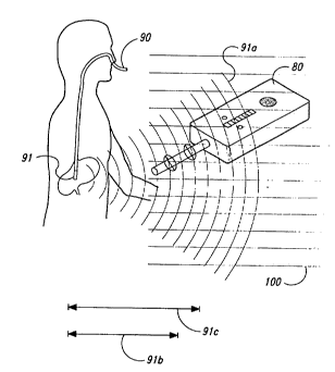
Detect;on of ~ Fee~; ng Tl~he
Referring to Figure 4, a feeding tube (90), with
a permanent magnet (91) located in its tip, includes an
elongated, tubular, main portion with a sealed magnet
chamber at its distal end, and an adapter at its proximal
end to allow connection to a source of feeding formula.
Side apertures at the distal end, above the magnet
chamber, extend from the inner tube lumen to the exterior
of the tube allowing the feeding formula to reach the
patient's stomach. The sealed magnet chamber contains a
cylindrical, rare earth, permanent magnet (91), of
approximate size 0.10 inches diameter by 0.50 inches in
length. The chamber is fused to the distal end of the
feeding tube with its long axis parallel to the long axis
of the feeding tube. The feeding tube and magnet chamber
are composed of a flexible polymer which is chemically,
biologically, and mechanically appropriate for purposes of
3~ gastroenteric feeding.
The feeding tube (90) is inserted into a
patient's nose, down the esophagus and into the stomach.
WO95/08130 ~ ~ ~ PCT~S94/10417
- 2171717
23
The detection apparatus (80) described in Example 1 above
and illustrated in Figure 3, is used to sense the magnet's
static magnetic field strength (9la) at two different
distances (9lb) and (9lc) while immersed in the earth's
ambient magnetic field (100). As the detection apparatus
(80) is moved about the patient's body, greater and lesser
magnetic field gradients are indicated. The feeding tube
(90) is located by moving the detection apparatus until
the greatest magnitude is indicated by detection apparatus
(80).
~ x~le 3
netection A~r~tl~
Referring to Figure 5, in a preferred
alternative embodiment of the apparatus of Example 1, the
first sensor (10) includes x, y, and z-axis sensors (101),
(102), and (103), respectively, while the second sensor
(20) includes x, y, and z-axis sensors (201), (202), and
(203), respectively. In this embodiment the sensors are
flux-gate toroidal sensors with an associated sensor
driver (not shown).
Referring to Figure 6, the first and second
sensor signals (11) and (21), the first and second
amplified signals (13) and (23), the first and second
detection signals (15) and (25), and the differential
signal (41) are vectors.
The first amplifier (12) includes x, y, and z-
axis amplifiers (121), (122) and (123). Similarly, the
second amplifier includes x, y, and z-axis amplifiers
(221), (222) and (223). In addition, the first integrator
(14) includes x, y, and z-axis integrators (141), (142)
and (143), while the second integrator includes x, y, and
z-axis integrators (241), (242) and (243). Finally, the
differential amplifier (40) includes x, y, and z-axis
differential amplifiers (401), (402) and (403).
The operation of the first and second sensors
(10) and (20), the first and second amplifiers (12) and
WO95/08130 , 2 1 7 1 7 1 7 PCT~S94110417
., ~ .. ,- - . --
24
(22), the first and second integrators (14) and (24), and
the differential amplifier (4), is the same as in Example
1, with the exception that in this preferred embodiment,
the signals (11), (21), (13), (23), (15), (25), and (41)
are vectors.
~ ple 4
netection ~p~r~tus with Wollnd-Core In~uct;ve Sensors
As noted above, the invention may be implemented
with analog, mixed-mode, or digital elements. In a
preferred embodiment, the detection apparatus detects the
static magnetic field strength gradient as a vector, as
opposed to a scalar.
Referring to Figure 7, a representative
embodiment includes a first and second sensor (10) and
(20), a first and second detector (207) and (206), and a
microprocessor (208).
The first sensor (10) includes an x, y, and z-
axis oscillator (226), (227) and (228) having associated
wound-core inductive sensors (226a), (227a) and (228a),
respectively. Similarly, the second sensor (20) includes
an x, y, and z-axis oscillator (216), (217) and (218)
having wound-core inductive sensors (216a), (217a) and
(218a), respectively. Further, the first detector (207)
includes an x, y, and z-axis frequency counter (246),
(247) and (248), while the second detector (206) includes
an x, y, and z-axis frequency counter (236), (237) and
(238).
The first and second sensor signals (11) and
(21), the first and second detection signals (15) and
(25), and the differential signal (41) are vectors. The
first sensor x, y, and z-axis oscillators provide the x,
y, and z components, respectively, of the first sensor
signal (11). Similarly, the first detector x, y, and z-
axis frequency counters provide the x, y, and zcomponents, respectively, of the first detection signal
(15). Likewise, the second sensor x, y, and z-axis
WO95/08130 ~s ~ _ ~ 2 1 7 l 7 1 7 PCT~S94/10417
oscillators provide the x, y, and z components,
respectively, of the second sensor signal ~21), and the
second detector x, y, and z-axis frequency counters
provide the x, y, and z components, respectively, of the
second detection signal (25).
The wound-core inductive sensors (216a), (217a),
(218a), (226a), (227a), and (228a) are high-permeability
magnetic cores wrapped with windings. Each wound-core
inductive sensor, together with its associated oscillator,
comprises an LR relaxation oscillator having a period
fixed by the inductance L of the sensor. Since the
inductance L of each sensor is a function of the static
magnetic field strength sensed by that sensor, the period
of the associated oscillator is a function of the same
static magnetic field strength.
Thus, the x, y, and z-axis frequency counters
(246), (247) and (248) receive the x, y, and z components,
respectively, of the first sensor signal (11), and the
period of these components is a function of the first
static magnetic field strength. Similarly, the x, y, and
z-axis frequency counters (236), (237) and (238) receive
the x, y, and z components, respectively, of the second
sensor signal (21), and the period of these components is
a function of the second static magnetic field strength.
Each frequency counter determines the frequency
of its associated first or second signal component. It
then provides that frequency to the microprocessor (208)
in the form of the first and second detection signals (15)
and (25). The microprocessor (208) determines the
magnitude of the detection signals (15) and (25) by
subtracting the second detection signal vector from the
first detection signal vector, summing the squares of the
components of the resulting difference vector, and taking
the square root of the resulting sum. The microprocessor
then provides the differential signal (41) to the
magnitude circuit.
-
WO95/08130 ~ ; 2 1 7 1 7 1 7 PCT~S94/10417 ~
From the foregoing, it will be appreciated that,although specific embodiments of this invention have been
described herein for purposes of illustration, various
modifications may be made without deviating from the
spirit and scope of the invention. Accordingly, the
invention is not limited except by the appended claims.