Note: Descriptions are shown in the official language in which they were submitted.
~WO 95/18640
PCT/US94/00205
1 1
Zr'LEXIBLE RETAINER FLANGE
FOR GA8TRO8TOMY TUBE
BACKGROUND OF THE INVENTION
This invention relates to a new and improved
gastrostomy device with a flexible, collapsible internal
retention dome. This retention dome improves the ease of
insertion by collapsing down in size as the gastrostomy
device is pulled down the esophagus of a patient during a
placement procedure. The collapsibility of the retention
dome also reduces the risk of tissue and stoma damage, and
reduces patient trauma during a subsequent external
traction removal procedure.
Various types of gastrostomy devices have been
installed in patients by means of a percutaneous insertion,
a surgical placement, a radiological placement or others.
The procedures employed generally follow those known as the
Sachs-Vine procedure, the Gauderer and Ponsky procedure,
and others. Typical patents describing these procedures
and publications of the technique are set forth in U.S.
Patents 4,861,334; 4,900,306; and 5,080,650.
Once installed, these devices are retained in place by
an internal retention member. Varios types of these
internal retention members currently exist, one type being
a molded or permanently attached flange element, and
another type being a collar and balloon.
Removal of gastrostomy devices are needed upon
conclusion of enteral nutrition of a patient, or if the
device were to be replaced with another enteral feeding
device (e. g. an inflatable, replaceable gastrostomy tube),
and various techniques are currently used for this removal
" procedure. These techniques include 1, cutting the
gastrostomy tube at skin level and retrieving the bumper
endosco icall 2. cuttin the
p y: g gastrostomy tube at skin
level and allowing the flange or collar to pass through the
WO 9S/18640 PCT/US94/00205
1 2
gastro-intestinal tract and be expelled by excretion; or,
3. physically pulling the internal retention device through
the patient's stoma. ,
There are problems associated with all three of the
above prior art removal techniques. For example, the ,
endoscopic retrieval process places the patient at high
risk during the procedure. Some of the risks involved are
esophageal tissue damage and possible blockage of the
trachea by the flange or collar upon retraction which could
result in death.
Another risk occurs in allowing the flange or collar
to pass through the gastrointestinal tract which may cause
intestinal or bowel blockage.
Still another risk arises when these prior art devices
are removed from a patient by means of external traction
techniques, since they have been known to cause
considerable trauma and excessive bleeding to patients.
Consequently, the device of the present invention is
ZO designed for placement by either of the aforementioned
percutaneous endoscopic or other types of procedures, and
improves upon prior art devices during placement of the
device and also during the external traction removal
procedure.
THE INVENTION:
According to the invention, a gastrostomy device is
disclosed which may be inserted into a patient by a
percutaneous endoscopic, radiological, surgical, or other
type of procedure. The device comprises a catheter tube,
a proximal portion of which is used as a means for
insertion, and a distal internal retention flange.
The internal retention flange includes a dome which is
designed to collapse down significantly in diameter when a
force is longitudinally applied along the axis of the
F
~_ WO 95/18640 ~ PCT/US94/00205
3
catheter tube. The collapsing aspect of this dome allows
for easier passage during placement of the device.
In normal catheter operation, the shape of the dome
enables it to conform to the contour of the patient's
stomach and provide a comfortable and secure internal
retention means. When the gastrostomy device is no longer
needed by the patient, or when replacement and substitution
is required by another enteral feeding device, the
collapsing dome of the retainer facilitates the external
traction removal procedure.
The shape and configuration of the dome, as described
herein, enables the dome to plicate when a force is applied
longitudinally along the axis of the catheter tube.
~5 The predictable manner in which this dome buckles
under a reduced amount of force is directly related to the
varying thicknesses that are designed into the dome. The
buckling or collapsing effect, and the compact shape which
the dome assumes during removal of the gastrostomy tube,
20 can alleviate much of the pain and trauma associated with
this procedure.
~N THE DRAWINGS'
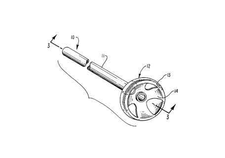
FIG. 1 is an external, perspective view of the
25 gastrostomy device of this invention;
FIG. 2 is a bottom plan view of FIG. 1 showing the
underside of the flange element;
FIG. 3 is a sectional view in side elevation taken
along lines 3 - 3 of FIG. 1;
30 FIG. 4 is an external view in side elevation and
partly in perspective showing the gastrostomy tube of this
invention following installation into a patient;
FIG. 5 is an external view in side elevation, partly
in perspective, showing the gastrostomy tube of this
35 invention as it is being withdrawn from a patient;
WO 95/18640 PCT/US94/00205
2 ~.'~ ~. ~ 3 &
1 4
FIG. 6 is an external view in side elevation showing
indicia markings on the exterior of the catheter; and,
FIG. 7 is an external view, partly in perspective of
another embodiment of this invention.
DESCRIPTION OF THE PREFERRED EMBODIMENTS:
The gastrostomy device 10 of this invention is shown
in FIG. 1, and comprises a hollow elongate catheter
0 tube 11, through which a patient is fed, and an attached
flange 12 at the distal end. The flange may be attached to
the catheter tube by heat sealing, adhesives, sonic
bonding, etc., or the flange 12 may be integrally formed
with the catheter.
~5 The flange and catheter components are constructed of
a biocompatible polymer such as a silicone elastomer,
silicone copolymer, polyurethane, etc. Either of these two
components may incorporate a white filler, or may omit a
filler and be relatively clear. Also, a radiopaque
20 material such as BaS04 may be incorporated into the distal
end of the catheter. As shown in FIG. 6, spacing indicia
are provided to enable location of the device during
installation, and during use.
Typically, the catheter has a durometer hardness of
25 about 40 - 60, with an outside diameter of about 14 - 20
French, an inside diameter of about 155 - 175 mils, and a
wall thickness of about 40 - 60 mils.
A dilating sheath of polyethylene, or ABS (not shown)
is attached to the end of the catheter to enable
30 manipulation of the device during installation.
Alternatively, a wire loop (not shown) may be used with a
dilating tip, and the wire is typically made of a medical
grade stainless steel and coated with a biocompatible
material such as a polyurethane.
35 Due to its plastic memory, in its usual or
unconstrained form, the flange 12 comprises an
CA 02171938 1998-11-06
WO 95/18640 PCT/US94100205 -
1 5
approximately hemispherically shaped dome 13. The dome is
about 20 - 35 mils thick and is reinforced with a cut-
out 14 about 20 - 25 mils thick and positioned on the
inside of the dome. The cut-out 14 has a cruciform shape
which defines four elements 15 spaced equally around the
interior of the hemisphere dome. The central area 16 of
the cut-out is continuous, and the dome reinforcement in
this central area is~ about 40-60 mils thick. The cut-
out 14 may be formed integrally while being insert molded,
or it may be formed separately and then attached t-, the
dome by heat or adhesive sealing, sonic bonding, etc.
It will be appreciated that the cruciform shape of the
cut-out 14 is not critical, and the cut-out may assume
15 various shapes, such as elliptical, rectangular,
triangular, asterism, striated, etc., provided it enables
collapse of the hemisphere dome from the periphery to the
central area 16 during removal of the gastrostomy device. -
As shown in FIGS. 5 & 6, a portion lla of the catheter
20 extends beyond the patient and will engage a closure. member
(not shown) , which when opened, connects to a feeding port;
this type of arrangement is well known in the art.
If desired, the external portion of the catheter can
be formed into a feeding port which functions along with a
25 gasket to produce a sliding friction fit and thereby
accommodate for peristaltic forces and other stomach
movements. U.S. Patents 4,666,433; 4,685,901; 4,701,163;
and, 4,798,592 show this arrangement. This embodiment
shown in Fig. 7 provides a catheter 20 attached at one
30 end to a dome element 21 of this invention, and includes
a feeding port 22 mounted at the opposite end of the
catheter. A closure plug 23 attached to a connector 24
fits into the feeding port when the device is not in use.
A gasket 25 provides a sliding friction fit along the
35 catheter and moves outwardly along the catheter in response
CA 02171938 1998-11-06
WO 95/18640 PCTIUS94100205 -
6
to peristaltic forces, stomach movements, etc., and to
prevent the device from being drawn into the patient s
stomach. Gasket 25 has a plurality of legs 26 which rest
against the user's body, and along with a plurality of air
vent bores 27, permit better air circulation and reduce the
presence of moisture.
Generally speaking a size differential of about 10 -
20 mils between the outside diameter of the catheter and
the insider diameterlof' the gasket bore is employed to
produce a sliding friction fit between the gasket and
catheter.
FIGS. 4 and 5 illustrate the gastrostomy tube 10 when
installed through a stoma 16 of a patient as shown in
~5 FIG. 4, and during commencement of its removal, shown in
FIG. 5. When installed, as shown in FIG. 4, the
gastrostomy tube permits feeding through the catheter 11,
while being retained within the stomach of the patient by
means of the flange 12. The retention characteristics of
20 the present gastrostomy tube in the patient are quite
adequate. When the gastrostomy tube 10 is retracted,
usually for replacement purposes, the flange commences to
collapse 17 as the retraction begins, and this initial
collapse can be felt by the health care worker, physician,
25 etc., who perform the procedure. Hence, following this
initial collapse, less subsequent force compared to prior
art devices, is required to pull the gastrostomy device
from the patient's body.
Removal of these prior art devices from the patient
30 frequently results in trauma, and often requires the use
of
masks and/or other coverings by medical workers when
performing this procedure. The gastrostomy device of the
present invention is less painful and traumatic to the
patient during its removal, and is less troublesome to
35 health care workers.