Note: Descriptions are shown in the official language in which they were submitted.
2172129
W095/08945 PCT~S94110623
MULTIPLE BIOPSY SAMP~ING DEVICE
Field of the Invention
This invention relates to t~k;~g samples of tissue
5 from the body ~or biopsy analysis.
Bac~Loul,d of the Invention
Tissue samples can be examined in a laboratory to
determine the presence of a pathological disorder (e.g.
malignancy). Often, the samples must be obtained from
0 deep within the body using a medical sampling instrument.
It is usually best to obtain several samples around the
location where the disorder is suspected so that the
prese~cP and progress of ~icP~, if any, can be
accurately determined. The samples must be catalogued
15 according to the location from which each sample is taken
and the integrity of the samples must be maint~;n~ for
the subsequent laboratory analysis.
SummarY of the Invention
In one aspect, the invention relates to an
20 instrument for obt~;n;~g tissue samples from a site deep
within the body. The instrument has an elongated
proximal portion that is constructed to follow a long,
torturous path to the site and has a distal end
constructed to sever and remove a tissue sample from the
25 body, including tissue specimens, polyps or the like.
The improvement includes that the instrument is
constructed to take multiple biopsy samples without being
withdrawn from the body. The instrument includes a
tissue sample retractor. The retractor is axially
30 movable between an extended tissue-engaging position and
a retracted position. There is an open passage into
which the retractor moves when moving from the extended
to the retracted position. The retractor has a distal
end portion constructed to engage tissue and apply axial
35 transporting force thereto while moving from the extended
2172129
W095/08945 PCT~S94/106
to the retracted position. The retractor is constructed
to be advanced and retracted repeatedly to acc~ te a
series of samples in the instrument.
In another aspect, the invention features an
5 instrument for obt~in;ng endoscopic tissue samples. The
instrument is sized and constructed to pass through the
working ~-h~nnel of an endoscope to take samples including
tissue specimens, polyps or the like, under endoscopic
guidance. The device has a distal end constructed to
lO sever and remove a tissue sample from the body. The
improvement includes the instrument constructed to take
multiple biopsy samples without being withdrawn from the
endoscope. The instrument includes a tissue sample
storage device with a tissue-penetrating element having a
15 distal barb formation. The barb formation is arranged to
facilitate entry into tissue during advancement against
the tissue and to resist distal dislodgment of the sample
after the barb has penetrated the tissue.
In another aspect, the invention features an
20 instrument for obt~;n;ng endoscopic tissue samples. The
instrument is sized and constructed to pass through the
working channel of an endoscope to take samples including
tissue specimens, polyps or the like, under endoscopic
guidance. The instrument has a distal end constructed to
2~ sever and remove a tissue sample from the body. The
improvement includes the instrument constructed to take
multiple biopsy samples without being withdrawn from the
endoscope. The instrument includes a tissue sample
storage device, wherein the storage device comprises a
30 helical cork-screw-like projection constructed to be
rotated to enter tissue.
In another aspect, the invention features an
instrument for obtaining endoscopic tissue samples. The
instrument is sized and constructed to pass through the
35 working channel of an endoscope to take samples including
~ 1 72~ 29
W095/08945 PCT~S9411~623
tissue specimens, polyps or the like, under endoscopic
guidance. The instrument has a distal end constructed to
sever and remove a tissue sample from the body. The
improvement includes the instrument constructed to take
5 multiple biopsy samples without being withdrawn from the
endoscope. The instrument includes a tissue sample
storage device. The sample storage device includes an
elongated tissue penetrating element of length sufficient
to accommodate at least three samples and constructed to
lO enable a specimen to be slidably advanced progressively
thereover away from a severing device of the instrument
as additional samples are taken as a result of pressure
transmitted through the previously taken samples during
the spearing action on the next sample, thereby to
15 prepare the instrument to take further samples.
In another aspect, the invention features an
instrument for obtaining endoscopic tissue samples. The
instrument is sized and constructed to pass through the
working channel of an endoscope to take samples including
20 tissue specimens, polyps or the like, under endoscopic
guidance. The instrument has a distal end constructed to
sever and remove a tissue sample from the body. The
improvement includes the instrument constructed to take
multiple biopsy samples without being withdrawn from the
25 endoscope. The instrument includes a severing device
with at least one pivotable jaw. The jaw has a pair of
pivotable jaw support portions lying close to respective
sides of a supporting structure, there being an open
space for multiple tissue sample storage between the
30 support portions.
Embodiments of the invention may include
combinations of the features above and also have one or
more of the following features. The instrument has a
severing device including at east one pivotable jaw.
35 The jaw has a pair of pivotable jaw support portions
W095l08945 2 1 ~27 2~ PCT~ss4/lo623
lying close to respective sides of a supporting
structure. There is an open space for multiple tissue
sample storage between the support portions. The
retractor is a tissue-penetrating element. The tissue-
5 penetrating element has a barb formation arranged tofacilitate entry into tissue during advancement against
the tissue and to apply axial transporting force to the
tissue during retraction movement. The tissue-
penetrating element is constructed and arranged to
10 penetrate a mid-portion of the sample of tissue being
taken. The penetrating element is elongated and
constructed to spear and securely store thereupon, in
stacked relationship, a series of tissue samples in the
order in which the samples have been taken. The element
15 is sufficiently long to store a series on the order of
three, e.g., five or more tissue samples. Multiple barbs
are disposed along the length of the element, constructed
to enable a specimen ~o be advanced progressively over
the barbs as additional samples are taken as a result of
20 pressure transmitted through the previously taken samples
during the spearing action on the next sample. The
retractor is a helical cork-screw-like projection
constructed to be rotated to enter tissue and constructed
to move axially to retract the tissue sample. The cork-
25 screw-like projection is sufficiently long to store
thereupon a series on the order of five or more tissue
samples, in the sequence in which the samples have been
taken. Helical threads extend along the length of the
projection along which previously-taken samples advance
30 when, upon further rotation, additional samples are
taken. The retractor is constructed to extend along the
side of a sample being severed and has a laterally-
extending dragger formation constructed to engage the
sample and apply proximally-directed transporting force
35 thereto. The retractor is constructed and arranged
- 2172129
W095/08945 PCT~S94/10623
relative to the open passage to drag successive severed
samples into the passage and stack them therein in the
order in which the samples have been taken. The severing
device includes at least one pivotable jaw and one
5 stationary jaw. The retractor is of generally tongue
form and during severing action lies along the stationary
jaw. The retractor is of wire form and has a distal hook
formation constructed to apply proximally-directed
transporting force to the sample. The retractor is an
10 axially displaceable grasper constructed to grasp the
tissue sample by pinching action for transport of the
sample. The grasper includes tong-like grippers
constructed to grasp and transport a tissue sample. The
grippers are closed upon a sample by axial movement of an
lS actuating tube slidingly disposed over structure
connected to the grippers. The movable retractor is
constructed to draw the severed sample into the passage
away from a severing device of the instrument to prepare
the instrument to take further samples. The severing
20 device has at least one actuatable cutting jaw. The jaw
is supported on the distal end of a tubular structure.
An internal portion of the structure provides space for
tissue sample storage. The severing device includes
opposed actuatable cutting jaws. The jaws are
25 constructed to be closed upon a sample by axial movement
of an actuating tube slidingly disposed over supporting
arms of the jaws. The jaw is pivotably supported by
supporting structure and control means extend along the
instrument for pivoting the jaw. The instrument includes
30 a severing device in the form of a snare loop projectable
from the instrument over tissue to be removed. The
ins ~L ~cnt is sized and
constructed to pass through the working channel of an
endoscope to take multiple samples under endoscopic
35 guidance without being withdrawn from the endoscope.
W O 95/08945 2 1 7 2 1 2 9 PC~rnUS94/10623
- 6 -
Embodiments may also include one or more of the
following features. The tissue penetrating element, upon
completion of taking of the samples, is constructed to be
detached from the instrument and be sent to the pathology
S laboratory with the samples intact upon the element in
the order in which the specimens were taken. The element
is constructed and arranged, upon completion of use and
withdrawal from the body, to extend distally beyond
sample severing mech~ism of the instrument to enable the
10 multiple samples to be removed therefrom.
Embodiments may also include one or more of the
following features. The in~LLu~cnt has a distal
supporting tube, a distal extremity of the tube
constructed to form a fixed jaw, and the pivotable jaw
15 has support arms lying close to respective sides of the
tube and being pivotably mounted with respect thereto.
The arms lie on the exterior of the tube. The arms lie
in the interior of and closely adjacent to respective
sides of the interior wall of the tube. A respective
20 short pin-formation pivotably mounts each arm to the
tube. A through-axle extend across the interior of the
tube, upon which the arms are mounted, open space being
provided adjacent the axle for storage of the tissue
samples.
Other features and advantages follow.
Brief DescriPtion of the Drawing
We first briefly describe the drawings.
Fig. 1 is a perspective view of an embodiment of
the invention being delivered into the body through an
30 endoscope;
Figs. 2-2c are cross sectional views that
illustrate the structure and use of an embodiment of the
invention; t
2 1 721 29
W095/08945 PC~S94/10623
Fig. 3 is a cross sectional view of another
embodiment of the invention;
Figs. 4-4f illustrate the structure and use of
another embodiment of the invention;
- 5 Figs. 5-5a are perspective and top views,
respectively, that illustrate another embodiment of the
invention;
Figs. 6-6a illustrate another embodiment of the
invention;
Figs. 7-7c and 8 are assembly views that
illustrate hinge arrangements for a moveable jaw
according to the invention;
Figs. 9-9c and 10-lOa illustrate tissue sample
retractor arrangements according to the invention;
Figs. 11-llc illustrate positioning of retractors
according to the invention;
Figs. 12-12d illustrate various jaw configurations
for use with forceps embodiments of the invention;
Fig. 13 illustrates another embodiment of the
20 invention;
Figs. 14-14d illustrate the structure and use of
yet another P~hoA; ment of the invention.
Description of the Preferred Embodiments
Referring to Fig. 1, the device 10 for multiple
25 biopsy sampling may be delivered into the body through
the channel of an endoscope device 11 (e.g., gastroscope,
sigmoidoscope, or colonoscope). The endoscope device
typically has a length of about 100-250 cm and a ~h~nne
diameter of 2.0 - 3.8 mm, typically about 2.8 mm. A
30 distal sampling portion 16 is extended from the endoscope
for cutting and storing a sample of tissue from a body
surface 18 of a patient (e.g. from a surface in the
gastrointestinal tract, bronchial tract, urinary tract,
reproductive organs, cardiac tissue, or the like). The
35 device has a diameter of preferably around 1.8 - 2.4 mm,
2 1 721 29
WO 95108945 PCIIUS94/10623
-- 8 --
typically about 2.3 mm or less, and is of sufficient
flexibility so it passes easily though the channel when
the endoscope follows a tortuous body passageway. The
endoscope includes other lumens for water, air, suction,
5 and viewing. Devices according to the invention can be
adapted to be i~.Lrud~ced to sites deep within the body by
other means. For example, a device can be configured
with a lumen so that it can be advanced over a guidewire,
e.g., in vascular applications. The device may be passed
10 through an illLLGd~cer or guiding catheter in, e.g.,
cardiac applications. The sampling and storage
arrangements may be useful in open su.yery applications.
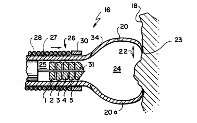
Referring to Figs. 2-2c, in an ~ho~;ment, the
sampling portion 16 includes a pair of jaw members 20,
15 2Oa which are movable with respect to each other (arrow
22) between an open and closed position. The outer edge
23 of the jaw members includes a sharp cutting edge for
cutting a sample of tissue from the body surface 18. Jaw
members 20, 2Oa are formed of an elastic material, such
20 as an elastic stainless steel, nitinol or the like, and
are biased outwardly so the jaws are open in the relaxed
configuration. The jaws encompass a space 24 in which a
sample is contained after it is cut from the surface 18.
The space 24 communicates with an open throat
25 region 25, just proximal of the jaws, where successive
prior samples 1, 2, 3, 4 and 5 are stored, while the next
sample, sample 6, is ta~en. The samples are stored in
the order in which they taken by use of a retractor 36,
which in this embodiment is a spear-form element that
30 pierces the samples through their centers. As will be
discussed in more detail below, the retractor can be
moved axially into the space 25 to retreive a sample cut
by the }aws by piercing it, then withdrawn proximally to
store the samples in the throat.
W095/08945 2 1 72 1 29 PCT/US94tlO623
_ g _
The throat 25 and jaws 20, 20a, may be defined by
a tubular member 27 that has been modified (e.g. slit
longit~ n~lly and worked) at its distal end to form jaws
20, 2Oa or the throat may be defined by a tube of a
5 different material to which the jaws are attached. There
may be two jaw members, as shown, or there may be more
than two jaw elements that fit together when the jaws are
closed. The jaws may have sides to completely enclose
the space 24 when the jaws are closed or the jaws may be
10 open, like a clipper, such that on}y the cutting edges at
the distal end of the jaws meet when closed. In use,
with the jaws biased open, the device is urged against
the tissue at a location where a sample is to be taken,
as shown in Fig. 2.
Referring particularly to Fig. 2a, the jaws are
closed by an axially movable, concentric sheath member
26. The sheath member, formed of a helical wire coil 28
for most of its length for increased flexibility,
includes at its most distal end a short, stiff bearing
20 member 30. By a suitable mech~nical control located
outside the body, the sheath is moved axially distally
(arrow 33) to cause the bearing member 30 to bear on the
outside portions 34 of the jaws 20, 20a to collapse them
into a closed position so the cutting edge 23 cuts or
25 edulses sample 6 from the body surface 18 of the patient.
Referring particularly to Fig. 2b, the elongated
spear-form retractor 36 is movable along its axis into
the space 24 en~o~rassed by the jaws for piercing the
next sample, sample 6. With the jaws closed, the sample
30 6 is pushed against the interior of the distal portions
of the jaws and onto the retractor. The pressure against
sample 6 is transferred to sample S and through the other
samples, thus displacing previous samples 1-5 axially
proximally along the body of the retractor.
rB
2 1 72~ 29
wos~/08s4s PCT~S94/106~
-- 10 --
Referring particularly to Fig. 2c, the spear-form
retractor 36 is then withdrawn axially to store samples
1-6 in the open throat 25, as illustrated. The tip of
the retractor is pointed to allow samples to be pierced
5 but also includes a barb 31 that prevents the sample from
slipping off durin~ retraction. The spear-form element
grasps and ;n~eYe~ successive samples 1-6 cut by the jaws
in the order in which they were taken. The procedure
above can be repeated so additional samples can be taken
10 without removing the device from the endoscope.
Other Embodiments
Referring to ~ig. 3, another ~ho~;ment of the
invention as shown. In this case, the retractor is a
forceps member 50, with jaws 51, 51a much smaller than
1~ the cutting jaws 20, 20a so the retractor can be moved
axially (arrow 52) in to the space 24 to grasp a sample
and pull it back into the throat for storage. The jaws
of the forceps member 50 may be biased in the open
position and closed by distal axial extension of an outer
20 concentric sheath 54, like the closure of the jaws 20,
20a with sheath 26. A spear element (not shown) may be
provided within the throat of the forceps 50, to hold
multiple samples in the throat 55, proximal of the jaws
51,51a.
Referring to Figs. 4-4f, another emhoA;ment is
shown. In this embodiment, a sampling forceps includes a
stationary jaw 60 and a movable jaw 62. The movable jaw
62 is attached to the body 64 of the device at two hinge
points 66, 67 and actuated between the open and closed
30 position by pull wires 63. Referring particularly to
Fig. 4f, a perspective view, the jaw 62 is hinged at
opposite sides of the body 64 to provide the open throat
area for storage of successive samples. The jaws may be
hinged on either the outside of the body, as shown, or
35 inside of the body. A movable or stationary retractor
2 1 721 29
Woss/o8s4s - PCT~S94110623
68, with a barb or undercut 69, is used to anchor the
device in tissue and hold samples after cutting.
Referring particularly to Fig. 4a, in use, the jaw
62 is opened and the retractor advanced into tissue 18.
(The retractor can as well be advance after the sample is
cut as shown in Figs. 2 et seq.) Previous samples are
moved axially proximally along the retractor. Referring
to Fig. 4b, using pull wire 63, the jaw 62 is closed and
a tissue sample 65 is cut and captured by the jaws. The
lO retractor is advanced forward axially to firmly hold the
tissue. Referring to Fig. 4c, the retractor is then
drawn axially proximally so that the tissue is located in
the throat h~hin~ the jaws. Referring to Fig. 4d, the
jaws are opened and the spear is re-advanced to take the
15 next sample. Referring to Fig. 4e, to remove the
multiple samples, after the device has been removed from
the body, the jaws are opened and the spear moved axially
distally (arrow 71) beyond the jaws where the samples can
be easily acrecc~. The samples can but need not be
20 removed from the spear. Rather, the end of the retractor
carrying the samples can be detached at a location
proximal of the samples (by cutting or by a reusable
attachment me~h~n; sm) and the samples, still indexed on
the spear according to the order they were taken, sent to
25 the lab.
In alternate emho~iments~ the retractor is
stationary and of extended length running from the throat
region to the space within the jaws. In embodiments, the
spear can be withdrawn proximally the full length of the
30 device to remove it and access the samples while leaving
the rest of the device in an endoscope.
Referring to Figs. 5 and 5a (top view), the jaw 62
may be operated by a single pull wire 70, attached
centrally to a common pivot arm 72.
21 72~ 29
WO 95/08945 PCI/US94/10623
Referring to Figs. 6-6a, the jaw 62 may be closed
using a coaxial sheath 74, which is slid forward (arrow
73, Fig. 6a). The jaw 62 may be biased open using a
spring at the pivot points or pulled open using a pull
5 wire.
Referring to Figs. 7-7c, the jaw 64 may be
attached to the body of the device using separate pins at
each pivot point. Pins 77 may be integral with the body
of the device, to protrude into a hole 78 in the jaw
(Fig. 7) or a pin 79 may protrude from the jaw into a
hole 81 in the body (Fig. 7a). One pivot point may have
one construction and the other pivot point the other
construction. Both the jaws and body may include holes
83,85 which are adapted for separate pins 87 (Fig. 7b) or
15 a single pin 76 may pass through the body of the device
(Fig. 7c). In the latter embodiment, the retractor can
pass to one or the other side of the pin. The pin 76
could also be modified to inclu~e rotatable extensions,
which like a paddle wheel, could draw samples from the
20 jaws and place them in the throat for storage.
Referring to Fig. 8, an elongated slot 80 or cam
configuration may be provided at the pivot point for
opening the movable jaw 64 wider and increasing cutting
force and action. In use, the jaw is opened and slid
25 distally to grab tissue. Then the jaw is closed by
rotation about the pivot point while drawing the jaw
proximally along the slot.
Referring to Figs. 9-9c, various additional
retractor embodiments are illustrated. Referring
30 particularly to Fig. 9, the retractor may be a preformed
wire member 66 with a hook end 68 which is used to snare
or trap the tissue so that it can be withdrawn into the
device. Referring to Fig. 9a, the retractor may be a
loop end device 70. By rotating the body of the
35 retractor about its axis, the loop can be rotated to
2172129
Woss/0894s PCT~S94/10623
catch tissue. The sample can then be withdrawn into the
throat by withdrawing the retrator axially. Referring to
Fig. 9b, the body of the member can be provided with a
number of small, axially separated barbs 89 that separate
and retain adjacent samples. Referring to Fig. 9c, the
retractor may be a rotatable spiral-form cork-screw
member 73 (shown symbolically) that collects and stores
the samples along the spiral surface by rotation of the
member about its own axis. The cork-screw-like
projection can be rotated to enter a tissue sample and
then withdrawn axially into the the throat. Helical
threads extend along the projection on which previous
samples advance when, upon further rotation, additional
samples are taken.
Referring to Figs. 10-lOa in another embodiment,
an axially movable tongue 90 is provided. The tongue is
shaped to conform to the inner contour of one of the
jaws, typically the stationary jaw 60. After the jaws
have cut the sample, the tongue can be drawn proximally
(arrow 91) to drag the sample into the throat for storage
(Fig. lOa).
In other embodiments, the distal end of the
retractor may be straight, without an undercut or barb,
e.g. the tip may be rounded. The retactor may include a
reduced diameter section proximal of the tip. The member
may be formed of metal, plastic, composite or
combinations thereof. Preferably, the member has
considerable length compared to its width for storing
multiple samples.
Referring to Figs. ll-llc, in cases where the
retractor is to be centered or otherwise carefully
positioned with respect to the axis of the device body, a
positioning plate 130 may be located in the body just
proximal of the throat. The positioning plate has an
aperture 132. The retractor passes through the aperture
W O 95/08945 2 1 72 1- 29 PC~r~US94/10623
- 14 -
distally. Proximally, a wire 134, integral or attached
to the retractor, extends to the proximal end of the
device to control axial movement of the retractor.
Referring to Figs. 12-12d, the jaws of the device
5 may be of a variety of designs for particular
applications. The jaws may have a straight plane through
the center (Fig. 12). The jaws may be angled with
respect to the center ~Fig. 12a). The jaws may follow a
curved plane through the center (Fig. 12b). The cutting
10 edges of the jaws may be jagged (Fig. 12a), serrated
(Fig. 12c) or razor-edged tFig. 12d). The jaws can be
provided with a heating means, such as an electrical
current, to assist in cutting.
Referring to Fig. 13, another embodiment is shown.
15 The device includes an open tubular member 100 capable of
providing suction in the direction (arrow 102) of the
proximal end of the device. A grasping spear-form member
104 stacks and stores successive samples as they are
drawn into the tubular member by the suction. Means
20 other than forceps may be used to cut the sample from the
body, e.g., suction alone.
Referring to Figs. 14-14d, another embodiment is
shown. The device includes a delivery catheter 120,
suitable for passage through an endoscope. The cather may
25 have a single, or preferably, multiple lumens. The
catheter 120 carries, in one of its lumens, a polypectomy
snare-type wire loop 122 and, in another lumen, a tissue
retention device such as a retractor 124 with a barb 125.
Referring to Fig. 14a, in use, the snare wire loop
30 is positioned around a polyp 126 attached to the wall of
the alimentary tract. Referring to Fig. 14b, the spear-
form element is advanced into the polyp until its barb
125 is completely within the polyp. Referring to Fig.
14c, the polyp is severed from the wall by actuating the
W095/08945 2 1 72 1 29 PCT~S94/10623
- 15 -
snare wire loop. Referring to Fig. 14d, the snare wire
loop is withdrawn into the catheter. Referring to Fig.
14d, the sample is then held by the spear-form member.
Additional samples can be taken and stored on the spear-
5 form member by repeating the above steps.
The grasping members shown in each of the
embodiments above can be used in each of the other
embodiments above. In general, suction can be used to
assist operation of any of the embodiments above.
A system for taking multiple biopsy samples is
taught in Chu "Instruments for Collecting Multiple Biopsy
Specimens", USSN 062,671, filed May 17, 1993, the entire
contents of which is hereby incorporated by reference.
Still other embodiments are within the following
15 claims.