Note: Descriptions are shown in the official language in which they were submitted.
2173316
WO 95/09659 PCT/US94/11304
LOCAL POLYMERIC GEL CELLULAR THERAPY
Background of the Invention
This invention is generally in the area of
methods of treating tissue defects and modulating
cell to cell interactions by administration of a
polymeric gel material containing bioactive
molecules to a tissue surface and the use of
certain ECM and RGD peptides.
The hollow or tubular geometry of organs
commonly has functional significance, for example,
in the facilitation of fluid or gas transport
(blood, urine, lymph, oxygen or respiratory gasses)
or cellular containment (ova, sperm). Disease
processes may affect organ tissue or its components
by encroaching upon, obstructing or otherwise
reducing the cross-sectional areas of the hollow or
tubular elements. Additionally, other disease
processes may violate the native boundaries of the
hollow organ and thereby affect its barrier
function and/or containment ability. These disease
processes include those which are induced by aging,
diet, injury, or activation of the coagulation,
complement and other inflammatory systems or the
development of a neoplasia or malignancy. The
ability of the organ or structure to properly
function can then be severely compromised. This is
particular evident in coronary artery disease,
where successful treatment initial may subsequently
be complicated by overproliferation of endothelium,
called restenosis, or vessel renarrowing or closing
after dilation.
A specific therapeutic strategy which would
= greatly benefit from an adjuvant treatment to
prevent cell migration is percutaneous transluminal
coronary angioplasty (PTCA, balloon angioplasty).
Balloon angioplasty has become the mainstay in the
.interventional therapy of advanced coronary and
peripheral,artery disease. While this procedure
~ 4.. WO 95/09659 217J 31 6 PCT/US94/11304
2
achieves the therapeutic goal of enlargement of the
diseased arterial lumen where a blockage occurs,
the therapy can itself damage the arterial wall and
cause alterations in arterial function. Restenosis
or vessel reclosure following balloon angioplasty
is the major limitation undermining a consistent
long-term success rate for this procedure. In
fact, the currently unmodifiable post-angioplasty
failure rate due to restenosis is 30 to 50%.
Intimal hyperplasia or thickening of the
vascular wall, a fundamental mechanism of
restenosis, is caused by stimulation of smooth
muscle cells within the wall, causing them to
migrate, proliferate, and coordinately secrete or
deposit extracellular matrix proteins. This
combination of smooth muscle cell migration and
matrix deposition, progressing toward the lumen,
and eventually encroaching upon it, is responsible
for restenosis. This SMC response to injury is
marked by a transformation of SMC phenotype from a
quiescent, contractile state to a synthetic,
proliferative state in a high percentage of the
medial SMCs. Another important event which occurs
following injury is that SMCs (both synthetic and
contractile SMCs) become migratory, moving from the
media to the intima.
The types of problems associated with
angioplasty are also characteristic of similar
treatment of other types of natural lumens,
including surgical correction and balloon dilation
of urinary and reproductive tract disorders, for
example, following prostate surgery, or treatment
by laparoscopy and balloon dilation of stenosis or strictured fallopian tubes,
as well as treatment of
openings arising from disease, surgery and trauma. Further, these
reobstructive problems also occur in
artificially or therapeutically created lumens or
WO 95/09659 21! 3316 PCT/US94/11304
3
pathways, such as in renarrowing of the
intrahepatic shunt formed in transjugular
intrahepatic portosystemic shunting procedure
, (TIPS).
As described in the literature, for
, example, U.S. Patent No. 5,213,580 to Slepian, pre-
formed polymeric materials can be inserted into
blood vessels and then contoured to fit the
surfaces of the vessels, providing protection of
the blood vessel and prevention of restenosis. As
described in U.S. Patent Nos. 5,126,141 and
5,135,751 to Henry, et al., aqueous, thermally
reversible gel compositions formed of a
polyoxyalkylene polymer and an ionic polysaccharide
can be applied to injured areas of the body to
prevent adhesions. These same type of
polyoxyalkylene polymers have also been used for
the local delivery of oligonucleotides (antisense)
to the surgically exposed surface of blood vessels
for treatment of restenosis, as described by
W093/01286 by Rosenberg, et al.
None of these, however, describe a means
for forming a polymeric material at or on a lumen
surface which can be used as a barrier of
controlled permeability or for controlled delivery
of a bioactive substance, nor can these materials
be targeted to a particular cell type. While the
prior art discloses useful treatments of damaged
lumen surfaces, it would be desirable to have
materials which could provide these additional
useful functions, especially controlled
permeability which would allow free exchange of
gases and nutrients or controlled diffusion of
macromolecules which are beneficial to the lumen
surface, as well as for controlled drug delivery to
the surface, for example, of growth factors or
antiinflammatories.
CA 02173316 2006-07-12
4
It is therefore an object of the present
invention to provide polymeric materials which are
initially amorphous, biocompatible, and can be
formed in situ.
It is a further object of the present
invention to provide polymeric materials of
controlled permeability which can be used as
selective barriers on lumen surfaces.
It is a still further object of the present
invention to provide materials which can be used
for controlled delivery of drugs and other
biologically active substances, either to tissue
lumen surfaces or into the lumens themselves.
Summary of the Invention
The present invention relates to a use for a
biocompatible polymeric material which can be applied to a
cell or tissue surface to alter cell to cell interactions,
wherein the polymeric material is applied in a first fluent
state and converted in situ to a second non-fluent state as a
function of temperature or the presence or removal of ibns,
and wherein the material in the second non-fluent state
selectively limits or controls passage of macromolecules,
microorganisms, and cells through the polymeric material in
the non-fluent state as a function of molecular weight.
The polymeric material may be applied to a
device selected from the group consisting of a prosthesis,
stent, catheter, graft and implant.
The polymeric material may further comprise a
polymer material chelator or ion exchange incorporated into
the polymer to remove calcium ions or lipids.
In a further aspect of the invention, the
material may incorporate compounds selected from the group
consisting of chemoattractant factors, growth factors,
antiangiogenic factors, antiproliferative compounds, and
antisecretory factors for use in a method for promoting tissue
repair or ingrowth at a site where tissue ingrowth may occur.
CA 02173316 2005-08-03
4a
Methods for creating in situ specific local
interactions or cellular interactions in living
tissue are disclosed. This is accomplished by
applying a fluent material which forms a local,
selectively permeable barrier, alone or in
combination with specific bioactive molecules,
directly to a site to be treated. Upon
application, the fluent material is conformed to
the tissue and converted to a less fluent state by
alteration in temperature, ion concentration,
application of shear force, or chemical or physical
polymerization or crosslinking. In one embodiment,
cellular interactions, such as formation of
thrombus, inflammation, or adhesions, are inhibited
by physically blocking cellular and/or
macromolecular interactions while allowing
selective permeability to nutrients, gases, and
other molecules. Permeability is controlled by
selection of the material, method of manufacture,
density, degree of crosslinking, molecular weight
of monomer units, incorporation of particulate or
other material, and degradability or non-
2173316
WO 95/09659 PCT/US94/11304
biodegradability of the polymeric material. In
another embodiment, the polymeric gel is provided
in combination with bioactive molecules, especially
= those providing contact guidance, or chemotactic or
5 haptotactic activity, which can be utilized to
= alter cell proliferation, migration, and
inflammatory reactions.
As demonstrated by the examples, a
synthetic barrier made of a biocompatible polymeric
material can be applied in vivo to a tissue or
cellular surface such as the interior surface of a
blood vessel or tissue lumen. The material may
also be applied to tissue contacting surfaces of
implantable medical devices. The polymeric
material is applied in the first fluent state to
the site to be treated using, for example, a
catheter, or by means of spraying or irrigation at
the time of surgery. The material is then
reconfigured to have intimate conforming contact
with the surface to be coated, and then maintained
under conditions which convert the material into
its second non-fluent state. The conversion may be
achieved either by active methods in which the
environment surrounding the material is altered by
the addition or removal of chemicals or energy, or
it may be by passive means in which, for example,
maintaining the material at the normal internal
body temperature of the patient causes the material
to undergo conversion into its non-fluent state.
The transition of the material from a fluent state
to a non-fluent state may be the result of a phase
change in which the material goes from a liquid
state to a solid state, by gelation, or in the
alternative, it may be the result of a viscosity
change with the material actually remaining in a
single phase.
WO 95/09659 21' 3"' 1" PCT/US94/11304
6
As part of these studies, it has now been
discovered that the extracellular matrix (ECM)
protein tenascin facilitates cell migration in vivo
for the treatment of diseased or injured tissues
and can be used alone or in combination with a
carrier such as the polymeric gel for localized
therapy.
Brief Description of the Drawings
Figure 1 is a schematic of the method of
the present invention.
Figure 2A is a cross sectional view of the
multilumen features of the catheter shown in Figure
2B. Figures 2B and 2C are expanded views of
catheters useful in the method described herein for
application of polymeric materials to the tissue
lumen surfaces.
Figures 3A-3G are schematics of photographs
of application of a polymeric material as described
herein within a mock hollow tubular organ. Figures
3A and 3B are schematics of the catheter and the
catheter being inserted into the tube; Figure 3C is
of the two balloons in the catheter being inflated
to seal off the vessel; Figure 3D is of the
polymeric material being injected into the tube;
Figure 3E is of the tube with the polymeric
material having gelled and the balloons deflated;
Figure 3F is of the catheter being removed to leave
a gel coating on the vessel walls with an interior
lumen or annual space; and Figure 3G is of the
lumen after the balloons are collapsed and
withdrawn from the coated vessel, and the material
has been smoothed and thinned by reapplication of
the distal occlusion balloon.
Figures 4A-4F are schematics of micrographs
of injection of polymeric material into isolated
bovine coronary arteries.
RECTIFIED SHEE,~ (RULE 91)
ISA / EP
WO 95/09659 2173316 . J PCT/US94/11304
7
Figure 5 is a schematic of a micrograph (250x)
of the cross-section of a gel coated artery with a
thin gel coating (lower left corner).
Figures GA and GB are scanning electron
micrographs of the intimal surface of rat carotid
= arteries following 60 minutes of reexposure to
blood post-injury; Figure 6A is the control balloon
abraded rat intimal (endoluminal) surface with
significant platelet, white cell and fibrin
deposition; Figure GB is the gel coated (Pluronic
F127, 25% w/v) arterial surface showing a
significant reduction in platelet, white cell and
fibrin deposition and adherence.
Figure 7 is a schematic of photographs of the
effect of gel coating on limiting the development
of arterial neointimal hyperplasia 14 days post-
injury.
Figure 8 is a graph of %- control migration
versus peptide concentration (mM) for cyclic RGD
(closed squares), GRGDdSP (a stronger inhibitor of
Sl integrins) (open squares), GRGDSP (linear RGD
peptide which inhibits Z. integrins) (closed
circles), and GRADSP (non-sense peptide) (open
circles).
Figure 9A is a graph of the percent control
migration of tenascin (1.0 g/ml) versus untreated
control.
Figure 9B is a graph of the percent control
migration for 1 g/ml tenascin in combination with
either anti-vitronectin receptor or anti-
fibronectin receptor, as compared with control
tenascin alone.
Figure 10 is a graph of the SMC surface bound
tenascin (i mol/10-6 cells) versus soluble tenascin
in culture media ( g/ml).
RECTIFIED SHEET ( RtJLE 91)
ISA / EP
WO 95/09659 2173316 PCT/US94/11304
8
Detailed Description of the Invention
As described herein, polymeric materials
are applied to the surface of tissue lumens to
provide a barrier having either a controlled
permeability to materials in the lumen, for example
blood, and/or controlled release of incorporated
bioactive agents.
Selection of Polymeric Materials
The basic requirements for the polymeric
material are biocompatibility and the capacity to
be applied in a fluent state then chemically or
physically reconfigured under conditions which can
be achieved in vivo to yield a non-fluent polymeric
material having defined characteristics in terms of
permeability and release of incorporated materials.
The polymeric materials can be applied as
monomers, macromers, polymers, or combinations
thereof, maintained as solutions, suspensions, or
dispersions, referred to herein jointly as
"solutions" unless otherwise stated. Although
capable of many forms in their non-fluent state,
organogels and hydrogels represent preferred
embodiments. Although non-degradable and
biodegradable materials can be used, biodegradable
materials are preferred. As used herein,
"biodegradable" is intended to describe materials
that are non-permanent and removed by natural or
imposed therapeutic biological and/or chemical
processes. For application to the interior of
blood vessels following angioplasty, it is
preferred to use polymers degrading substantially
six months after implantation; for prevention of
adhesions or controlled release following treatment for injury or surgery, the
degradation should be
correlated with the time required for healing, i.e., generally in excess of
six days but less than
six months.
2173316
WO 95/09659 PCT/US94/11304
9
The polymeric materials are selected from
those materials which can be polymerized or their
viscosity altered in vivo by application of
exogenous means, for example, by application of
light, ultrasound, radiation, or chelation, alone
. or in the presence of added catalyst, or by
endogenous means, for example, a change to
physiological pH, diffusion of calcium ions
(alginate) or borate ions (polyvinyl alcohol) into
the polymer, or change in temperature to body
temperature (37 C).
As used herein, a hydrogel is defined as an
aqueous phase with an interlaced polymeric
component, with at least 60%, preferably at least
75%, more preferably with 80% or more, and as a
specific example, with 90% of its weight as water.
The following definition is from the Dictionary of
Chemical Terms, 4th Ed., McGraw Hill (1989):
Hydrogel: a colloid in which the disperse phase
(colloid) has combined with the continuous phase
(water) to produce a viscous jellylike product, for
example, coagulated silicic acid.
An organogel is defined as an organic phase
with an interlaced polymeric component, with at
least 60%, preferably at least 75%, more preferably
with 80% or more, and as a specific example, with
90% of its weight as organic solvent. Preferred
solvents include non-toxic organic solvents,
including but not limited to dimethyl sulfoxide
(DMSO), and mineral and vegetable oils.
Suitable materials are commercially
available or readily synthesizable using methods
known to those skilled in the art. These materials
include:
CA 02173316 2006-07-12
1. Materials which change from a first fluent
state to a second non-fluent state as
a function of temperature.
Poly(oxyalkylene) polymers and copolymers
5 such as poly(ethylene oxide)-poly(propylene oxide)
(PEO-PPO) or poly(ethylene oxide)-poly(butylene
oxide) (PEO-PBO) copolymers, and copolymers and
blends of these polymers with polymers such as
poly(alpha-hydroxy acids), including but not
10 limited to lactic, glycolic and hydroxybutyric
acids, polycaprolactones, and polyvalerolactones,
can be synthesized or commercially obtained. For
example, polyoxyalkylene copolymers are described
by U.S. Patent Nos. 3,829,506; 3,535,307;
3,036,118; 2,979,578; 2,677,700; and 2,675,619.
Polyoxyalkylene copolymers are sold by BASF
and others under the tradename PluronicsTm.
Preferred materials include F-127, F-108, and for
mixtures with other gel materials, F-68. These
materials are applied as viscous solutions at room
temperature or lower which solidify at the higher
body temperature.
Other materials with this behavior are
known in the art, and can be utilized as described
herein. These include KlucelT"' (hydroxypropyl
cellulose), and purified konjac glucomannan gum.
Polymer solutions that are liquid at an
elevated temperature but solid or gelled at body
temperature can also be utilized. A variety of
thermoreversible polymers are known, including
natural gel-forming materials such as agarose,
agar, furcellaran, beta-carrageenan, beta-1,3-
glucans such as curdlan, gelatin, or
polyoxyalkylene containing compounds, as described
above. Specific examples include thermosetting
biodegradable polymers for in vivo use described in
CA 02173316 2006-07-12
11
U.S. Patent No. 4,938,763 to Dunn, et al.
Thixotropic and pseudoplastic polymers
exhibit shear thinning, whereby the polymer becomes
more fluent under shear, and then reverts to a
high-viscosity or gelled form on cessation of
shear. A preferred example of a material altering
viscosity from a liquid to a gel upon exposure to
shear or other physical forces is the naturally
occurring hyaluronic acid, most preferably of a
high molecular weight in the range of 300,000
daltons or more, at concentrations of about 1% or
more. Hyaluronic is present in joints where it
acts to absorb shock and lubricate the moving
surfaces. This can also be crosslinked ionically,
as discussed below.
2. Materials which change from a first fluent
state to a second non-fluent state as
a function of the presence or removal of ions.
Tissue and blood contain numerous anions
and cations, at regulated conditions of pH, ionic
strength and osmolarity, which can induce the
gelation or local precipitation of polymers.
Several divalent ions including calcium, barium,
magnesium, copper, and iron are normal constituents
of the body tissues and blood. These ions can be
used to ionically crosslink polymers, for example,
alginates and derivatized alginates and kappa,
lambda, and iota carrageenans will gel in the
presence of calcium ions. Other carboxylated and
sulfated polymers such as hyaluronic acid, heparin,
carboxymethyl cellulose, cellulose sulfate, xanthan
gum, and pectin and various natural gums such as
traganth, can substantially increase in viscosity
in the presence of divalent cations. Monovalent
ions can gel gellan; potassium can gel kappa
carrageenan. Chitosan is soluble in mildly acidic
conditions, and will gel at physiological pH or
with phosphate or sulfate ions. Organogels can
CA 02173316 2006-07-12
12
also be formed using these procedures. Typically
the gelling polymer is dissolved in a tissue-
compatible non-aqueous solvent and applied to
tissue, where the polymers gels or precipitates as
the organic solvent is removed by diffusion.
Materials which form polymers upon removal
of ions, such as the salts of certain monomers or
polymers, can also be used, where the salt diffuses
or is diffused out of the monomer solution at the
time of application to the tissue to be treated, or
by addition of chelators such as
ethylenediaminetetraacetic acid, EDTA, a chelating
agent used to as an anticoagulant.
20
30
CA 02173316 2006-07-12
13
Any of the foregoing materials can be mixed
with other materials to improve their physiologi-cal
compatibility. These materials include buffers,
physiological salts, conventional thickeners or
viscosity modifying agents, fillers such as silica
and cellulosics, and other known additives of
15
25
35
2173316
WO 95/09659 PCT/US94/11304
14
similar function, depending on the specific tissue
to which the material is to be applied.
Determination of Permeability of Polymeric
Materials
The polymeric material is designed to
achieve a controlled permeability, either for
control of materials within the lumen or for
release of incorporated materials. There are
basically three situations that the polymeric
material is designed to achieve with respect to
materials present in the lumen: wherein there is
essentially passage of only nutrients (small
molecular weight compounds) and gases from the
lumen through the polymeric material to the tissue
lumen surface or vice versa; wherein there is
passage of nutrients, gases and selected
macromolecules, including proteins and peptides;
wherein there is passage of nutrients, gases,
macromolecules and cells; and wherein the polymeric
material serves as a barrier to passage. As used
herein, "controlled porosity" refers to a defined
porosity allowing passage only of certain intended
molecules, or preventing passage of any molecules.
The molecular weight ranges of these materials are
known and can therefore be used to calculate the
desired porosity. For example, a macromolecule can
be defined as having a molecular weight of greater
than 1000 daltons; cells generally range from 600-
700 nm to 10 microns, with aggregates of 30-40
microns in size.
This controlled permeability function of
the polymeric material may be useful not only for
direct transfer at one location but also for
trapping or selective permeability downstream in a
tissue lumen. For example, if an organ or tissue
lumen secretes an endocrine factor upstream from
the polymeric material, the polymeric material can
serve as a selective trap to concentrate the
t fi ('
WO 95/09659 2173JZ1' PCT/US94/11304
15 / O
factor, to effect controlled release of the factor,
or to prevent passage of the factor to the site
covered by the polymeric material.
Solidification of polymeric material, by
gelation, viscosity change, phase change or
polymerization, is generally referred to as
"solidification" and yielding a "solidified
material". Methods of achieving porosity control
in the solidified material are known in the art.
An excellent review of controlled release systems
and fabrication technology is provided in
"Controlled Release Systems: Fabrication
Technology" Vol. II, Dean Hsieh, Editor, Chapter 3
"Gels for Drug Delivery" by David W. Woodford and
Dean S.T. Hsieh pp. 42-57 (CRC Press, Florida), the
teachings of which are incorporated herein.
Typically, porosity control is achieved by
selection of the material to be solidified, i.e.,
chemical composition, molecular weight,
availability of groups for crosslinking; the degree
of crosslinking of the polymer: ionic strength,
osmolarity and pH of the polymer solution; addition
of viscosity modifying agents such as sorbitol,
glycerin or sucrose; addition of lipids or highly
charged polymers to alter surface binding to cells
and proteins; and incorporation of water-insoluble
organic material or particles. The latter can be
used to form composites that have increased
strength or form a gradient sieve.
Polymeric materials can also be applied in
layers of different or gradient porosity, or
encapsulating bioactive materials, in the same or
staggered layers for cyclic release. Release of
incorporated biologically active materials is
described below in more detail.
Incorporation of Bioactive Agents
1. Selection of Bioactive Agents
WO 95/09659 2173316 PCT/US94/11304
16
A wide variety of bioactive agents can be
incorporated into the polymeric material. These
can be physically or chemically incorporated into
the polymeric material. Release of the physically
incorporated material is achieved by'diffusion
and/or degradation of the polymeric material;
release of the chemically incorporated material is
achieved by degradation of the polymer or of a
chemical link coupling the agent to the polymer,
for example, a peptide which is cleaved in vivo by
an enzyme such as trypsin, thrombin or collagenase.
In some cases, it may be desirable for the
bioactive agent to remain associated with the
polymeric material permanently or for an extended
period, until after the polymeric material has
degraded and removed from the site.
In the broadest sense, the bioactive
materials can include proteins (as defined herein,
including peptides unless otherwise specified),
saccharides, polysaccharides and carbohydrates,
nucleic acids, lipids, gangliosides, and synthetic
organic and inorganic materials.
Specific materials include antibiotics,
antivirals, antiinflammatories, both steroidal and
non-steroidal, antineoplastics, anti-spasmodics
including channel blockers, modulators of cell-
extracellular matrix interactions including cell
growth inhibitors and anti-adhesion molecules,
enzymes and enzyme inhibitors, anticoagulants
and/or antithrombotic agents, growth factors, DNA,
RNA, inhibitors of DNA, RNA or protein synthesis,
compounds modulating cell migration, proliferation
and/or growth, vasodilating agents, and other drugs
commonly used for the treatment of injury to
tissue. Specific examples of these compounds
include angiotensin converting enzyme inhibitors,
prostacyclin, heparin, salicylates, nitrates,
WO 95109659 2173316 PCTIUS94/11304
17
calcium channel blocking drugs, streptokinase,
urokinase, tissue plasminogen activator (TPA) and
anisoylated plasminogen activator (TPA) and
anisoylated plasminogen-streptokinase activator
complex (APSAC), colchicine and alkylating agents,
and aptomers. Specific examples of modulators of
cell interactions include interleukins, platelet
derived growth factor, acidic and basic fibroblast
growth factor (FGF), transformation growth factor B
(TGF !3), epidermal growth factor (EGF), insulin-
like growth factor, and antibodies thereto.
Specific examples of nucleic acids include
antisense and ribozymes. Specific examples of other
bioactive agents include modified extracellular
matrix components or their receptors, and lipid and
cholesterol sequestrants.
In a preferred embodiment, the bioactive
materials are selected to provide chemotactic
activity, haptotactic activity, or contact guidance
for cells. Chemotaxis is defined as directed
migration in response to a concentration gradient
of a soluble attractant, i.e., in the gel. A
definition is provided in "The Molecular and
Cellular Biology of Wound Repair" ed. R.A.F. Clark
and P.M. Henson ed.,(Plenum Press, NY 1988) Chapter
13. J.B. McCarthy, Sas, and Furcht, the teachings
of which are incorporated in. Haptotaxis is
defined as the directed migration along an adhesion
gradient. Information comes from the substratum;
as described herein, by incorporation into the
polymeric material of molecules that direct the
behavior of the cells. Examples include
extracellular matrix proteins such as laminin,
fibronectin, vitronectin or collagen, or peptides
derived therefrom or having an effect on binding to
the proteins, such as the RGD peptides described in
the following examples. Contact guidance refers
2173316
WO 95/09659 PCT/iJS94/11304
18
to the physical direction of cells, through
grooves, fissures, or pores of the polymeric
material, or by incorporation within the polymeric
material of particles, ribbons, or fibers which
direct cell growth. An example is regeneration of
nerve fibers, which does not occur in the absence
of physical guidance, as in the form of a sheath.
in applications where multiple polymer
layers are used, different pharmacological agents
can be employed in different polymer layers to
achieve specific effects.
Optional additions to the polymeric
material such as barium, iodine or tantalum salts
for X-ray radio-opacity allow visualization and
monitoring of the coating.
Cells can also be incorporated into the
polymeric solution as a suspension which forms a
gel at the tissue surface that allows the cells to
grow and in some cases to proliferate. The cells
can be living (whether naturally occurring or
produced through recombinant DNA technology),
artificial cells, cell ghosts (i.e., RBC or
platelet ghosts), or pseudovirions, to serve any of
several purposes. For example, the cells may be
selected to produce specific agents such as growth
factors at the local tissue location.
Cells incorporated in the material may also
be progenitor cells corresponding to the type of
tissue at the treatment location or other cells
providing therapeutic advantages. For example,
liver cells might be incorporated into the
polymeric material and implanted in a lumen created
in the liver of a patient to facilitate
regeneration and closure of that lumen. This might
be an appropriate therapy in cases where diseases
(e.g. cirrhosis, fibrosis, cystic disease or
malignancy) results in non-functional tissue, scar
WO 95/09659 2173316 PCT/US94/11304
19
formation or tissue replacement with cancerous
cells. Similar methods may be applied to other
organs as well.
2. Physical Incorporation of Bioactive
Agen ts .
In most cases, it is possible to physically
incorporate the bioactive agent by mixing with the
material prior to application to the tissue surface
and polymerization. The material can be mixed into
the monomer solution to form a solution, suspension
or dispersion. In one embodiment, the bioactive
agent can be encapsulated within delivery devices
such as microspheres, microcapsules, liposomes,
cell ghosts or pseudovirions, which in themselves
effect release rates and uptake by cells such as
phagocytic cells.
3. Chemical Incorporation of Bioactive
Agents.
Bioactive agents can be chemically coupled
to the polymeric material, before or at the time of
polymerization. In the preferred embodiment, the
bioactive agents are chemically coupled prior to
administration of the polymeric material to the
tissue surface. Several polymeric biocompatible
materials are amenable to surface modification in
which surface bound bioactive molecules/ligands
exhibit cellular binding properties. These methods
are described by Tay, Merrill, Salzman and Lindon
in Biomaterials 10, 11-15 (1989), the teachings of
which are incorporated herein by reference.
Covalent linkages can be formed by reacting
the anhydride or acid halide form of an N-protected
amino acid, poly(amino acid) (two to ten amino
acids), peptide (greater than 10 to 100 amino
acids), or protein with a hydroxyl, thiol, or amine
group on a polymer. The amine groups on the amino
acid or peptide must be protected before forming
the acid halide or anhydride, to prevent self-
WO 95/09659 21733' 6 PCT/US94/11304
condensation. N-protection is well known by those
skilled in the art, and can be accomplished by use
of various protecting groups, such as a
carbobenzoxy (CBZ) group.
5 The term 1 protecting group" as used herein
refers to a moiety which blocks a functional group
from reaction, and which is cleavable when there is
no longer a need to protect the functional group.
Examples of functional groups include, but are not
10 limited to, amino, hydroxy, thio, and carboxylate
groups. Examples of protecting groups are well
known to those skilled in the art.
A carboxylate-containing compound can
contain various functional groups, such as hydroxy,
15 thio, and amino groups, that can react with an acid
halide or anhydride. These functional groups must
be protected before forming an acid chloride or
anhydride to avoid self-condensation. After
formation of the acid chloride or anhydride, and
20 subsequent reaction with the hydroxyl, thiol, or
amino group(s) on another molecule, the protecting
group can be removed in a 'deprotecting1 step. The
N-protected amino groups can be deprotected by
means known to those skilled in the art. Any
hydroxy or thio groups on these compounds must be
protected so as not to react with the acid halides
or anhydrides. Examples of suitable protecting
groups for alcohols include but are not limited to
trialkyl silyl groups, benzyl ethers, and
tetrahydropyranyl ethers. These groups can be
protected by means known to those skilled in the
art, and can be subsequently deprotected after the
esterification is complete. Examples of protecting
groups can be found in Greene, T.W., and Wuts.,
P;.G.M., "Protective Groups in Organic Synthesis,
2d Ed., John Wiley & Sons, Inc., pp. 317-318
(1991), hereby incorporated by reference.
2173316
WO 95/09659 PCTIUS94/11304
21
A non-limiting method for preparation of
acid halide derivatives is to react the carboxylic
acid with thionyl chloride, preferably in benzene
or toluene with a catalytic amount of DMF. A known
method for producing anhydrides is to react the
carboxylic acid with acetic anhydride. In this
reaction, as acetic acid is formed, it is distilled
out of the reaction vessel. Peptides can be
covalently bound to the polymeric material, for
example, when the polymeric material is a polymer
of an alpha hydroxy acid such as poly(lactic acid),
by protecting the amine functionality on the
peptide, forming an acid halide or anhydride of the
acid portion of the polymer, reacting the acid
halide or anhydride with free hydroxy, thiol, or
amine groups on the polymer, then deprotecting the
amine groups on the peptide to yield polymer having
peptide bound thereto via esterification,
thioesterification, or amidation. The peptide can
also be bound to the polymer via a free amine using
reductive amination with a dialdehyde such as
glutaraldehyde.
The ester groups on a polyester surface can
be hydrolyzed to give active hydroxy and carboxyl
groups. These groups can be used to couple
bioactive molecules. Preferably, before converting
the active carboxylate group to the acid halide or
anhydride form, the active hydroxy group is
protected to avoid reaction with the resulting acid
halide or anhydride. As a non-limiting example,
the active hydroxy group can be protected as a
benzyl ether. The active carboxyl group can then
be converted to the acid halide or anhydride, and
reacted with a hydroxy or amino group on a second
compound to form an ester or amide linkage. The 0-
protected hydroxy group can then be deprotected.
WO 95/09659 217 3 3 1 6 PCT/US94/11304
22
Polyanhydrides can be partially hydrolyzed
to provide carboxyl groups. The resulting carboxyl
groups can be converted to acid halides, which can
be reacted with amino acids, peptides, or other
amine containing compounds with binding properties
and form an amide linkage.
Polyesters and polylactones can be
partially hydrolyzed to free hydroxyl and carboxyl
groups. The hydroxyl groups can be protected by
means known to those skilled in the art, and the
carboxyl groups converted to acid halides. The
acid halides can be reacted with amino acids,
peptides, or other amine containing compounds with
binding properties and form an amide linkage.
Alternatively, if the hydroxyl groups are
primary or secondary hydroxyl groups, they can be
oxidized to aldehydes or ketones, and reacted with
amines via reductive amination to form a covalent
linkage.
Polyamides can be partially hydrolyzed to
provide free amine and carboxylic acid groups. The
amine group can then be reacted with an amino acid
or peptide in which the amine groups have been
protected, and the carboxyl groups have been
converted to acid halides. Alternatively, the
amine groups on the polyamide can be protected, and
the carboxyl groups converted to acid halides. The
resulting acid halides can then be reacted directly
with the amine groups on amino acids or peptides.
Polyalcohols with terminal hydroxy groups
can be appended with amino acids or peptides. One
first protects the amine groups, then converts the
carboxyl groups on the amino acid or peptide to
acid halides. The acid halide can be reacted
directly with the hydroxy group to provide an ester
linkage.
WO 95/09659 2173316 PCT/US94/11304
23
The acid halides described above can also
be reacted with thiol groups to form thioesters.
Application of the Polymeric Materials
1. Administration of polymeric material
to tissue surfaces.
In general terms, the polymeric material is
a biocompatible polymeric material having a
variable degree of fluency in response to a
stimulus, as described above. The material is such
that it is substantially non-fluent in vivo upon
completion of the coating process. The material,
in its fluent form, is positioned in contact with a
tissue or cellular surface to be coated and then
stimulated to render it non-fluent, as described
above. The fluent phase of the polymeric material
is applied using catheters, endoscopes, syringes,
or sprays, depending on the tissue lumen surface to
which it is applied. Such devices are known to
those skilled in the art.
The coating typically will be applied using
some type of catheter, such as a modified PTCA
catheter. The material is preferably applied using
a single catheter with single or multiple balloons
and lumens. The catheter should be of relatively
low cross-sectional area. A long thin tubular
catheter manipulated using fluoroscopic guidance is
preferred for providing access to the interior of
organ or vascular areas.
The tissues involved may be those organs or
structures having hollow or tubular geometry, in
which case the polymeric products are deposited
within the naturally occurring lumen.
Alternatively, the tissue may be a normally solid
organ in which a cavity has been created either as
a result of a surgical procedure, a percutaneous
intervention, an accidental trauma, or disease.
Examples of hollow vessels include the aorta,
coronary arteries, veins and lymphatic vessels.
2173316
WO 95/09659 PCT/US94/11304
24
Examples of hollow organs include the heart, the
eye, intestine, fallopian tube, uterus, kidney or
the bladder. In addition many organs have
component structures which are hollow such as the
trachea (lung), the biliary duct (gall bladder), or
the pancreatic duct (pancreas). In addition to
organs around hollow geometrics many solid organs
possess internal "true" spaces, such as cavities,
cavernous sinuses or lumens, or "potential" spaces,
following a disease process which creates the
space, i.e., the interior of a necrotic tumor.
Once the fluid phase of the polymeric
material has been applied, the fluid state of the
material is reconfigured to form a coating or
"paving" layer in intimate and conforming contact
with the surface. The resulting paving layer can
have a sealing function, i.e., it forms a coating
of sufficiently low porosity that it excludes
macromolecules (i.e., less than 53 Angstroms for a
small protein up to 2000 Angstroms for a rod such
as myosin) and cells (600 nm for platelets up to 30
to 40 microns for large cells). The coating
preferably has a thickness on the tissue surface on
the order of 0.001-1.0 mm, however, coatings having
a thickness outside this range may be used as well.
By appropriate selection of the material employed,
using materials commercially available, and methods
for crosslinking that are known to yield a specific
percent crosslinking and porosity, and of the
configuration of the paving material, the process
can be tailored to satisfy a wide variety of
biological or clinical situations.
The polymeric materials may be applied in
custom designs, with varying thicknesses, lengths,
and three-dimensional geometries (e.g. spot,
stellate, linear, cylindrical, arcuate, spiral) to
achieve varying finished geometries. Further, the
WO 95109659 2173316 PCT/US94l11304
process may be used to apply material to the inner
surfaces of hollow, cavernous, or tubular
biological structures (whether natural or
artificially formed) in either single or multi-
5 layer configurations. The process may also be
used, where appropriate, to occlude a tissue lumen
completely.
2. Application of Polymeric Material to
Isolated Cells and cell aggregates.
10 The polymeric material may also be applied
to cellular surfaces, for example to coat or
encapsulate individual or multiple cells such as
blood components, smooth muscle cells, endothelial
cells and tumor cells that are being removed and
15 are treated to prevent attachment if accidently
detached and left in the patient. In general, this
methodology would be used to isolate the treated
cells.
In a second embodiment, the polymeric
20 material is used to protect and attach isolated
cells or cell aggregates to an area within the body
where cell attachment, growth and/or proliferation
is desirable. One process involves first inserting
a catheter into a lumen within a diseased organ
25 segment. The lumen can be a native vessel or it
can be a man-made lumen. A polymeric plug is
introduced into the lumen. The catheter is then
removed, leaving the plug in place to act as a
focus for new growth stemming from cells implanted
along with the polymeric plug. If the desire is
for a more tubular structure, the plug can be
appropriately reconfigured.
3. Representative Devices for application
of polymeric material.
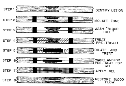
Figure 1 is a schematic of the process for
applying polymeric material to a tissue lumen. In
step 1, a lesion in the lumen is identified and
isolated. In step 2, a catheter, optionally a
. ~. . :=
WO 95/09659 2173316 PCT1US94/11304
26
balloon catheter consisting of a tubular shaft
which includes one or more balloons, is inserted
into the lumen. In the preferred embodiment for
the treatment of blood vessels, the distal
occlusion balloon is used to occlude the distal end
of a treatment site. In embodiments where the
lumen can be rinsed clean, for example at an end
artery or in the gastrointestinal tract or lungs,
it is not necessary to inflate the balloon. In any
case, the treatment site is cleared of blood,
mucous, or other extraneous material, as shown in
step 3. The site may then be treated with drugs,
for example a drug inhibiting responsiveness to
mechanical stimuli or cell proliferation, as shown
in step 4. In step 5, if appropriate, the lesion
itself is treated by expansion of the balloon, in
the case of an arterial plaque, or by other
mechanical, thermal, optical, photochemical,
ultrasonic, or radiation means. As shown in step
6, the site is again treated with drugs and/or
washed or compounds to increase adhesiveness
applied. In step 7, the solution for forming the
polymeric material at the tissue surface is applied
and polymerized or solidified. In some embodiments
the catheter includes a "mold core" which is used
to shape the polymeric material so that it covers
only the area to be treated in a thin layer. The
central mold core member may be able to adjust
size, i.e., for a balloon it may be underinflated
to not occupy the maximum space, thereby leaving
room for the polymeric material. The polymeric
material may be shaped as a uniform layer, or
patterned or segmented as desired. In step 8, the
catheter is removed and flow of material through
the polymeric coated lumen restored.
Two other embodiments of delivery catheters
that can be utilized for application of the
2173316
WO 95109659 PCT/US94/11304
27
polymeric material are shown in Figures 2A, 2B and
2C. Figure 2A is a single entity with means for
entering a tissue lumen, isolating a zone, washing,
applying a drug, adhesive and/or a polymeric
material and a core forming member arid/or dilating
member. The catheter 11 is constructed with two
isolation balloons 10, 14, and a central dilating
or molding balloon 12, as well as a plurality of
lumens and an attached reservoir 16 for delivering
washing fluid, drug, adhesive and/or polymer. A
detailed cross-section enlargement of the tip of
the application device is shown in Figure 2A. Two
isolation balloons 18, 22 are constructed out of
elastomeric material, i.e., latex, krayton or C-
flex or thermoplastic polymers such as
polyethylene, polyolefin co-polymer, polyethylene
terepthalate, or nylon. The balloons 18, 22 are
attached to a multi-lumen shaft 43 including a
central lumen 42 running the length of the device
to allow flushing or passage over a guide wire (not
shown). A central mold-core balloon 20 is
fabricated out of similar materials to those
forming the isolation balloons 18, 22, or from less
compliant materials so that it opens to a
designated dimensions without a continuous stretch
or expansion via creep of the balloon material. In
addition, lumens exist for filling the isolation
balloon 24, 26 and for instilling, filling or
removing fluid from the dilating or mold core
balloons 32, 34. In addition, there are lumens 30,
36 for instilling fluid into the isolation zone.
Lumens 38, 40 are used to instill fluid or remove
fluid from the isolation zone. This device
provides a means to instill, perfuse, or superfuse
a zone.
Figure 2C shows another catheter 45
encompassing two telescoping members 46 within 44.
WO 95/09659 2173316 PCT/US94/11304
28
Zone isolation balloons 50 and 52 and a central
mold core and/or dilating balloon 54, as well as
instillation or aspiration ports 56, provide an
alternative means for applying polymeric material.
The material may also be applied to the
surface to be coated by spraying, extruding or
otherwise internally delivering the material in a
fluent form via a delivery device having single or
multiple lumens.
Application of the coating material may be
accomplished by extruding a solution, dispersion,
or suspension of monomers, polymers, macromers, or
combinations thereof through a catheter to coat or
fill a tissue or cellular surface, a tissue lumen
or a hollow space. The formation of the coating
can be controlled by introducing crosslinking
agents, gelling agents or crosslinking catalysts
together with the fluent material and then altering
the conditions such that crosslinking and/or
gelling occurs. Thus, when a balloon catheter is
used, a flow of heated or chilled fluid into the
balloon can alter the local temperature to a level
at which gelling or cross-linking of introduced
material is induced, thereby rendering the material
non-fluent. Localized heating or cooling can be
enhanced by providing a flow of heated or chilled
liquid directly onto the treatment site. Thermal
control can also be provided, however, using a
fluid flow through or into the balloon, or using a
partially perforated balloon such that temperature
control fluid passes through the balloon into the
lumen. Thermal control can also be provided using
electrical resistance heating via a wire running
along the length of the catheter body in contact
with resistive heating elements. This type of
heating element can make use of DC or radio
frequency (RF) current or external RF or microwave
217 3 316
WO 95/09659 PCT/US94/11304
29
radiation. Other methods of achieving temperature
control can also be used, including light-induced
heating using an internal optical fiber (naked or
= lensed). Similar devices can be used for
application of light, ultrasound, or irradiation.
Catheter bodies are made of standard
materials, including metals such as surgical steel
and thermoplastic polymers. Occluding balloons may
be made from compliant materials such as latex or
silicone, or non-compliant materials such as
polyethylene terephthalate (PET). The expansible
member is preferably made from non-compliant
materials such as PET, (PVC), polyethylene or
nylon. If used, the balloon catheter portion of a
dilatation may optionally be coated with materials
such as silicones, polytetrafluoroethylene (PTFE),
hydrophilic materials like hydrated hydrogels and
other lubricous materials to aid in separation of
the polymer coating.
Medical Indications for Treatment
1. Treatment of Lumen Surfaces
In addition to treatment of arteries, the
method described herein can be utilized for other
applications such as paving the interior of veins,
ureters, urethras, bronchi, biliary and pancreatic
duct systems, the gut, nasolacrimal ducts, sinus
cavities, the eye, and eustachian, spermatic and
fallopian tubes. The process can be used to
provide a paving layer in the context of
transjugular intrahepatic portosystemic shunting
procedure (TIPS), dialysis grafts, arterio-venous
fistulae, and aortic and other arterial aneurysms,
as well as in the treatment of abrupt vessel
reclosure post PCTA, the "patching" of significant
vessel dissection, the sealing of vessel wall
"flaps" either secondary to catheter injury or
2173316
WO 95/09659 PCT/US94/11304
spontaneously occurring, and the sealing of
aneurysmal coronary dilations associated with
various arteritidies.
The ultimate in vivo geometry of the
5 material dictates the final function of the
coating. The thinner applications allow the
polymer film to function as a coating, sealant,
partitioning barrier, bandage, and/or drug depot.
The hollow or cavernous geometry present in
10 many body components has functional significance.
Such geometry facilitates fluid (blood, urine,
lymph, bile) gas, a cellular (ovary, spleen)
containment or transport. These hollow vessels,
organs and organ components are typically composed
15 of several tissue layers. Generically these organs
are composed of an inner cellular layer typically
functioning as a barrier layer, one or several
middle functional layers containing muscularis,
glands or other functional tissue, and an external
20 supportive or stromal covering layer.
Disease may effect the innermost layer of
these hollow organs and thereby violate their
barrier function. Diseases can be either: (1)
systemic with overall diffuse constitutional
25 manifestations, (2) systemic with localized
specific intra-organ focal involvement, or (3)
localized only with definitive.regional intra-organ
involvement. Examples of such diseases include
spontaneous plaque rupture, unstable angina, non-
30 cardiogenic pulmonary edema, sepsis, and
erosive/infiltrative tumors.
2. Manipulation of Cell-Cell Interactions
The methods described herein restore the
barrier function, and/or provided controlled drug
delivery, thereby providing a method for treatment
for these disorders. The polymeric material can
also served as a trophic layer, an adhesive layer,
2173316
WO 95/09659 PCT/US94/11304
31
as a coating of other therapeutic intraluminal
devices, as an absorbing layer, as a sequestrant,
or chelator.
As described above, in a particularly
preferred embodiment, the polymeric material is
used to apply an effective amount of bioactive
molecules such as chemotactic molecules,
haptotactic molecules or molecules providing
contact guidance, to a site where the bioactive
molecules would otherwise not reach in an effective
dosage. In the case of cell to cell interactions,
the polymeric materials provide a substrate that is
analogous to the cell surfaces on which these
molecules are normally found and therefore appear
to be significantly more effective than
administered in the same dosage in the absence of
the polymeric material.
Materials such as attachment peptides,
selectin receptors and carbohydrate molecules such
as Sialyl Le", can be used which serve to attract
and bind specific cell types, such as white cells
and platelets. Materials such as fibronectin,
vitronectin, and collagen, can be used to non-
specifically bind cell types, to facilitate cell
migration and thereby to enhance healing. Growth
factors and modulators of cell growth,
proliferation and migration are particularly
useful. For example, one may incorporate into the
polymeric material a chemoattractant factor to
cells such as PDGF or matrix proteins, i.e.,
fibronectin, laminin, fibrin, or type IV collagen,
which will then facilitate cell ingrowth for wound
repair or a gap or rent resulting from disease.
Extracellular Matrix Components
During the past two decades, the base
knowledge of cell adhesion and migration in
extracellular matrices (ECMs) at the molecular
WO 95/09659 217.J .I t 6 PCT/1JS94/11304
32
level has expanded rapidly. Early efforts in this
area of research concentrated on the adhesion-
promoting ECM protein fibronectin (FN). Studies
which employed limited proteolysis of FN revealed a
120 KD polypeptide fragment of FN which supported
cell adhesion in a way similar to the whole
molecule. This fragment existed as a domain
embedded in the FN molecule and was designated the
cell-binding domain. Further sequence analyses and
peptide mapping of the FN cell-binding domain
yielded a minimal sequence which maintained cell-
binding activity in the tetrapeptide Arg-Gly-Asp-
Ser (RGDS).
The biological interaction of the RGDS
sequence with cell-surface fibronectin receptors
was revealed by demonstrating that synthetic RGDS-
containing peptides in solution could competitively
inhibit fibroblast cell spreading on fibronectin-
coated substrates. Soluble RGDS also inhibited the
direct binding of radiolabeled fibronectin to
fibroblastic cells in suspension. These
competition studies indicated that the RGD sequence
is critical for the cell adhesive function of the
parent molecule.
After the RGD cell adhesion recognition
site in fibronectin was identified, the sequences
of other cell adhesion proteins were examined for
related signals. Other proteins known to carry
functional RGD sequences include the platelet
adhesion proteins fibrinogen, vitronectin and von
Willebrand factor, osteopontin, and laminin. These
findings imply that RGD is a ubiquitous cell
adhesion signal.
Specific RGD peptides are described in U.S.
Patent Nos. 4,517,686 to Ruoslahti, et al.,
4,589,881 to Pierschbacher, et al., 5,169,930 to
Ruoslahti, et al., 5,149,780 to Plow, et al.,
WO 95/09659 2173316 PCT/US94/11304
33
4,578,079 to Ruoslahti, et al., 5,041,380 to
Ruoslahti, et al., and Pierschbacher and Ruoslahti,
J. Biol. Chem. 262(36), 17294-17298 (1987), Mohri,
= et al., Amer. J. Hem. 37:14-19 (1991), Aumailley,
et al., FEBS 291(1), 50-54 (1991), Gurrath, et al.,
Eur. J. Biochem. 210, 911-921 (1992), and
Scarborough, et al., J. Biol. Chem. 268(2), 1066-
1073 (1993), the teachings of which are
incorporated herein.
Laminin is a large adhesive glycoprotein
found in basement membranes which promotes cell
adhesion, migration, differentiation, and growth
(Kleinman, et al., J. Cell Biochem. 27:317-325
(1985); Kleinman, et al., Biochem. 25:312-318
(1986); Beck, et al., FASEB J. 4:148-160 (1990).
LN is composed of three chains designated A(Mr =
400 kD) , Bi (Mr = 210 kD) , and B2 (Mr = 200 kD) .
All three chains of the murine protein have been
cloned and sequenced (Sasaki & Yamada, J. Biol.
Chem. 262:17111-17117 (1987); Sasaki, et al., Proc.
Natl. Acad. Sci. USA 84:935-939 (1987); Sasaki, et
al., J. Biol. Chem. 263:16536-16544 (1988), and
several adhesion-promoting sites were identified on
the molecule. Several synthetic peptides based on
sequences have been described as having biological
activities similar to those of the whole laminin
molecule. A nonapeptide CDPYIGSR as well as the
pentapeptide YIGSR, from the B1 chain were shown to
promote cell attachment and migration (Graf, et
al., Cell 48:989-996 (1987), Biochem. 26:6896-6900
(1987)). Further studies have shown that YIGSR-
containing peptides can inhibit angiogenesis and
tumor metastasis (Grant, et al., Cell 58:933-943
(1989), Iwamoto, et al., Science 238:1132-1134
(1987), Sakamoto, et al., Cancer Res. 51:903-906
(1991). Other peptides include PDSGR and IKVAV.
WO 95/09659 2173316
PCTlUS94/11304
34
The YIGSR peptide class of adhesion ligands
is a good example of a class of compounds which can
be utilized for the treatment of diseases where
cell proliferation and migration in the affected
tissues occurs. While YIGSR peptides have been
shown to selectively inhibit specific cell-ECM
interactions, they must reach their preselected and
specific target tissues in order to be
therapeutically effective. Systematic
administration of YIGSR would typically be an
unsatisfactory therapeutic strategy since
significant interference with normal cell-ECM
interactions as well as those of targeted cells
would occur. A more appropriate therapy would be
to deliver YIGSR locally to the targeted site.
Integrin receptors for ECM
Isolation of RGD-directed cell-surface
receptors for various cell adhesion proteins from
many cell types has been performed using affinity
chromatography on SepharoseTM carrying the
appropriate, covalently bound, adhesion protein.
Cell-surface adhesion receptors from cell extracts
were observed to specifically bind to these columns
and were eluted with RGD-containing peptide
solutions. The use of fibronectin as the affinity
ligand yielded a receptor that was a heterodimer
with a 160 kD a-subunit and a 140 kD 13-subunit.
Similar affinity chromatography experiments have
yielded distinct heterodimeric RGD-directed
receptors specific for vitronectin and a platelet
receptor with affinities for fibrinogen and
fibronectin. It was realized that the
heterodimeric structure was characteristic of RGD-
directed receptors, with a-subunits ranging between
140 and 160 kD and t3-subunits ranging between 90
and 140 kD. These RGD receptors, known as
2173316
WO 95/09659 - PCTIUS94/11304
integrins, form the integrin superfamily of cell-
surface adhesion proteins.
The integrin superfamily is an important
= and well characterized group of cell-surface
5 receptors for both cell-substrate and cell-cell
adhesion. Integrins are characteristically
membrane-spanning heterodimeric protein complexes
consisting of an a-subunit and a B-subunit.
Fourteen distinct a-subunits and 11 B-subunits have
10 currently been isolated and identified, and several
a13 combinations have been observed. Integrin
complexes containing B1 and B3 submits generally are
involved in cell adhesion to the extracellular
matrix, while the B2 integrins are involved in cell-
15 cell adhesion.
Integrins typically bind to cell adhesion
proteins via the rather highly conserved sequence
Arg-Gly-Asp X (RGDX), where X is variant depending
on the particular cell adhesion protein. It was
20 observed that by varying this flanking residue, the
affinity of the RGDX ligand for particular
integrins was modified, but selectivity for
specific integrins was not achieved. Further
studies indicated that cyclization of RGDX-
25 containing peptides created a ligand which was
highly selective for integrin avB3, the vitronectin
receptor. Other studies confirmed that RGD
sequences that are conformationally constrained
within cyclic peptides bound with higher affinity
30 and selectivity for integrin avB3 than linear RGD
sequences. Extracellular administration of cyclic
RGD peptides has been shown to inhibit cell
adhesion and migration on vitronectin-coated
substrates in vitro.
35 A recent in vitro study examined the role
of B1 and vB3 integrin receptors in promoting SMC
adhesion and migration on substrates coated with
1 1. ~ .
WO 95/09659 2173316 PCT/US94/11304 ~
36
fibronectin (FN), laminin (LN), vitronectin (VN),
type I collagen (I), and type IV collagen (IV).
Using functionally blocking antibodies directed
against specific integrin complexes, Clyman et al.,
Exp. Cell Res. 200:272-284 (1992), found that SMC
adhesion on the FN-, LN-, VN-, I-, or IV-coated
substrates depended exclusively on functioning B1
integrins and that SMC migration on these
substrates depended to a large extent on the avB3
integrin.
Ligand affinity chromatography and
immunoprecipitation analyses identified a unique
series of B1 integrins binding to each matrix
component: FN a581 a3B1 av81) , LN (a181, a781) ,
VN(av81), I(a181, a2f31) , and IV (a181) . The 83
integrin, avB3, was observed to bind to all of the
adhesion proteins tested (FN, LN, VN, I, and IV).
These studies suggested that induction of SMC
migration required a switch from an immobile state,
consisting of stable B1 integrin interactions with
the ECM, to a mobile state, where cells form
transient interactions with the ECM via integrin
av83, and that cyclic RGD should be a potent
inhibitor of SMC migration since it could
specifically block integrin avB3 interactions with
the ECM. This has now been demonstrated, as shown
by the following examples.
Tenascin
Tenascin is an unusually large hexameric
ECM protein of molecular weight greater than 1000
kDa, when compared to other ECM proteins such as
fibronectin which is dimeric and 400 kDa. Electron
microscopy of tenascin molecules reveals a
characteristic six-armed structure with a central
globular domain, as reported by Chiquet-Ehrismann,
FASEB J. 4:2598-2604 (1990). The distribution of
tenascin in tissues is much more restricted than
.~~. 2173316
= WO 95/09659 PCT/US94/11304
37
that of laminin and fibronectin. Recent evidence
has revealed that tenascin is transiently expressed
in many developing organs during organogenesis, in
the stroma of specific tumors, and in adult tissues
during wound healing, as reported by Mackie, et
al., J. Cell Biol. 107:2757-2767 (1988), Chuong and
Chen, Am. J. Path. 138:427-440 (1991). These
findings have led to investigations of the
functional role of tenascin during these processes.
The earliest studies of tenascin function
primarily focused on functional domains of tenascin
and their effects on cell-matrix adhesion or
interactions of tenascin with ECM components, as
described by Chiquet-Ehrismann (1990; 1991). A
major cell binding functional domain of tenascin
was mapped with a monoclonal antibody, mAb Tn68, by
Chiquet-Ehrisman, et al., Cell 53:383-390 (1988).
The Tn68 epitope peptide has been demonstrated by
Prieto, et al., J. Cell Biol. 119:663-678 (1992),
to promote fibroblast adhesion when it is adsorbed
to culture substrates. In contrast to the
adhesion-promoting activity of the Tn68 epitope
peptide, the whole tenascin molecule inhibits
fibronectin- or tenascin-mediated cell adhesion in
vitro. Further studies by Prieto, et al., have
shown that the monoclonal antibody directed against
the Tn68 epitope, mAb Tn68, blocks the inhibition
of tenascin-mediated cell adhesion on fibronectin
substrates. These studies indicate that the TN68
epitope is anti-adhesive in its native state within
the whole tenascin molecule but can promote cell
adhesion as a peptide fragment.
More recent functional mapping studies of
tenascin revealed four independent cell binding
domains along the arm of a tenascin molecule, as
diagrammed below. The fibrinogen-like domain at
the C-terminal knob of the arm and a domain
WO 95/09659 2173 316 pCT/US94/11304 =
38
containing fibronectin type III repeats II-VI
promote cell adhesion, as described by Prieto, et
al. The EGF-like repeats and the last two
fibronectin type III repeats were observed to
inhibit cell adhesion. Other studies reported by
Aukhil, et al., J. Biol. Chem. 268:2542-2553
(1993), revealed that a domain containing
fibronectin type III repeats IV-V and the
fibrinogen domain had heparin-binding as well as
cell-binding activities. These studies provide a
basis for the multifunctional role of tenascin in
the ECM and guidelines for isolating receptors that
mediate various cellular responses to tenascin.
Fibrino f n domoii
EOr domains i il iiiiyy vi vll YIII
1 2 4 3 i 7 i ! 0 1 P. 3 OOH
III do~nalns
Flbroneotln Type
Disulflds linkaps to oentral cors
=Adapted from Aukhil et al. 1993 (Hatched regions depict alternatively splieed
domains)
Other studies have investigated the
interaction of tenascin with ECM components. There
is increasing but inconsistent evidence that
tenascin binds to proteoglycans, collagens, and
fibronectin. In studies which report binding of
tenascin to other ECM components, the interaction
of tenascin with these components is generally
weak, as reviewed by Faissner, et al., J.
Neurochem. 43:1004-1015 (1990); Lightner and
Erickson, J. Cell Sci. 95:263-277 (1990). The
physiological relevance of these studies is that
tenascin may be easily removed from the ECM and
become associated with the cell surface when it
binds to cell surface receptors.
Detailed in vivo studies by Bourdon and
Ruoslahti, J. Cell Biol. 108:1149-1155 (1989), on
the mechanism of tenascin-mediated attachment of
the human glioma cell line U251MG revealed that
integrin-type adhesion receptors were involved.
2173316
WO 95/09659 PCT/US94/11304
39
Ligand affinity chromatography of cell membrane
extracts and subsequent gel electrophoresis
revealed a heterodimeric cell surface protein which
bound to a tenascin affinity column matrix. This
protein complex was specifically eluted by peptides
containing the RGD sequence which is recognized by
integrins. Western blot analysis identified one
subunit in the dimeric complex as integrin fl1. More
recent studies by Sriramaro, et al., J. Cell Sci.
105:1001-1012 (1993) and Joshi, et al., J. Cell
Sci. 106:389-400 (1993), have shown that
endothelial cell adhesion and spreading is mediated
by integrins ay(31 and aõ,63 respectively. The
tenascin binding site for integrin aõ/33 was the
sequence S GDMS within the third fibronectin type
III domain. The interaction of integrin a2a1 was
not RGD-dependent and no binding sequence was
determined for this receptor.
Cell attachment to the fibrinogen domain of
a tenascin arm was observed by Aukhil, et al. not
to be integrin-dependent since cell adhesion via
this domain was not divalent cation-dependent or
inhibited by soluble RGD peptides. Inhibition of
cell attachment to the fibrinogen domain was
observed when cells were treated with soluble
heparin or heparitinase. These results suggested
that cell attachment to the fibrinogen domains of
tenascin were mediated by cell surface
proteoglycans.
Studies by Mackie, et al., Am. J. Path.
141:377-388 (1992), demonstrated that tenascin
expression in large and small arteries from
normotensive Wistar-Kyoto (WKY) rats was at low
levels throughout the tunica media and at higher
levels only at branching sites. In contrast to the
expression patterns in normotensive rats, high
levels of tenascin was observed to be dispersed
2173316
WO 95/09659 PC'd'/US94/11304
focally throughout the tunica media of arteries
from spontaneously hypertensive WKY rats. Further
in vitro studies by Mackie and Scott-Burden, Am. J.
Path. 142:659 (1993), with WKY rat aorta SMC
5 cultures revealed that increased expression of
tenascin mRNA and protein was inducible by
angiotensin II, transforming growth factor-(31, and
platelet-derived growth factor. Another in vitro
study by Sharifi, et al., J. Biol. Chem. 25:23910-
10 23915 (1992), confirmed the stimulatory effect of
angiotensin II on SMC tenascin mRNA expression.
These studies suggest that increased focal
expression of tenascin by vascular SMCs is
associated with chronic hypertension and may
15 mediate angiotensin II-induced changes in vascular
structure associated with chronic hypertension.
Hedin, et al., Am. J. Path. 139:649-656
(1991), observed that proliferating, synthetic
phenotype SMCs in the neointima of balloon-injured
20 rat carotid arteries secreted detectable levels of
tenascin. In contrast, they found no detectable
levels of tenascin in the media of normal and
balloon-injured rat carotid arteries. Further
studies by this group demonstrated that SMCs in
25 culture would deposit tenascin in the matrix as
they transformed from the contractile phenotype of
freshly isolated cells to a synthetic state. It
was concluded from this work that tenascin
production in vivo and in vitro was induced
30 concomitantly with the transition of SMC phenotype
from the contractile to the synthetic state. These
studies correlated increased expression of tenascin
in the vessel wall with chronic hypertension or in
response to vascular injury. To date, however, no
35 functional role for tenascin has been described in
the prior art for vessel wall disease or injury.
WO 95/09659 21 7 3 3 1 U PCT/US94/11304
41
As demonstrated in the following examples,
tenascin has now been demonstrated to stimulate
injury-induced SMC migration in vitro. The initial
results show that integrin a,93 is important for
tenascin-stimulated SMC migration. The subsequent
results demonstrate that integrin aõ/33, the
predominant integrin mediator of SMC migration, is
a SMC surface component which actively binds
tenascin.
It is therefore possible to inhibit SMC
migration by inhibition of the interaction between
tenascin and integrins on SMCs, especially aõ83.
TopicaZ Delivery of Adhesion Ligands
The cyclic RGD peptide class of adhesion
ligands is a good example of a class of compounds
which could be utilized for the treatment of
diseases where cell proliferation and migration in
the affected tissues occurs. While cyclic RGD
peptides have been shown to selectively inhibit
specific cell-ECM interactions, they must reach
their preselected and specific target tissues in
order to be therapeutically effective. Systematic
administration of cyclic RGD would typically be an
unsatisfactory therapeutic strategy since
significant interference with normal cell-ECM
interactions as well as those of targeted cells
would occur. The quantity of peptide which would
be required for efficacy would also be enormous. A
more appropriate therapy is to deliver cyclic RGD
locally to the targeted site, using the polymeric
gel described above.
Chemotactic and Growth Factors
In a preferred example for endothelial
cells, heparin, macrophage chemotactic factor
(Banda, et al., Proc. Natl. Acad. Sci. USA 78:7773-
7777 (1982)), basic FGF or tumor angiogenesis
factor can be used to facilitate repair post
2173316
WO 95/09659 PCTIUS94/11304 ~
42
angioplasty, atherectomy, stenting or vascular
surgery. In a preferred example for treatment of
bladder cancer following administration of
chemotherapeutic agents such as BCG, EGF is applied
in a gel to coat the bladder. EGF can be similarly
applied in a polymeric gel following crysotherapy
of the cervix to facilitate re-epithelization.
To aid in organ repair, a paste or layer of
gel incorporating growth factors can be applied
adjacent to injured organs to enhance organ
regrowth after disease or surgery. Embryonic
cardiomyocytes plus growth factor can be seeded in
a polymeric gel in artificial lumens decreased in
diseased, for example, myocardium following heart
failure or infarction, for cell repopulation and
creation of "mini-organs" of contractile function.
Thinning with eventual rupture of the septum and
the creation of a VSD leads to communication
between the ventricles and the acute onset of heart
failure with significant associated mortality.
Currently, DacronTM patches are inserted to
stabilize blood flow and pressure but they leave a
large zone in the septum which is non-functional.
Using the polymeric material applied to the
composite in combination with bioactive molecules
can facilitate repair and regrowth.
Incorporation of Cells to produce Factors
Chief cells of the parathyroid can be
incorporated into a polymeric gel and locally
implanted to form islands of local parathyroid
hormone production following parathyroid
destruction or removal associated with thyroid
removal, which is a particularly significant
problem following resulting in altered calcium and
phosphate metabolism.
In addition to functional alteration and
disease processes in tubular organs many non-
WO 95/09659 2173316 PCT/US94/11304
43
tubular organs and tissue surfaces may also undergo
a change in either function or structure due to
aging, disease or injury. As an example, if a
tumor is found on a tissue surface in an internal
organ, current therapy involves local surgical
excision to create a disease free margin. To
prevent further disease progression into the normal
zone either external, often toxic, chemotherapy is
administered or the patient is subjected to
radiation therapy. These therapies result in many
side effects and are frequently of limited
effectiveness. Using the method described herein,
one can locally apply a coating of polymeric
material to a tissue surface which alone, or by
incorporated anti-proliferative chemotherapeutic
agents or bioactive substances limit the ingrowth
of tumor cells. One can also utilize bioactive
molecules which selectively favor the ingrowth of
normal parenchymal cells or the overgrowth of
epithelial cells.
The polymeric material is particularly
useful as an enhancement to healing fpllowing
normal surgical procedures where the wound is
closed using sutures or staples.
The present invention will be further
understood by reference to the following non-
limiting examples.
Example 1: In vitro application of polymer to a
mock hollow organ.
A catheter was inserted into a mock blood
vessel constructed from a clear plastic TygonTM
tube. The distal occlusion balloon was expanded to
define a treatment site, and PluronicTM F127 in its
fluent form was injected into the vessel through
the catheter. A mold core balloon was inflated,
and the PluronicT"s gel material was allowed to warm
and gel. Finally the balloons were deflated and
2173316 PCT/US94/11304
WO 95/09659
44
the catheter was withdrawn, leaving a gel coating
on the interior surface of the "vessel" lumen.
As shown in detail in Figures 3A-3G, Figure
3 reveals an actual example of use of the balloon
application catheter as outlined in Figure 2C above
for the application of a flowable gel polymer in a
thick hollow tubular structure. A telescoping gel
paving catheter is shown in Figure 3A. This
catheter consists of a proximal hollow shaft 64
with an end annular occlusing balloon 58 (i.e., the
proximal occlusion balloon). Telescoped within the
proximal shaft is a second smaller shaft 62 with an
attached distal occluding balloon 60 and a mold
core or gel paving balloon 66. In Figure 3B the
catheter assembly is placed within the lumen 68 of
a mock hollow tubular organ, in this case clear
TygonTM tubing.
In Figure 3C the proximal and distal
occluding balloons are shown inflated, isolating a
zone 70 of the hollow tubular organ.
In Figure 3D, a flowable polymeric solution
72 has been instilled within the isolation zone.
In this example PluronicTm F127 (25% v/v) at 4 C was
utilized with the solution colored with dissolved
Toluidine Blue for visibility. In Figure 3E the
polymer has warmed and gelled, thereby gelling the
instilled fluid. Note that the proximal and distal
occlusion balloons have been deflated yet the
colored polymer 74 remains contained within the
zone, demonstrating its gelled nature.
In Figure 3F the application catheter has
been removed leaving a gel coating 76 in the zone
with a central hollow lumen 78.
In Figure 3G a thinner coating of the
polymer 80 is seen which has been achieved via a
second retrograde passing of the distal occlusion
balloon 60 through the coated zone further
WO 95/09659 2173316 PCTNS94/11304
smoothing and forming the gel to yield a thin
coating.
Example 2: In vitro application of polymer to an
isolated blood vessel segment.
5 A segment of a blood vessel was excised.
The catheter was inserted into the interior of the
vessel. A chilled Pluronic gel F127 in its fluent
form was injected through the catheter into the
space between the catheter and the vessel wall, and
10 the mold core balloon was expanded. Once the
polymer had warmed to a temperature sufficient to
cause gelling, the mold core balloon was deflated
and the catheter removed.
Figures 4A-4F are micrographs showing
15 application of a gel coating to isolated bovine
coronary arteries using a "mold-core" central
catheter. In Figure 4a a segment of an isolated
bovine coronary artery 82 is seen in cross-section.
In Figure 4B a mold core catheter 84 has been
20 placed centrally within the lumen. In Figure 4C a
flowable colored polymeric solution 86 (PluronicTM
F127 25% (w/v) plus Toluidine Blue) has been
instilled by injection into the lumen occupying the
space defined by the mold core balloon and the
25 endoluminal surface of the vessel. In Figures 4D
and 4E, upon gelation of the polymer and removal of
the catheter, a thin annular coating of polymer gel
88, 90 is seen in intimate contact on the vessel
endoluminal surface. In Figure 4F the gel coated
30 or paved artery is seen under magnification (6x)
and a thin endoluminal gel layer 92 is identified
which is adherent and conforming to the underlying
arterial wall 94.
The resulting tissue surface is paved with
35 a pluronic gel in a manner which coats the surface
and fills and conforms irregularities on the
surface. Further, the deployed interior surface of
the gel is smooth, thereby providing a barrier
WO 95/09659 2173 3 16 PCT/US94/11304
46
layer having a rheologically advantageous surface
with improved blood flow.
Figure 5 is a micrograph of a frozen cross-
section (250x) of a gel coated bovine coronary
artery. A thin layer of gel 96, formed as
described above, is seen in intimate conformal
contact with the underlying endoluminal (intimal)
surface 98 of the vessel.
Example 3: Thermoreversible polyether hydrogels
reduce the thrombogenicity of injured
arterial intimal surfaces in vitro and
ex vivo.
Polymeric Endoluminal Paving is a generic
method of applying thin layers of biodegradable
polymers to the endoluminal surface of hollow body
structures. The applied polymer layers may
function as temporary wall supports, barriers or
localized sustained drug delivery vehicles.
Studies to date utilizing structural polyesters in
the vasculature have demonstrated that endoluminal
paving layers may be effectively applied in situ
via localized catheter-based thermoforming, being
structurally stable, wall-supportive and
hemocompatible.
As an extension of the paving method recent
studies have examined the feasibility of applying a
layer of non-structural polymeric hydrogels to
arterial endoluminal surfaces to act as a short
term barrier, locally reducing injured arterial
surface thrombogenicity. Studies using
biodegradable and erodible polyethers (PE) were
conducted to determine the blood compatibility of
PE gels in vitro, the ability of PE gels to reduce
the thrombogenicity of acutely injured arterial
surfaces in vitro, and the ability of endoluminal
PE gels to thromboprotect injured arterial surfaces
ex vivo.
Materials and Methods:
2 173316
PCTlCTS94/11304
WO 95/09659
,.. , l.. . .
47
Spin-case gel films of PE on glass, and
strips of intima-abraded freshly explanted rat
aorta (ex vivo) (Ao) on which PE gels were formed,
were placed in a parallel plate flow chamber and
exposed to overflowing heparinized (2'U/ml)
mepacrine-labeled (10 mM) fresh human blood (100
sec-1 shear rate, 37 C). Surfaces were examined
after 5 min. using epi-fluorescence videomicroscopy
and the number of adherent platelets (pl),
aggregates of 3-10 pl (pa) and thrombi (t) per
1000x field were measured. Films of Gelatin on
glass and non-coated abraded rat aorta served as
controls.
Results:
Surface n pl/field pa/field t/field
Gelatin (reference) 3 132.8 t 38.8 12.8 t 3.1 6.8 2.9
PE gel on glass 3 1.40 t 0.4', 0t 0'
Abraded Ao (control) 4 204.8 t 44 23.8 t 6.3 6.5 t 1.0
Abraded Ao + PE gel 6 1.6 f 0.5 0~ 0
* (p<0.05)
Similar results were obtained with other
polymeric materials including neutralized
poly(acrylic acid), pH 7.4 (CarbopolTM 934 and 940,
B.F. Goodrich).
Example 4: Thermoreversible polyether hydrogels
reduce the thrombogenicity of injured
arterial intimal surfaces and
subsequently limit the eventual
development of neointimal hyperplasia
in vivo.
Acute thrombosis of injured arterial
intimal surfaces is a potentially serious
complication following angioplasty, thrombolytic
therapy and stent placement. A study was conducted
to determine whether formation of thermoreversible
WO 95/09659 2173316 PCTIUS94/11304
48
polyether hydrogels directly on injured arterial
subintimal surfaces would limit subsequent platelet
deposition and thrombus formation in vivo.
Materials and Methods
Bilateral carotids in five rats were
exposed, segments of paired arteries isolated via
atraumatic clips, washed free of blood with normal
saline and balloon abraded (2fr Fogarty x 3). In
one carotid per animal, chosen to be the control
vessel, blood flow was then restored. In the
corresponding experimental carotid a layer of
polyether hydrogel was then applied to the
endoluminal surface following which blood flow was
restored. Note: In the experimental vessel care
was taken to prevent injured surface re-exposure to
blood until after the polymer was applied.
Following one hour of blood re-flow, animals were
heparinized (200 U/kg), sacrificed via anesthesia
overdose, bilateral carotids washed with saline and
pressure fixed (90 mm Hg, 2% glutaraldehyde) in
situ. Vessels were then excised, examined grossly,
and via stereomicroscopy and scanning electron
microscopy.
Results:
Group n Platelets/hpf (3000x)
Abraded (Control) 5 90 8
Abraded + PE gel 5 *5 t 9
* (p <0.05)
The results shown in Figures 6A (control)
and 6B, demonstrate that there is almost no
deposition of platelets, white cells and fibrin on
2173316
WO 95/09659 PCT/US94/11304
49
the treated vessel, in contrast to the untreated
control.
The analysis showed that 7 1 thrombi were
detected on control aortas. No thrombi were seen
on gel coated injured arterial surfaces. 205 44
single platelets were detected on the control
surfaces versus 2 1 platelets on the coated
artery. (p<0.05)
Figures 7A and 7B are representative
examples of histologic cross-sections of rat
carotid arteries harvested 14 days post balloon
abrasion injury. The artery 102 in Figure 7B is an
example of a control artery that had been balloon
abraded and allowed to heal for fourteen days,
without receipt of a gel coating following injury.
There is significant neointimal thickening 110 with
an almost doubling in thickness compared with the
underlying media 106.
In comparison, the gel treated artery 100
in Figure 7A has a significant reduction in
neointima 108. This artery was coated with
PluronicTM F127 (25% w/v) gel and then re-exposed to
overflowing blood and allowed to heal for fourteen
days.
Conclusions:
Polyether gels are hemocompatible and
provide a surface which is minimally platelet
activating. Thermoreversible polyether hydrogel
layers formed directly on injured arterial
surfaces, either in vitro or in vivo, create an
effective physical barrier layer limiting platelet
deposition and thrombus formation with an overall
reduction in intimal surface thrombogenicity.
Example 5: Delivery of adhesion receptor ligands
or other adhesion receptor modulators
to a selected local site in vivo as a
treatment of disease.
=S , _ .'.
~ = _
WO 95/09659 211 3316 PCTIUS94/11304
A study demonstrating local delivery of a
cyclic RGD peptide inhibits neointimal hyperplasia
following balloon injury was conducted as follows
to assess whether one could provide a method of
5 local delivery of cyclic RGD to an injury site in a
vessel wall in vivo, i.e. a site where PTCA was
performed, so that localized inhibition of intimal
SMC migration would occur which could effectively
reduce intimal hyperplasia. Specifically, a study
10 was conducted to determined whether interference=
with integrin-matrix interactions in the arterial
wall, through localized delivery of a cyclic
integrin antagonist peptide, would alter the degree
of neointimal hyperplasia development at 14 days in
15 a rat balloon injury model.
The left carotid artery in 10 rats (male,
350 g) was balloon abraded (2Fr fogarty x3). In
five of the ten rats the integrin antagonist cyclic
peptide GPenGRGDSPCA (cRGD) was mixed to a
20 concentration of 1 mM Pluronic gel (500 l) and
locally applied to the adventitia of the injured
artery. The five untreated rats served as
controls.
At 14 days the rats were sacrificed,
25 carotid arteries pressure fixed and mean intima and
media thickness determined. The mean intima/media
ratio (I/M) of control balloon abraded arteries was
2.09 0.54. The mean intima/media ratio was 0.17
0.10 in the abraded cRGD treated arteries
30 (p<0.001).
These results demonstrate that local
application of cRGD peptide leads to a 92%
reduction in the degree of hyperplasia. There was
no significant change in media thickness between
35 the groups. The localized application of an
integrin antagonist to the arterial wall following
balloon injury modifies the normal healing response
2173316
WO 95/09659 PCT/US94/11304
51
resulting in a significant reduction in neointimal
hyperplasia development.
Example 6: Comparison of the efficacy of locally
delivered linear RGD peptide compared
with cyclic RGD peptide in limiting
post-injury neointimal hyperplasia.
Cell-matrix interactions, mediated via cell
surface integrins and extracellular matrix protein
ligands, have been shown to regulate cell phenotype
and function. As described in the foregoing
examples, interference with integrin-matrix
interactions in the arterial wall, through
localized delivery of a cyclic integrin antagonist
peptide, GPenGRGDSPCA (cRGD), resulted in a 92%
inhibition in the development of neointimal
hyperplasia at 14 days in a rat balloon injury
model. It remains unclear whether SMC integrin avB3
interaction with the ECM is the predominant
mechanism for post-injury SMC migration and
development of neointimal hyperplasia or if B1
integrins are also important for the post-injury
SMC response.
This study addresses the issue by comparing
the efficacy of locally delivered linear peptide
GRGDSPCA which inhibits B1 integrins more strongly
than B3 integrin, versus cRGD, a cyclic peptide
(GPenGRGDSPCA) which targets B3 integrins, in
limiting 14 d post-injury neointimal hyperplasia.
Materials and methods.
In 14 rats (male, 350 g) the left carotid
artery was balloon abraded (2fr Fogarty x3).
Linear (4/14 rats) or cyclic RGD (5/14) was locally
applied at 1 mM to the injured artery in an
adventitial PluronicTM gel (500 l). Untreated rats
(5/14) served as controls. At 14 days rats were
sacrificed, carotid arteries pressure fixed and
mean intima and media thickness determined.
Results.
Tõ ~ t=
2173316
WO 95/09659 PCT/US94/11304
52
The mean intima/media ratios (I/Ms) in
balloon abraded cyclic and linear RGD-treated
arteries were 0.17 0.10 (p<0.001) and 1.95 0.32
(p<0.007) respectively. In control abraded
arteries, the I/M was 2.09 0.,54. Local
application of cRGD peptide lead to a 92% reduction
in the degree of hyperplasia whereas linear RGD-
treatment resulted in no significant reduction.
Results are shown in Figure 8 for three linear
peptides, two known B1-integrin inhibitors, a non-
sense peptide, and the cyclic RGD peptide, which
were studied under similar conditions.
Discussion. B3 integrin inhibitors, such as
cRGD, can effectively reduce neointimal hyperplasia
development, whereas B1 integrin inhibitors, i.e.,
linear RGD, fail to limit hyperplasia. Therefore,
the interaction of SMC B3 integrins with vessel wall
ECM must be more important than Bi integrin
interactions for post-injury migration and
subsequent development of neointimal hyperplasia.
Cell integrin-matrix interactions may be an
additional viable target for pharmacologic
manipulation aimed at limiting injury-induced
restenosis.
Example 7: Local Delivery of a Non-Integrin cell
matrix receptor binding peptide
inhibits Neointimal Hyperplasia
following balloon injury.
The interaction of cells with the
extracellular matrix protein laminin, mediated
partially through a cell associated 69 kD non-
integrin receptor, has been shown to regulate cell
phenotype and function. Interference with this
interaction via laminin peptide fragments has been
shown to limit migration of neural crest cells and
experimental metastasis.
Materials and Methods.
2173316
WO 95/09659 PCT/US94/11304
53
In this study it was determined whether
interference with laminin-69 kD receptor
interactions in the arterial wall, through
localized delivery of a laminin !31 chain peptide
fragment, would alter the degree of neointimal
hyperplasia development at 14 days in a rat balloon
injury model. In 10 rats (male, 350 g) the left
carotid artery was balloon abraded (2fr. Fogarty
x3). In 5/10 rats the linear nonapeptide CDPGYIGSR
amid (YIGSR amide) was locally at 1 mM to the
injured artery in an adventitial PluronicTM gel
(5001ambda). Untreated (5/10) served as controls.
At 14 days rats were sacrificed, carotid arteries
pressure fixed and mean intima and media thickness
determined.
Results.
The mean intima/media ratio (I/M) of
control balloon abraded arteries was 2.09 + 0.54.
In abraded YIGSR amide treated arteries the I/M
ratio was 0.22 0.16 (p<0.001). Local application
of the nonapeptide YIGSR amide lead to an 89%
reduction in the degree of hyperplasia. There was
no significant change in media thickness between
the groups.
Discussion
The localized application of a nonapeptide
fragment of the laminin !31 chain, CDPGYIGSR amide,
to the arterial wall following balloon injury,
modifies the normal healing response, resulting in
a significant reduction in neointimal hyperplasia
development. Smooth muscle cell non-integrin
laminin receptor-laminin interactions may be an
additional viable target for pharmacologic
manipulation aimed at limiting restenosis following
vascular injury.
Example 8: Modification of Cell Migration With
Tenascin.
WO 95/09659 2173316 PCT/US94/11304
54
Increased levels of the ECM (extracellular
matrix protein) tenascin have recently been
detected in arterial neointima following balloon
injury. The effect of exogenous tenascin on
injury-induced smooth-muscle cell (SCM) migration
was therefore examined to further understand the
role of tenascin in arterial injury. Confluent
cultures of rat aortic SMCs (passage 2-4) were
wounded by a single scrape with a wooden
applicator, washed with fresh medium (DMEM + 10%
FCS) and incubated in medium plus tenascin (1
microgram/milliliter). Controls were treated
identically but incubated without tenascin. At 24
hours, cell migration was determined by measurement
of mean distance travelled by cells from the
wound's edge. Migration was also assessed in the
presence of blocking antibodies for the integrins
av03 and a501 anti-VNR and anti-FNR, respectively,
to determine the role of these integrins in
promoting tenascin-mediated post-injury migration.
Antibodies were added immediately post-injury at
concentrations known to be maximally inhibitory.
In six samples, the mean percent migration
tenascin-treated samples relative to untreated
controls (100%) was 182.6 20.1%. This is
significant at the p<0.01 level. In the integrin-
blocking study, the percent migration relative to
tenascin-treated controls (100%) was 33.6 7.2%
(p<0.01, n = 5) for anti-av03, and 94.7 4.1 (p =
not significant, n=5) for anti-a5(31. These studies
were corroborated by measurement of the binding of
tenascin, at the same concentration, to controls
and to cells treated with the two antibodies.
Control binding was 53 1 femtomole/106 cells;
anti-VNR reduced the binding to 29.7 3.8, while
anti-FNR had no effect (50.3 2.8).
2173316
WO 95/09659 PCT/US94/11304
These results demonstrate that treatment
with soluble tenascin significantly increases
migration of scrape-wounded smooth muscle cells.
Anti-av/3l-antibody has no effect on tenascin-
5 stimulate migration, but a3(31 significantly reduces
migration, essentially blocking the tenascin
effect. These results suggest a role for tenascin
in the stimulation of post-injury SMC migration,
and further suggest that the migratory response is
10 at least partially mediated by integrin av#3.
Example 9: Hydrogel-Based Local Delivery of a
cRGD peptide for Inhibition of Smooth
Muscle Cell Migration.
A study was conducted which shows that
15 hydrogel-based local delivery of a cRGD peptide
leads to greater inhibition of smooth muscle cell
migration compared with direct short term peptide
exposure. This study, as a model of local in vivo
delivery, compared the effect of hydrogel-based
20 versus direct short term delivery of an anti-
migratory peptide on smooth muscle cell (SMC)
migration following scrape wound injury in vitro.
Materials: Sixteen SMC monolayers (rat
aorta) were scrape-wounded and thin layers of a
25 hydrogel consisting of a poly(ethylene glycol)-
lactic acid block co-polymer containing cyclic RGD
peptide (1 mM) were photopolymerized directly on
four of the sixteen monolayers which were then
covered with media. In four other monolayers 1 mM
30 cRGD in media (MEM) was directly applied, without
any hydrogel. Drug-free hydrogel alone was applied
in four of sixteen monolayers or media alone was
applied in four of sixteen monolayers as controls.
Following 10 minutes of incubation (37 C), the
35 media of all monolayers was discarded, cultures
washed and further incubated in drug-free media for
24 hrs. Cultures were fixed, stained and the area
of SMCs migrating from the wound line for both the
WO 95/09659 217 3 3 l 6 PCT/US94111304
56
gel and direct treatment groups was measured and
reported as a migration index relative to gel alone
controls (Migration Index = Area,,,,,mmt/Area,~ual)
Results: The migration index of cRGD
hydrogel-treated monolayers was 0.27 + 0.09 versus
0.93 + 0.08 for direct 10 minutes cRGD exposure
(p<0.01). Hydrogel-based delivery lead to an
additional 66% reduction in the degree of SMC
migration compared to direct 10 minutes peptide
exposure. Gel alone did not limit migration
compared to media alone controls.
Conclusion: Hydrogel-based delivery of an
SMC anti-migratory peptide lead to enhanced local
efficacy, with greater reduction in SMC migration
compared with direct short term peptide exposure
alone. Delivery of promising anti-restenosis
agents via polymeric hydrogel delivery vehicles
should provide a method for enhancing local drug
efficacy beyond that achievable with direct
catheter-based exposure alone.
Example 10: Retention of cRGD at the Target Tissue
Site, and In Vivo Efficacy.
Radiolabelled cRGD was used to study the
retention of cRGD in gels in arteries. Male
Sprague-Dawley rats (375 g) were abraded in their
carotid arteries by three passages of a 2fr Fogarty
catheter as described above. The right carotids
were clamp isolated and washed to remove blood. A
thin gel layer was deposited in these arteries
essentially according to Hill-West, et al., by
exposure of the artery to 0.02 mg/ml Eosin Y,
followed by washing to remove excess eosin. Gel-
forming macromer, consisting of 10% w/v acrylated
polyethyleneglycol lactate ester in isotonic
buffer, was instilled into the artery. The
macromer solution was administered without additive
(gel control), or containing 5 mM cRGD, labelled
L
WO 95/09659 21r331 1U PCT/US94/11304
57
with 35S to about 5 Ci/mole. The gel was
photopolymerized at 514 nm for 30 sec.
Some arteries were kept clamped for 2 hrs,
and then the amount of radioactivity was compared
to controls. The percent of radioactive RGD
retained at 2 hrs was 100%, which shows that the
gel did not leak out of the region. When 35S cRGD-
containing gels were exposed to normal blood
circulation for 2, 24 and 72 hours, the relative
.10 amount of retained cRGD was about 4%, 4%, and 1%,
respectively. This is a significant retention of a
low-molecular weight material, and may represent
diffusion into the wall of the artery.
Efficacy of the cRGD application was
evaluated by sectioning of arteries and measurement
of media: intima ratio at 14 days. With no gel,
the ratio was 2.5, indicating hyperplasia of the
media. In gel-only controls, the ratio was 0.28.
In cRGD-treated arteries, this was further reduced
to 0.14. Thus, polymer alone is effective in
suppressing hyperplasia of the arterial media and
cRGD in combination with polymer is even more
effective than polymer in suppressing hyperplasia
of the arterial media.
Example 11: Localized therapy for cell-
extracellular matrix interactions.
A study was conducted to examine whether
interference with laminin-69 kD receptor
interactions in the arterial wall, through
localized delivery of a laminin bi chain peptide
fragment, would alter the degree of neointimal
hyperplasia development at 14 days in a rat balloon
injury model.
Methods: The left carotid artery was
balloon abraded (2fr Fogarty x3) in ten rats (male,
350 g). In five out of ten rats the linear
nonapeptide CDPGYIGSR amide (YIGSR amide) was
locally applied at 1 mM to the injured artery in an
..r.~,.:~ 2173316
WO 95/09659 PCT/US94/11304
58
adventitial PluronicTM (polyethylene oxide-
polypropylene glycol) gel (4001). Five untreated
rats (5/10) served as controls. At 14 days rats
were sacrificed, carotid arteries pressure fixed
and mean intima and media thickness determined.
Results: The mean intima/media ratio (I.M)
of control balloon abraded arteries,was 2.09
0.54. In abraded YIGSR amide treated arteries the
I/M was 0.22 0.16 (p<0.001). Local application
of the nonapeptide YIGSR amide lead to a 89%
reduction in the degree on hyperplasia. There was
no significant change in media thickness between
the groups.
Conclusion: The localized application of a
nonapeptide fragment of the laminin bi chain,
CDPGYIGSR amide, to the arterial wall following
balloon injury modifies the normal healing response
resulting in a significant reduction in neointimal
hyperplasia development. Smooth muscle cell non-
integrin laminin receptor - laminin interactions
may be an additional viable target for
pharmacologic manipulation aimed at limiting
restenosis following vascular injury.
Example 12: Tenascin as a Pro-Migratory Agent for
Therapeutic Applications.
Tenascin has now been demonstrated to
stimulate injury-induced SMC migration in vitro.
As an initial step, the effect of soluble
tenascin on injury-induced SMC migration in vitro
was investigated. SMC migration was induced in
vitro by wounding a cell monolayer similar to the
method described by Stewart, et al., Br. J. Exp.
Path. 60:582-588 (1979). Rat aorta SMCs were
seeded at a high density in 24-well culture dishes
coated with type I collagen and were incubated for
24 h. To induce cell migration, the monolayers
were scrape-wounded with a wooden applicator stick.
Progression of cell migration into the wound zone
2173316
WO 95/09659 PCT/US94/11304
59
could readily be determined because the original
wound line was clearly distinguishable from the
leading edge of migrating cells. Immediately
following scrape injury, soluble tenascin (1 g/ml)
was added to each experimental culture well.
Control wells were wounded but did not receive
soluble tenascin. Following wounding, cells were
incubated for 24 h to allow for cell migration to
proceed at a measurable distance from the wound
site. At the 24 hour post-wound time point, cells
were fixed in 4% paraformaldehyde (2 h, room temp.)
and stained with 1% toluidine blue (2 min). Each
culture well was examined at 100x magnification via
brightfield microscopy (Zeiss, Axiovert). Images
generated by a video camera were digitized and
processed with a MacintoshTm IIci host computer
equipped with a frame-grabber board and image
processing software (NIH ImageTM). A closed
perimeter of the zone of migrating cells in the
wound site was traced with a digitizing tablet
(SummagraphicsTM) for each field examined and the
area encompassed by migrating cells was quantitated
using an integration routine in the imaging
software package. Five fields from the wound site
were examined for each sample and triplicate
samples were examined for experimental and control
groups. SMC migration for each experimental group
was expressed as a percentage of control migration.
It was observed that the SMC migration rate
increased by 80% when soluble tenascin (1 g/ml)
was present, as shown in Figure 9A. Further
studies indicated that soluble tenascin levels
above 2.0 g/ml disrupted cell adhesion, which is
indicative of the characteristic anti-adhesive
property of tenascin.
In a second study, the in vitro wound model
was used to determine the role of integrin a03
21733 l 6
WO 95/09659 PCT/US94/11304
(traditionally known as the vitronectin receptor
(VNR)) and integrin a5(31 (traditionally known as the
fibronectin receptor (FNR)) in the promotion of
tenascin-stimulated SMC migration. For this study,
5 cells were seeded in 24-well culture'dishes and
wounded as described above. Experimental samples
were preincubated 30 min. prior to wounding with
functionally blocking rabbit antiserum (GIBCO/BRL)
directed against either integrin aõ03 (VNR) or
10 integrin a5(31 (FNR) at 100 g/ml, a concentration
which has previously been determined by Massia and
Hubbell, J. Cell Bio. 114:1089-1100 (1991), to
maximally block integrin function. As a non-
immunoreactive preimmune sham control, selected
15 samples were preincubated with normal, preimmune
rabbit serum. All samples were incubated in the
presence of soluble tenascin (1 g/ml) immediately
following wounding up to the 24h post-injury
experimental end point. At the 24 h end point,
20 samples were fixed, stained, and analyzed as
described above.
In this second study, it was observed that
antiserum directed against VNR (anti-VNR antiserum;
100 g/ml, maximal inhibitory concentration)
25 reduced the SMC migration rate to 33.3 1.7% of
control migration, which is indicative of the
maximal inhibitory capacity of the antiserum, as
shown in Figure 9B. In contrast, anti-FNR at its
maximal inhibitory concentration reduced the SMC
30 migration rate to only 83.3 3.3% control
migration, a value that much less than the
inhibitory capacity of the antiserum. These
results suggest that integrin a,03 is important for
tenascin-stimulated SMC migration.
35 A whole cell radioligand binding assay was
utilized to determine whether tenascin specifically
binds to the surface of SMCs. Briefly, tenascin
WO 95/09659 2173316 PCT/US94/11304
61
was radiolabeled with t-butoxy carbonyl-L[3jS)
methionine-N-hydroxysuccinimidyl ester and the
specific activity of the labeled product was
determined. SMCs were harvested and suspended in
serum-free medium at a concentration of 106
cells/ml. Cell suspensions in 50 l aliquots were
incubated with serial concentrations of soluble
tenascin from 0.2 g/ml to 4.0 g/ml for 1 h at RT.
Cell suspensions were then washed and the
radioactivity in each sample was determined by
liquid scintillation counting. The amount of bound
tenascin in each sample was calculated based on the
specific activity of the radiolabeled tenascin.
Specific binding was observed to increase
as the soluble tenascin levels were increased from
0.4 to 4.0 g/ml, as shown in Figure 10). At 1.0
g/ml of soluble exogenous tenascin, a level which
is promigratory for SMCs, the level of specifically
bound tenascin was 53 1 fmol/106 cells, or
approximately 3 x 10⁴ bound tenascin molecules per
cell.
A second whole cell radioligand binding
study examined the effect of preincubation (15 min,
room temperature) with antibodies directed against
integrin a,03 and a$Q, on specific binding of
tenascin to SMCs when incubated with 1 g/ml
tenascin. These antibodies were the same blocking
antibodies used in the above described migration
assay pilot studies. Antibody concentrations were
at levels which have been observed to maximally
inhibit the function of these integrins in cultured
rat aortic SMCs. Preincubation of SMCs with anti-
avQ3 resulted in a marked decrease in specific
binding of tenascin, from 53.0 1.0 fmoi/106 cells
in untreated cells to 29.7 3.8 in anti-aõa3-
treated cells. Anti-asfll treatment did not
significantly reduce tenascin binding since
2173316
WO 95/09659 PCT/US94/11304
62
specific binding was at 50.3 2.8 fmol/106 cells in
this treatment group (Figure 10). These results
demonstrate that integrin aõ03, the predominant
integrin mediator of SMC migration, is a SMC
surface component which actively binds tenascin.
Modifications and variations of the present
invention will be obvious to those skilled in the
art from the foregoing detailed description. Such
modifications and variations are intended to come
within the scope of the following claims.
uu ---- ---- -~- _ _,
;
, .h . -- ------ ---
~,.. , -- -
--
~
-~~~--
- ----
..