Note: Descriptions are shown in the official language in which they were submitted.
2173361
WO 95/10607 PCT/US94/11616
-1-
A
ANTISENSE OLIGONUCLEOTIDE GENERATORS
Technical Field
This invention relates to gene regulation
technologies, gene therapies, and methods of measuring
triplex binding. Specifically the invention relates to
the use of U6-type RNA polymerase III promoters in
constructs that produce intracellular oligonucleotides,
particularly antisense, triplex-binding, and/or ribozyme
RNA transcripts, which are relatively free of disruptive
RNA secondary structure in their binding domains.
Specifically the invention also relates to a method for
screening oligonucleotide sequences that are candidates
for triplex formation with a double-stranded DNA target
site.
Background
There are three main strategies for using
oligonucleotides to affect gene regulation. These
WO 95/10607 2173361 PCTIUS94/11616
-2-
strategies involve the use of antisense oligonucleotides,
triplex (or antigene) oligonucleotides, and ribozymes.
Each strategy has its inherent advantages and
limitations, and each has a growing base of experimental
successes.
Antisense Oligonucleotides
Antisense oligonucleotides are the earliest
examples of oligonucleotide-based approaches to gene
regulation. Not only were they the first to be tested
for biological activity in the laboratory and the clinic,
but also the antisense concept may have first arisen in
prokaryotes. Naturally occurring antisense RNA has been
detected in E. coli bacteria as well as in colEl and
IS10 plasmids. In these strains, regions of transcribed
RNA in the antisense orientation serve to regulate the
translation of RNA in the sense orientation (Inouye, M.
(1988) Gene 72:25-34; and Simons, R.W. and Kleckner, N.
(1988) Annual Rev. Gen. 22:567-600). Similar naturally
occurring antisense regulation strategies have now been
identified in several eukaryotic genes as well (Bentley,
D.L. and Groudine, M. (1986) Nature 321:702-706;
Kimelman, D. Gene regulation: Biology of Antisense RNA
and DNA, R.P. Erickson, J.G. Izant, eds. (Raven Press,
New York) pp. 1-10). Given that the normal messenger RNA
(mRNA) transcribed from DNA is referred to as the "sense"
RNA strand, oppositely oriented RNA are termed antisense
RNA. Antisense oligonucleotides, then, refer to specific
sequences of DNA or RNA which can bind in a Watson-Crick
fashion to a sequence on a target mRNA.
In forming a double-stranded region on the
mRNA, subsequent steps of protein synthesis may be
interrupted by any of a variety of mechanisms.
Interruption may occur by sterically blocking ribosome
assembly or, progression, sterically blocking intron/exon
WO 95/10607 2173361 PCT/1JS94/11616
-3-
junctions and splice-sites needed for the processing of
premature mRNA, or by invoking the cellular enzyme RNAse
H that specifically cleaves mRNA in mRNA/DNA hybrids.
The potential of antisense oligonucleotides to enact
specific inhibition of protein synthesis is reflected in
the tremendous number of publications which have appeared
in the nearly 20 years since the earliest report of an
antisense effect against the Rous Sarcoma Virus
(Zamecnik, P.C. and Stephenson, M.L. (1978) Proc. Natl.
Acad. Sci. USA 75:280-284; and for general reference on
antisense oligonucleotides see: Moffat, A.S. (1991)
Science 253:510-511; Chrisey, L.A. and Hawkins, J.W.
(1991) Biopharm. 36-42; Oligonucleotides: Antisense
Inhibitors of Gene Expression (1989) J.S. Cohen, ed.
(MacMillan Press, London); and Stein, C.A. and Cheng,
Y.C. (1993) Science 261:1004-1012). Antisense RNA and
DNA has been demonstrated to lead as much as 95%
inhibition of specific mRNA translation. In some cases,
the antisense strategy has led to upregulated levels of
the corresponding protein (Williard, R.L. et al. (1994)
Gene (in press))
A single gene encoded in DNA can, and most
often will, be transcribed multiple times, giving rise to
many copies of mRNA. In turn, a single mRNA molecule
can, and most often will, be translated multiple times
giving rise to many copies of the corresponding protein.
Therefore, amplification can take place both at the DNA->
=mRNA level, and also at the mRNA->protein level.
Antisense oligonucleotides can be very effective at
blocking the translation of RNA and reducing the amount
of protein synthesized, but they must be present within
the cell in sufficient numbers to account for the
previous DNA->mRNA amplification step. If a cell
contains a compensatory response to lower levels of a
given protein, feedback may upregulate the transcription
WO 95/10607 PCT/US94/11616
2173361 4_
of the corresponding RNA, increase mRNA stability, or
signal_for an increase in ribosomal assembly -- all of
which may serve to diminish the efficacy of an antisense
approach. Given this constraint, it is unlikely that
100%- inhibition of target gene expression could be
achieved. To achieve this maximal level of inhibition,
one must backstep and posit a strategy in which mRNA
transcription (and thus the first level of amplification)
is prevented.' Such a strategy can be theoretically
achieved by triple helix-forming (triplex)
oligonucleotides.
Triplex Oligonucleotides
The development of triplex oligonucleotides as
inhibitors of gene expression is receiving considerable,
albeit delayed, attention. In 1957, Felsenfeld, Davies,
and Rich described a surprisingly stable structure
composed of three homopolymeric nucleic acid strands
(Felsenfeld, G. et al. (1957) J. Am. Chem. Soc.
79:2023-2024). Two of these strands formed normal
Watson-Crick hydrogen bonds, while the third strand was
associated by what are now called Hoogsteen hydrogen
bonds. Later studies confirmed and elaborated upon these
findings, and determined that a pyrimidine-rich third
strand could reside in the major groove of
homopurine/homopyrimidine double-stranded DNA or RNA
without perturbation of underlying Watson-Crick
base-pairs. The binding was found to be pH-dependent
(when cytosine residues were involved), and oriented
parallel to the corresponding purine strand of the
double-stranded helix (Lyamichev, V.I. et al. (1986) J.
Biomol. Struct. and Dynam. 3:667-669; and Moser, H.E. and
Dervan, P.B. (1987) Science 238:645-650). Nearly three
decades passed before several laboratories began to use
triplex oligonucleotides both to create a new class of
WO 95/10607 2173381 PCT/US94/11616
O
-5-
sequence-specific DNA cleaving tools (Moser, H.E. and
Dervan, P.B. (1987) Science 238:645-650; Strobel, S.A.
and Dervan, P.B. (1990) Science 249:73-73; Strobel, S.A.
and Dervan, P.B. (1991) Nature 350:172-174; and Dervan,
P.B. Oligonucleotides: Antisense Inhibitors of Gene
Expression, J.S. Cohen, ed. (Macmillan Press, London)
pp. 197-210) as well as to mediate specific gene
regulation at the level of the promoter (Durland, R.H. et
al. (1991) Biochem. 30:9246-9255; Duval-Valentin, G. et
al. (1992) Proc. Natl. Acad. Sci. USA 89:504-508; and
Maher, L.J. et al. (1989) Science 245:725-730).
Subsequently, a number of reports emerged
citing the ability of a sequence-specific pyrimidine-rich
oligonucleotide to inhibit the binding of DNA binding
proteins and in vitro gene transcription and elongation
(Maher, L.J. et al. (1992) Biochem. 31:70-81; Young SL et
al. (1991) Proc. Natl. Acad. Sci. USA 88:10023-10026; and
Cooney, M. et al. (1988) Science 241:456-459). In
addition to the pyrimidine-rich third strand binding
motif previously described, a second triplex binding
motif was identified involving a purine-rich third strand
bound in a Mg+'-dependent, pH-independent fashion
(Chamberlin, M.J. and Patterson.D.L. (1965) J. Mol. Biol.
12:410-428; Cooney, M. et al. (1988) Science 241:456-459;
and Beal, P.A. and Dervan, P.B. (1991) Science
251:1360-1363). Orientation was shown to be antiparallel
when the third strand was composed of G and A residues
and dependent upon GpA steps in the target sequence when
the third strand was composed of G and T residues (Sun,
J.S. et al. (1991) C.R. Acad. Sci. Paris Serial III
313:585-590). A growing number of reports of in vivo
inhibition of gene expression have been cited using
triplex oligonucleotides of this second binding motif,
while only triplex oligonucleotides conjugated to
crosslinking agents have shown similar capabilities when
WO 95/10607 PCTIUS94111616 Is
21'736.
-b-
using the first binding motif (Postel, E.H. et al. (1991)
Proc. Natl. Acad. Sci. USA 88:8227-8231; McShan, W.M. et
al. (1992) J. Biol. Chem. 267:5712-5721; Ing, N.H. et al.
(1993) Nuc. Acids Res. 21:2789-2796; Roy, C. (1993) Nuc.
Acids Res. 21:2845-2852); and Grigoriev, M. et al. (1993)
Proc. Natl. Acad. Sci. USA 90:3501-3505).
While the majority of reports which cite
triplex-based repression of transcription both in vitro
and in vivo have utilized DNA triplex oligonucleotides on
DNA double-stranded targets, recent research indicates
that the thermodynamics of binding may favor an RNA third
strand over a DNA third strand should a pyrimidine-rich
binding motif be chosen (Roberts, R.W. and Crothers, D.M.
(1992) Science 258:1463-1467; and Escude, C. et al.
(1993) Nuc. Acids Res. 21:5547-5553). However, RNA
purine-rich oligonucleotides have not been shown to form
triplex structures with DNA double-stranded targets under
any known conditions (Escude, C. et al. (1993) Nuc. Acids
Res. 21:5547-5553; and Noonberg, S.B. et al. (1994)
BioTechniques 16:1070-1073), but have been shown to form
triplex structures with RNA homopurine/hompyrimidine
duplexes and DNA/RNA hybrid duplexes (Chamberlin, M.J.
and Patterson, D.L. (1965) J. Mol. Biol. 12:410-428).
Such results underscore the importance of differing sugar
backbones on triplex formation and stability.
A great deal of research effort and resources
are being devoted to triplex oligonucleotides due to
their potential to abolish completely the expression of a
specific protein, especially when conjugated to
intercalating agents which can crosslink the underlying
DNA at a given triplex site (Grigoriev, M. et al. (1993) =
Proc. Natl. Acad. Sci. USA 90:3501-3505; and Helene, C.
and Toulme, J.J. in Oligonucleotides: Antisense
Inhibitors of Gene Expression, J.S. Cohen, ed. (Macmillan
Press, London) pp. 137-172). Whether the triplex
2173361 WO 95/10607 PCT/US94/11616
-7-
structure is permanent or in a binding equilibrium,
triplex formation may inhibit the expression of a given
gene in a variety of mechanisms: steric interference
with transcription factor binding and assembly on
promoter targets, alteration of duplex rigidity and
inability for nonadjacent DNA binding proteins to
associate, alteration of major and minor grooves to
prevent neighboring transcription factor binding, and
inhibition of elongation of the transcription complex
producing truncated mRNA transcripts. In addition, a
covalent complex may induce DNA repair elements to cleave
out the linkage and thus inactivate a regulatory region
of a gene.
Ribozymes
First identified within the RNA of Tetrahymenae
(Kruger, K. (1982) Cell 31:147-157), ribozymes are able
to perform self-cleavage reactions without additional
protein enzyme or catalytic elements. This feature
suggests that ribozymes may represent the first
self-replicating entity. Ribozymes have now been
isolated (primarily from viral sources) in various
species of plants, fungi, and animals, all sharing the
same capacity for catalytic activity without additional
protein co-factors (Foster, A.C. and Symons, R.H. (1987)
Cell 49:211-220). Their possible role in selective gene
regulation stems from the ability to couple the catalytic
RNA center domain to flanking RNA sequences designed to
target an mRNA molecule by Watson-Crick base-pairing.
Thus, the ribozyme can bind and cleave a target mRNA
without self-impairment. Like antisense oligonucleotides,
ribozymes also act on mRNA as opposed to DNA, and
therefore do not affect the initial amplification step of
transcription. And like antisense oligonucleotides, it
may be near impossible to achieve 100% inhibition of
WO 95/10607 PCTIUS94/11616
2173361
-8-
target gene expression using a ribozyme strategy.
However, unlike antisense oligonucleotides, ribozymes
have the potential to quickly, permanently, and
repetitively inactivate substrate mRNA. These
differences may allow for the detection of significant
biological activity at far lower concentrations
intracellularly.
As with antisense and triplex oligonucleotides,
a growing number of reports of ribozyme-mediated
suppression of protein synthesis are appearing (Cotten,
M. and Birnstiel, M.L.(1989) EMBO J. 8:3861-3868; Sarver,
M. et al. (1990) Science 247:1222-1224; Cameron, F.H. and
Jennings, P.A. (1989) Proc Nati Acad Sci USA
86:9139-9143; and Haseloff, J. and Gerlach, W.L. Nature
334: 585-591) .
Increasing attention has been drawn to
employing antisense, ribozyme, and triplex forming
oligonucleotides in strategies for specific gene
regulation. Oligonucleotides may specifically bind to
viral DNA and RNA sequences involved in viral replication
and pathogenicity, cellular oncogenic DNA and RNA
sequences involved in neoplastic cell proliferation and
differentiation, and various protein coding DNA and RNA
sequences involved in disease pathophysiology.
Oligonucleotides are thought to have potential utility in
antiviral, anticancer, and antiprotein therapeutics.
Historically, oligonucleotides have been
introduced into cells in one of three ways: by addition
to the extracellular media, by invasive techniques such
as electroporation or microinjection, or by integration
of antisense mRNA vectors into the host chromosome. Each
of these methods has its advantages and limitations. The
first method, the addition of oligonucleotides to the
media bathing the cells, induces little cellular damage
and delivers short nucleic acid sequences, but the
WO 95/10607 2133 1. PCT/US94/11616
-9-
mechanisms of oligonucleotide uptake have poor efficiency
and the cost of oligonucleotide synthesis is significant.
The second class of techniques, microinjection
= and electroporation, can greatly increase the efficiency
of uptake of small oligonucleotides into the cell, but
= may cause significant cellular injury and disruption of
normal cellular function. The third class of techniques,
integration of the target gene and RNA polymerase II
promotor in an antisense orientation via plasmids or
retroviral vectors, can provide stable transcripts in
healthy cells, but these transcripts retain their full
length and can be expected to display considerable
secondary structure, masking key antisense regions. In
addition, RNA polymerase II transcripts are not
continually produced, and transcriptional frequency is
quite low in comparison to RNA products from
polymerases I and III.
Interest in the biological activity of
triplex-forming oligonucleotides has been steadily
increasing, owing in part to their potential as
artificial repressors of gene expression (Helene, C.
(1991) Anticancer Drug Design 6:569-584; Maher III, L.J.
et al. (1989) Science 245:725-730; Postel, E.H. et al.
(1991) Proc. Natl. Acad. Sci. USA 88:8227-8231; and Ing,
N.H. et al. (1993) Nucleic Acids Res. 21:2789-2796) as
well as mediators of site-specific DNA cleavage (Moser,
H.E. and Dervan, P.B. (1987) Science 238:645-650; and
Strobel, S.A. and Dervan, P.B. (1991) Nature 350:172-
174).
The potential of triplex and antisense
oligonucleotides to inhibit selectively protein synthesis
from a specified target gene has generated significant
enthusiasm for their development as experimental
therapeutics. Inhibition of expression of
virally-derived proteins (Zamecnik & Stephenson (1978)
WO 95/10607 PCT/US94/11616
-10-
2173361
Proc. Natl. Acad. Sci. USA 75: 280-284; Agrawal, S.
(1991) In Prospects for Antisense Nucleic Acid Therapy
of Cancer and AIDS. Eric Wickstrom, ed. (Wiley-Liss, New
York), pp. 143-158; and McShan, W.M. et al. (1992) J.
Biol. Chem. 267: 5712-5721) or endogenously activated
oncogenes that contribute to cancer induction and/or
progression (Helene, C. (1991) Anticancer Drug Design 6:
569-584; Postel, E.H. et al. (1991) Proc. Natl. Acad.
Sci. USA 88: 8227-8231; and Calabretta, B. (1991) Cancer
Research 51: 4505-4510) represent two particularly active
areas of applied research, although the technology is
also a powerful basic science research tool for the
functional assessment of specific genes in cellular
growth and differentiation (Simons, M. et al. (1992)
Nature 359: 67-70).
While sufficient evidence indicates that
oligonucleotides can cross the multiple cellular membrane
barriers needed to reach their intracellular targets
(Loke, S.L. et al. (1989) Proc. Natl. Acad. Sci. USA 86:
3474-3478; Yakubov, L. et al. (1989) Proc. Natl. Acad.
Sci. USA 86: 6454-6458; Wu-Pong, S. et al, (1992)
Pharmaceutical Research 9: 1010-1017; and Noonberg, S.B.
et al. (1993) J. Invest. Dermatol. 101: 727-731), a
growing number of reports suggest that this uptake
process is highly inefficient and may exhibit cell-type
specificity and heterogeneity (Noonberg, S.B. et al.
(1993) J. Invest. Dermatol. 101: 727-731; Krieg, A.M. et
al. (1991) Antisense Research and Development 1: 161-171;
and Iverson, P.L. et al. (1992) Antisense Research and
'30 Development 2: 211-222). In addition, imaging studies
demonstrate that the typical pattern of oligonucleotide
uptake results in oligonucleotide compartmentalization
within punctate vesicles believed to be of endosomal
origin (Loke, S.L. et al. (1989) Proc. Natl. Acad. Sci.
USA 86: 3474-3478; and Yakubov, L. et al. (1989) Proc.
WO 95/10607 2173361 PCT/US94/11616
-11-
Natl. Acad. Sci. USA 86: 6454-6458), sequestered from
their DNA or RNA targets, and subject to eventual
lysosomal fusion and nuclease degradation. Rapid
extracellular degradation has also been noted (Wickstrom,
E. (1986) J. Biochem. Biophys. Meth. 13: 97-102).
= Biological activity is thought to arise from the small
fraction of full-length oligonucleotide that either
escapes from endosomes and rapidly accumulates in the
nucleus, or ehters the cytoplasm by another process and
similarly accumulates intranuclearly. Oligonucleotide/
nucleic acid target interactions can thus occur en route
to or within the nucleus.
Current strategies for the delivery of nucleic
acid for antisense gene regulation fall into 1 of 2 major
classes: direct extracellular addition of short DNA
oligonucleotides (or analogues thereof) to cell culture
media (as described in the previous chapter), or cellular
gene transfection which is transcribed by RNA polymerase
II into a long antisense transcript (or mRNA with
antisense insert). Strategies for the delivery of
nucleic acids for triplex gene regulation fall entirely
into the first class, while strategies for the delivery
of nucleic acid for ribozyme gene regulation fall
entirely into the second class. Both classes have clear
advantages, but both are also handicapped by limitations
which have yet to be adequately circumvented (Table I).
WO 95/10607 PCT/US94/11616
2."3611
-12-
Table I.
Advantages and Limitations to Current Nucleic Acid Delivery Strategies
Advantages Limitations
Oligodeoxyribonucleotides Easy introduction Uptake can be
to cells. inefficient,
heterogeneous.
Minimal secondary Rapid intra/extracellular
structure concerns. degradation.
No transfection Possible toxicity.
required.
Accommodates Noncontinuous
chemical administration.
analogues
Can be used for High cost of synthesis
antisense or triplex and purification.
strategies.
Polymerase II transcripts Intracellular Variable expression.
expression.
Can be inducible. Rapid intracellular
degradation.
Can be made Long transcripts with
permanent. variable start and stop
positions.
Inexpensive. Considerable secondary
structure can mask
binding regions.
Perhaps the biggest advantage of extracellular
addition of oligonucleotides translates into the biggest
disadvantage of intracellular generation of polymerase II
transcripts --namely the length of the nucleic acid
delivered. Oligonucleotides are, by their very name,
short fragments of nucleic acid, and are thus generally
capable of weak or predictable secondary and/or
intermolecular structures. As such, their binding
WO95110607 2173361 PCT/US94/11616
-13-
regions are expected to display increased accessibility
to an intracellular nucleic acid target (whose
accessibility may be difficult to predict). In contrast,
polymerase II transcripts are generally very long (>1
kilobase) with highly variable trailing 3' sequences.
These long and variable transcripts inevitably lead to
complex secondary and tertiary structures which may mask
key binding sequences. In most cases, a structural
prediction of these long transcripts cannot be accurately
determined.
Analogously, perhaps the biggest advantage of
intracellular generation of polymerase II transcripts
translates into the biggest disadvantage of extracellular
addition of oligonucleotides -- namely the site of
nucleic acid delivery. Polymerase II transcripts are, by
their very nature, generated intracellularly and thus do
not need to cross the cell membrane barrier. For nuclear
targets, the transcript is already in the correct
subcellular compartment, while for cytoplasmic targets,
the transcript must be actively or passively transported
across the nuclear membrane. In contrast,
extracellularly-added oligonucleotides must first cross
the cell membrane, a process which has been shown to be
inefficient, heterogeneous, cell-type specific, and to
often lead to endosomal sequestration and subsequent
lysosomal degradation. In addition, nucleic acid from
both strategies are also susceptible to rapid degradation
by cellular endonucleases and exonucleases, and both
strategies may give rise to low or variable intracellular
nucleic acid concentrations.
A logical question then becomes, can one design
a system which combines the major advantages of both
strategies while eliminating the major disadvantages? In
positing such a system, one can put forth the following
design criteria:
WO 95/10607 PCT/US94/11616
2173361
-14-
1. The system must be capable of delivering
short nucleic acids of a pre-specified sequence and
length to allow for adequate secondary structure
prediction.
2. The system must be capable of delivering
nucleic acid intracellularly.
3. The system must be capable of delivering
nucleic acids of sufficient intracellular stability
against nuclease degradation.
4. The system must be capable of delivering
nucleic acids in high yield without cell-type
specificity.
Summary of the Invention
We have developed a system, here termed an
"oligonucleotide generator", or an "in vivo
oligonucleotide generator" for intracellular generation
of short sequence-specific oligonucleotides in extremely
high yield for the purposes of gene regulation. The
invention provides for a continuous and abundant supply
of short genetic fragments for use in any of a variety of
gene regulation strategies.
According to the oligonucleotide generator
invention, RNA polymerase III based promoter and
terminator genetic sequences are used with one or more
antisense, ribozyme, or triplex forming oligonucleotides
as the coding regions. The constructs in some
embodiments of the invention are provided with self-
complementary ends to enhance stability.
We have developed a method, here termed
"triplex blotting", for detection of triplex-forming RNA
using radiolabeled double-stranded DNA probes within a
background of total cellular RNA. Triplex blotting
provides for detection of single cellular or in vitro
PCT/US9 3/11616
WO 95/10607 2173361
-15-
generated RNA species, electrophoretically separated and
immobilized on filters with radiolabeled triplex-forming
double-stranded DNA probes.
The triplex blotting invention provides for
comparison of relative binding affinities of various
triplex-forming RNAs, for screening potential RNA
sequences for triplex formation with double-stranded DNA
targets, and for confirming the specificity of triplex
formation of a DNA target probe within total cellular
RNA. The benefits of triplex blotting include:
sensitive and specific detection of homopurine/
homopyrimidine RNA sequences; rapid screening of duplex
DNA target sequences against triplex forming RNAs, with
direct comparison of relative binding affinities; and
confirmation of specificity of triplex formation amidst a
background of total cellular RNA.
The present invention encompasses, constructs
for generating a specific oligonucleotide within a cell,
which construct comprises a nucleotide sequence from
which the transcript is the specific oligonucleotide,
said nucleotide sequence being flanked in the 5'
direction by a stabilizing region and in the 3' direction
by a termination sequence, and a promoter, which
initiates transcription by RNA polymerase III, and which
promoter is in the 5' direction from the stabilizing
region.
The'present invention further encompasses
oligonucleotide generators, comprising from 5' to 3':
(a) a U6-type RNA polymerase III promoter; (b) a specific
nucleotide sequence from which a specific oligonucleotide
can be transcribed; and (c) a termination sequence;
wherein the components of the oligonucleotide generator
are operably linked; and wherein the oligonucleotide
generator is capable of being transcribed by RNA
CA 02173361 2006-09-20
69790-71
-16-
polymerase III to produce a transcript comprising the
specific oligonucleotide.
The present invention encompasses methods for
generating oligonucleotides intracellularly, comprising
administering an oligonucleotide generator of the
invention, in a form that permits entry of the
oligonucleotides into a target cell.
The present invention encompasses generator
vectors, comprising from 5' to 3': (a) a U6-type
promoter; (b) a stabilizing region from which a hairpin-
forming sequence can be transcribed; and (c) a
termination sequence; wherein the components of the
generator vector are operably linked.
The present invention encompasses methods of
measuring triplex formation, which method comprises: (a)
attaching a single-stranded nucleic acid to a solid
support; (b) contacting the solid support with a fluid
comprising a labeled double-stranded probe; (c)
separating the unbound probe from the solid support; and
(d) quantifying the amount of labeled double-stranded
probe bound to the solid support.
The present invention encompasses methods of
measuring triplex blotting, which method comprises: (a)
attaching a double-stranded nucleic acid to a solid
support; (b) contacting the solid support with a fluid
comprising a labeled single-stranded probe; (c)
separating the unbound probe from the solid support; and
(d) quantifying the amount of labeled single-stranded
probe bound to the solid support.
CA 02173361 2010-04-16
69790-71
-16a-
In another aspect, the invention provides a
chimeric construct for generating a specific regulatory
RNA molecule within a cell, which chimeric construct
comprises a nucleotide sequence from which the transcript is
the specific regulatory RNA molecule, said nucleotide
sequence being flanked in the 5' direction by a stabilizing
region and in the 3' direction by a termination sequence,
and a promoter, which initiates transcription by
RNA polymerase III, and which promoter is in the
5' direction from the stabilizing region.
In another aspect, the invention provides a
chimeric oligonucleotide generator, comprising from
5' to 3': (a) an U6-type RNA polymerase III promoter; (b) a
specific nucleotide sequence from which a specific
regulatory RNA molecule can be transcribed; and (c) a
termination sequence; wherein the components of the chimeric
oligonucleotide generator are operably linked; and wherein
the chimeric oligonucleotide generator is capable of being
transcribed by RNA polymerase III from which the transcript
is the specific regulatory RNA molecule.
In another aspect, the invention provides use of a
chimeric oligonucleotide generator as described above for
continuously generating oligonucleotides intracellularly,
wherein said oligonucleotide generator is a chimeric
oligonucleotide generator as described above in a form that
permits entry of the oligonucleotides into a cell.
In another aspect, the invention provides a chimeric
generator vector for constructing an oligonucleotide generator
capable of being transcribed by RNA polymerase III to produce
a transcript comprising a specific regulatory RNA molecule,
comprising from 5' to 3': (a) a U6-type promoter; (b) a
stabilizing region from which a hairpin-forming
CA 02173361 2007-07-20
69790-71
-16b-
sequence can be transcribed; and (c) a termination sequence;
wherein the components of the chimeric generator vector are
operably linked.
In another aspect, the invention provides the
chimeric generator vector as described above, further
comprising from 5' to 3': a first restriction enzyme site;
and a second restriction enzyme site; wherein the first and
second restriction enzyme sites are operably linked and
positioned between the stabilizing region and the
termination sequence.
Brief Description of the Drawings
Figure 1 is a diagram presenting an overview of
the oligonucleotide generator system according to the
invention, and provides an example of its use.
WO 95/10607 2173361 PCT/US94/11616
-17-
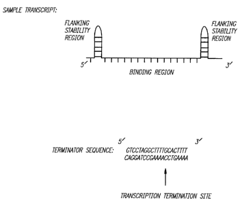
Figure 2 is a diagram illustrating in further
detail the invention shown in overview in Fig. 1,
demonstrating exemplary transcript configurations and
promoter and terminator sequences according to the
invention.
Figure 3 is a diagram showing a double-stranded
DNA probe and triplex forming RNAs. The 50 base-pair
double-stranded probe corresponds to bases -68 to -19 on
the HER2/c-erb B2/neu proto-oncogene. The region in bold
refers to the homopurine/homopyrimidine tract involved in
triplex formation. The CU-rich and GA-rich RNA sequences
correspond to pyrimidine (parallel) or purine
(antiparallel) third strand triplex binding motifs,
respectively.
Figure 4 is a second diagram presenting an
overview of the oligonucleotide generator system
according to the invention. The structure of the U6
snRNA gene, the chimeric U60N gene and the resulting U60N
transcript are shown. Fig. 4A. shows the U6 gene has
three critical promoter elements necessary for efficient
transcription, a 5' self-complementary hairpin sequence
sufficient for capping, and a string of 5 thymidine
residues necessary for termination. Fig. 4B demonstrates
the elements which were retained in the construction of
the chimeric gene, while its internal sequence was
mutated to produce twn unique restriction sites for
inserting oligonucleotide sequences (bold line). Fig. 4C
shows the resulting oligonucleotide which may retain the
5' hairpin, followed by the oligonucleotide (bold line)
and the native U6 uridine-rich sequence.
Figure 5 is a half-tone reproduction of
Northern blots which demonstrate the production and
nuclear localization of the U60N transcript. For Fig.
5A, MDA453 and 293 cells were transfected with either 10
g of the chimeric U6ON gene or 10 g of promoterless
WO 95/10607 PCT/US94/11616
217336:1
-18-
plasmid DNA. Total cellular RNA was isolated 48 hours
later followed by Northern blotting with both U6 and U60N
radiolabeled probes. For Fig. 5B, MDA453 cells were
transfected with increasing quantities of the chimeric
gene followed by RNA isolation at 48 hours and Northern
blotting as described above. All transfections contained
40 g total DNA, with promoterless plasmid DNA
supplementing the chimeric gene as necessary. For Fig.
5C, MDA453 cells were transfected with 10 gg of the
chimeric gene and after 48 hours, RNA was separated into
nuclear and cytoplasmic fractions. The nuclear fraction
shown above contained the U60N transcript along with the
native U6 snRNA. All Northern blots were generated from
l0 g of RNA loaded/well.
Figure 6 is a graph and a half-tone
reproduction of a Northern blot which demonstrates the
kinetics of U60N expression. For Fig. 6A, 293 cells were
transfected with 20 jig of the chimeric gene and total
cellular RNA was isolated at 0, 3, 6, 9, and 12 hours
time points. After Northern blotting (10 mg RNA
added/well) and autoradiography with a U60N radiolabeled
probe, the U60N bands were cut from the filter and
scintillation counted. For Fig. 6B, MDA453 cells were
transfected with 20 mg of the chimeric gene and total
cellular RNA was isolated at 48, 72, 96, 120, 144, and
168 hour time points. Northern blotting followed with 20
jig of RNA added/well.
Figure 7 is a graph and a half-tone
reproduction of a Northern blot which demonstrates the
intracellular stabilities of the chimeric gene and the
U60N transcript. For Fig. 7A, cell counting in parallel
with the RNA isolations of Figure 6B allowed the U60N
band densities to be normalized to account for the
dilutional effects of cell division. Normalized band
densities were plotted as a function of time to determine
S WO 95110607 2173361 PCT/US93/11616
-19-
the rate of chimeric gene degradation (or inactivation).
For Fig 9B, 293 cells were transfected with 5 jig of the
chimeric gene and after 48 hours, cellular transcription
was halted by a 10 g/ml treatment of Actinomycin D. At
0, 0.5, 1, 2, and 4 hour time points, RNA was isolated.
Northern blotting (20 g RNA/well) with U6 and U60N
radiolabeled probes, followed by densitometry of the U60N
bands, allowed for the determination of U6ON half-life.
Figure 8 is a half-tone reproduction of a
Northern blot demonstrating the immunoprecipitation of
the U60N transcript with a 5' 'y-monomethyl phosphate cap.
Total cellular RNA samples isolated after a transfection
with 20 g of U60N in 293 cells were immunoprecipitated
with 0.5 mg of a U6 cap-specific antibody as previously
described (Gupta et al. (1990), J. Biol. Chem. 265:19137-
19142. Eric Wickstrom, ed. (Wiley-Liss, New York),
pp. 143-158). Each immunoprecipitation required 20 g of
initial total cellular RNA. Immunoprecipitated RNA was
used for Northern blotting with U6 and U60N radiolabeled
probes.
Figure 9 is diagram of two predicted secondary
structures for two RNA transcripts and a half-tone
reproduction of a Northern blot, demonstrating the
effects of the insert sequence on secondary structure and
intracellular transcript levels. For Fig. 9A, RNA
secondary structures and associated energies were
predicted for two different constructs, U6CTcon and U6AS,
using the program RNAFOLD (Martinez, H. (1990) Methods in
Enzymolocty, 183, 306-317). Upper case letters refer to
base-pairings, lower case letters refer to mismatches and
colons refer to bulged regions. The energies of U60N and
mU6, which were found to have similar structural profiles
to U6CTcon and U6AS, respectively, are given in
parentheses. For Fig 11B, RNA secondary structure was
predicted for 4 different oligonucleotide transcripts and
WO 95/10607 PCT/US94/11616 41
-20-
the corresponding chimeric genes were constructed. 20 g
of the_chimeric genes were transfected into MDA453 cells,
followed by Northern blotting (20 g RNA added/well) 48
hours later with a U6 probe and a probe for each of the
possible RNA transcripts.
Figure 10 is a diagram of the HER2 proto-
promoter map with a triplex RNA oligonucleotide.
oncogene
Figure 11 is a graph representing the effects
of the U6ON and U6CTcon oligonucleotide generators on CAT
activity in cells expressing CAT from an HER2 promoter.
Figure 11A illustrates the CAT activity arising from the
minimal 125 bp HER2 promoter and the stronger 500 bp HER2
promoter in MDA453 and MCF-7 cells 48 hours after
co-transfection with 20 g of either U6ON or U6CTcon.
Promoterless plasmid DNA was used to equalize DNA
concentrations. Both U6ON and U6CTcon RNA
oligonucleotides demonstrated virtually 100k
downregulation of the minimal promoter and roughly 65 to
70 %- downregulation of the stronger 500 bp promoter.
Figure 11B illustrates the HER2/CAT downregulation of the
stronger 500 bp promoter by triplex and antisense RNA
oligonucleotides after co-transfection with 10 Etg of
either U6ON or U6AUG.
Figure 12 is two graphs and two half-tone
reproductions of Northern blots, which demonstrate the
effect of the U6ON generator on endogenous HER2 mRNA and
cell growth in MDA 453 cells. Figure 12A is a Northern
blot, which compares the levels GAPDH and HER2 RNA from
48 to 96 hours after transfection with the U6ON
oligonucleotide generator, as compared with control DNA.
Figure 12B is a graph demonstrating the HER2 RNA values
from Figure 12A after normalization for the GAPDH levels.
Figure 12C demonstrates the temporal pattern of U6ON
expression after transient transfection. Figure 12D
WO 95/10607 21733 6 1 PCTIUS94/11616
-21-
demonstrates the cell growth rate in U6ON transfected
cells versus control cells between 48 hours and 96 hours.
Figure 13 is a diagram detailing the promoter,
termination, and capping factors of the chimeric gene
(oligonucleotide generator). This drawing illustrates
= schematically a possible configurations of the needed
transcriptional factors of both the U6 gene and the
chimeric RNA-oligonucleotide producing gene.
Figure 14 is a half-tone reproduction of a
Northern blot and a graph which demonstrate the dose
dependence of gene transfection on U6 and U6ON in 293
cells. Cells were transfected with 40 g of DNA which
contained 5 to 40 g of the chimeric gene or promoterless
plasmid DNA. RNA was isolated 48 hours after
transfection. After Northern blotting with U6 and U6ON
probes (Fig. 14A), densitometry was performed to
quantitate the relative downregulation of U6 with
concurrent upregulation of U6ON and the data was
presented in figure 16B. Error bars arise from standard
deviation of densitometry data and slight inequalities in
RNA loading, normalized by ethidium bromide staining.
Figure 15 is a half-tone reproduction of a
Northern blot, demonstrating that U6 levels vary as a
function of RNA oligonucleotide stability. 293 cells
were treated with 20 g of the gene for U6AS (lanes 1-4)
or U6ON (lanes 5 to 8) followed by RNA isolation at 48
hour, 72 hour, 96 hour, and 120 hours time points. U6AS
has previously been shown to be an unstable RNA
oligonucleotide while U6ON has been shown to be a stable
and 5' capped RNA oligonucleotide.
Figure 16 is a half-tone reproduction of a
Northern blot, demonstrating that U6ON can downregulate
U6 stability. 293 cells were transfected with 20 g of
the chimeric gene for U6ON followed 48 hours later by 10
g/ml treatment with Actinomycin D to mediate total
WO 95/10607 PCT/US94/11616
21733 1
-22-
transcription arrest. RNA isolations followed at 0 hour,
0.5 hour, 1 hour, 2 hour, 4 hour, and 8 hour time points
followed by Northern blotting.
Figure 17 is a graph demonstrating that U6
stability is titratable by U60N levels. The experiment
presented in Figure 16 was repeated using 10 Ag and 5 Ag
chimeric gene transfection doses. After performing
individual Northern blots, data was amassed and
quantitated by densitometry.
Figure 18 is a series of three graphs,
illustrating the results of dynamic simulation of U6 and
U60N expression in 293. The dynamic modelling program
Stella was used to solve the differential equations
described in the Examples section. Data was then output
to Cricket Graph software and graphically displayed.
Fig. 18A shows steady-state levels of U6 expression.
Fig. 18B shows transient and steady-state levels of U60N
expression. Fig 20C shows transient and steady-state
levels of expression of U6 after reduction of transcript
stability by U60N production.
Figure 19 is a graph demonstrating the dose
dependence of U60N on 7SK, U1, and U3 RNA levels. The
nylon filter used to generate Figure 14A for 293 cells
was stripped at 70 C in prehybridization buffer for 40
minutes followed by reprobing with 7SK, U1 and U3 probes.
Quantitative data was obtained by densitometry analysis.
Figure 20 is a series of three graphs,
demonstrating the stabilities of 7SK (Fig. 18A), U1 (Fig.
18B), and U3 (Fig. 18C) RNA in the presence of U60N. The
nylon filters used to generate Figure 17 for 20 Ag and 5
Ag gene transfections were stripped and reprobed as
described for the previous figure. Quantitative data was
obtained by densitometry analysis.
Figure 21 is a graph demonstrating the effect
of RNA oligonucleotide stability on 7SK and U1 RNA
WO 95/10607 21733 61 PCT/US94/11616
-23-
levels. 20 g of the gene for U6ON, U6CTcon, U6AS, or
promoterless plasmid control DNA was transfected in
MDA453 cells followed by RNA isolation and Northern
blotting at 48 hours with 7SK and U1 probes. U6ON and
U6CTcon are stable RNA oligonucleotides while U6AS is
unstable. Quantitative data was obtained by densitometry
analysis.
Figure 22 is a graph demonstrating the effect
of UGON on GAPDH RNA levels. MDA453 cells were
transfected with 20 g of U6ON, U6AS or promoterless
plasmid control DNA followed by RNA isolations at 48
hour, 72 hour, and 96 hour time points. Agarose gel
Northern blotting (as opposed to polyacrylamide Northern
blotting) with a GAPDH probe and densitometry followed.
Figure 23 is a set of two graphs, demonstrating
the effect of U6ON on co-transfected (3-HCG. MDA453 cells
were co-transfected with 20 g of a chimeric
oligonucleotide-producing gene or promoterless plasmid
DNA followed by Q-HCG quantitation by an
immunoradiometric assay described in the Examples below.
Two representative experiments are provided to
demonstrate the variability in 0-HCG expression, but lack
of correlation with RNA oligonucleotide presence, levels,
lengths, or stabilities.
Figure 24 is a diagram illustrating a
double-stranded DNA probe and triplex forming RNAs. The
43-bp double-stranded DNA probe corresponds to bases -76
to -34 on the HER2/c-erb B2/neu proto-oncogene promoter.
The region in bold refers to the
homopurine/homopyrimidine tract involved in triplex
formation. The CU-rich RNA sequence corresponds to the
pyrimidine-rich (parallel) third strand triplex binding
motif.
Figure 25 is a half-tone reproduction of an
autoradiogram, demonstrating triplex blotting with in
WO 95/10607 PCTIUS94/11616
.2173361 -24-
vitro generated RNA. CU-rich, GA-rich, or control RNA
strands were generated in vitro by T3 or T7 RNA
polymerases from approximately 0.2 pmoles of linearized
plasmid. Equal aliquots from these reactions were added
to formamide loading buffer, fractionated by
electrophoresis and Triplex blotted as described in
Example 18.
Figure 26 is a set of two half-tone
reproductions of autoradiograms, demonstrating triplex
blotting versus Northern blotting with SKBR3 total
cellular RNA. Cells were transfected with either 0 g,
10 g or 20 Ag of the modified U6 plasmid which generates
an 82 nucleotide triplex-forming CU-rich RNA
oligonucleotide (lanes 2 and 3) or 20 g of promoterless
plasmid DNA (lane 1). Figure 26A shows equal amounts of
total cellular RNA (11 gg per lane) which were added to
formamide loading buffer, fractionated by electrophoresis
and Triplex blotted as described in Example 17. Figure
26B shows the same filter after stripping at pH 7.5 and
reprobing by Northern blot with radiolabeled
single-stranded DNA generated by random priming.
Figure 27 is a set of two diagrams displaying
(Fig. 27A) the sequence of the mU6 parent vector from
which oligonucleotide-producing genes are made, and (Fig.
27B) the sequence of the U6ON generator.
Detailed Description of the Invention
An improved method for delivery of
oligonucleotides, preferably antisense or triplex
oligonucleotides, was developed in order to circumvent
the obstacles of extracellular degradation, cellular
uptake, and intracellular sequestration. This new method
is sufficiently general to provide for ribozyme delivery
as well. The strategy was designed with the following
WO 95/10607 21733 61 PCT/US94/11616
-25-
criteria in mind: (a) oligonucleotides should be
generated in high yield within the cell nucleus without
significant cell type specificity; (b) they should be
sufficiently stable; (c) they should contain minimal
secondary structure that could mask binding regions; and
(d) they should be of a pre-determined and well-defined
sequence and length.
These criteria were satisfied by constructing
one of the preferred embodiments of the oligonucleotide
generator invention, containing regulatory regions from
the human U6 small nuclear RNA (snRNA) gene and a
synthetic double-stranded insert bearing the
oligonucleotide to be generated. U6 snRNA, which
normally functions in conjunction with several small
nuclear riboproteins (snRNPs) in the splicing of
premature messenger RNA (Manniatis & Reed (1987) Nature
325: 673-678), is transcribed in high yield by RNA
polymerase III, requires only upstream promoter sequences
for initiation, and terminates cleanly upon reaching a
string of 4-6 thymine residues (Kunkel, G. et al. (1986)
Proc. Natl. Acad. Sci. USA 83: 8575-8579; Reddy, R. et
al. (1987) J. Biol. Chem. 262: 75-81; Kunkel & Pederson
(1989) Nucleic Acids Res. 17: 7371-7379; and Willis, I.M.
(1993) European J. of Biochem. 212: 1-11.) Transcript
stability is strongly enhanced by 5' y-monomethyl
phosphate capping (Singh & Reddy (1989) Proc. Natl. Acad.
Sci. USA 86: 8280-8283; and Singh, R. et al. (1990) Mot.
Cell Biol. 10: 939-946) which is directed by a 5'
self-complementary hairpin followed by a conserved
hexameric AUAUAC sequence (Shumyatsky, G. et al. (1993)
Nucleic Acids Res. 21: 4756-4761). Within this
description of the present invention the abundant
production, intranuclear localization, kinetics of
expression, capping, and insert-specific stability of
WO 95/10607 + PCTIUS94/11616
2173361 -26-
transcripts generated from an oligonucleotide generator
of the invention were characterized.
In a general aspect, the oligonucleotide
generators of the invention encompass a construct for
producing a specific oligonucleotide within a cell, which
construct comprises (a) an U6-type RNA polymerase III
promoter; (b) a specific nucleotide sequence from which
the specific oligonucleotide is transcribed; and (c) a
termination sequence; wherein the components of the
construct are operably linked and positioned from 5' to
3' in the order of (a), (b), and (c).
The terms "U6-type RNA polymerase III promoter"
and "U6-type promoter" are used interchangeably herein to
refer to a promoter which is able to initiate
transcription by RNA polymerase III from a position
upstream of the transcribed DNA. "U6-type promoters"
have been referred to in the literature, in at least one
instance, as RNA polymerase III, type III promoters
(Willis, I. (1993) FEBS 212: 1-11). The "U6-type
promoter" contains regulatory elements which are
necessary and sufficient to facilitate transcription by
RNA polymerase III, but these regulatory elements are not
themselves transcribed. Thus, U6-type RNA polymerase III
promoters include the following promoters:
naturally-occurring U6 from higher order eukaryotes (Das
et al. (1988) EMBO J. 7(2): 503-512), 7SK (Murphy et al.
Cell 51:81-87), H1 RNA gene (Hannon, G. et al. (1991) J.
Biol. Chem. 266(34): 22796-22799), U3 snRNA genes in
plants (Marshallsay C. et al. (1992) Plant Molecular
Biology 19(6): 973-983), and MRP gene (Yuan, Y. and
Reddy, R. Biochem. et Biophys. Acta 1089(1): 33-39), as
well as any recombinant promoter sequence which is able
to initiate transcription by RNA polymerase III without
itself being transcribed. Preferably,
217336.
WO 95/10607 PCT/US94/11616
-27-
naturally-occurring U6 promoter is used as the U6-type
RNA polymerase III promoter.
Recombinant U6-type promoters for use in the
oligonucleotide generators of the invention will usually
have the distal sequence enhancer, proximal sequence
element and TATA box as displayed in Figure 4B. The
proximal sequence element and TATA box are required for
Polymerase III transcription, and the relative position
of these elements to each other and the start site are
relatively inflexible. The distal sequence enhancer,
however, can be deleted in part or completely, as well as
moved closer to or farther away from the proximal
sequence element, particularly if a reduction in the
level of transcription is desired.
The oligonucleotide generators of the present
invention can be used to facilitate delivery of
oligonucleotides to any type of eukaryotic cell. The
choice of a specific U6-type promoter is made on the
basis of the target cell (cell to be transfected with the
generator). U6-type promoters derived from a species
that is closely related or at least has a similar
promoter sequence to that of the target cell type are
preferably used if maximal transcription of the specific
oligonucleotide is desired. For example, human U6
promoters, when integrated into an oligonucleotide
generator of the invention, would provide higher levels
of transcription in human cells, but would be less
effective in plant cells. Conversely, a plant U6
promoter would provide higher levels of oligonucleotide
-30 production in a plant than a mammalian system.
In a general aspect, the invention also
features a construct for generating a specific
oligonucleotide within .a cell, which construct includes a
nucleotide sequence from which the transcript is the
specific oligonucleotide, flanked on the 5' end by a
WO 95/10607 PCTIUS94/11616
2173361
-28-
stabilizing region and on the 3' end by a termination
sequence, and a promoter at the 5' end of the stabilizing
region.
In preferred embodiments, the promoter and the
stabilizing region are derived from a RNA polymerase III
gene, and in particular embodiments it is derived from a
human U6 small nuclear RNA gene. In particularly
preferred embodiments the stabilizing region comprises
the first approximately 25 nucleotides of the human UG
small nuclear RNA gene, as this includes a portion that
forms a stable hairpin in the product and makes a "cap"
that prevents degradation of the product at the 5' end.
Alternatively, any hairpin-forming sequence can be used,
and needn't be derived from a native source.
Generally, the optional "stabilizing region" of
the oligonucleotide generator can be of any length,
so long as the resulting RNA transcript of the
oligonucleotide generator is predicted to form a hairpin
structure by a computer program that models and predicts
secondary structure of RNA. These computer programs
include, for example, the algorithm described in
Example 9. While longer stabilizing regions can be used,
they are generally between about 16 and about 50
nucleotides in length. Most preferably the segments of
the stabilizing region that are predicted to form base
pairs in the resulting RNA transcript are continuous and
perfectly complimentary, containing no mismatches.
However, such mismatches are tolerated within the
segments of the stabilizing region that are predicted to
'30 form base pairs, so long as they do not completely
disrupt the hairpin structure as predicted by the
computer modeling program. Usually the mismatched bases
are less than about 1 in 5 of the nucleotides in the
complimentary regions of the predicted hairpin structure,
and almost always less than about 1 in 4.
= WO 95/10607 173381 PCT/US94/11616
-29-
A "stabilizing region", when present, is
preferably able to reduce the rate of intracellular
degradation for the resulting RNA transcript as compared
with an identical RNA transcript that does not contain
the region transcribed from the stabilizing region.
Methods of measuring the intracellular degradation rates
of RNA are known in the art, and include the methods
described in Example 11.
As described above there is a portion of the 5'
end of the U6 gene that "makes a 'cap' that prevents
degradation of the product at the 5' end", and is
included in some preferred embodiments of the
oligonucleotide generators of the invention. The
stability of the resulting RNA transcripts is strongly
enhanced by this 5' y-monomethyl phosphate capping, which
is directed by a 5' self-complementary hairpin followed
by a conserved hexameric AUAUAC or AUAUCC sequence,
preferably AUAUAC sequence, in the RNA transcript. Thus,
when present in a oligonucleotide generator or generator
vector of the invention the sequence on the coding strand
for the capping segment is ATATCC or ATATAC. This
optional "capping segment" in the oligonucleotide
generators of the invention is usually only operable when
it is immediately downstream of the hairpin structure of
a stabilizing region, although the first two nucleotides
of the capping segment may form part of the hairpin
structure. When the "capping segment" is present, the
hairpin structure of the stabilizing region is preferably
about 20 to about 30 nucleotides in length, more
preferably about 20 nucleotides in length, most
preferably the hairpin structure from the 5' end of the
U6 transcript.
The "specific oligonucleotide" may be any
oligonucleotide that is desired to be transcribed within
the cell and includes, for example, a triplex-forming
WO 95/10607 21733 61 PCT/US94/11616
-30-
oligonucleotide, an antisense oligonucleotide, a
ribozyme, or a combination of these. Thus, the
oligonucleotide generators of the present invention can
be used to deliver RNA oligonucleotides for any purpose.
For example, the oligonucleotide generators of the
invention may be used to deliver tumor suppressing RNAs
(Rastinejad, F. et al. (1993) Cell 75: 1107-1117; and
Wickens, M. and Takayama, K. (1994) Nature 367: 17-18).
Particularly favorable results are obtained using the
construct of the invention for intracellular production
of oligonucleotides in the size range between about 10
and about 60 nucleotides (and more particularly in the
range between about 20 and 50 nucleotides), although the
success of the invention is not strictly dependent upon
the length of the oligonucleotide product.
Oligonucleotide products are usually less than about 500
nucleotides in length.
In preferred embodiments, the termination
region includes, in addition to a termination sequence
(e.g.. TTTT), a sequence between the termination sequence
and the 3' end of the oligonucleotide sequence, to
provide a 3' tail on the product; this aids in protecting
the product from degradation at the 3' end; and the tail
may be constructed to form a hairpin or other protective
structure. Thus, the termination region includes at a
minimum a transcription termination sequence recognized
by RNA polymerase III, i.e. a stretch of four to six
thymine nucleotides on the coding strand of the
oligonucleotide generator.
The optional region of the oligonucleotide
generator that provides the "3' tail" on the RNA
transcript may be designed such that it forms a hairpin
of any size, generally between about 16 and about 50
nucleotides in length. Also this region may be designed
to form a lariat structure by base pairing with the
2173361
S WO 95/10607 PCT/US94/11616
-31-
nucleotides transcribed from a stabilizing region or a
region of the transcript that is upstream of the specific
oligonucleotide. Preferably, when a lariat forming 3'
tail is used, the oligonucleotide generator also provides
for the transcription of a hairpin structure in the 5'
stabilizing region immediately followed by a capping
segment. Thus, some of the RNA transcript may be capped
with 5' 'y-monomethyl phosphate prior to the formation of
the more thermodynamically stable lariat structure.
The oligonucleotide generators of the invention
can optionally be designed such that it produces a
transcript with a predicted lariat conformation, wherein
the stem of the lariat formed by the Watson-Crick base
pairing of a 3' tail in the termination region and a
portion of the transcript which is upstream of the
specific oligonucleotide. The stem of the lariat
structure may be of any length as long as a stable lariat
structure is predicted by a computer program that models
and predicts secondary structure of RNA. These computer
programs include, for example, the algorithm described in
Example 9. While longer stem regions can be used, the
lariat stem is generally between about 8 and about 30
nucleotides in length. Most preferably, the stem of the
lariat is predicted to form continuous base pairs for the
stems entire length, containing no mismatches. However,
such mismatches are tolerated within the segments of the
lariat stem that are predicted to form base pairs, so
,long as they do not completely disrupt the lariat
structure as predicted by the computer modeling program.
Usually the mismatched bases are less than about 1 in 5
of the nucleotides in the lariat stem, and almost always
less than about 1 in 4 of the nucleotides in the lariat
stem.
Thus, the present invention encompasses
oligonucleotide generators, comprising from 5' to 3': (a)
WO 95/10607 PCT/US94/11616
2173361 -32-
an U6-type RNA polymerase III promoter; (b) a specific
nucleotide sequence from which a specific oligonucleotide
can be transcribed; and (c) a termination sequence;
wherein the components of the oligonucleotide generator
are operably linked; and wherein the oligonucleotide
generator is capable of being transcribed by RNA
polymerase III to produce a transcript comprising the
specific oligonucleotide; and further comprising: a 5'
tail from which a first lariat-forming sequence can be
transcribed; and a 3' tail from which a second lariat-
forming sequence can be transcribed; wherein the 5' tail
is operably linked and positioned between the U6-type RNA
polymerase III promoter and the specific nucleotide
sequence; wherein the 3' tail is operably linked and
positioned between the specific nucleotide sequence and
the termination sequence; wherein the oligonucleotide
generator is capable of being transcribed by RNA
polymerase III to produce a transcript comprising from 5'
to 3' the first lariat-forming sequence, the specific
oligonucleotide, and the second lariat-forming sequence;
and wherein the transcript is predicted to from a stable
lariat structure by Watson-Crick base pairing between the
nucleotides of the first lariat-forming region and the
second lariat-forming region.
In particularly preferred embodiments, the
stabilizing and termination portions of the construct are
derived from the same source, and the construct is made
-by providing a vector containing, in sequence but not
necessarily contiguous sequence, a promoter of the
U6-type, a stabilizing 5' portion of the source gene
(which may preferably be a part of a type III gene such
as the first -25 nucleotides of the human U6 gene), a
XhoI site, a NsiI site, and a 3' portion of the source
gene including at least a termination sequence (which may
preferably be a part of the same type III gene such as a
WO 95/10607 2 1 7 3 3 6 1 PCTIUS94/11616
-33-
-20 nucleotide 3' portion of the human U6 gene). Of
course, the XhoI and NsiI restriction sites can be
replaced with any first and second unique restriction
enzyme sites to facilitate insertion of the specific
nucleotide sequence.
Thus, in another general aspect, the present
invention encompasses vectors which comprise any of the
oligonucleotide generators described herein with the
specific nucleotide sequence removed. These "generator
vectors" may be used to construct an oligonucleotide
generator of the invention. Preferably these generator
vectors comprise two unique restriction enzyme sites, one
on each side of the position of the generator vector into
which the specific nucleotide sequence must be inserted
to form an oligonucleotide generator of the invention.
These unique restriction enzyme sites, when present,
serve to facilitate insertion of the specific nucleotide
sequence into the generator vector. More preferably the
two restriction enzyme sites are not recognized by the
same enzyme.
The U6 gene was chosen to provide the
regulatory components for some of the preferred
embodiments of the invention, as it is transcribed in
nearly all mammalian cells in high yield; is transcribed
constitutively; requires only upstream promoter sequences
for transcription; and initiates and terminates cleanly
and precisely (Kunkel, G. et al. (1986) Proc. Natl. Acad.
Sci. USA 83: 8575-8579; Reddy, R. et al. (1987) J. Biol.
Chem. 262: 75-81; Kunkel & Pederson (1989) Nucleic Acids
Res. 17: 7371-7379; and Willis, I.M. (1993) European J.
of Biochem. 212: 1-11.) In addition, the U6 gene
contains a sequence-specific signal that directs the 5'
capping of transcripts by a T-monomethyl phosphate which
greatly augments transcript stability (Singh & Reddy
(1989) Proc. Natl. Acad. Sci. USA 86: 8280-8283; Singh,
WO 95/10607 PCT/US94/11616
2143361 -34-
R. et al. (1990) Mol. Cell Biol. 10: 939-946; and
Shumyatsky, G. et al. (1993) Nucleic Acids Res. 21:
4756-4761). The output of this gene results in the
abundant intracellular production of short,
sequence-specific RNA oligonucleotides containing a 5'
nuclease-resistant y-monomethyl phosphate cap.
In another general aspect, the invention
features a method for intracellularly generating an
oligonucleotide of interest in a subject or in cells or
tissues derived from a subject, including administering
to the subject the construct according to the invention
in a form that permits entry of the construct into the
subject's cells. The administration may be carried out
by any of various techniques known, for example, in the
art of gene therapy (see, for example, J.W. Larrick and
Kathy L. Burck, Gene Therapy, Application of Molecular
Biology, Elsevier, Holland, 1991); or by administration
in immunoliposomes, according to techniques known in the
immunotherapeutic art, wherein the immunoliposome may be
targeted to, and its contents delivered into, a
particular cell type (such as, for example, a breast
cancer cell); or by localized injection at a site for
treatment; or by way of mucosally-lined passages; or via
the airways, for example; all depending upon the
particular treatment.
Advantages of the oligonucleotide generator of
the invention include high yield and continuous
production of oligonucleotides within the cell, and
minimal secondary structure within binding regions on the
oligonucleotide products, except where the secondary
structure is an important functional part of a ribozyme
contained in the specific oligonucleotide.
Viral integration into the chromosome of the
cell can, according to the invention, confer permanence
WO 95/10607 - 217336-1 PCTIUS94111616
-35-
to the oligonucleotide-based antiviral, anticancer, or
antiprotein gene regulation.
The boxed regions in Figure 2 (i.e. A box, TATA
box) represent natural invariant promotor and terminator
sequences with their lengths in base pairs identified
below. The straight line regions represent areas where
sequence-specific oligonucleotide portions can be
inserted. These oligonucleotide portions may be any
oligonucleotide to be transcribed, for example, triplex
forming oligonucleotides, antisense oligonucleotides,
ribozymes, or a combination thereof. The
oligonucleotides can be designed for binding to different
regions of different DNA or RNA targets, to different
regions of the same DNA or RNA target, or to the same
region of the same DNA or RNA target. Decisions as to
vector design would be based upon whether the
experimenter wanted to hit multiple targets broadly or a
single target intensely. In addition, as shown in
Figure 2, self complementary ends can be generated on the
oligonucleotide which may form small double-stranded
hairpin loops. Such double-stranded ends will protect
against exonuclease activity, prolong oligonucleotide
half-life within the cell, and prevent other
oligonucleotide secondary structures from masking key
binding regions.
Once the promotor and oligonucleotide sequences
have been attached in the correct orientation, they can
be inserted into a viral vector (such as an adenovirus or
retrovirus) and integrated into the chromosomes of the
cells of interest as, for example, the methods described
by Sullenger et al. (1990) Molecular and Cellular Biology
10(12): 6512-6523. The result of the integration would
produce a continual and large supply of short length
oligonucleotides generated by the cell's own
transcriptional machinery based upon natural RNA
WO 95/10607 2173361 PCTIUS94/11616
-36-
polymerase III promotion initiation, and termination
sequences. By varying the multiplicity of viral vector
infection, the degree of gene regulation can be
modulated.
The advantages of such a RNA polymerase III
system for generating antisense and triplex forming
oligonucleotides are manifold. RNA polymerase III
transcribes at a nearly constant rate and high frequency
in almost all mammalian cell types, in marked contrast to
the widely used RNA polymerase II based systems which
transcribe at lower frequencies and are highly variable
with time and cell type. The RNA polymerase III
transcription initiation and termination processes are
also highly efficient allowing for clean transcription
start and stop sites, usually within 1-2 nucleotides.
This feature too stands in contrast with RNA
polymerase II approaches which generate widely varying
transcript lengths with long polyadenylated tails and
often hundreds to thousands of trailing 31 nucleotides.
In addition, since RNA polymerase III normally generates
transcripts that are both selectively transported to the
cytoplasm (rRNA, tRNA) and maintained in the nucleus
(various snRNA), it may be possible to utilize similar
sequences to keep the designed oligonucleotide sequences
primarily in the cell compartment of interest.
Several reports have emerged citing the ability
of a transfer RNA (tRNA) gene to be used as a carrier for
an antisense oligonucleotide (Izant, J.G. (1992) In Gene
Regulation: Biological Activity of Antisense RNA and
DNA. R.P. Erickson, J.G. Izant, eds. Raven Press (New
York), pp. 183-196; and Sullenger, B. et al. (1990) Mot.
Cell. Biol. 10: 6512-6523). In one case the
oligonucleotide was placed within one of the hairpin
loops in the internal region of the gene (Izant, J.G.
(1992) In Gene Regulation: Biological Activity of
WO 95/10607 PCT/US94/11616
-37-
Antisense RNA and DNA. R.P. Erickson, J.G. Izant, eds.
Raven Press (New York), pp. 183-196), in another
instance, the oligonucleotide was placed on the 3'
tailing region of the tRNA (Sullenger, B. et al. (1990)
Mol. Cell. Biol. 10: 6512-6523). Both reports
demonstrate an antisense effect against a target mRNA
using these RNA transcripts, but did not result in the
RNase H-mediated cleavage of the target. As with the U6
system, these' approaches utilize polymerase III for
oligonucleotide production. However, in both of these
systems, the oligonucleotide is within a much larger
sequence containing key promoter elements. Consequently,
the antisense binding sequence is constrained to regions
where the tRNA can be transcribed normally, and is likely
masked by structure of the much larger tRNA sequence, or
interfere with intragenic promoter function. In
addition, these tRNA strategies may result in
oligonucleotides which are close enough in structure to
native tRNA molecules to disrupt translation of mRNA into
proteins or result in the transport of the chimeric
oligonucleotide out of the nucleus.
By contrast, U6 and 7SK -based systems require
no internal promoter as opposed to the tRNA chimeric
genes, and thus can be composed almost entirely of the
oligonucleotide of interest. This feature is important
in determining and designing the secondary structure of
the transcript to maximize the oligonucleotide binding
sequence while also maximizing stabilizing flanking
sequences. In one preferred embodiment, a 5' U6 flanking
'30 sequence was maintained to invoke the enzyme(s) which
recognize and cap the transcript. Another technique
which may provide both oligonucleotide accessibility and
stability is to build in a 5' and 3' self-complementary
hairpin, creating a lariat-like structure with the
oligonucleotide within the loop. As the RNA is small,
s i
WO 95/10607 21733 6 1 PCT/US94/11616
-38-
secondary structure prediction algorithms have utility in
the design of these transcripts. The U6 system also has
nearly all of the native U6 deleted from the resulting
chimeric gene. This feature eliminates the possible
sequestering of oligonucleotide within the active
spliceosome, as well as eliminates possible toxicity from
dysfunctional chimeric U6 RNA that can carry out only a
subset of the functions of the native U6 RNA.
There is another chimeric gene that has been
developed by others, which utilizes the promoter of U2
snRNA gene (Izant, J.G. (1992) In Gene Regulation:
Biological Activity of Antisense RNA and DNA. R.P.
Erickson, J.G. Izant, eds. Raven Press (New York), pp.
183-196). Like the U6-based chimeric gene, the promoter
is entirely upstream, but unlike the UG-based chimeric
gene, it is transcribed by RNA polymerase II. These
chimeric genes also retain nearly all of the native U2
gene which is considerably longer and more complex in
structure than is the tRNA gene. Thus, accurate
predictions of structure and accessibility of these
oligonucleotides are significantly more difficult to
obtain a priori. However, the kinetics of expression,
stability, intracellular localization, and absolute RNA
transcript levels have not been characterized in these
chimeric genes.
In the oligonucleotide generators of the
invention, the optional use of viral integration allows
for the modulation of infection with the oligonucleotide
generator, and thus the amount of transcripts generated.
=30 Such a feature will be invaluable in generating
dose-response curves to a given target. Viral
integration will also generate cells that are healthy and
otherwise functioning and replicating normally, while
producing the constant supply of oligonucleotides. As
opposed to plasmid transfection techniques, the
2173361
WO 95/10607 - PCT/US94/11616
-39-
integration is permanent and does not require continuous
selective pressure. Once the producer lines of virally
integrated cells are generated, a variety of primary
cells and cell lines can be infected, a variety of
combination and concentrations of infection can be tried,
and the effect of various strategies of gene regulation
can be compared (i.e. antisense versus triplex).
An additional advantage arises from the
capability to hit multiple targets by the use of more
than one oligonucleotide sequence within a transcript.
Multiple sequences could target the start codon, a splice
site, and the ribosomal binding sequence within a single
mRNA, thus increasing the likelihood of blocking
subsequent translation of that protein. Alternatively,
multiple mRNA`s could be used as targets leading to
downregulation of multiple proteins along a common
pathway.
We have designed, constructed, and tested a
system for the intracellular generation of short
sequence-specific oligonucleotides in extremely high
yield for the purposes of gene regulation. The system,
when transfected into cells by electroporation, produces
the desired oligonucleotide in high quantity. We have
analyzed the production of our transcript by both
Northern and RNAse Protection assays, and both analyses
show that the quantity of oligonucleotide produced is at
least several orders of magnitude higher than the
quantity of a typical messenger RNA. However, should
lower concentrations be warranted, smaller doses of our
system result in a lower quantity of the oligonucleotide.
Experimentation has also shown that the oligonucleotide
is sufficiently stable (i.e., it is composed of only one
strong band; no degradation products are observable), and
is of an exact "user-definable" length.
WO 95/10607 217 3 6 PCT/US94/11616
1
-40-
This system has the advantage of user
flexibility in oligonucleotide sequence, length, and
yield not capable with current intracellular nucleic acid
delivery techniques. Its intracellular location and
seemingly constant production bypass uptake and
sequestration concerns which limit the utility of current
extracellular nucleic acid delivery techniques. This
system is also compatible with viral integration for
conferring permanence in oligonucleotide generation. We
expect our system to result in enhanced capacity for gene
regulation by antisense, triplex, or any other
application of oligonucleotides in cells.
The in vivo oligonucleotide generator according
to the invention can be used for permanently
downregulating or upregulating cellular proteins of
interest. Examples of effective antisense, triplex-
forming, and rybozyme oligonucleotides, as well as
strategies for developing new efficacious
oligonucleotides are known in the art and are thus not
described exhaustively herein. Examples of proteins for
which downregulation might be indicated include
transplantation antigens (e.g. ICAM, MHC classes I and II
antigens), hormones, cellular adhesion molecules,
clotting proteins, oncogene products, and proteins of
viruses which form latent infections in human, including
but not limited to Herpes, HIV, CMV, and human papilloma
viruses. Information concerning the use of
oligonucleotides to downregulate transplantation antigens
is found in PCT/US 93/00797. Examples of proteins for
which upregulation might be indicated include
underexpressed proteins in deficiency states.
The in vivo oligonucleotide generator according
to the invention can be used for control of viral,
parasitic or mycotic replication, by targeting key
replication and protein sequences (such as for example
WO 95/10607 2 1 7 3 3 6 1 PCT/US94/11616
-41-
the T antigen in SV40) and to prevent viral infection by
downregulating necessary attachment proteins.
The in vivo oligonucleotide generator according
to the invention can be used to block the proliferative
effect of cellular or viral oncogenes such as myc, myb,
or fos and therefore reduce or prevent neoplasm growth
and tissue invasion.
The in vivo oligonucleotide generator according
to the invention can be used to create new genetically
altered organisms, cells and tissues, useful for example
in agriculture and in human and veterinary medicine.
In the Examples section below we demonstrate
that the levels of production of the RNA transcripts,
e.g. U6ON, produced by the oligonucleotide generators of
the invention can rival and even exceed those of the
native U6 snRNA (5 x 10⁴ to 5 x 10⁶ copies/cell) and,
like native U6 RNA, these RNA oligonucleotides can be
capped and remain intranuclear in controlled
concentrations which may range from 160 M to 16 mM.
U60N production occurs rapidly upon transfection and can
still be detected up to one week after transfection, as
50% inactivation of the parent plasmid from steady-state
requires approximately 4 days. This long-lived
production suggests that in slowly growing cell
populations, longer time points (i.e., longer than the
typical 48 to 72 hours) may be used for measuring a
biological response from a transient transfection of the
oligonucleotide generator. In addition, this long-lived
production may allow for the detection of biological
effects of antisense or triplex oligonucleotides after
transient transfection, even when the target mRNA and/or
protein is fairly stable. For example the half-life of
U60N transcripts is estimated to be 1 hour; however,
transcript stability is dependent upon the sequence of
the oligonucleotide insert.
A, 3
WO 95/10607 217336 1 PCTIUS94/11616
-42-
The sequence of the insert is able to affect
the ability of the transcript to retain the initial 5'
hairpin structure, and thus the ability to obtain a 5'
cap. Correlations have been observed between RNA
secondary structure predictions and experimental
determinations of transcript levels containing different
oligonucleotide insert sequences. When the algorithm
predicted the loss of the 5' hairpin, dramatic decreases
in transcript-levels were seen experimentally after
electroporation and Northern blotting. Low transcript
levels reflect either a decrease in production or an
increase in degradation. As the U6 gene has consistently
been shown to require only upstream promoter sequences
for transcription (Kunkel & Pederson (1989) Nucleic Acids
Res. 17: 7371-7379; and Willis, I.M. (1993) European J.
of Biochem. 212: 1-11), the low transcript levels seen
with some oligonucleotide insert sequences cannot be due
to a decrease in production, and must therefore are due
to an increase in degradation. U6 stability has been
attributed primarily to its 5' cap and its extensive
hybridization with self+U4 (Terns, M.P. et al. (1993)
Genes and Development 7: 1898-1908). As all of the
chimeric oligonucleotide generator genes have the
U6/U4+self hybridization regions deleted, it becomes more
likely that differences in stability are due to the
presence or absence of a 5' cap. Finally, correlations
between modeling studies and experimental data point
.toward the retention of a 5' hairpin structure as a means
of linking oligonucleotide sequence, retention of the 5'
cap, transcript stability, and thus, absolute transcript
levels. Consequently, in the design of an
oligonucleotide insert, the overall secondary structure
of the RNA transcript has importance in optimizing
transcript stability and steady-state transcript levels.
WO 95/10607 2173361 PCTIUS94/11616
-43-
There are a variety of potential applications
for a system which generates sequence-specific short RNA
oligonucleotides in high yield within the cell nucleus.
For example, antisense oligonucleotides can be generated
intracellularly in levels several orders of magnitude
greater than typical sense mRNA molecules and far greater
than antisense mRNA generated by more traditional vectors
that rely on RNA polymerase II for transcription. In
addition, the ability to produce short transcripts
minimizes the chances that the binding region for a
targeted biological effect is masked by secondary
structure which can occur with much larger antisense mRNA
transcripts that do not have pre-determined length or
sequence.
The present invention also provides a means for
the generation of intranuclear triplex RNA
oligonucleotides. Pyrimidine-rich triplex RNA
oligonucleotides which bind in a parallel fashion with
respect to the corresponding purine strand of a
homopurine/homopyrimidine duplex (Felsenfeld, G. et al.
(1957) J. Am. Chem. Soc. 79:2023-2024, have a greatly
increased binding affinity over their triplex DNA
oligonucleotide counterparts (Roberts & Crothers (1992)
Science 258: 1463-1467; and Escude, C. et al. (1993)
Nucleic Acids Res. 21: 5547-5553). The high
concentration of a triplex RNA oligonucleotide which is
both generated and retained in the nucleus in vast excess
over its DNA duplex target may drive triplex binding to a
critical element on a gene promoter, and block subsequent
gene expression.
In addition to antisense and triplex
oligonucleotides, the vectors of the present invention
may be used in generating longer length ribozyme
transcripts for use in binding and cleaving target mRNA.
Combinations of triplex and antisense oligonucleotides as
WO 95/10607 2173361
-44- PCTIUS94/11616
well as ribozymes targeted to a single gene may also
yield synergistic approaches to the selective repression
of gene expression.
Other potential uses of the present invention
include the quenching of specific single-stranded nucleic
acid binding proteins by short RNA sequences, or
generating self-complementary RNA hairpins that can mimic
known DNA binding consensus sequences, thus quenching
specific DNA binding transcription factors. These
embodiments of the present invention rely on the nuclear
localization of abundant and sufficiently stable
oligonucleotides with short and fully defined sequences
to allow for reasonable approximation of secondary
structure.
Triplex Blotting
In another aspect, the invention features a
method for screening oligonucleotide sequences that are
candidates for triplex formation with a double-stranded
DNA target site, including steps of identifying a
sequence corresponding to the target site, producing and
isolating oligonucleotide sequences that appear likely to
bind to the target site, prehybridizing and hybridizing
the isolated oligonucleotide sequences with a labeled
double-stranded probe having the sequence of the target
site, removing unbound probe from the oligonucleotide
sequences, and detecting the label to identify
probe-bound oligonucleotides.
In its most general aspects the triplex
blotting invention described herein embodies two
different methods of measuring triplex formation and the
strength of triplex binding. The first method comprises
(a) attaching a single-stranded nucleic acid to a solid
support; (b) contacting the solid support with a fluid
comprising a labeled double-stranded probe; (c)
WO 95/10607 21733 6 1 PCT/US94/11616
-45-
separating the unbound probe from the solid support; and
(d) quantifying the amount of labeled double-stranded
probe bound to the solid support. The second method
comprises (a) attaching a double-stranded nucleic acid to
a solid support; (b) contacting the solid support with a
fluid comprising a labeled single-stranded probe; (c)
separating the unbound probe from the solid support; and
(d) quantifying the amount of labeled single-stranded
probe bound to the solid support.
The attachment of single and double-stranded
nucleic acids to solid supports can be accomplished by
any method known in the art including, but not limited
to, Northern blotting transfer techniques, slot or dot
blotting techniques, or by the nucleic acid chip
technology of Affymax. The single-stranded nucleic acid
used in the triplex blotting method of the invention can
be either DNA or RNA. The double-stranded nucleic acid
used in the triplex blotting method of the invention can
be DNA:DNA, DNA:RNA, or RNA:RNA duplexes. In all cases
modified nucleotides, and nucleotide linking groups
(groups to replace internucleotide phosphodiester
linkages) can be used.
The label on the labeled single and
double-stranded probes can be any nucleic acid label
known in the art including, but not limited to,
radionuclides (e.g. 32P and "S), fluorescent dyes, and
biotin.
The separation of the unbound probe from the
solid support can be accomplished by any washing
technique known in the art for use with Northern,
Southern and slot blotting. Such washing techniques may
include multiple washes at increasing stringencies while
monitoring the ratio of specifically bound labeled probe
to background labeled probe, for example with a geiger
counter for probes labeled with 32P as is typically done
WO 95/10607 2173361 -46- PCT/US94/11616
with Northern blotting. Increasing stringency can be
accomplished by reducing the concentration of salts
generally, and Na* particularly, by increasing the
temperature, or by increasing the pH.
The quantification of the amount of labeled
single- or double-stranded probe bound to the solid
support may be preformed by any method known in the art
for the quantification of the particular label used. For
example in the case of a radionuclide the quantification
can be performed by autoradiography followed by
densitometry or scintillation counting.
In one preferred embodiment of the triplex
blotting invention described herein, is for use in drug
screening assays. For such assays, the single-stranded
nucleic acid is attached to the solid support and the
double-stranded nucleic acid is labeled and used as a
probe. In this case the probe is designed to mimic a
particular target site for triplex binding. A large
number of single-stranded nucleic acids with different
sequences are attached to one or more solid supports in
an ordered and cataloged manner. The solid support is
contacted with a fluid comprising the labeled
double-stranded probe/target site. After the unbound
probe is separated from the solid support, the amount of
labeled double-stranded probe bound to each different
single-stranded nucleic acid is quantified. The
single-stranded nucleic acid with the highest amount of
labeled double-stranded probe bound to its position on
the solid support is the best candidate for a triplex-
forming oligonucleotide.
The practice of the present invention will
employ, unless otherwise indicated, conventional
techniques of protein chemistry, molecular biology,
microbiology and recombinant DNA technology, which are
CA 02173361 2006-09-20
69790-71
-47-
known to one of ordinary skill in the art. Such
techniques are explained fully in the literature. See,
e.g., R.K. Scopes, Protein Purification Principles and
Practice, 2nd ed. (Springer-Verlag 1987); S. Colowick and
N. Kaplan, Eds., Methods in Enzymology (Academic Press,
Inc.); Sambrook, Fritsch & Maniatis, Molecular Cloning: A
Laboratory manual, 2nd ed. (Cold Spring Harbor Laboratory
Press 1989); M.J. Gait, Ed., Oligonucleotide Synthesis
(1984); and D.M. Weir and C.C. Blackwell, Eds., Handbook
of Experimental Immunology, Vols. I-IV (Blackwell
Scientific Publications 1986).
Described below are examples of the present
invention which are provided for illustrative purposes,
and not to limit the scope of the present invention. in
light of the disclosure, numerous embodiments within the
scope of the claims will be apparent to those of ordinary
skill in the art.
Examples
Example 1
Initial Characterization of
In Vivo Oligonucleotide Generator
Expression and Lack of Toxicity
Tests of the system using several different
cell populations (breast cancer MDA453 cells; human
embryonic kidney 293 cells) and have produced comparable
results. Normal U6 small nuclear RNA which utilizes
identical transcriptional assemblies were unaffected by
WO 95/10607 3 6 1 PCT/US94/11616
21''7-3
-48-
transfections of up to 20 g of the chimeric ON-producing
gene; cells producing the oligonucleotide grow normally
and demonstrate no morphologic changes at these levels of
transfection.
Kinetics
We have examined how fast the oligonucleotide
generator can produce RNA oligonucleotides after cell
transfection with the parent plasmid. We electroplated
cells and examined total RNA at 12 hours, 24 hours, and
48 hours post-transfection. Using a system substantially
as described above, the oligonucleotide generator reaches
a near steady-state level at about 12 hours
post-transfection. These data indicate that the
oligonucleotide is formed rapidly, and therefore may have
immediate activity once the system is introduced into
cells.
Dose-response
We have examined the effect of increasing the
amount of the oligonucleotide generator in cells with
respect to RNA oligonucleotide levels in cells 48 hours
after transfection. To summarize, when cells are
transfected with 0, 5, 10, 20, or 40 micrograms of the
generator using a system substantially as described
above, the amount of RNA oligonucleotide isolated from
cells increases linearly. Thus, it is possible to drive
cells to produce enormous levels of a single RNA
oligonucleotide. At 40 microgram transfections, we
estimate that we can produce on the order of 1-5 million
copies per cell. This level is very far in excess of
that achievable with typical mRNA species.
Effect of the Oligonucleotide Generator on the Cells
3 6 1
WO 95/10607 2173 PCT/US94/11616
-49-
As the oligonucleotide generator produces such
high levels of RNA oligonucleotides in the cell, we
examined the effect of the oligonucleotide generator on
two parameters, namely, cell growth and normal U6 RNA
levels. After transfection of 10' cells with 0, 5, or 10
micrograms of an oligonucleotide generator substantially
as described above, no effect on the ability of cells to
divide and grow normally was detected, and no significant
effect on native U6 levels, which might have indicated
competition for transcription factors, was detected.
However, at levels of transfection z 20 g of the U60N
generator, cellular levels of U6 RNA decreased. Further
analysis indicated that this decrease was due to a
lessened stability of the cellular U6 RNA.
Example 2
Targeting the HER2 Proto-Oncogene Promoter with Triple
Helix Forming Oligonucleotide
Oligonucleotides designed to form local triple
helices with the HER2 promoter and interfere with
transcription factor binding may potentially inhibit HER2
receptor expression and growth of HER2 overexpressing
breast cancers.
We have identified, using promoter deletion
mapping, a 125 base-pair core proximal promoter region
capable of conferring greater than 30 fold variation in
HER2 transcriptional activity. Within this region are
several putative protein binding sites (including CAAT,
TATA, and a GAGGAA ets-related response element),
essential for promotor activity, which both surround and
overlap a 41 base-pair purine-rich sequence. Gel
mobility shift experiments demonstrate that this
41 base-pair sequence is compatible with triple helix
WO 95/10607 21733
-50- PCT/US94/11616
=
formation by both parallel (pyrimidine-rich) and
antiparallel (purine-rich) triplex binding motifs.
We tested oligonucleotides ranging in length
from 26 to 41 nucleotides. Increasing oligonucleotide
length corresponds with increasing gel retardation,
however triplex formation is relatively unaffected by
length, demonstrating only sequence-specificity.
Antiparallel triplex formation is strongly dependent upon
Mg- (0.1 to 10mM), while parallel triplex formation has
an additional sensitivity to pH, demonstrating
significant loss of hybridization from pH 7.0 to 8Ø
These triple helix forming oligonucleotides can inhibit
binding of ets-related proteins such as PU.1 to the HER2
promoter.
in addition, transfection of synthetic genes
capable of constitutively generating short triple helix
forming oligonucleotide transcripts is testing the
ability of these oligonucleotides to downregulate both
native HER2 receptor levels as well as a co-transfected
HER2-driven chloramphenicol acetyltransferase reporter
gene. Triple helix forming oligonucleotides and effect
of HER2 downregulation on proliferation and
tumorigenicity of HER2-positive breast cancer cells have
a therapeutic potential.
Example 3
Construction of the Chimeric U6ON Gene
The human U6 gene cloned within the SmaI site
of pGeml (Promega, Madison, WI), along with a mutant
human U6 gene with bases +25 to +55 replaced by an XhoI
restriction site (with A/C substitution at base 24) were
generously provided by G. Kunkel and T. Pederson (Kunkel
& Pederson (1989) Nucleic Acids Res. 17: 7371-7379). The
mutant U6 gene was recloned into a pBluescript
(Stratagene, La Jolla, CA) vector to produce
WO 95110607 21733 61 PCT[US94/11616
-51-
single-stranded phage and to allow two site-directed
mutations at bases +86 and +88 (T to G and G to A,
respectively) to create a unique NsiI restriction site.
This plasmid, mU6, was then recloned back into pGEM1, cut
with XhoI and NsiI, and religated with a synthetic 38 bp
duplex fragment bearing 5' XhoI and 3' NsiI compatible
overhanging ends (Keystone Laboratories, Menlo Park, CA).
Incorporation of the synthetic oligonucleotide was
verified by Maxam and Gilbert dideoxy DNA sequencing.
The resulting transcript arising from this vector, U60N,
is 25 nucleotides shorter than native U6 RNA (82 vs.
107). Sequences of the upper strand of U60N and other
various inserts are as listed:
U60N:
5' TCGACTCCTCTTCCTCCTCCACCTCCTCCTCCCATGCA 3'
U6CTcon:
5' TCGACCTCCCTTCCCTTCCCTTCCCCTTCCTCCATGCA 3'
U6AS:
5' TCGACATGAGCATTCATCAGGCGGGCAAGAATGTGATGCA 3'
MU6:
5' TCGAGCATGGCCCCTGCGCAAGGATGACACGCAAATGCA 3'
Figure 4 illustrates schematically the
structure of the native U6 snRNA gene, the modifications
involved in generating the chimeric gene, and the
resulting RNA oligonucleotide transcript, U60N. As
shown, the upstream promoter and enhancer regulatory
regions, the initial 25 bp (with A/C substitution at base
24), and the terminal 19 bp of the native U6 gene were
retained in the chimeric gene. Mutagenesis and
restriction digest removed the remaining native U6
internal sequence in order to create a hybrid of native
and synthetic sequences in a gene designed to express any
oligonucleotide of interest. As shown in Figure 4C, the
WO 95/10607 PCT/US94/11616
2173361
-52-
transcribed RNA oligonucleotide was designed to retain
the original 5' hairpin, in order to obtain the 5'
y-monomethyl phosphate cap. This initial sequence is
followed by the sequence-specific oligonucleotide and the
native U6 uridine-rich 3' terminus. The total length of
the resulting transcript is a function of the synthetic
oligonucleotide inserted, a 38 bp duplex was inserted
yielding a U6ON of 82 nucleotides.
Example 4
Cell Culture and Gene Transfection
The human embryonic kidney cell line, 293, and
the human breast cancer cell line, MDA453 (ATCC,
Rockville, MD), were transfected by electroporation (250
V, 960 F) with 5 g to 40 g of the chimeric gene or
promoterless plasmid DNA. Cell viability after
transfection ranged from 40-609 (with 293 cells showing
slightly higher tolerance to electroporation than MDA453
cells) and was unaltered by increasing gene transfection
dosage up to 40 g/107 cells. 293 cells were cultured in
minimal essential media with Earle's basic salt solution,
10%- fetal calf serum supplemented with 100 U/ml
penicillin and streptomycin in 5% CO2 incubators. MDA453
cells were cultured in Leibovitz L-15 media with 10%
fetal calf serum supplemented with 100 U/ml penicillin
and streptomycin in the absence of CO2. Where indicated,
cell counts were obtained by Coulter counting.
Example 5
RNA Isolation and Northern Blotting
Total cellular RNA was isolated 48 hours after
transfection by the guanidinium isothiocyanate/cesium
chloride centrifugation technique (Glisin, V.R. et al.
WO 95/10607 2173361 PCTIUS94/11616
-53-
(1974) Biochemistry 13: 2633-2643). RNA (10-20 g, as
indicated in Brief Description of the Drawings) was
electrophoresed in 8% polyacrylamide/7 M urea gels,
electroblotted onto nylon filters (Amersham, Arlington
Heights, IL) in 8 mM Na2HPO4/17 mM NaH2PO4 buffer for 3
hours at 350 mA, and then UV cross-linked onto the
filters for 2 min. Probes to detect native U6 and the
generated RNA oligonucleotide were radiolabeled by
random-priming from an 800 bp BamHI/EcoRI fragment taken
from the original U6 gene or the chimeric gene within
pGeml. After membrane hybridization and autoradiography,
bands were either quantitated by scanning densitometry or
cut from the filter for scintillation counting.
To prepare nuclear and cytoplasmic RNA
fractions, transfected cells were electroporated with 10
g of the chimeric gene and after 48 hours, cells were
washed twice in phosphate buffered saline (PBS) without
calcium or magnesium, and the nuclei extracted by gentle
hypotonic lysis (Maniatis, T. (1989) Molecular Cloning:
a laboratory manual. T. Maniatis, E. Fritsch,
J. Sambrook, eds. Cold Spring Harbor Press (New York:
Cold Spring Harbor)). After 15 seconds of vortexing and
5 min at 4 C, nuclei were pelleted and rewashed in PBS.
RNA from the nuclear pellets and the aqueous cytoplasmic
fraction was separately extracted in 4 M guanidinium
isothiocyanate/cesium chloride.
Example 6
Cellular Transcription Arrest
Intracellular stabilities of U6ON and normal U6
were assessed by halting cellular transcription with 10
g/ml of Actinomycin D (Sigma, St. Louis, MO)
administered to cell cultures 48 hours after
transfection. Cells were harvested and total cellular
1 6 1 6
WO 95/10607 2173361
-54- PCT/US9-1/1
RNA was isolated at 0, 0.5, 1, 2, and 4 hour time points
after Actinomycin D treatment. Northern blotting was
performed to quantitate U6 and U6ON transcript levels.
Example 7
RNA immunoprecipitation
293 cells were transfected with 20 g of the
chimeric gene or promoterless plasmid DNA, and after 48
hours, total cellular RNA was isolated. 20 g of this
RNA was used for immunoprecipitation with 0.5 mg of a 5'
-y-monomethyl phosphate cap-specific antibody, generously
provided by R. Reddy. Incubation and precipitation
conditions were followed as previously described for this
antibody (Gupta, S. et al. (1990) J. Biol. Chem. 265:
19137-19142).
Example 8
RNA Secondary Structure Prediction
Proposed secondary structures of the RNA
oligonucleotides were obtained using the Martinez
algorithm RNAFOLD (Martinez, H. (1990) Meth. in Enzymol.
183: 306-317). In all models, loop destabilization was
allowed and a maximum "bulge" size of 30 nucleotides was
permitted.
Example 9
Intracellular Generation of RNA Oligonucleotides
Generation in Two Cell Types
MDA453 and 293 cells were transfected with
either 10 g of the chimeric U6ON gene or 10 g of
promoterless plasmid DNA as described in Example 5.
WO 95/10607 2173361 PCTIUS94111616
-55-
Total cellular RNA was isolated 48 hours later followed
by Northern blotting with both U6 and U6ON radiolabeled
probes as described in Example 6. In Figure 5A, the
intracellular generation of this sequence-specific RNA
oligonucleotide, U6ON, is shown in two different human
cell lines, MDA453 and 293, following transfection with
g of the chimeric gene. In this experiment and in
other replications of this experiment with other human
cell types, no cell-type specificity in production has
10 been observed in any of 5 different human cell lines
tested. However, transcript levels varied in accordance
with the amount of chimeric gene transfected within the
range of 5 to 40 g plasmid DNA per 107 cells.
Linear Correlation Between Transcription Rate and
Transfection Dose
MDA453 cells were transfected with increasing
quantities of the chimeric gene followed by RNA isolation
at 48 hours and Northern blotting as described above in
Examples 5 and 6. All transfections contained 40 g
total DNA, with promoterless plasmid DNA supplementing
the chimeric gene as necessary. Figure 5B illustrates
that within this 8-fold range of transfected gene dosage,
a near 100-fold linear variation in U6ON transcript
levels is observed. Using native U6 RNA levels (known to
be present at roughly 0.5 x 10⁶ copies per cell
(Sauterer, R. et al. (1988) Exptl. Cell Research 176:
344-359)) as a marker, densitometry was performed to
compare the U6ON bands at each transfection level with
the U6 band at the 5 g gene transfection level.
Previous results have shown that native U6 transcript
levels do not vary upon transfection with 0, 5, or 10 g
U6ON gene transfection.levels, but do show transient
decreases in transcript levels after transfection with 20
to 40 g of the U6ON gene). This analysis yields
WO 95/10607 PCT/US94/11616
-56-
2173361
estimates of steady-state intracellular U6ON transcript
levels to range from 5 x 10⁴ to 5 x 10⁶ copies/cell at 48
hours post-transfection, depending upon quantity of gene
transfected. If nuclei are assumed to be spherical and
to have an average diameter of 10 m, these values
correspond to an intranuclear (see Fig. SC) concentration
ranging from 160 M to 16 mM. These calculations assume
an even distribution of the U6ON gene throughout the
electroporated cell population, as is found for the U6
gene. Thus, errors resulting from this assumption may
lead to higher actual intracellular transcript
concentrations.
Localization to the Nuclear Fraction
MDA453 cells were transfected with 10 g of the
chimeric gene and after 48 hours, RNA was separated into
nuclear and cytoplasmic fractions as described in Example
6. The nuclear fraction shown above contained the U6ON
transcript along with the native U6 snRNA. All Northern
blots were generated as described in Example 6 from 10 g
of RNA loaded/well. As shown in Figure 5C, when RNA from
gene transfected or mock transfected MDA453 cells is
separated into nuclear and cytoplasmic fractions, U6ON is
found predominantly in the nuclear fraction, along with
native U6. U6ON could not be detected to any significant
extent in the cytoplasmic fraction. Moreover, the
relative ratio of U6 to U6ON found in the nuclear
fraction mirrors the ratio found in total cellular RNA
samples.
-30
WO 95/10607 21 7 3 3 6 = PCT/US94/11616
-57-
Example 10
Kinetic Analysis of U60N Expression
Figure 6 illustrates the rapid production,
steady-state levels, and decaying expression of U6ON in
293 cells and MDA453 cells.
Rapid Production and Steady State Levels
293 cells were transfected with 20 g of the
chimeric gene and total cellular RNA was isolated at 0,
3, 6, 9, and 12 hours time points. After Northern
blotting (10 g RNA added/well) and autoradiography with
a U60N radiolabeled probe, the U60N bands were cut from
the filter and scintillation counted. As shown in Figure
6A, analysis over the first 48 hours post-transfection
shows that U60N expression from the chimeric gene begins
within 3 hours post-transfection, and reaches
steady-state levels in less than 10 hours. Between 12
hours and 48 hours post-transfection, steady-state U60N
levels are constant.
Decaying Expression
MDA453 cells were transfected with 20 g of the
chimeric gene and total cellular RNA was isolated at 48,
72, 96, 120, 144, and 168 hour time points. Northern
blotting followed with 20 g of RNA added/well. The
Northern blot shown in Figure 6B demonstrates the decline
in U60N transcript levels out to 168 hours
post-transfection where production is diminished but
still readily detectable.
35
PCT/US94/11616
WO 95/10607
2173361 -58-
Example 11
Intracellular Stabilities of the Chimeric
Gene and the U6ON Transcript
Estimation of U6ON Degradation Rate
Cell counting in parallel with the RNA
isolations of Figure 6B allowed the U6ON band densities
to be normalized to account for the dilutional effects of
cell division. Normalized band densities were plotted as
a function of time to determine the rate of chimeric gene
degradation (or inactivation).
Specifically, intracellular stability of the
transfected gene was estimated under the assumption that
the observed decline in U6ON transcript levels between 48
hours and 168 hours (Figure 6B) arises predominantly from
two major causes: plasmid degradation (or functional
inactivation) and the dilutional effect of cell division
(given the equal amount of RNA loaded per lane). The
dilutional effect of cell division was accounted for by
cell counting in parallel with RNA isolation from 48
hours to 168 hours, and normalizing the Northern blot
density values by these cell counts. (Normalized band
density = [absolute cell count/cell count at 48 hours]
raw band density) The rate of plasmid degradation (or
inactivation) was then estimated as the amount of time
required for transcript levels to diminish by 50% from
steady-state (48 hours) levels. Cell counting revealed a
38 hour average doubling time from 48 hours to 120 hours,
after which time, cell confluence was reached. From 120
hours to 168 hours, absolute cell counts declined
slightly. As shown in Figure 7A, there was a relatively
constant plasmid degradation (or inactivation) rate after
this normalization procedure, suggesting a zero-order
decay process with plasmid half-life (T112) determinations
dependent ox the initial (48 hours) plasmid levels.
WO 95/10607 2173361
PCT/US94/11616
-59-
Thus, given the above assumptions and constraints, after
a 20 g transfection of the chimeric gene into 10' cells,
approximately 50% of the chimeric gene remains functional
after 96 hours (4 days). Variations of this estimate in
different cell types would be expected.
Determination of U6ON half-life
293 cells were transfected with 5 g of the
chimeric gene and after 48 hours, cellular transcription
was halted by a 10 g/ml treatment of Actinomycin D, as
described in Examples 5 and 7. At 0, 0.5, 1, 2, and 4
hours time points, RNA was isolated as described in
Example 6. Northern blotting (20 g RNA/well) with U6
and U60N radiolabeled probes, followed by densitometry of
the U6ON bands, allowed for the determination of U60N
half-life, as described in Example 6. Thus, the
intracellular T1/2 of the U60N transcript was directly
measured by halting cellular transcription with
Actinomycin D treatment 48 hours after transfection with
5 g of the chimeric gene, and monitoring the decay of
intensity from the U60N band in Northern blots. The
native U6 band was also monitored as a control since its
intracellular T1/2 is known to be 16-24 hours (Sauterer,
R. et al. (1988) Exptl. Cell Research 176: 344-359; and
Terns, M.P. et al. (1993) Genes and Development 7:
1898-1908). Figure 7B demonstrates the decline in U60N
band intensity between 0 hours and 4 hours following
transcription arrest, indicating an intracellular T1/2 of
approximately 1 hour after quantitation by densitometry.
This analysis also confirms the prolonged stability of
native U6. Similar U60N T,/2values were obtained when
the experiment was repeated with 10 g and 20 g gene
transfections. Such results indicate that increases in
absolute U60N steady-state transcript levels do not
WO 95/10607 PCTIUS94/11616
2173361 -60-
affect U6ON T112 determinations, consistent with a
first-order process of transcript degradation.
Example 12
Measurement of 5' y-Monomethyl Phosphate Capping for U6ON
To determine whether the retention of the
capping signal of native U6 in the U6ON gene allowed for
the production of capped UGON transcripts, RNA
immunoprecipitations with a 5' y-monomethyl phosphate
cap-specific antibody were performed. This antibody has
previously been shown to be specific for U6, 7SK, and
several other unidentified transcripts which contain this
unique 5' cap.
Total cellular RNA samples isolated after a
transfection with 20 yg of U6ON in 293 cells were
immunoprecipitated with 0.5 mg of a U6 cap-specific
antibody as previously described (Gupta, S. et al. (1990)
J. Biol. Chem. 265: 19137-19142) and as reviewed in brief
in Example 8. Each immunoprecipitation required 20 gg of
initial total cellular RNA. Immunoprecipitated RNA was
used for Northern blotting with U6 and U6ON radiolabeled
probes as described above in Example 6. As shown in
Figure 8, the U60N transcript is specifically recognized
and immunoprecipitated by this antibody after a 20 g
gene transfection in 293 cells, despite an A/C
substitution at base 24. The relative decline in native
'U6 transcripts immunoprecipitated in the presence of U6ON
may be attributed to limiting levels of the antibody, a
transient decrease in U6 transcript levels at this higher
transfection dose, or competition with U6 RNA for capping
enzyme(s) and/or substrates.
WO 95/10607 2 17 MI PCT/US94/11616
Example 13
Insert Sequence-Specific Effects on Transcript Secondary
Structure and Intracellular Transcript Levels
As seen in Figure 7, immunoprecipitating total
cellular RNA with a U6 cap-specific antibody confirmed
that the U6ON obtains the 5' y-monomethyl phosphate cap
structure found on native U6 RNA. However, insert
sequences which favor disruption of the initial 5'
hairpin for a longer and more stable stem-loop secondary
structure may reduce overall transcript stability, and
thus steady-state transcript levels.
Transcript Secondary Structure
As shown in Figure 9A, the conformational
output and associated energies of the RNA secondary
structure prediction algorithm RNAFOLD (Martinez, H.
(1990) Meth. in Enzymol. 183: 306-317), given two
different oligonucleotide insert sequences, U6CTcon and
U6AS. Despite the same initial nucleotide sequence
derived from native U6 in both transcripts, the expected
ability to retain the 5' initial hairpin within this
sequence differs as a result of the downstream insert
sequence. The overall structure and energy values
obtained for U6ON and mU6, mirror U6CTcon and U6AS,
respectively. Using this structure prediction program, a
variety of chimeric genes which generate transcripts that
are predicted'to prefer one conformation over the other
were then designed and constructed.
Predicted Secondary Structure Affects Intracellular
Transcript Levels
RNA secondary structure was predicted for 4
different oligonucleotide transcripts and the
corresponding chimeric genes were constructed. 20 g of
the chimeric genes were transfected into MDA453 cells,
WO 95/10607 2173361 PCT/US94/11616 =
-62-
followed by Northern blotting (20 g RNA added/well) 48
hours later with a U6 probe and a probe for each of the
possible RNA transcripts.
As shown in Figure 9B, when the algorithm
predicts that the 5' hairpin is disrupted by downstream
secondary structure (as in U6AS and mUG), steady-state
transcript levels are drastically reduced. Only at very
long film exposures (6 days) can the bands corresponding
to mU6 and U6AS be observed.
Ten chimeric gene constructs have been created
to test the hypothesis that the insert sequence can
affect intracellular transcript stability and thus
steady-state transcript levels by interfering with the
formation of the initial 5' hairpin (6 of which the
algorithm predicts to retain the initial 5' hairpin, and
4 of which the algorithm predicts to disrupt the initial
5' hairpin). Of these 10 constructs, 8 conform to the
pattern of expression and stability shown in Figure 9B in
both MDA453 cells and 293 cells. The two constructs
which did not conform were designed to generate stable
RNA transcripts, but upon transfection and Northern
blotting were found to generate unstable transcripts.
All constructs designed to generate unstable transcripts
gave rise to unstable transcripts. These disparities may
arise from limitations in predicting a preferred RNA
state from two competing states in vivo.
Example 14
Targeting the HER2 Proto-Oncogene Promoter
With Triplex RNA Olicronucleotides
Over the past two decades, multidisciplinary
contributions from genetics, biology, and clinical
medicine have led to a greatly improved understanding of
the molecular mechanisms which underlie cancer induction
21733$1
WO 95/10607 PC fUS94/11616
-63-
and progression. Central to this improved understanding
is the recognition of the role of proto-oncogenes in the
control of cell growth and differentiation (Varmus, H.E.
(1984) Ann. Rev. Gen. 18:553-612; Bishop, J.M. (1987)
Science 235:305-308; Park, M. and Vande Woude, G. (1989)
Cancer: Principles and Practice of Oncology, 3rd ed., In
DeVita, V.T., Hellman, S, and Rosenberg, S.A. (eds), pp.
45-66; and Greenberg, M.E. and Ziff, E.B. (1984) Nature
311:433-436). There is considerable data linking the
amplification and overexpression of a proto-oncogene with
altered cell growth phenotypes (Erisman, M.D. et al.
(1985) Mol. Cell. Biol. 5:1969-1976; and Brodeur, G.M. et
al. (1984) Science 224:1121-1124).
In support of this link is evidence that
several known proto-oncogenes encode growth factor
receptors which couple extracellular protein stimuli
(through receptor ligands) with intracellular signal
cascades that lead to the synthesis of RNA and DNA
necessary for increased cell growth and proliferation.
One such proto-oncogene is known as HER2/c-erb B2/neu
(hereafter referred to as HER2) which encodes a 185 kD,
1255 amino acid transmembrane receptor (Akiyama, T. et
al. (19??) Science 232:1644-1646). The amplification
and/or overexpression of HER2 is detected in 11% to 41%
of human breast carcinomas (Gullick, W.A. et al. (1991)
Brit. J. Cancer 63:434-438), and correlates strongly with
increased breast tumor aggressiveness and reduced patient
survival (Slamon, D.J. et al. (1987) Science
235:177-182).
The detection of HER2 amplification and/or
overexpression in a significant percentage of breast
carcinomas as well as the ability of HER2 to act as a
potent prognostic factor of both overall and disease-free
patient survival, are two compelling arguments in support
of continued research on this proto-oncogene. An
WO 95/10607 PCTJUS94/11616 =
2173361
64-
additional argument stems from recent pre-clinical and
clinical evidence that downregulation of HER2 receptor
levels by anti-HER2 monoclonal antibodies may have
therapeutic value in managing HER2-positive breast cancer
patients (Park, J.W. et al. (1991) Genes, Oncogenes, and
Hormones: Advances in Cellular and Molecular Biology of
Breast Cancer, Dickson, R.B. and Lippman (eds), pp.
193-211). Apart from the limited efficacy that may be
anticipated from an antibody-based therapy, recent
identification of HER2-positive tumor cells resistant to
downregulation by anti-HER2 antibodies (Scott, G.K. et
al. (1993) Mol. Cell. Biol. 13:2247-2257) demonstrates
the need for a strategy which targets HER2 at the level
of the gene rather than at the level of the mature
protein.
The HER2 Promoter and Triplex RNA Oligonucleotides
A triplex approach to HER2 downregulation was
chosen because of the unique HER2 promoter structure and
because of a lack of information on suitable mRNA target
sequences. Previous attempts at HER2 downregulation by
antisense oligonucleotides were only partially, if at
all, successful (Christopher C. Benz & Debu Tripathy,
personal communication), and the secondary structure of
HER2 has not been sufficiently well characterized to
predict accessible mRNA sequences. Therefore, production
of an anti-HER2 anti-sense RNA oligonucleotide was ruled
out. Production of an anti-HER2 ribozyme was similarly
ruled out both because of a lack of a priori knowledge of
susceptible target sequences, as well as because of a
necessity of producing a much longer RNA oligonucleotide
containing the entire ribozyme.
However, both the sequence and
structural/functional elements of the HER2 promoter have
been well characterized (Scott, G.K. et al. (1994) J.
2173361 WO 95/10607 PCT/US94111616
-65-
Biol. Chem. 269: 19848-19858; Ishii, S. et al. (1987)
Proc. Natl. Acad. Sci. USA 84: 4374-4378; and Hollywood,
D.P. and Hurst, H.C. (1993) EMBO J. 12: 2369-2376) and
were uniquely suited for a triplex approach to gene
regulation. The critical region of the HER2 promoter has
been localized to a 125 base-pair sequence which is
capable of conferring a > 30-fold variation in HER2
transcriptional activity (Scott, G.K. et al. (1994) J.
Biol. Chem. 269: 19848-19858). Within this proximal
promoter illustrated in Figure 10 were several putative
protein binding sites (including CART, TATA, and a GAGGAA
ets-related response element) which surround a 28
base-pair homopurine/homopyrimidine sequence (with 1
central A/T inversion).
Given the structure of the HER2 promoter,
should triplex formation occur in vivo, the net result of
transcriptional repression could occur through one of
several mechanisms. The RNA oligonucleotide could
compete directly for binding on the promoter with one or
more essential DNA binding transcription factors; the
non-triplex-forming 5' or 3' tails of the RNA
oligonucleotide could sterically interfere with
protein/DNA binding or protein/protein associations; the
RNA oligonucleotide could alter duplex rigidity, prevent
DNA looping and inhibit association of CAAT and TATA
binding proteins; and finally, triplex formation could
alter the dimensions of the major and minor grooves and
prevent or decrease protein binding on neighboring sites.
While the consensus binding sites illustrated
'30 in Figure 10 did not directly overlap the triplex binding
sequence, DNA methylation interference assays on the
GAGGAA ets-related response element have demonstrated
partial DMS methylation protection upstream to the last
few guanines of the triplex target (Scott, G.K. et al.
(1994) J. Biol. Chem. 269: 19848-19858). In addition,
WO 95/10607 PCT/US94/11616 =
21733j1 -66-
DNase I protection assays have shown clear footprinting
within the triplex binding site (Hollywood, D.P. and
Hurst, H.C. (1993) EMBO J. 12: 2369-2376), and a
hypersensitivity site within the GAGGAA response element.
Moreover, initial experiments have demonstrated that DNA
oligonucleotide-directed triplex formation on the HER2
promoter using a GT purine-rich binding motif could
inhibit transcription factor binding to the ets-response
element, and that this inhibition was enhanced in the
presence of both nucleotide and non-nucleotide 5' "tails"
attached to the triplex oligonucleotide. These "tails,"
were not found to confer increased inhibition through an
increase in triplex binding affinity, but more likely
through an increase in steric interference at the
adjacent ets binding site. Such similar effects could
also occur with the RNA 5' and 3' "tails" of U6ON.
Finally, a recent report has demonstrated that triplex
formation with a DNA oligonucleotide could inhibit
transcription from the HER2 promoter in vitro
(Ebbinghaus, S.W. et al. (19??) J. Clin. Invest.
92:2433-2439). These findings support the hypothesis
that triplex formation on this promoter could mediate
HER2 transcriptional repression in vivo.
The U6ON was tested for its ability to
downregulate HER2 promoter activity. In the first set of
experiments, the activity of the promoter was monitored
by expression'of a co-transfected reporter
chloramphenicol acetyl transferase (CAT) gene to which
different HER2 promoters was fused. This bacterial CAT
gene is not found within the human genome; therefore, its
expression in transfected cells was indicative of a
functioning promoter. Two different promoter constructs
were tested: the first. was the minimal promoter extending
125 base-pairs from the transcriptional start site, and
the second was a larger and stronger promoter extending
WO 95/10607 2173361 PCT/US94/11616
-67-
500 base-pairs from the transcriptional start site.
Experiments were performed in both HER2-positive cells
(MDA453) and HER2-negative cells (MCF-7). The effect of
the anti-HER2 triplex RNA oligonucleotide, U60N, was then
compared with an anti-CAT antisense RNA oligonucleotide,
U6AUG. These experiments provided an initial comparison
of triplex and antisense data to a common gene.
In the second set of experiments, the activity
of the promoter was monitored by examination of
endogenous levels of HER2 RNA from 48 to 96 hours after
transfection with the chimeric gene for U6ON or control
DNA. As HER2 encodes a transmembrane growth factor
receptor, RNA isolations were performed in parallel with
cell counting to monitor the effect of U6ON on cell
growth, and to correlate HER2 mRNA downregulation with
growth rate alterations.
A variety of breast cancer cell lines exist
which overexpress HER2 to varying extents. The HER2
overexpression can arise from either increased HER2
transcriptional activity or HER2 gene amplification.
These phenomena may in fact be causally related, although
the exact mechanism through which this might occur has
not been clearly delineated. MDA453 is a prototypical
HER2-positive cell line which demonstrates both HER2
overexpression and HER2 gene amplification. These cells
are easily transfected by a variety of methods, are
relatively homogenous in size and shape, and have
previously been demonstrated to be capable of driving
transcription from a reporter gene fused to HER2 (Scott,
G.K. et al. (1994) J. Biol. Chem. 269:19848-19858). For
these reasons they were chosen as a model HER2-positive
cell line for testing the effects of RNA oligonucleotides
on HER2 promoter activity. MCF-7 cells are also easily
transfected, are widely used in breast cancer research,
and have previously been shown to be incapable of driving
WO 95/10607 PCT/US94/11616
-68-
significant transcription from a reporter gene fused to
HER2 (Scott, G.K. et al. (1994) J. Biol. Chem.
269:19848-19858). For these reasons they were chosen as
a model HER2-negative cell line for testing the effects
of RNA oligonucleotides on HER2 promoter activity.
Materials and Methods
Unless otherwise noted the material and methods
used in Example 14 are described in the preceding
Examples.
Construction of U6AUG
The chimeric gene producing an RNA
oligonucleotide referred to as U6AUG was designed as an
antisense CAT oligonucleotide to a particularly GC-rich
sequence just upstream from the CAT gene stop site. The
ATG start site could not be used as an antisense RNA
oligonucleotide as it is surrounded by runs of T
residues. However, that sequence was used to generate
U6mini. The Martinez RNA secondary structure prediction
algorithm was implemented to ensure that the GC-rich
antisense oligonucleotide did not interfere with the
formation of the initial 5' hairpin. Northern blotting
with 10 to 20 gg gene transfections in MDA453 cells then
confirmed that the RNA oligonucleotide, U6AUG, was indeed
abundantly produced, stable, and gave rise to an
expression profile similar to that described for U6ON.
The sequence of the upper strand of the inserted duplex
fragment was:
5' TCGACCGCCCCGCCCTGCCACTCATCGCAGTACATGCA 3'
The digestion, ligation, and sequencing of this gene was
as described in the Examples above.
WO 95110607 - 217 3 3 61 PCT/US94/11616
-69-
MCF-7 cell culture
MCF-7 cells were maintained before and after
electroporation in DME medium with 1 g/L glucose,
supplemented with 10% fetal calf serum, 100 U/ml of
penicillin/streptomycin, and 10 g/ml insulin at 37 C,
5t C02 .
Q-HCG quatitation
Determination of cellular (3-HCG was performed
48 hours after MDA453 cell electroporation with 2 g of
the /3-HCG gene added to 20 g of the chimeric
oligonucleotide producing gene or promoterless control
DNA using the Tandem R total 0-HCG immunoradiometric
assay (Hybritech, San Diego, CA). In this kit,
anti-g-HCG antibodies are coupled to plastic beads which
are then incubated with the supernatant from transfected
cells. A secondary I125 radiolabeled anti-/3-HCG antibody
is then added, and after a 1 hour incubation at 37 C,
beads are washed in 0.01% sodium azide and quantitated by
a gamma counting.
CAT Assays
Each CAT assay used 10 to 20 jig of the chimeric
gene co-transfected by electroporation with 20 g of the
CAT gene and 2 g of the Q-HCG gene per approximately 3 x
106 cells. Total DNA concentrations were constant for
all electroporations by supplementing where necessary
with promoterless plasmid DNA. Cells were harvested 48
hours after transfection by freeze thaw lysis in 100 mM
Tris, pH 7.6, 2% Triton X. Cell lysates were then
collected and microcentrifuged (12,000xg) for 10 min at
4 C, and supernatants were heated to 65 C for 10 min to
inactivate cellular proteases. Each sample was then
overlaid with 100 mM Tris, pH 7.6, 1 mM chloramphenicol
(Sigma), and 1 ACi of [3H]-acetyl coenzyme A (Dupont/NEN
WO 95/10607 PCTIVS94/11616
2173361
-70-
Research Products), followed by the addition of 5 ml of
Econofluor water immiscible scintillation fluid
(Dupont/NEN Research Products). Samples were immediately
scintillation counted for 1 min to obtain baseline
values, followed by incubation at 37 C and counting at
regular intervals (20-30 min) for 2 to 3 hours. CAT
activity values were obtained by normalizing the slope of
counts vs. time for each sample with corresponding a-HCG
values obtained as described above.
Effect of RNA oligonucleotides on HER2-driven CAT
Expression
Figure 11A illustrates the CAT activity arising
from the minimal 125 bp HER2 promoter and the stronger
500 bp HER2 promoter in MDA453 and MCF-7 cells 48 hours
after co-transfection with 20 g of either U6ON or
U6CTcon. Promoterless plasmid DNA was used to equalize
DNA concentrations. Both U6ON and U6CTcon RNA
oligonucleotides demonstrated virtually 100
downregulation of the minimal promoter and roughly 65 to
70 % downregulation of the stronger 500 bp promoter. As
expected, no CAT activity was found in MCF-7 cells in
either the presence or absence of RNA oligonucleotides.
Interestingly, (3-HCG levels, which were used to normalize
cell populations for transfection efficiency were not
similarly downregulated. Thus, the HER2/CAT
downregulation cannot be attributed to a general
downregulation of protein synthesis. Successive
repetitions of this CAT assay using these two chimeric
gene constructs with the 500 bp promoter have produced
similar patterns of downregulation (ranging from roughly
45% to 75k).
Figure 11B compares HER2/CAT downregulation of
the stronger 500 bp promoter by triplex and antisense RNA
oligonucleotides after co-transfection with 10 g of
WO 95/10607 2173361 PCT/US94/11616
-71-
either U60N or U6AUG. The antisense RNA oligonucleotide
produced 57% downregulation whereas the U60N produced 49%
downregulation. Successive repetitions of this CAT assay
have demonstrated similar patterns of downregulation.
While absolute levels vary, in each case the antisense
RNA oligonucleotide continually demonstrates 5 to 15%
greater downregulation than the triplex RNA
oligonucleotide. As proper controls are still lacking,
the significance of these observations has yet to be
determined.
Figure 12 illustrates the effect of U60N on
endogenous levels of HER2 mRNA and cell growth in MDA453
cells. As seen from the Northern blot (Figure 12A),
GAPDH levels are not significantly altered by the
presence of the RNA oligonucleotide from 48 to 96 hours
after gene transfection. Note also that no indication of
unspliced GAPDH or HER2 could be detected despite a 20 g
gene transfection, suggesting that U6 was present and
functioning within the spliceosome. However, after
normalization with GAPDH levels, a relative decrease in
HER2 levels was seen in the U60N transfected cells when
compared to cells transfected with equal amounts of
promoterless plasmid DNA (Figure 12B). This decrease in
HER2 RNA was most prominent at 48 hours (47.5%
downregulation) and 72 hours (50.8% downregulation), but
declines in significance at 96 hours (24.1%
downregulation). Given the long t11 of intracellular
HER2 mRNA (roughly 24 hours), complete promoter shut down
immediately upon transfection could only lead to a
maximum of 75% downregulation. Thus, the nearly 50%
downregulation at 48 hours indicated that initially, the
promoter was operating at only one-third of its normal
rate.
This pattern of high HER2 downregulation at 48
hours and 72 hours and low HER2 downregulation at 96
WO 95/10607 PCT/US94/11616
2173361
-72-
hours HER2 was reflective of the corresponding decline in
intracellular U6ON levels at 48 h, 72 h, and 96 hours
shown in the Northern blot of Figure 12C. As
demonstrated in Figure 6B, a 20 g U6ON transfection in
MDA453 cells lead to nearly equal levels of UG and U6ON
at 48 hours post-transfection, with U6 slightly reduced
from normal levels. Earlier rough calculations estimated
an intracellular U6ON concentration of 5 x 10⁵ copies of
U6ON/cell leading to an intranuclear UGON concentration
of 1.6 mM. That such mass accumulation of an RNA
oligonucleotide within the nucleus appears to affect the
production of HER2 without affecting the production of
GAPDH demonstrates a degree of specificity of U6ON.
Cell counts obtained in parallel with RNA isolations
(Figure 12D) showed a corresponding decrease in cell
growth in U6ON transfected cells versus control cells
between 48 hours and 72 hours (11.7% versus 28.8%), but a
rebound in growth rate between 72 hours and 96 hours (85%-
versus 65%).
Example 15
Effect of Intracellular RNA Oligonucleotides on
Native U6 Levels: Evidence that U6 Transcription and
Capping are Uncoupled in Vivo
The effect of producing high concentrations
(160 M to 16 mM) of intranuclear RNA oligonucleotides
generated from a plasmid bearing the regulatory promoter,
capping, and termination sequences of the human U6 gene
on native U6 RNA levels was studied. Native U6 RNA
levels were unaffected by transfections of up to 20 gg of
the chimeric oligonucleotide-producing gene, but U6 RNA
levels decreased substantially after transfections of 20
g to 40 g of the chimeric gene. These effects were
21733131
WO 95/10607 PC`iIUS94/11616
-73-
seen within 24 hours after gene transfection and
persisted unchanged up to 48 hours after gene
transfection. U6 RNA levels returned to native levels
after 72 to 96 hours. These decreases in U6 RNA were
determined to result primarily from a decrease in U6 RNA
stability rather than a decrease in U6 RNA production.
U6 RNA stability was found to be titratable in the
presence of increasing levels of stable RNA
oligonucleotides bearing a 5' y-monomethyl phosphate cap,
but were normal and unchanged for unstable RNA
oligonucleotides whose secondary structure may inhibit
capping. These results on U6 RNA differential stability
in the presence of capped RNA oligonucleotides strongly
suggest that in vivo transcription and capping are
uncoupled for U6 RNA.
Short, sequence-specific RNA oligonucleotides
can be generated in high yield within the cell nucleus
using an expression system derived from the U6 gene. As
the U6 gene requires only upstream elements.for
initiation and elongation by RNA Polymerase III, and
stops cleanly upon reaching a string of 4 or more thymine
residues (Kunkel G, et al. (1986) Proc. Natl. Acad. Sci.
83:8575-8579; Reddy R, et al. (1987) J. Biol Chem.
262:75-81; Kunkel G and Pederson T. (1989) Nuc. Acids
Res. 17:7371-7379), its internal sequence can be removed
and be replaced by an oligonucleotide sequence designed
to target cellular DNA or RNA. In addition, the
retention of an initial native U6 hairpin and the
majority of an adjacent hexameric sequence will allow for
efficient 5' y-monomethyl phosphate capping (Singh, R.
and Reddy, R. (1989) Proc. Nati. Acad. Sci. USA
86:8280-8283; Singh, R. et al. (1990) Mol. Cell. Biol.
10:939-946). As all of the regulatory elements of U6 are
retained in the chimeric oligonucleotide-producing gene,
one could expect that as the quantity of gene transfected
WO 95/10607 217336.1
-74- PCT/US94/11616
is increased, one or more of the regulatory elements of
U6 would come under limiting supply. The experiments of
this section seek to determine which of these elements is
most limiting, and at what transfected gene dose this
element becomes limiting.
U6 is a unique mammalian gene as it appears to
represent an intermediate between standard class II genes
and class III genes (Dahlberg, J.E. and Lund, E. (1992)
Science 254:1462-1463). U6 is transcribed by RNA
polymerase III and stops at runs of thymines; however,
unlike almost all other class III genes, it contains no
functional internal control region. While there is a
region within the gene with homology to the "box All
sequence of other class III genes, it is either vestigial
or coincidental since it can be deleted without loss of
wild type expression (Kunkel, G. and Pederson, T. (1989)
Nuc. Acids Res. 17:7371-7379). In addition, like
standard class II genes, the U6 promoter contains a TATA
box (located at position -30) which is essential for
promoter activity. Interestingly, the exact location of
this TATA box is slightly altered with respect to the
position of class II gene TATA boxes, and alteration of
this U6 TATA box converts the gene from a class III gene
to a class II gene (Lobo, S.M. and Hernandez, N. (1989)
Cell 58:55-67). As in class II and class III genes, as
well as class I genes, U6 transcription requires a TATA
box binding factor (TBP) (Rigby, P.W. (1993) Cell
72:7-10). This factor is a subunit within the
transcription factor TFIIIB also essential for
initiation.
In addition to RNA polymerase III and TFIIIB,
U6 transcription requires an as yet unpurified factor
referred to as the proximal sequence element binding
factor (PBP) which binds strongly to a highly conserved
element adjacent to the TATA box (located at -60). This
WO 95/10607 173361 PCT/US94/11616
-75-
element has been shown to confer species specificity to
the U6_gene, as well as to ensure initiation at the
proper starting nucleotide (Simmen, K.A. et al. (1992) J.
Mol. Biol. 223:873-884; and Goomer, R.S. and Kunkel, G.R.
(1992) Nuc. Acids Res. 20:4903-4912). The last known
promoter element necessary for efficient U6 transcription
is referred to as the distal control region (DCR) which
resides at roughly -244 to -149 and contains binding
sites for the transcription activator Octl (Danzeiser,
D.A. et al. (1993) Mol. Cell. Biol. 13:4670-4678). Two
other transcription factors, TFIID and TFIIA have been
shown to stimulate U6 expression in vitro, but their
roles in in vivo U6 expression have not been elucidated
(Simmen, K.A. et al. (1991) EMBO J. 10:1853-1862). All
of these U6 promoter elements are also found in the
promoter of the chimeric oligonucleotide-producing gene,
and all of these factors are expected to play identical
roles in the initiation and elongation of the RNA
oligonucleotide.
The chimeric gene also shares the initial 25
nucleotides and adjacent hexameric sequence (with 1 C to
A mismatch at base position 24) and potential for 5'
capping with the U6 gene. Thus, both the recently
purified capping enzyme (Shimba, S. and Reddy, R. (1994)
J. Biol. Chem. 269:12419-12423) and the capping
substrate(s) are expected to function identically for
both genes. A final shared element is the
Lupus-associated (La) antigen (Rinke, J. and Steitz, J.
(1982) Cell 49:149-159). This 55 kD protein has been
shown to bind transiently to the uridine-rich tail of U6.
It may function to protect the 3' end of nascent
transcripts from nuclease degradation, or to participate
in 3' post-transcriptional modifications such as the
addition of uridine residues and a terminal 2'-3' cyclic
phosphate (Lund, E. and Dahlberg, J.E. Science
17 3 13 PCTIUS94/11WO 95/10607 616
-76-
255:327-330). Regardless of activity, it may be expected
to function in both nascent U6 as well as nascent RNA
oligonucleotides. A summary of the factors shared by
both genes and/or transcripts is provided in Figure 13.
As the entire U6 sequence from +26 to +87 was
deleted in creating the chimeric
oligonucleotide-producing gene, putative catalytic
elements and U4 and U2 hybridization regions within this
deleted sequence are not found within the RNA
oligonucleotide (Datta, B. and Weiner, A.M. Nature
352:821-824; and Sontheimer, E.J. and Steitz, J.A. (1993)
Science 262:1989-1996). As these elements are essential
for U6 splicing activity within the splicing apparatus,
neither splicing nor association with U2 or U4 are
expected with the RNA oligonucleotide.
By monitoring U6 RNA levels and U6 RNA
stability in the presence and absence of both the capped
(UGON) and the presumably non-capped (U6AS) RNA
oligonucleotides, information was obtained on the
dose-dependent effects of RNA oligonucleotide production.
At a 20 gg of transfected chimeric UGON gene, a
dose-dependent decrease in total U6 RNA levels was
observed. This decrease was attributed primarily to a
decrease in U6 RNA stability rather than a decrease in U6
production as no decrease in U6 RNA levels was found upon
transfection with a20 g of the chimeric U6AS gene. U6
stability has previously been described as resulting from
the nuclease-resistant 5' cap and the extensive 3'
hybridizations (Terns, M.P. et al. (1993) Genes and Devt.
7:1898-1908). Since U6ON can be 5' capped but cannot
hybridize with itself or with, competition for capping
enzymes and/or cofactors is assumed, and the 5' capping
apparatus is deemed the factor in most limiting supply.
The most likely explanation -- that the capping
apparatus was saturated and thus RNA stability is reduced
S WO 95/10607 2173361 PCT/US94111616
-77-
at high levels of U60N gene transfection, but not at high
levels of U6AS gene transfection -- was tested by
examining the half-life, t1/2, of U6 RNA in the presence
of U60N. Figure 16 illustrates the dramatic reduction in
t1/2 seen 48 hours after a 20 E.l.g U60N gene transfection
followed by transcription arrest by Actinomycin D in 293
cells. The normal t1/2 value for U6 has previously been
determined to range from 16 to 24 hours. The t1/2 value
estimated after scanning densitometry of this Northern
blot was roughly 3 hours. A similar dramatic decrease in
U6 stability (9-fold) has previously been reported in
characterizing capped vs. non-capped in vitro generated
U6 RNA after microinjection into Xenopus oocytes
(Shumyatsky, G. et al. (1993) Nucleic Acids Res 21:
4756-4761).
Materials and Methods
All relevant details on cells and cell culture,
electroporation, transcription arrest, RNA isolation,
Northern blotting, and secondary structure prediction are
described in the Examples above. The capped
intracellular RNA oligonucleotide consists of the same
CU-rich sequence referred to as U60N. The non-capped
intracellular RNA oligonucleotide consists of the same
mixed nucleotide sequence as that used in Figure 9 and is
similarly referred to as U6AS. The following Method is
pertinent to Example 15 in particular.
Dynamical Modeling of U6 and U60N
Simple linear first and second order differential
equations were derived to describe the expression of both
U6 and U60N, and dynamical simulations were performed
using the modelling program STELLA (High Performance
Systems, Hanover, NH). In these models, steady-state
values of RNA levels were assumed to be a function of
WO 95/10607 78- PCTIUS94/11616 2173361 only two variables, production (i.e.,
from transcription)
and decay (i.e., from nuclease degradation). Production,
P, was assumed to be a function of the initiation rate
constant, Ki, and gene copy number, gcn, and the decay
rate constant Kd, was determined from the experimentally
derived values of t1/2, assuming a first order model of
degradation.
d [U6] /dt = Ki*gcn - Kd* [U6] = P - Kd* [U6]
d [U60N] /dt = Ki*gcn - Kd* [U60N]
For both U6 and U60N, the initiation rate constant, Ki
was assumed to be constant and to represent constitutive
transcription. The rate of elongation was eliminated
from these models as the process of initiation was
assumed to be the rate limiting step in transcription.
Thus, the rate of initiation was assumed to represent the
rate of production of a single complete U6 or U6ON RNA
molecule from its genomic template. For U6, the gene
copy number, and thus P was held constant, whereas for
U60N, the gene copy number was assumed to be directly
coupled and vary linearly with gene transfection dose.
Initial values of U60N were set to 0 and initial values
of U6 were set to the normal value of 0.5 x 10⁶
copies/cell. U6 gene expression was assumed to be
equally present in 100 s of the cell population and to be
unaffected by the physical process of electroporation.
Electroporation was assumed to result in U60N gene
expression with a transfection efficiency of 70's of the
cell population. Differential equations were solved by
Euler's method.
Results
Figure 14A and 14B demonstrate the effect of
increasing quantities of the transfected gene for U60N in
WO 95/10607 2173361 PCTIUS94/11616
-79-
human 293 cells. Results demonstrate that the levels of
U6 RNA remain relatively constant up to 10 to 20 g
transfection doses, but cause a decrease in U6 RNA levels
at transfection doses of 20 to 40 g. The RNA in each
case was isolated 48 hours after electroporation;
however, isolation at 24 hours also produced identical
trends of U6ON upregulation with concurrent U6
downregulation.
Example 13 presented evidence that the sequence
of the inserted oligonucleotide can effect the stability
of the resulting intracellular RNA transcript by
interfering with formation of an initial hairpin and
disrupting the necessary signal for capping (Figures 9A
and 9B). Several genes were designed to contain this
interfering oligonucleotide sequence, the prototype of
which was termed U6AS. The generation of this unstable
U6AS transcript was used to determine whether this
dose-dependent decrease in total U6 RNA levels was a
result of a decrease in production (i.e., saturation of a
requisite transcription factor or polymerase) or an
increase in degradation (i.e., greater accessibility to
exonucleases and/or endonucleases).
Figure 15 illustrates the differences in U6 RNA
profiles 48 hours to 120 hours after transfection with 20
g of either the U6AS gene or the U60N gene in 293 cells.
(The pattern was also verified with another stable RNA
oligonucleotide, U6CTcon.) Whereas U6 RNA levels
remained relatively unaffected by the presence of the
unstable and presumed non-capped U6AS, U6 RNA levels were
drastically reduced and then rebound in the presence of
the stable capped U60N. Given that U6AS and U60N differ
only in the 28 nucleotide internal oligonucleotide
sequence and their ability to retain the initial 5'
hairpin needed for capping, not in the upstream promoter
or termination signals, saturation of transcription
WO 95/10607 21ry PCTIUS94/11616 I
factors, RNA polymerase III, the La antigen, or the
poorly characterized post-transcriptional modification
apparatus were unlikely as an explanations for the
disparity in U6 levels.
Figure 17 demonstrates graphically the profiles
of U6 degradation as U6ON gene transfection doses were
reduced from 20 g down to 5 g. As the dose was
lowered, the U6 RNA stability increased to its native
level, consistent with the trends presented in Figure 14.
The stability of U6ON after a 20 g gene transfection
dose was shown in the graph, but similar profiles were
seen after 10 g and 5 jig gene transfections. No effect
on normal U6 RNA stability was seen 48 hours after a 20
g U6AS gene transfection. Transcription arrests were
not carried out for time points longer than 8 hours to
avoid significant artifacts due to cellular dysfunction
(such as reduced levels of cellular nucleases) as a
result of the Actinomycin D treatment.
Figure 18 demonstrates the simulated output of
U6 and U6ON given different parameters of decay rate
constant (derived from different ti/z values) and gene
copy number (derived from transfection dose). While
these models were highly simplified, they corroborate
previously determined experimental parameters of U6ON
expression; they provided broad estimates as to the gene
copy number of both the human U6 gene and the
electroporated U6ON gene, and their rates of
transcription initiation; and they gave schematic
representation to the effect of large quantities of U6ON
upon U6.
In Figure 18A, the steady-state production of
U6 is displayed. This RNA has previously been
demonstrated to be present at roughly 0.5 x 10⁶
copies/cell with a tl/2 value of roughly 24 hours. Using
these values, a production flow rate, P, of 4
WO 95/10607 217 3 PCT/US94/11616
-81-
transcripts/second was derived. The exact copy number of
the human U6 gene was not known explicitly due to the
presence of many pseudogenes, but was estimated to range
between 2 and 10 based on comparisons with other species
(Saluz, H. et al. (1988) Nuc. Acids Res. 16: 3582; and
Das, G. et al. (1987) J. Biol. Chem. 262: 1187-1193).
This lead to an estimate of transcription initiation
rate, Ki, of 0.4 to 2 initiations/second/gene copy. As
the contribution to transcript production by elongation
was assumed negligible, Ki estimations referred to the
rate of production of a completed full length transcript
from a single gene copy. These Ki estimations were
reasonably consistent with those previously made for the
U1 and U2 genes, present in similar intranuclear
concentrations with similar t1/2 values (Dahlberg, J.E.
and Lund, E. (1988) Structure and function of major and
minor small nuclear ribonucleoprotein particles,
Springer-verlag, Berlin).
In Figure 18B, the profile of U60N expression
after a 20 g gene transfection was depicted using Kd and
Ki*gcn values experimentally derived in Example 14 from
production rates (Figure 7A) and t1/2 values (Figure 7B),
respectively. Implementing these parameters and
simulating production for 48 hours produces a
steady-state production levels very similar to that shown
in Example 10 (Figure 5B) i.e. slightly above U6 levels.
Here, the Ki*gcn value was roughly 100. If constitutive
expression is assumed, Ki was identical for U6 and U60N,
and therefore 0.4 to 2 initiations/second/gene copy.
This lead to an estimation of 50 to 250 functional
U60N-producing genes/cell after transfection in the
electroporation conditions described above.
In Figure 18C, the profile of U6 expression
following transfection with 20 g of the U60N-producing
gene was plotted. The effect of production of U60N on U6
WO 95/10607 _ 2173361 PCT/US94/11616
-82-
RNA was modeled as a first order exponential smoothed
step function of degradation from an initial value
derived from a 24 hour t1/2 to a final value derived from
a 3 hour t1/2. As expected, upon transfection, U6 RNA
levels dropped by a factor of 8, and the subsequent U6
expression profile demonstrated a second order appearance
(illustrated by slight overshoot at 12 h). In addition,
the experimental observation that this new steady-state
value was reached by 24 hours is consistent with this
model. The reduction in U6 RNA levels modeled solely by
the impact of reduced RNA stability, in comparison with
the experimentally-derived observations presented in this
and previous Examples, support further the notion that
reduced U6 RNA stems primarily from reduced stability
rather than reduced production.
From the results presented above, several
important conclusions can be drawn. Firstly, upon
transfection with a 20 g of a gene for stable (capped)
RNA oligonucleotides, total U6 RNA levels were decreased.
Secondly, this decrease in U6 RNA could be attributed
primarily to a decrease in RNA stability rather than a
decrease in RNA production. Thirdly, a saturation of
capping enzymes and/or cofactors with subsequent
production of uncapped U6 RNA was the most likely
explanation for the decrease in U6 RNA stability.
Finally, these conclusions taken together support the
inference that transcription and capping are uncoupled in
the U6 gene.
The observation of decreased levels of U6 RNA
24 to 48 hours after transfection with z 20 g of the
chimeric U60N-producing gene placed a clear upper limit
on the gene dosage above which cells were affected by the
production of the stable RNA oligonucleotide. This 20 g
value was estimated in two unrelated cell lines, 293 and
MDA453, suggesting that the type of cell transfected
S WO 95/10607 2173361
PCT/US94/11616
-83-
probably did not play a large role in defining this upper
limit. However, the conditions of electroporation (i.e.,
the voltage and capacitance that sets the dimensions of
the electrical pulse) would be expected to play a large
role. In these experiments, a setting of 250 V, 960 F
led to a cell survival rate post-electroporation of
roughly 40 to 60 %. At higher voltage settings, the
survival percentage would be reduced, but those surviving
might contain higher doses of transfected gene, leading
to possible reductions of U6 RNA levels at lower than 20
g doses.
Several lines of evidence lead to the
conclusion that the reduction in U6 RNA was a result of
decreased RNA stability rather than decreased RNA
production. In strongest support is the finding of
dramatically reduced U6 RNA t1/2 in the presence of a high
concentrations of U6ON. Thus, a decrease in U6 RNA must
play a role in.overall decreased levels, but does it play
the primary role? The observation that U6 RNA levels
were not reduced in the presence of similar transfection
doses with a gene for an unstable RNA oligonucleotide
lends additional support to the above hypothesis. Given
all previous evidence of needing only upstream U6
promoter sequences and downstream termination sequences
for proper initiation and termination of transcription,
one can assume that the unstable RNA oligonucleotide
(U6AS) was generated equally as often and used the same
transcription factors as the stable RNA oligonucleotide
(U6ON). Given equal gene copy numbers of U6AS and U6ON,
each should have bound to the same amount of
transcription factors if there existed no system of
negative feedback control for U6. To date, no such
regulatory system has. been described.
Saturation of the capping apparatus appears the
most likely cause of decreased total U6 RNA in the
WO 95/10607 PCTIUS94/11616 0
217336
-84-
presence of abundant levels of capped U6ON RNA. U6 RNA
stability has previously been attributed primarily to its
5' cap and its substantial 3' hybridizations. Since UEON
contains no regions amenable to self or U4
hybridizations, but does obtain the 5' cap, the dramatic
decrease in U6 stability should therefore resulted from
the loss of the 5' cap. In further support, evidence of
an 8- to 9-fold loss of U6 RNA stability has previously
been reported for intracellular non-capped U6 by two
independent laboratories (Terns, M.P. et al. (1993) Genes
and Devt. 7: 1898-1908; and Shumyatsky, G. et al. (1993)
Nuc. Acids Res. 21: 4756-4761). This reduction
corresponded well to the 8-fold decrease in U6 RNA t112
seen upon production of high concentrations of U6ON.
Therefore, if the above two properties are indeed
primarily responsible for the 24 hour t1/2 of U6 RNA, the
contribution of extensive hybridization with self and U4
RNA to the stability of native U6 RNA can be estimated as
roughly 3-fold.
To date, all other known capped RNA (including
mRNA and other small nuclear U-rich RNA such as U1, U2,
etc.) are thought to have transcription coupled to
capping (Reddy R. et al. (1992) Pharm Ther. 54: 249-267).
If this U6 RNA with reduced stability does represent
non-capped U6, this finding demonstrates for the first
time in vivo (and corroborates observations in vitro
(Gupta et al. (1990) J. Biol. Chem. 265: 9491-9495) that
transcription and capping are uniquely uncoupled for the
U6 gene.
=30
WO 95/10607 2173361 - PCT/US94/11616
-85-
Example 16
Effect of Intracellular RNA Oligonucleotides
On General Properties of
Transcription and Translation
The effect of U6ON expression on 7SK, Ui, U3,
and GAPDH RNA levels and (3-HCG protein levels was studied
to make an initial assessment of possible toxic effects
of the chimeric oligonucleotide-producing gene on
cellular transcription and translation. Whereas levels
of U6 RNA are significantly decreased upon transfection
with a 20 g of a chimeric gene producing a stable,
capped RNA oligonucleotide, no such effects were seen in
any of the above RNA species. Relative levels were
equally unaffected by the inherent stability of the
intracellularly-generated oligonucleotide. Transcription
arrests with Actinomycin D demonstrated that the presence
of an RNA oligonucleotide did not perceptively alter the
> 24 hour t,/2 of the small nuclear RNA. No increases in
GAPDH RNA levels were seen at successive time points 48
hours to 96 hours after transfection as the
oligonucleotide-producing gene was becoming increasingly
diluted within cells. Expression of the
oligonucleotide-producing gene also did not demonstrate
any effect on cellular translation as measured by
immunoradiometric protein quantitation of a
co-transfected exogenous gene for (3-HCG. Taken together,
these results suggest that neither stable nor unstable
intracellularly generated RNA oligonucleotides
significantly alter the cellular properties of
transcription or translation.
Constitutive production of abundant
intracellular RNA oligonucleotides from an exogenous gene
source may be expected to utilize a significant quantity
of cellular energy and resources. A variety of cellular
WO 95/10607 PCT/US94/11616
2173361 -86-
factors, both general and specific, could, in theory, be
overwhelmed by the increased demands brought about by
high doses of the oligonucleotide-producing gene.
Evidence for saturation by U6ON of factors involved in
the 5' capping of U6, with subsequent decrease in RNA
stability, is one example of how a cell might be
adversely affected by the RNA oligonucleotides. This
adverse effect was found to diminish as transfection
dose, and thus intracellular U6ON levels, were decreased,
in Example 15 above.
To extend this investigation of possible
cellular toxicity to include the more general processes
of cellular transcription and translation, the expression
of four other genes were monitored. Two of these genes,
7SK and Ui, have basal promoter structures similar to U6,
and thus the oligonucleotide-producing gene. The other
two genes, U3 and GAPDH, share relatively few promoter
features with U6 and the oligonucleotide-producing gene.
The study of the RNA expression from these four genes
provided a means for monitoring not only polymerase and
transcription factor availability, but also nuclease and
nucleotide saturation.
The structural features of the 7SK promoter has
previously been described as nearly identical to those
found in U6 (Wasserman DA and Steitz, JA. (1991)
Molecular and Cellular Biology 11: 3432-3445; and Murphy
S, et al. (1987) Cell 51: 81-87). Both contain a TATA
box, a PSE, as well as the upstream DCR. Both are
transcribed by RNA polymerase III and terminated by a
series of thymine residues. In addition, both receive
the unique 5' non-nucleotide y-monomethyl phosphate cap.
More recent studies, however, suggest that despite the
high degree of homology in promoter structure and
polymerase-specificity, the critical TBP is complexed
differently in the two genes and are not mutually
WO 95/10607 2173361 PCT/US94/11616
-87-
exchangeable (Surig, D. et al. (1993) Gene Expression 3:
175-185). Aside from the U6 gene itself, 7SK represents
the gene with the greatest degree of promoter similarity
and transcriptional requirements to that of the
oligonucleotide-producing gene. The function of this RNA
gene product is still unknown.
The spliceosomal U1 gene is transcribed by RNA
polymerase II, rather than RNA polymerase III, and does
not contain a'TATA box; however, it does share the same
requirement for the PSE and the DCR. Interestingly, the
same PBP can activate both promoters, but, as was found
for 7SK, the TBP resides in different complexes -- TFIIB
vs TFIIIB (Bernues J, et al. (1993) EMBO J. 12:
3573-3585). This difference is thought to provide the
means for differential polymerase selectivity. Both U1
and 7SK are nuclear (but nonnucleolar), abundant,
constitutively expressed, and stable (tl/2 Z 24 h) as is
U6. In addition, U1 is functionally related to U6 in the
formation of an active spliceosome. Thus, the structural
and functional similarities of 7SK and U1 to U6 and the
oligonucleotide-producing gene make them important genes
to monitor for signs of transcriptional dysfunction.
U3 is also a small nuclear RNA gene, but does
not function within the mRNA spliceosome, and is confined
in location to the nucleolus (Carmo-Fonseca, M., et al.
(1991) EMBO J. 10: 195-206). It is believed to play an
important role in the splicing of ribosomal RNA.
Glyceraldehyde-3-phosphate dehydrogenase (GAPDH) is a
translated gene encoding an enzyme within the glycolytic
pathway of glucose metabolism. Both of these genes are
transcribed by RNA polymerase II and share only few
common transcriptional factors with U6 or the
oligonucleotide-producing gene. In addition, neither RNA
reside primarily in the same subcellular component as U6
or the RNA oligonucleotide. The RNA from these genes
WO 95/10607 PCTIUS94/11616
2173361
-88-
allow monitoring of availability of more general
transcription factors or substrates, which would lead to
RNA downregulation, and nucleases, which would lead to
RNA upregulation.
To test for interference with the translational
apparatus, a co-transfected, non-native gene was
introduced. Monitoring of protein levels from this gene
provided unambiguous information about the ability to
synthesize a protein from a genomic template. Artifacts
based upon differential or extended stabilities of
endogenous proteins or mRNA cannot arise with this
approach. In addition, the rapid expression of the RNA
oligonucleotide upon transfection allowed for an
examination of protein induction in the presence of
virtually steady-state levels of the RNA oligonucleotide.
Using the above described gene products as
potential markers of RNA oligonucleotide-induced cellular
toxicity, indications of RNA downregulation, upregulation
or altered stability were examined. Neither general nor
specific effects of RNA oligonucleotide production on
cellular transcription could be detected in any of the
above RNA, even at concentrations previously found
detrimental to U6. Protein synthesis was similarly
unaffected by the expression of the RNA oligonucleotides
(stable oligonucleotides or unstable oligonucleotides).
These findings suggest that, upon initial investigation,
only U6, the gene most closely related to the
loligonucleotide-producing gene, was adversely affected by
RNA oligonucleotide expression. Other more distantly
related cellular RNA appear to retain normal levels of
expression and degradation in the presence of RNA
oligonucleotide expression, and the protein synthesis
pathway appears to function normally.
Materials and Methods
WO 95/10607 217 3 3 6 t PCT/US94/11616
-89-
See the Examples above for details on
construction of the chimeric genes, cells and cell
culture, electroporation, transcription arrest, RNA
isolation and polyacrylamide gel Northern blotting. The
following Materials and Methods are relevant to this
Example.
Agarose gel Northern blotting
A 1% agarose gel was prepared in ix
3-(n-morpholino)propanesulfuric acid (MOPS) with
formaldehyde and diethylpyrocarbonate (DEPC) treated
water. Total cellular RNA was prepared for
electrophoresis with the addition of lx MOPS, 13.2 '
formaldehyde, 36.8 % formamide, and 5% bromophenol
blue/xylene cyanol loading buffer. Samples were heated
for 10 min at 65 C followed by the addition of 0.045
g/ml ethidium bromide. The gel was electrophoresed in
ix MOPS at a constant 100 V, washed in DEPC-treated water
and photographed. RNA was transferred to Hybond N nylon
filters (Amersham, Arlington Heights, IL) by passive
capillary transfer in lOx standard sodium citrate (SSC)
overnight, followed by UV crosslinking for 2 min.
Hybridization and washing conditions were as described in
Example 6.
Construction of U6mini
The chimeric gene producing an RNA
oligonucleotide referred to as U6mini was designed to
contain strings of 4 thymines within the oligonucleotide
insert region (at base positions 31-34 and 40-43) to
terminate the transcript prematurely. The sequence of
the upper strand of the inserted duplex fragment was:
5' TCGACTTTTCTCCATTTTAGCTTCCTTAGCTCCTGATGCA 3'
WO 95/10607 PCT/US94/11616
2173361 -90-
The digestion, ligation, and sequencing of this gene was
as described in the previous chapter, Materials and
Methods, "Construction of the chimeric gene."
f3-HCG quatitation
Determination of cellular Q-HCG was performed
48 hours after MDA453 cell electroporation with 2 jig of
the $-HCG gene added to 20 g of the chimeric
oligonucleotide producing gene or promoterless control
DNA using the Tandem R total j-HCG immunoradiometric
assay (Hybritech, San Diego, CA). In this kit,
anti-f-HCG antibodies were coupled to plastic beads which
are then incubated with the supernatant from transfected
cells. A secondary I125 radiolabeled anti-,6-HCG antibody
was then added, and after a 1 hour incubation at 37 C,
beads are washed in 0.01% sodium azide and quantitated by
a gamma counting.
Results
Figure 19 illustrates the lack of perturbation
in 7SK, Ui, or U3 RNA levels following transfection with
increasing doses (5 to 40 g DNA/107 cells) of the
chimeric gene for U6ON in 293 cells. This gene has been
demonstrated to generate RNA oligonucleotide levels
ranging from 5 x 10⁴ to 5 x 10⁶ copies/cell and at higher
levels was demonstrated to lead to a downregulation of U6
RNA. The absence of any detectable small nuclear RNA
downregulation argues strongly that neither polymerases
nor TBP or PBP transcription factors were in limiting
supply. The absence of detectable RNA upregulation
argues analogously that cellular nucleases were not
saturated by the presence of additional RNA
oligonucleotides.
Figure 20 illustrates a similar lack of
perturbation in 7SK, Ui, or U3 RNA stability. The nearly
WO 95/10607 PCT/US94/11616
-91-
constant levels of these small nuclear RNA following
transcription arrest 48 hours after electroporation with
either 5 g or 20 g of the chimeric U60N-producing gene
demonstrated that these RNA retained their normal z 24
hour t112. This finding was not surprising for U1 or U3
RNA which obtain a trimethyl guanine 5' cap which has
proven to contribute substantially to RNA stability.
This cap is structurally distinct from that found on U6
and U6ON, and has been believed to be coupled to the
process of transcription (Reddy R. et al. (1992) Pharm
Ther. 54: 249-267). However, 7SK RNA is believed to
obtain the same 5' y-monomethyl phosphate cap found on U6
and U6ON. By the same reasoning used to explain U6
downregulation,.one might expect that the saturation of
the capping apparatus should similarly reduce 7SK
stability and therefore overall 7SK levels. However,
certain factors could explain this disparity. Most
importantly, the increase in RNA stability afforded by
the presence of the 5' cap has been shown to be far
greater for U6 than for 7SK (>3-fold difference in cap
dependence at 8 h) (Shumyatsky G et al. (1993) Nucleic
Acids Res 21: 4756-4761). Secondly, the absolute t1/2
value has not been adequately determined for 7SK. If it
is substantially larger than that of U6, one may need to
carry out the transcription arrests for longer than 8
hours to see an effect. Finally the 330 nucleotide
length of 7SK may involve more secondary structure or
protein binding sites than the 108 nucleotide length of
U6, protecting the RNA from more substantial degradation.
The absence of an effect on the inherent
stability of the generated RNA on 7SK and U1 RNA levels
is demonstrated in Figure 21. In the Examples above U6ON
and U6CTcon were both found to be stable and abundant RNA
oligonucleotides, whereas U6AS was found to be highly
unstable and present at only barely detectable levels.
WO 95/10607 2173361 PCT/US94111616
-92-
Neither 7SK nor Ui RNA levels were altered perceptively
by the different RNA oligonucleotide sequences,
stabilities, or overall levels in MDA453 cells. Cellular
GAPDH RNA levels were similarly unaffected by a 20 g
transfections of the chimeric gene for U6ON or U6AS
monitored 48 to 96 hours post-transfection as seen in
Figure 22.
Figure 23 illustrates two representative
examples of ,6'-HCG protein quantitation 48 hours after
co-transfection with 20 g of various chimeric
oligonucleotide-producing genes or promoterless control
DNA in MDA453 cells. The presence of substantial levels
of /3-HCG attests to the presence of an intact protein
synthesis pathway after transfection with different
chimeric genes and subsequent production of RNA
oligonucleotides. U6mini refers to construct which
terminates at 34 nucleotides. Absolute levels of the
protein were highly variable, but did not demonstrate any
detectable trend correlating with oligonucleotide levels
or oligonucleotide stability. Variations were assumed to
result primarily from differences in transfection
efficiency, cell survival rate post-electroporation, and
experimental error. As expected, Q-HCG levels were found
to be positively affected by total co-transfected DNA
concentrations; however, supplementing DNA concentrations
with U6ON or promoterless control DNA gave rise to
similar increases in total 13-HCG levels (data not shown).
A first analysis of the possible cellular
toxicity arising from intracellular production of RNA
oligonucleotides in high concentrations was undertaken in
Examples 16 and 17. Upon transient transfections with
increasing doses of a variety of chimeric genes, adverse
effects were seen only in the U6 gene. This adverse
effect took the form of a decrease in U6 stability
leading to a decrease in overall U6 RNA levels; however,
WO 95/10607 2 1 7 3 3 6 1 PCT/US94/11616
-93-
it was present only after transfection doses of z 20
g/10' cells and only with chimeric genes giving rise to
stable RNA oligonucleotides. Lowering the transfection
dose relieved both the decrease in stability as well as
the decrease in overall U6 RNA levels. Interestingly,
even in total cellular RNA samples demonstrating a
significant decrease in U6 RNA from normal levels, no
longer length non-spliced RNA from either the GAPDH or
the human HER2 gene (as described in Example 14) could be
detected. Thus, it is possible that lowered U6 levels
would not negatively impact the general health of a cell.
The finding that U6 is normally in 2-3 fold excess over
U4 and U5 (found with U6 in bi-and tri-small nuclear
riboprotein complexes) is consistent with this
possibility (Sauterer R, et al. (1988) Exptl Cell
Research 176: 344-359).
The conditions upon which U6 stability was
decreased, along with the amount by which it was
decreased, suggest strongly that the 5' capping apparatus
can be saturated by large amounts of stable RNA
oligonucleotides giving rise to a population of uncapped
U6 RNA. This suggestion, while not yet definitively
proven, leads to in vivo evidence that in U6, capping and
transcription are uncoupled. Such evidence is consistent
with a previous report demonstrating in vitro evidence of
uncoupling (Gupta et al. (1990) J. Biol. Chem. 265:
9491-9495), as well as with a previous report that the
activity of a purified capping factor on labeled U6 RNA
could be quenched by excess unlabeled U6 RNA (Shimba S.
and Reddy R. (1994) J. Biol. Chem. 269:12419-12423).
No other adverse effects of RNA oligonucleotide
production could be detected in several other small
nuclear RNA with differing transcription factor,
polymerase, and capping requirements. U1, U3, and 7SK
demonstrated no apparent upregulation or downregulation
-94- PCTIUS94/11616
WO 95/10607 2173361
upon transfection with increasing doses on a variety of
chimeric genes. Levels of the "house-keeping" RNA,
GAPDH, were similarly unaffected by oligonucleotide
production as were protein levels arising from a
non-natural co-transfected gene for $-HCG. These results
imply that, upon first analysis, transient transfection
with subsequent production of high concentrations of an
RNA oligonucleotide does not significantly impair the
general properties of cellular transcription or
translation. Further experimentation with cells stably
transfected with the chimeric gene will provide much
needed additional information on toxicity.
Example 17
Specificity of Triplex Formation with Intracellularly
Generated RNA Oligonucleotides
Triplex formation with RNA oligonucleotides and
double-stranded DNA provides a means of controlling gene
expression from specific promoters and/or creating more
selective DNA cleaving agents. The development of a
novel technique, Triplex blotting, designed to detect RNA
species capable of triplex formation with radiolabeled
double-stranded DNA probes within a background of total
cellular RNA is described. Triplex blotting offers a new
approach for screening potential RNA sequences for
triplex formation with double-stranded DNA targets, for
comparing relative binding affinities of various
=30 triplex-forming RNAs, and for confirming the specificity
of triplex formation of a DNA target probe within total
cellular RNA. In addition, the technique allows for
repeated probing of the same filter while varying
critical hybridization conditions such as pH,
temperature, or ionic strength.
= WO 95/10607 2173361
PCT/US94/11616
-95-
Interest in oligonucleotides designed to form
triple_helices on double-stranded DNA has been steadily
increasing, primarily due to their potential as
artificial repressors of gene expression (Helene, C.
(1991) Anticancer Drug Design 6:569-584; Ing, N.H. et al.
(1993) Nuc. Acids Res. 21:2789-2796; Maher III, L.J. et
al (1989) Science 245:725-730; and Postel, E.H. et al.
(1991) Proc. Natl. Acad. Sci. USA 88:8227-8231) and their
potential as mediators of site-specific DNA cleavage
(Moser, H.E. and Dervan, P.B. (1987) Science 238:645-650;
and Strobel, S.A. and Dervan, P.B. (1992) Met. Enzymol.
216:309-321). While the majority of reports cite
activity from DNA oligonucleotides, recent reports
demonstrate that triplex formation with RNA
oligonucleotides may result in a marked increase of free
energy of formation over their DNA oligonucleotide
counterparts (Roberts, R.W. and Crothers, D.M. (1992)
Science 258:1463-1467).
A general technique is described in this
Example for the detection of specific RNA molecules
capable of triplex formation by a method referred to as
Triplex blotting. Similar in nature to standard Northern
and Southern blotting techniques, Triplex blotting allows
for the detection of a single species of cellular or in
vitro generated RNA by triplex formation on radiolabeled
double-stranded DNA probes. The benefits of triplex
blotting include the sensitive and specific detection of
homopurine or homopyrimidine RNA sequences, the rapid
screening of duplex DNA target sequences against
potential triplex forming RNAs (with direct comparison of
relative binding affinities), and the confirmation of
specificity of triplex formation amidst a background of
total cellular RNA. Moreover, a single blot can be
probed multiple times allowing for direct comparison of
triplex binding in a variety of hybridization conditions.
WO 95/10607 PCT/US94/11616
2173361
Materials and Methods
In vitro generation of triplex RNA
CU-rich RNA was generated in vitro from a
pBluescript (Stratagene, La Jolla, CA) derivative in
which the multicloning site was replaced with a 28
base-pair synthetic oligonucleotide duplex. When
linearized with SstI and transcribed by T3 RNA polymerase
(GIBCO BRL/Life Technologies, Gaithersberg, MD) the
transcript 5' UCC UCU UCC UCC UCC CCC UCC UCC UCC C 3'
was generated. When linearized with KpnI and transcribed
by T7 RNA polymerase (GIBCO BRL) in the opposite
orientation, the corresponding antisense GA-rich strand
was generated. Both transcripts also contained
approximately 12 nucleotides of flanking RNA derived from
cloning and the polymerase start sequences. Control RNA
was generated by linearizing pBluescript with EcoRl in
the center of its multicloning site and transcribing with
T3 RNA polymerase to give similar full length
transcripts. All restriction enzymes were purchased from
GIBCO/BRL.
in vivo generation of triplex RNA oligonucleotides
Sequence-specific CU-rich RNA oligonucleotides
were generated intracellularly after electroporation with
10 or 20 g of the chimeric gene producing the RNA
oligonucleotide U60N per 107 cells. Details on the
construction of this chimeric gene is found in the
Examples above. Control SKBR3 cell populations were
electroporated with 20 g of promoterless plasmid DNA.
RNA was isolated 48 hours after transfection by the
guanidine isothiocyanate/CsCl method (Glisin, V.R. et al.
(1974) Biochem. 13:2633-2643).
Cells and cell cul ture
CA 02173361 2006-09-20
69790-71
-97-
Intracellular triplex RNA oligonucleotides were
generated in the human breast cell line, SKBR3 (ATCC,
Rockville, MD). These cells were grown in McCoy's medium
without Tricine supplemented with 101i fetal calf serum
and 100 U/ml of penicillin/ streptomycin, and were
maintained before and after transfection at 5% CO2.
Transfection occurred by electroporation (Biorad,
Cambridge, MA) using settings as previously described in
Example 5.
Radiolabeling the duplex probe
The double-stranded probe for Triplex blotting
was generated by annealing a 43-mer oligonucleotide with
a short 10-mer primer and extending with a 12p_ labeled
dCTP, 0.5 mM dATP, dTTP, and dGTP and Exo- Klenow
fragment (Stratagene, La Jolla, CA) for 25 minutes
followed by a 10 minute chase with 0.5 mM cold dCTP.
The radiolabel was constrained to the strand unable to
form a Watson-Crick duplex with the generated RNA
oligonucleotide. The 43 base-pair duplex corresponds to
-76 to -34 of the human HER2 promoter within which is a
28 base-pair triplex target (Figure 24).
Triplex blotting
Triplex blots were obtained by first
electrophoretically fractionating in vivo or in vitro
generated RNA on 7 M urea, 6% polyacrylamide gels,
transferring to nylon Hybond-N*filter (Amersham,
Arlington Heights, IL) by electroblotting in a buffer
composed of 17 mM NaH2PO4/8 mM Na2HPO41 followed by UV
crosslinking for 2 min. Filters were prehybridized for 1
hour at 20 C in a solution containing 500 mM sodium
acetate, 5 mM EDTA brought to pH 5.5 with glacial acetic
acid (5x NAE), 5x Denhardt's solution, 1% sodium dodecyl
sulfate (SDS), and 2õ g/ml of salmon sperm DNA.
*Trade-mark
WO 95/10607 PCT/US94/11616
2173361 -98-
Prehybridization was followed by overnight hybridization
with 10 pmoles of the radiolabeled probe in the same
solution at room temperature with gentle shaking.
Filters were subsequently washed twice with shaking in 2x
NAE pH 5.5, 0.1 % SDS for 20 minutes at room temperature.
Figure 24 illustrates the double-stranded
triplex target within the HER2 proximal promoter and the
CU-rich, GA-rich, and control RNA sequences. Note the
presence of a single mismatch within the
homopurine/homopyrimidine region. CU-rich triplex RNA
and DNA oligonucleotides have been shown to bind tightly
to double-stranded DNA in a pH-dependent fashion,
oriented parallel to the purine strand in the major
groove (Felsenfeld, G. et al. (1957) J. Am. Chem. Soc.
79:2023-2024; and Moser, H.E. and Dervan, P.B. (1987)
Science 238:645-650). GA-rich triplex DNA
oligonucleotides have been demonstrated to bind in a Mg++
dependent fashion, oriented antiparallel to the purine
strand in the major groove (Beal, P.A. and Dervan, P.B.
(1991) Science 251:1360-1363). Neither GA-rich nor
GU-rich triplex RNA oligonucleotides have been documented
to form triple helices with double-stranded DNA under any
known conditions (Skoog, J.U. and Maher, L.J. (1993) Nuc.
Acids Res. 21:2131-2138).
Given the orientational preferences and the
homopurine/pyrimidine nature of the triplex
oligonucleotides, both GA-rich and CU-rich triplex RNAs
could be generated in vitro from a single plasmid with
oppositely directed bacterial promoters. The control RNA
was generated by run-off transcription from pBluescript
linearized at the center of the multicloning site. After
in vitro transcription, RNA samples were
electrophoretically fractionated, transferred and
crosslinked to a nylon filter, and hybridized with the
32P-labeled double-stranded DNA probe bearing the triplex
WO 95/10607 2 17
~. PCT/US9 3/11616
-99-
target sequence. Figure 25 demonstrates the sensitive
and precise detection of triplex-forming CU-rich RNA by
triplex blotting. GA-rich and mixed-sequence control RNA
are not detected as they are unable to form triplex
structures. Parallel experiments using 32P-labeled UTP
or GTP in the in vitro transcription reaction confirmed
that the transcripts' mobility corresponded to those of
the triplex RNA oligonucleotides.
This Triplex blotting technique was tested on
total cellular RNA samples after triplex RNA transcripts
were generated intracellularly through a plasmid bearing
the U6 gene with its internal 25-87 nucleotide sequence
replaced by the CU-rich triplex-forming RNA sequence
shown in Figure 24. The U6 gene has previously been
shown to be transcribed by RNA polymerase III, however it
does not contain any intragenic control regions
characteristic of other class III genes (Kunkel, G.R. et
al. (1987) Proc. Natl. Acad. Sci. USA 83:8575-8579; and
Kunkel, G.R. and Pederson, T. (1989) Nuc. Acids Res.
18:7371-7379). Therefore, the internal U6 sequence can
be replaced by a triplex-forming (or antisense) sequence
and hybrid RNA sequences can be generated intra-
cellularly. The generated triplex RNA oligonucleotide
has previously been referred to as UGON.
In Figure 26a, SKBR3 cells were transfected
with either 10 or 20 g (lanes 2 and 3) of the modified
U6 triplex-forming RNA generating plasmid, and after 48
hours RNA was isolated, fractionated and blotted as
performed with the in vitro generated RNA. Control SKBR3
cell populations were transfected with 20 g of
promoterless plasmid DNA (lane 1). Figure 26a
demonstrates that the double-stranded probe cleanly
detects the specific triplex-forming RNA species, U6ON,
amidst the background of total cellular RNA. This
filter was then stripped in 0.5x NAE, pH 7.5 at 70 C for
WO 95/10607 2173361 PCT/US94/11616
-100-
1 hour and reprobed by a standard Northern blot to verify
transcript generation and mobility (Fig 28b).
To determine optimal binding conditions or to
investigate stringency of triplex interactions, ionic
strength or pH can be altered in the hybridization
buffer. For instance, hybridization and washing at pH
7.5 eliminated virtually all triplex signal from both the
in vitro generated RNA filter of Figure 25 as well as the
in vivo generated RNA filter of Figure 26.
These results clearly demonstrate the
feasibility, sensitivity, and specificity of Triplex
blotting to detect triplex RNA and establishes this
technique as a new tool to aid researchers investigating
the potential biological applications of triplex
technology.
The results presented in this Example
accomplish several major goals. The ability of CU-rich
RNA sequences but not GA-rich RNA sequences to form
triplexes with a double-stranded
homopurine/homopyrimidine DNA target is confirmed. This
target sequence (corresponding to a 28 bp region of the
HER2/c-erbB2/neu proto-oncogene promoter) and CU-rich
triplex RNA are thus justified for use in further
investigation of the in vivo effect of an intracellularly
generated triplex RNA oligonucleotide.
The technique of Triplex blotting also confirms
the specificity of triplex binding to the target
sequence. Within a background of total cellular RNA,
only the CU-rich in vivo generated RNA is detected by the
duplex probe. Thus, probability is reduced that another
cellular RNA transcript will interfere by triplex
interactions with the target sequence. This technique
also offers a means of testing the optimal pH,
temperature, and ionic strength conditions for RNA
triplex oligonucleotides as has been previously tested
W WO 95/10607 2173361 PCT/US94/11616
-101-
for DNA triplex oligonucleotides. The distinct advantage
of this new method over those currently in use (such as
gel refardation assays and DNase I footprinting) is the
ability to repeatedly change hybridization conditions on
the same filter instead of needing to repeat the entire
experiment with each new set of conditions, thus reducing
experimental error. Finally, Triplex blotting offers a
means to test not only the specificity of triplex
formation, but also the stringency of triplex formation.
By creating in vivo generated CU-rich triplex RNA
oligonucleotides with various mismatches, the tolerance
to these mismatches can be studied in a variety of
hybridization conditions.
20
30