Note: Descriptions are shown in the official language in which they were submitted.
~173508
- 1 - TYO47
BACKGROUND OF THE INVENTION
1. Field of the Invention:
The present invention relates to a medical
device. More particularly, the present invention relates
to a medical device used for a medical prosthesis and an
artificial tissue which are implantable and tissue-
regenerative, such as a pledget, a bolster, a patch
graft, a suture, wound and burn dressings, a donor-site
_.
skin graft, a post-operative antiadhesive, an artificial
blood vessel, an artificial urethra, an artificial
ureter, an artificial esophagus, an artificial trachea,
an artificial membrana tympani, and an artificial oral
mucosa.
2. Description of the Related Art:
In the field of surgery, when damage, abnormali-
ty, dysfunction or the like occurs in a certain site of
body tissue, the defective tissue is conventionally and
frequently repaired by anaplerosis and prosthesis using
an artificial substance as a medical device substituting
the function of the tissue, for curing the tissue,
preventing adhesion of the tissue and restricting the
abnormal development of the tissue. The above-mentioned
medical devices are required to have: biocompatibility in
blood, biological fluid and/or body tissue; physical
properties such as strength, elongation, softness and
flexibility, and chemical and biological safety necessary
for the portion and condition to be applied and suture
and anastomosis; and suitable operativeness.
Generally, existing tissue-derived materials,
whether homologous or heterologous, are conventionally
2173508
- 2 - TY047
used because of their acceptable safety and efficacy
characteristics, even though undesirable conditions such
as immunological rejection, blood coagulation, tissue
hypertrophy, keloid or the like may be caused in some
cases when enthesis is conducted. The tissue-derived
materials include medical devices derived, for example,
from human cerebral dura mater, human fascia lata, horse
pericardium and pig pericardium.
On the other hand, synthetic polymer materials
are also widely used as medical devices because such
materials have excellent physical properties which can be
easily controlled. However, many of the synthetic
polymer materials are inferior to the aforementioned
tissue-derived materials in terms of biocompatibility,
bioaffinity and the like. Moreover, these synthetic
polymer materials lack physiological function, for
example, tissue regeneration. Therefore, the synthetic
polymer materials merely substitute the body tissue with
foreign substances. Thus, a novel medical device pos-
sessing the above-mentioned excellent characteristics is
still required.
As medical devices made of such tissue-derived
materials or synthetic polymer materials, the following
materials have been conventionally developed.
For example, Japanese Patent Publication No. 3-
4229 relates to a medical prosthesis utilizing human
amnion. US Patent No. 4,361,552 relates to a burn
dressing in which a crosslinked human amnion is used.
Furthermore, Japanese Patent Publication No. 58-52662
relates to a structure for covering damage made of an air
2173508
_ 3 _ TY047
permeable cloth substrate on which collagen-dispersed gel
is carried. Japanese National Publication No. 61-502129
relates to a collagen-based biodegradable matrix for use
in the topical application of an external or inter~al
wound.
However, none of the above-mentioned medical
devices described in the publications sufficiently
satisfies all of the above required properties. The
above-mentioned medical devices satisfy only a specific
property among biocompatibility, strength, flexibility,
operativeness for operation, but they do not satisfy the
other properties such as biocompatibility, strength,
flexibility and operativeness. Otherwise, even though
some of the above-mentioned medical devices may possess
all properties to a certain level, the levels are insuf-
ficient for each of the required properties.
More specifically, for example, human amnion
described in Japanese Laid-Open Patent Publication No. 3-
4229 comprises cells and cytoplasm such as an epithelium
layer and a fibroblast layer. Therefore, there is a
possibility that serious side-effects are caused due to
the activity of a slow virus, the activity of prion which
is a pathogen causing Creutz feldt-Jakob syndrome and an
immunological rejection. If the human amnion is suffi-
ciently crosslinked, for example, by glutaraldehyde to
decrease these medical risks and to improve the proper-
ties of material, the modified human amnion cannot be
absorbed in the body, and as a result, a foreign sub-
stance is present in the body such as the case of
TeflonTM. Thus, chemically modified materials are disad-
vantageous in that the materials permanently remain in
217~508
_
TY047
-- 4
the body and are further encapsulated by surrounding
tissue, and the encapsulated tissue portion is thickened
and enlarged with the elapse of time. Consequently,
disadvantageous disorders, such as adhesion between the
peripheral tissues are caused. In this way, the above-
mentioned conventionally medical devices are not medical
devices for homologous transplantation.
A medical prosthesis made of a human cerebral
dura mater, which has cell tissues, has been used for
several decades, and is accepted as a medical prosthesis
for homologous transplantation. However, it is not
legally accepted to be applied to a biological region
except for the human cerebral dura mater. Moreover, it
has been recently reported that a serious side effect,
that is epilepsy, occurs after the prosthesis on human
cerebral dura mater. Furthermore, since the medical
prosthesis made of a human cerebral dura mater is col-
lected from a human body, the material is disadvantageous
in its poor ability of supply and extremely high cost.
Thus, the medical prosthesis made of a human cerebral
dura mater has a critical defect that it is not equally
offered in terms of medical welfare.
When a defective part undergoes anaplerosis or
prosthesis by exsection in an abdominal organ, it is
impossible to suture the defective part as it is in the
case where the organ is a feeble organ such as the liver.
On the other hand, when a defective part undergoes
anaplerosis or prosthesis by exsection in a bone, signif-
icant strength is required to suture and fix the defec-
tive portion. In such a case, a suture reinforcing
material excellent in strength and flexibility is re-
2173508
_ 5 _ TY047
quired.
Thus, a medical device, which is excellent in
strength, softness and flexibility and has bioabsorption
ability, is required. For example, a mesh fabric made of
polyglycolic acid is used as a medical device satisfying
the above conditions. However, since the mesh fabric is
permeable, it disadvantageously leaks body fluid, for
example, bile from a gallbladder, from an organ to which
the mesh fabric is applied.
In prosthetic therapy of an affected portion, a
medical device is required to satisfy the following
conditions: capability of preventing liquid and gas in
the applied portion from being leaked and lost; easiness
to be sutured; capability of reinforcing by suture;
bioabsorption to promote regeneration and self-repair of
tissue of an affected part; excellent biocompatibility;
excellent operativeness, for example, easiness to treat
in surgical operations in terms of adhesion to a defec-
tive part; stably supply at reasonable cost.
SUMMARY OF THE INVENTION
The medical device of the present invention
consists essentially of stratum compactum of tissue
membrane.
In one embodiment of the present invention, the
tissue membrane is human amnion.
In another embodiment of the present invention,
a matrix pattern on a top face of the stratum compactum
2173508
- 6 - TYo47
is asymmetrical with respect to a matrix pattern on a
bottom face thereof.
In still another embodiment of the present
invention, the medical device is membranous.
In still another embodiment of the present
invention, the medical device is fibrous.
In still another embodiment of the present
invention, the medical device is tubular.
According to one aspect of the present invention,
the composite medical device of the present invention
comprises a medical material consisting essentially of
stratum compactum of tissue membrane and a bioabsorbable
material.
In one embodiment of the present invention, the
bioabsorbable material is polyglycolic acid, polylactic
acid, or a copolymer including glycolic acid or lactic
acid as main components.
In another embodiment of the present invention,
the bioabsorbable material is a mesh-like material having
an average diameter of a pore in the range of about 100
to about 2000 ,um.
In still another embodiment of the present
invention, the medical material is membranous and the
bioabsorbable material is flat sheet-shaped, and the
composite medical device has a sandwich-like structure in
which the bioabsorbable material is interposed between a
- 2l73sas
_ 7 _ TY047
pair of membranous medical materials.
In still another embodiment of the present
invention, the medical material is membranous, and the
medical material is reinforced with stitching of a
fibrous material made of the bioabsorbable material.
According to another aspect of the present
invention, a method for producing a medical device con-
sisting essentially of stratum compactum of tissue mem-
brane, includes the steps of: separating tissue membrane
including stratum compactum from tissue; sterilizing or
disinfecting the separated tissue membrane; and removing
all other layers except the stratum compactum from the
sterilized or disinfected tissue membrane using an
enzyme.
Thus, the invention described herein makes
possible the advantages of: (1) providing a medical
device excellent in bioaffinity and biocompatibility; (2)
providing a medical device capable of effectively com-
pleting regeneration and self-repair of tissue of a
defective portion, which is an effective substitute for
the defective portion along with regeneration of tissue
of the defective portion and then is degraded to be
absorbed in a human body or to be excreted so as not to
remain in the body; (3) providing a medical device
excellent in operativeness in surgical operations, which
provides simplified operations and reduced operation time
by enabling both manual and instrumental suture and
anastomosis and obviating drainage; (4) providing a
composite medical device, which is obtained by effec-
tively reinforcing a medical device having the above
- 2173508
TY047
-- 8 --
excellent effects, useful for anaplerosis and prosthesis
of an affected site and repairing a defective site; (5)
providing a method for producing a medical device having
the above-mentioned excellent effects; and (6) providing
a medical device and a composite medical device providing
pharmaceutical economic efficiency capable of reducing
the medical expense by facilitating the treatment of a
defective part, reducing hospitalization and rehabilita-
tion time periods of a patient, reducing a time period
required for a surgical operation and simplifying surgi-
cal operations.
These and other advantages of the present inven-
tion will become apparent to those skilled in the art
upon reading and understanding the following detailed
description with reference to the accompanying figures.
BRIEF DESCRIPTION OF THE DRAWINGS
Figure 1 is a cross-sectional view showing the
constitution of human amnion.
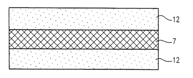
Figure 2 is a cross-sectional view showing an
example of the structure of a composite medical device
according to the present invention, which consists of a
three-layered material containing a reinforcing membrane.
Figure 3 is a plane view showing an example of a
composite medical device according to the present inven-
tion, which is reinforced by stitching.
Figure 4 is an enlarged partial plane view
showing a stitch pattern of a peripheral area a shown in
~1735~
TYO47
g
Figure 3.
Figure 5 is an enlarged partial plane view
showing a stitch pattern of a central area b shown in
Figure 3.
Figure 6 is a cross-sectional view taken along a
line IV-IV in Figure 4.
DESCRIPTION OF THE PREFERRED EMBODIMENTS
A medical device according to the present inven-
tion consists essentially of stratum compactum of tissue
membrane, more specifically, stratum compactum of connec-
tive tissue membrane. In the present invention, the
wording "consists essentially of stratum compactum" means
that a medical device only includes the stratum compactum
but does not include any other components of the tissue
membrane such as epithelium, basement membrane and
fibroblast. The medical device may, however, include
these other components at such an extremely small amount
that these components remain only as a trace. The
stratum compactum has acellular nature. The stratum
compactum of the tissue membrane comprises a collagen
layer including type I, type III, type IV, type V and
type XVI collagens, in which collagen fibers are present
in a finely reticulate form. The medical device accord-
ing to the present invention is obtained by removing
tissue membrane containing stratum compactum from body
tissue and further removing the stratum compactum there-
from. The tissue membrane includes human cerebral dura
mater, human fascia lata, horse pericardium, pig pericar-
- 2173508
TYO47
-- 10 --
dium and human amnion. Preferably, a medical device
according to the present invention is made of human
amnion. Figure 1 is a cross-sectional view showing the
constitution of human amnion (NANKODO Corporation Limit-
ed; KISO TO RINSYO (Basic Medicine and Clinical Medi-
cine), 1981, "Placenta", See page 31, Figure 34 for
amnion at the 13th week of pregnancy) .
With reference to Figure 1, the human amnion
comprises: epithelium and basement membrane 1 (both
collectively denoted by the reference numeral 1); stratum
compactum 2 having a width of about 10 ,um; and fibroblast
3. A medical device according to the present invention
consists essentially of stratum compactum 2, excluding
epithelium, basement membrane 1 and fibroblast 3 from
human amnion. As described later, the components except
for stratum compactum 2 are essentially eliminated by
conducting the following treatments on human amnion. A
method for obtaining a medical device, in particular, a
membranous medical device, according to the present
invention will be described in detail below taking human
amnion as an example.
First, a fetal membrane alone is removed and
separated from fresh placenta, umbilical cord and the
like of an uninfected woman in the state of maternity
immediately after parturition. Blood is immediately
removed from the separated fetal membrane by washing with
a physiological saline solution defined by the Japanese
Pharmacopoeia No. C-1365, i.e., an isotonic sodium chlo-
ride solution.
The fetal membrane from which blood is removed
2173S08
TY047
-- 11 --
is desalinizated and washed with aseptic non-pyrogenic
purified water. The desalinizated and washed fetal
membrane is allowed to stand in 0.1% benzalkonium chlo-
ride solution defined by Japanese Pharmacopoeia No. C-370
for 24 hours or more. It is considered that a treatment
in the benzalkonium chloride solution described above
causes separation among membrane layers constituting the
fetal membrane such as amnion, chorionic membrane,
decidua capsularis and decidua basalis, disinfection and
sterilization, and denaturalization of the cell-contain-
ing layers.
The amnion separated by such a treatment is
subjected to ultrasonication using aseptic non-pyrogenic
purified water.
Next, the thus obtained amnion is subjected to
enzymatic treatment. The enzyme includes ficin which is
one of a number of thiol proteases. The amnion is
immersed in 0.2 M phosphate buffer solution, pH of about
7.0 to about 7.5, preferably, pH of about 7.4, containing
0.01~ ficin at room temperature for 24 hours. Subse-
quently, the amnion treated with ficin is subjected to
ultrasonic washing using aseptic non-pyrogenic purified
water.
The thus obtained membranous material substan-
tially only includes stratum compactum, and excludes all
components except for stratum compactum from amnion.
This is confirmed by the following features of the
resultant membranous material.
(1) No cell-containing layer is found in the
- ~73508
TY047
- 12 -
membranous material through microscopic observation.Moreover, a matrix structure in which a pattern on a
surface is asymmetrical with respect to a matrix pattern
on a bottom face, which is assumed to be possessed by
stratum compactum included in human amnion, is observed.
(2) A ratio of the thickness of stratum compactum
to the total thickness of human amnion is in the range of
about 70 to about 80%. In the case where a thickness of
the membranous material, obtained by the above-mentioned
treatment, is measured for 30 times, and the calculated
average ratio of the thickness is 77%.
(3) The membranous material is acellular and is
composed of collagens, which include collagens of type I,
type III, type IV, type V and type XVI (J. Biochem. 112,
856-863 (1992)).
The above-mentioned membranous material may be
further subjected to sterilization, disinfection,
crosslinking and modification.
The sterilization or disinfection treatment
includes heating including a treatment using an auto-
clave, W irradiation, electron beam irradiation, gammairradiation and a treatment with ethylene oxide gas.
The crosslinking or modification includes a
process of a crosslinking reaction using glutaraldehyde
carbodiimide or succinic anhydride.
Since the above membranous material is strong
enough, the membranous material can be used as a medical
2173508
TY047
- 13 -
device without conducting any further treatments orprocesses. The membranous medical device can be used as
a medical product such as a donor-site skin graft, and
wound and burn dressings. The membranous medical device
is also used as a medical prosthesis for a defective part
of a pleura in combination with fibrin coagulation.
Safety and effectiveness of the membranous medical device
as a homograft allowing the regeneration and self-repair
of tissue can be demonstrated through animal experiments.
In one embodiment, the medical device according
to the invention can be fibrous. The fibrous material
includes filaments, threads and the like. For example,
the threads can be obtained by cutting the membranous
material or the membranous material immersed into gela-
tine or collagen solution into strips and twisting the
strips into threads. Furthermore, a plied thread can be
formed by plying and twisting the obtained threads. This
fibrous material is physically crosslinked, i.e., entan-
gled, or chemically crosslinked to form an extremelystrong thread or piled thread. The resultant fibrous
medical devices can be used as medical devices for
homologous transplantation.
In another embodiment, the membranous material
may be formed into a tubular member. The tubular member
can be formed by, for example, a filament winding method.
The filament winding method includes the steps of:
winding the thread or the piled thread prior to being
crosslinked around a bar-shaped core material in a
layered manner in a wet state so as not to leave any
space; crosslinking the wound material by the addition of
a crosslinking agent, resulting in a tubular material;
~173508
TY047
- 14 -
and removing the bar-shaped core material therefrom. A
strip material obtained by cutting the membranous materi-
al produced by the above method into a strip also can be
used as a starting material instead of the thread in the
filament winding method. The tubular medical device may
be used for an artificial urethra, an artificial ureter,
an artificial esophagus, an artificial trachea or an
artificial blood vessel used for homologous transplanta-
tion.
In the case where a membranous medical device is
used for prosthesis of a portion of an abdominal organ to
be subjected to an operation, the strength of the membra-
nous device merely made of stratum compactum may be
insufficient for performing prosthesis. Therefore, the
membranous material made of stratum compactum may be
reinforced with a reinforcing material so as to be used
as a composite medical device.
The reinforcing material used in the present
invention is a bioabsorbable material. The bioabsorbable
materials can be either naturally-occurring or synthetic.
In a preferred embodiment, the bioabsorbable material is
polyglycolic acid, polylactic acid or a copolymer includ-
ing components thereof (i.e., glycolic acid and/or lactic
acid) as main components.
Although the reinforcing material can be a cloth
including a textile and a knitting fabric, a fibrous
material or a mesh-like material, the form of the rein-
forcing material is not particularly limited. In one
preferred embodiment of the invention, the reinforcing
material is a mesh-like material having an average pore
2173~08
TY047
- 15 -
diameter in the range of about 100 to about 2000 ,um.
As an example of a composite medical deviceaccording to the present invention, a sandwich-like
layered structure is shown in Figure 2. The composite
medical device has a sandwich-like structure in which a
bioabsorbable material 7 is interposed between a pair of
membranous medical materials 12 each consisting essen-
tially of stratum compactum. Preferably, the composite
medical device according to the present invention has
such a structure that a mesh-like bioabsorbable material
is interposed between a pair of membranous materials made
of stratum compactum of human amnion. Any method may be
used as a method for laminating the membranous medical
materials 12 each made of stratum compactum and the
membranous bioabsorbable material 7. For example, roller
compression can be used.
The above-mentioned sandwich-like layered struc-
ture is produced, for example, as follows. First, a pairof membranous materials immersed into gelatine or colla-
gen solution and a mesh fabric made of polyglycolic acid
immersed into gelatine or collagen solution interposed
therebetween. Then, gelatine or collagen molecules which
are uniformly dispersed through the three membranous
materials are chemically crosslinked or physically
crosslinked, i.e., entangled, resulting in a sandwich-
like layered structure integrally formed by crosslinked
gelatine or collagen.
In another preferred embodiment, the membranous
medical material is reinforced by stitching the membrane
made of stratum compactum with a fibrous material made of
- 2173508
TY047
- 16 -
the bioabsorbable material.
A particularly preferred fibrous material made of
the bioabsorbable material is a fibrous material made of
polyglycolic acid, polylactic acid or a copolymer includ-
ing components thereof (i.e., glycolic acid and/or lactic
acid) as main components. A twisted thread which is ob-
tained by twisting a strip formed by cutting the membrane
made of stratum compactum, preferably, stratum compactum
of human amnion and a plied thread which is formed by
plying and twisting the twisted threads can also be used.
The stitch patterns used herein include, but are
not limited to, running stitch and zig-zag stitch. A
region where the membranous medical devices is to be
reinforced by stitching is not limited. A stitching
process can be performed in a peripheral area of the
membranous medical device or the entire membranous
medical device. Figure 3 is a plane view showing an
example of a composite medical device according to the
present invention.
A peripheral area a of the composite medical
device shown in Figure 3 is stitched in running stitches,
for example, along four sides of the composite medical
device. The peripheral area a corresponds to a region
between a line 2 mm inside and a line 20 mm inside from
the outer circumference of four sides of the membranous
material 22. Figure 4 is an enlarged partial plane view
showing the stitch pattern in the peripheral area a. As
shown in Figures 4 and 6, stitches are done so that an
nterval between one stitch line and another stitch line
is 4 mm and an interval between adjacent stitches is 2
2 1 7 3 5 0 8 TYO47
- 17 -
mm. Figure 6 is a cross-sectional view taken along a
line IV-IV in Figure 4.
A central area b surrounded by the peripheral
area a can be stitched in running stitches both in
vertical and horizontal directions. Figure 5 shows an
enlarged partial plane view showing the stitch pattern in
the central area b. As shown in Figure 5, both in
vertical and horizontal directions, stitches are done so
that an interval between one stitch line and another
stitch line is 10 mm and an interval between adjacent
stitches is 6 mm.
The composite medical device reinforced by
stitching is excellent in biocompatibility, bioaffinity,
strength and flexibility as well as easiness to treat and
strength for suture. If a membranous medical material is
reinforced by other membranous reinforcing materials, a
complicated process is required to tightly join the
reinforcing material and the medical material together.
Even when the reinforcing material and the medical
material are successfully joined together, the reinforced
medical device becomes unnecessarily thick. On the other
hand, the composite medical device reinforced by being
stitched according to certain embodiments of the present
invention is scarcely thickened. Thus, the resultant
composite medical device is excellent in flexibility
allowing the medical device to correspond to any shape,
and therefore is excellent in easiness to treat.
The medical device according to the present
invention consists essentially of stratum compactum of
tissue membrane. In general, tissue is formed by cell
2173~08
TY047
- 18 -
growth utilizing stratum compactum as a substrate.
According to the present invention, the medical device is
used while keeping a matrix of the stratum compactum of
the tissue membrane as it is. Therefore, the medical
device allows the regeneration, growth and self-repair of
defective tissue.
Furthermore, the medical device according to the
present invention is excellent in strength and easiness
to treat, and therefore is excellent in operativeness in
surgical operations. Thus, it is possible to facilitate
the treatment in a patient and to allow the patient to
leave hospital earlier than usual. The medical device
according to the present invention is suitably used for
a medical prosthesis and an artificial tissue which are
implantable and tissue-regenerative, such as a pledget,
a bolster, a patch graft, a suture, a donor-site skin
graft, a post-operative antiadhesive, an artificial blood
vessel, an artificial urethra, an artificial ureter, an
artificial esophagus, an artificial trachea, an artifi-
cial membrana tympani, and an artificial oral mucosa.
The present invention provides the excellent medical
device as described above, thereby facilitating the
treatment.
In addition, the human amnion has been treated as
medical waste after parturition, and no use of the human
amnion has been found. Therefore, it is possible to
recover the human amnion as a post-delivery waste. In
this way, a large amount of raw material is available at
low cost.
The composite medical device according to the
- 2173508
TY047
-- 19 --
present invention can have appropriate flexibility which
is almost equal to that of a material before being
reinforced. Therefore, the composite medical device is
excellent in easiness to treat for prosthesis and
anaplerosis of a defective part of the body. The compos-
ite medical device not only sufficiently satisfies the
requirements such as biocompatibility, bioaffinity,
strength and flexibility but also has strength for
suture. The resultant composite medical device has
remarkable effects on repair of an excised or incised
site of an organ such as the liver, pancreas, spleen or
gallbladder and prosthesis of a sutured site of bronchus
as a medical prosthesis for homologous transplantation
having suture reinforcing ability.
Although the present invention will be described
below by way of examples, the present invention is not
limited thereto.
Examples
(1) Working conditions
A. Working environment
1. Working place
All steps except for ultrasonication are con-
ducted on a clean bench placed in a sterilized room(Class 100).
2. Clothing of an operator
An operator puts on sterilized cap, mask, dust-
free garments and shoes in another room before enteringthe clean room.
All operations are conducted while the operator
- 21~3508
TY047
- 20 -
wears a pair of sterilized disposable rubber gloves.Whenever the operator enters and goes out from the clean
room or the operator touches an unsterilized substance,
the rubber gloves should be substituted by another pair
of sterilized rubber gloves.
B. Water to be used for production and the like
1. Water used for production is always treated according
to the following-procedure.
Water is filtrated using a prefilter and an
ultrafiltration membrane. The filtrated water is period-
ically collected, and then is subjected to limulus lysate
test, to detect endotoxin. The water, from which no
endotoxin is detected, is used for production.
2. The apparatus should be sufficiently sterilized with
75% ethanol to prevent bacteria growth therein when not
used for a long time.
3. Containers and tools used in each step are sufficient-
ly sterilized with 75% ethanol and preserved until their
use by immersing into 0.1% benzalkonium chloride solu-
tion.
(2) A production method using amnion of human fetal
membrane
The membranous medical device was produced
according to the following procedures.
An intermediate membrane obtained by,each step
was preserved in 0.1% benzalkonium chloride solution.
2173508
TY047
- 21 -
1. Separation of amnion from fetal membrane
A container made of stainless steel is filledwith 0.1% benzalkonium chloride solution. Then, a human
fetal membrane is placed within the container. The human
fetal membrane is bathed in the solution with hands
wearing rubber gloves so as to peal off amnion. The
obtained amnion is sufficiently washed with water,
thereby obtaining a first intermediate membrane.
2. Removal of foreign substances (1)
The first intermediate membrane is spread over a
plastic plate so as to remove any milky white casein-like
substances. Then, the membrane is sufficiently washed
with water, thereby obtaining a second intermediate
membrane.
3. Ficin treatment
First, 0.2 M phosphate buffer solution, pH 7 is
prepared using sodium hydrogenphosphite and sodium
phosphite. Then, sodium chloride is added to the buffer
solution so as to be 0.9 v/w~, thereby obtaining a buffer
solution used in Examples of the present invention.
Next, 5 liter of the buffer solution is poured into a
container made of stainless steel. Then, the container
is sealed so that the buffer solution is sterilized at
120C for 30 minutes. After cooling the buffer solution,
2.5 g of sodium azide is dissolved into the buffer
solution. Then, 0.5 g of ficin is dissolved into the
obtained solution. Ten second intermediate membranes are
put into the container, and are allowed to stand at room
temperature for 24 hours. Thereafter, the intermediate
membranes are sufficiently washed with water, thereby
obtaining third intermediate membranes.
~173~08
TY047
- 22 -
It is preferred that Step 3 is conducted within24 hours, preferably, several hours, more preferably, one
hour after completion of Step 1.
4. Removal of foreign substances (2)
The third intermediate membrane is spread over a
plastic plate so as to remove any milky white casein-like
substances by rubbing the surface with a plastic strip.
Then, the membrane is sufficiently washed with water,
thereby obtaining a fourth intermediate membrane.
It is preferred that Step 4 is conducted within
24 hours, preferably, several hours, more preferably, one
hour after completion of Step 3.
5. Attachment of frames
A rectangular frame made of polypropylene having
an inner dimension of 24 cm long by 33 cm broad is placed
on a plastic plate. The fourth intermediate membrane is
spread over the frame, and another frame made of
polypropylene having the same dimension is superimposed
thereon. Then, the two frames and the fourth intermedi-
ate membrane are fixed with clips.
6. Ultrasonication
The fourth intermediate membrane fixed with the
clips is suspended in a vessel made of stainless steel.
The fourth intermediate membrane is subjected to ultra-
sonication for 15 minutes using a ultrasonic generator
while overflowing water.
7. Packing
The membrane obtained by the above steps is
- ` 2173~08
TY047
- 23 -
impregnated with 0.1% benzalkonium chloride solution.Then, the membrane is put into a sterilized polyethylene
bag which is in turn thermally sealed.
The resultant membrane consists essentially of
stratum compactum.
(3) Quality of the obtained stratum compactum membrane.
The thus obtained medical device consisting
essentially of stratum compactum has the following quali-
ties.
1. The membrane is transparent or semitranspar-
ent.
2. No foreign substance is found to be attached
to the membrane through observation using a loupe of 10
magnifications.
3. The membrane was placed into an Erlenmeyer
flask, and 100 ml of physiological saline was added
thereto. The Erlenmeyer flask was sealed with an cap
made of aluminum and was heated at 70C for 24 hours.
After being cooled, the membrane was removed from the
Erlenmeyer flask so that a remaining solution serves as
a test solution. The test solution was tested in a
pyrogen test No. B-329 according to Japanese Pharmacopoe-
ia. The test solution was evaluated as suitable in the
test.
4. The membrane was aseptically picked up from
the package, and was partially cut into small pieces with
a pair of sterilized scissors under aseptic conditions.
2173508
TY047
- 24 -
Then, 5 g of the pieces of the membrane was put into atest tube containing 140 ml of thioglycolate medium for
sterility test. A bacterial limit test was conducted in
accordance with sterility test No. B-391 according to
Japanese Pharmacopoeia. The membrane was evaluated as
suitable in the bacterial limit test.
For a fungus limit test, on the other hand,
about 1 g of the pieces of the membrane obtained by the
same procedure as mentioned above was put into a 200 ml
Erlenmeyer flask containing 40 ml of glucose peptone
medium. The fungus limit test was conducted in accor-
dance with sterility test No. B-391 according to Japanese
Pharmacopoeia. The membrane was evaluated as suitable in
the fungus limit test.
(4) Application of the resultant stratum compactum mem-
brane
4.1
The resultant stratum compactum membrane which is
a medical device of the present invention was sealed
within a bag, and sterilized by gamma irradiation at 2.5
megarad rayage.
The medical device having the following proper-
ties: the membranous medical device can be used as a
medical product such as a donor-site skin graft, and
wound and burn dressings; the membranous medical device
can be used as a medical prosthesis for a defective part
of a pleura in combination with fibrin coagulation; and
the membranous medical device has safety and effective-
ness as a homograft allowing regeneration and self-repair
2173508
TY047
- 25 -
of tissue of an affected part. The usefulness of the
membranous medical device in combination with the fibrin
coagulation as a medical prosthesis was confirmed through
the animal experiment as described below.
The membranous medical device was evaluated using
pleuras of female beagle dogs (n=4) as follows. The
pleura was cut into a piece measuring 2 x 2 cm2 using a
scalpel under the anesthesia by Nembutal intravenous
injection and inhalation of halothane to be peeled off.
Then, 500,ul of Liquid B (i.e., a mixture of a calcium
chloride solution and powdered thrombin) was added
dropwise onto the bleeding defective site with a syringe.
Then, a square medical device described above measuring
3 x 3 cm2, which had been washed with distilled water,
air-dried, and then sterilized with ethylene oxide gas,
was placed on the defective site. Furthermore, 500,ul of
Liquid A (i.e., a mixture of an aprotinin solution and a
powdered human fibrinogen containing factor XIII) was
added dropwise onto the medical device to proceed fibrin
coagulation (Beriplast, Behringwerke AG). After one,
five and ten minutes following the application of the
medical device onto the defective part, internal pressure
of respiratory tract was elevated to cause pulmonary
fistula in order to examine the substitution effect of
the medical device. As a control, the fibrin coagulation
was formed by overlaying Liquid A and Liquid B on the
defective site of pleura. The results are shown in Table
below.
2173508
TYO47
- 26 -
Internal pressure at which pulmonary
fistula is caused (cmH2O)
Individual
One minute Five minutes Ten minutes
Number
after appli- after appli- after
cation cation application
1 35 45 55
2 20 25 60
3 15 30 35
4 15 40 45
Mean 20+8.7 34.0+8.2 46.0+5.1
10 +standard
deviation
Control 20 30 30
4.2
The membrane obtained in item 4.1 was cut into
strips. The obtained strips were twisted into threads.
The threads were plied and twisted into a thread. After
being sterilized, these obtained threads can be used as
sutures for homologous transplantations.
-
A tubular member was formed in accordance with a
filament winding method. The membrane obtained in 4.1
was cut into strips. The strip was wound around a
TeflonT~bar-shaped core material having an outer diameter
of 3 mm in a layered manner in a wet state so as not to
leave any space. The wound strip was immersed into 0.2~
glutaraldehyde solution for 15 minutes so as to cause a
- 217~508
TY047
- 27 -
crosslinking reaction, or was dried and heated at 105Cfor 24 hours, resulting in a tubular material made of a
crosslinked material. Then, the bar-shaped core was
removed therefrom. The obtained tubular material was
sterilized, resulting in a tubular medical device.
Another tubular medical device was formed in the
same manner as described above using the thread obtained
in item 4.2.
The resultant tubular medical devices can be used
for an artificial urethra, an artificial ureter, an
artificial esophagus, an artificial trachea or an
artificial blood vessel for homologous transplantation.
4.4
First, 20 g of purified gelatine defined by
Japanese Pharmacopoeia No. D-524 was weighed, and was
dissolved into 500 ml of hot water. Water is added so as
to obtain 2% gelatine solution. Then, a mesh fabric made
of polyglycolic acid having a pore of a diameter of 300
,um was immersed into the 2% gelatine solution, and was
pulled up therefrom. Then, the mesh was superimposed on
the membrane made of stratum compactum (22 cm wide and 31
cm long) obtained in item 4.1, which was spread over a
bench. Another membrane was further superimposed on the
mesh fabric. The resulting layered material was pressed
by a glass roller so that the three layers were suffi-
ciently adhered to each other with gelatine. The ob-
tained layered material was fixed to a polypropyleneframe.
The layered material fixed to the polypropylene
~173508
TY047
- 28 -
frames was suspended within a pressure-reducing, drying
and heating apparatus under aseptic conditions. Aseptic
dried air at 105C was allowed to flow and circulated
throughout the apparatus for 24 hours. The obtained
layered material was not pealed off even in water. The
layered material was crosslinked by immersing it into a
solution containing 500 ml of 0.02 M borate buffer,
pH 9.0 and 100 ml of 5% succinic anhydride solution in
acetone at room temperature for 2 hours so as to cause
succination. After termination of the reaction, the
layered material was sufficiently washed with water and
dried. Then, the layered material was subjected to a
gamma irradiation at 2.5 megarad rayage. As a result,
the reinforced layered medical device was obtained. The
structure of the thus obtained composite medical device
according to the present invention was shown in Figure 2.
The composite medical device has a sandwich-like struc-
ture in which the mesh-like bioabsorbable material 7 for
reinforcement is interposed between a pair of membranous
medical materials 12 each made of stratum compactum.
The thus obtained composite medical device can
exhibit remarkable effects on repair and prosthesis of an
excised or incised site of an organ such as liver,
pancreas, spleen and gallbladder and a sutured site of a
organ of bronchus as a medical prosthesis for homologous
transplantation having suture reinforcing ability.
4.5
A solution of collagen at a concentration of
0.15% by weight was prepared using a buffer solution
containing urea and creatine hydrochloride. The layered
reinforced medical device was obtained by the same method
- 2173508
TY047
- 29 -
as that in item 4.4 except that the collagen solution wasused instead of the purified gelatine solution.
The obtained composite medical device can be used
for the same application as that of the composite medical
device in item 4.4.
4.6
The membranous material 22 made of stratum
compactum was prepared according to the process described
in item 4.1. The membranous material 22 was rectangular,
which measured 22 cm by 31 cm. The membranous material
22 was stitched as shown in Figures 3 through 6 under
aseptic conditions. A polyglycol acid thread having a
size equivalent to No. 40 count was used as a thread 4.
The peripheral area a of the composite medical
device shown in Figure 3 was stitched in running stitches
along four sides of the composite medical device. The
peripheral area a corresponds to a region between a line
2 mm inside and a line 20 mm inside from the outer
circumference of four sides of the membranous material
22. Figure 4 is an enlarged partial plane view showing
the stitch pattern in the peripheral area a. As shown in
Figures 4 and 6, stitches were done so that an interval
between one stitch line and another stitch line is 4 mm
and an interval between adjacent stitches is 2 mm.
Figure 6 is a cross-sectional view taken along a line IV-
IV in Figure 4.
The central area b (a corresponding to a region
inside a line 20 mm inside from the outer circumference
of the membranous material 22) was stitched in running
2173~08
TY047
- 30 -
stitches both in vertical and horizontal directions.Figure 5 shows an enlarged partial plane view showing the
stitch pattern in the central area b. As shown in Figure
5, both in vertical and horizontal directions, stitches
were done so that an interval between one stitch line and
another stitch line was 10 mm and an interval between
adjacent stitches was 6 mm.
The resultant stitched membranous material was
immersed in 2% purified gelatine solution. Then, the
membranous material was pressed by a roller made of
stainless steel on a stainless plate so as to reduce
unevenness of stitches. After having been sufficiently
washed with water, the membranous material was fixed
between a pair of polypropylene frames with clips and was
aseptically dried at 105C, resulting in a composite
medical device. A thickness of the obtained composite
medical device was 16 ,um.
4-7
The same procedure described in item 4.6 was
repeated except that a zig-zag stitch pattern is used as
a stitch pattern instead of the running stitch pattern.
A composite medical device having a thickness of 15 ~m
was obtained.
Various other modifications will be apparent to
and can be readily made by those skilled in the art
without departing from the scope and spirit of this
invention. Accordingly, it is not intended that the
scope of the claims appended hereto be limited to the
description as set forth herein, but rather that the
claims be broadly construed.