Note: Descriptions are shown in the official language in which they were submitted.
CA 02173718 2000-OS-19
1
ULTRASOUND TRANSMISSION MEMBER HAVING IMPROVED
LONGITUDINAL TRANSMISSION PROPERTIES
Field of the Invention
l0 The present invention relates generally to medical devices
and more particularly to an improved ultrasound transmission
member for transmitting ultrasonic energy from an extracorporeal
ultrasound generating device to a location within a mammalian
body.
Related Application
This patent application is related to the subject matter of
U.S. Patent No. 5,304,115.
Background of the Invention
The prior art has included a number of ultrasonic devices
for ablating, destroying or removing obstructive material within
anatomical structures of the body, such as blood vessels.
Examples of devices which purportedly utilize ultrasonic energy,
alone or in conjunction with other treatment modalities, to
remove obstructions from anatomical structures include those
described in United States Patent Nos. 3,433,226 (Boyd),
3,823,717 (Pohlman, et al.), 4,808,153 (Parisi), 4,936,281
(Stasz), 3,565,062 (Kuris), 4,924,863 (Sterzer), 4,870,953 (Don
Michael, et al.), 4,920,954 (Alliger, et al.), and 5,100,423
(Fearnot) as well as other patent publications W087-05739
(Cooper), W089-06515 (Bernstein, et al.), W090-0130 (Sonic Needle
Corp.), EP316789 (Don Michael, et al.), DE3,821,836
(Schubert) and DE2,438,648 (Pohlman).
In particular, flexible ultrasound-delivering catheters
have been utilized to recanalize blood vessels
CA 02173718 2000-OS-19
2
which have become obstructed by atherosclerotic plaque and/or
thrombotic matter.
Previously United States Patent No. 5,304,115 describes
percutaneously insertable ultrasound delivering catheters which
are useable to ultrasonically ablate or remove obstructive matter
from blood vessels. As disclosed in U.S. Patent No. 5,304,115,
1o such ultrasound delivery catheters may be constructed of a
flexible catheter sheath having an elongate ultrasound
transmission member or wire extending longitudinally
therethrough. The cross-sectional dimension of the ultrasound
transmission member may be tapered or narrowed near the distal
end of the member. While such tapering or narrowing of the cross-
sectional diameter of the ultrasound transmission member will
typically decrease its rigidity and improve its bendability at
the region of the taper or narrowing, such tapering or narrowing
of the ultrasound transmission member carries with it a resultant
2o increase in amplitude of the ultrasonic energy being transmitted
through such narrowed or tapered region. Such increase in
amplitude at the narrowed or tapered region may give rise to an
increased likelihood of breakage or fracture of the ultrasound
transmission member.
To facilitate use of ultrasonic ablation techniques within
small tortuous blood vessels or other anatomical structures, it
is desirable to develop small-diameter ultrasound-delivery
catheters which are sufficiently pliable and bendable, at
least in their distal regions, to navigate tortuous anatomical
3o configurations without undue likelihood of breakage or fracture
of the ultrasound transmission member during use.
WO 95/10233 ~ 8 PCT/US94111550
3
In view of the foregoing, there remains a need in the
art for development of new ultrasound transmission members
having improved pliability or bendability with minimal
likelihood of breakage or fracture.
Summary of the Invention
In accordance with the present invention, there 'is
provided an ultrasound transmission member having at least
four regions of differing cross-sectional dimension. The
main proximal region of the member is of substantially
continuous first cross-sectional dimension or diameter.
The second region of the member extends distally from the
distal end of the first region thereof, and is downwardly
tapered to a (continuously or in a step-wise fashion) from
said first cross-sectional dimension to a second cross-
sectional dimension smaller than said first cross-sectional
dimension. A third region of the member extends distally
from the distal end of the second region and is of a
substantially continuous cross-sectional dimension
preferrably equal to said second cross-sectional dimension.
The fourth region of the member extends distally from the
distal end of the third region and is outwardly tapered
(continuously or in a step-wise fashion) to a fourth cross-
sectional dimension, said fourth cross-sectional dimension
being larger than the continuous cross-sectional dimension
of said third region.
Further in accordance with the invention, a sleeve,
sheath or other damping member may be positioned around
the
third region of the ultrasound transmission member to
dampen or limit transverse side-to-side vibration of the
third region during operation.
Still further in accordance with the invention, the
ultrasound transmission member may be formed of various
materials including superelastic metal alloy. A presently
preferred superelastic metal alloy is a nickel titanium
CA 02173718 2000-OS-19
4
alloy containing 50.8 atomic per cent nickel/balance titanium.
Still further in accordance with the invention, the
ultrasound transmission member of the foregoing character may be
incorporated into a flexible ultrasound catheter, said ultrasound
catheter being insertable into a blood vessel or other anatomical
structure for purposes of delivering ultrasonic energy to an
anatomical structure within the mammalian body.
1o Still further in accordance with the invention, the
ultrasound transmission member of the foregoing character may be
incorporated into a guidewire, or other elongate housing or body
for purposes of carrying ultrasonic vibration therethrough.
Therefore, various aspects of the invention are as follows:
An ultrasound transmission member coupleable to an
ultrasound generating device for transmitting ultrasound from
said ultrasound generating device to a location within a
mammalian body, said ultrasound transmitting member comprising:
an elongate member having a proximal end, distal end, and at
least four regions of differing cross-sectional dimension, said
four regions of said elongate member comprising:
i) a first region extending distally from the
proximal end of the member and having a substantially
continuous first cross-sectional dimension;
ii) a second region extending distally from the
distal end of said first region, said second region being
tapered to a second cross-sectional dimension smaller than
said first cross-sectional dimension;
iii) a third region extending distally from the distal
3o end of said second region, said third region being of a
substantially continuous third cross-sectional dimension,
said third cross-sectional dimension being substantially the
same as said second cross-sectional dimension; and
iv) a fourth region extending distally from, the
distal end of said third region, said fourth region being
CA 02173718 2000-OS-19
4a
tapered to a fourth cross-sectional dimension larger than
said third cross-sectional dimension.
A further object of an aspect of the invention is an
ultrasound transmission member coupleable to an ultrasound
generating device for transmitting ultrasound from said
ultrasound generating device to a location within a mammalian
body, said ultrasound transmitting member comprising:
an elongate member having a proximal end, a distal end, and
1o at least four regions of differing cross-sectional dimension,
said four regions of said elongate member comprising:
i) a first region extending distally from the
proximal end of the member and having a substantially
continuous first cross-sectional dimension;
ii) a second region extending distally from the
distal end of said first region, said second region being
tapered to a second cross-sectional dimension smaller than
said first cross-sectional dimension;
iii) a third region extending distally from the distal
2o end of said second region, said third region being of a
substantially continuous third cross-sectional dimension,
said third cross-sectional dimension being substantially the
same as said second cross-sectional dimension;
iv) a fourth region extending distally from the
distal end of said third region, said fourth region being
tapered to a fourth cross-sectional dimension larger than
said third cross-sectional dimension; and
v) a dampening member disposed about said third
region to dampen transverse side-to-side vibrational
3o movement in said third region, said dampening member
comprising a sheath.
A further object of an aspect of the invention is an
ultrasound catheter comprising:
an elongate flexible catheter sheath having a distal end, a
proximal end, and a hollow lumen extending longitudinally
therethrough; and
CA 02173718 2000-OS-19
4b
an ultrasound transmission member extending longitudinally
through the lumen of said catheter sheath, said ultrasound
transmission member comprising:
an elongate member having a proximal end, a distal end, and
at least four regions of differing cross-sectional dimension,
said four regions of said elongate member comprising:
i) a first region extending distally from the
proximal end of the member and having a substantially
1o continuous first cross-sectional dimension;
ii) a second region extending distally from the
distal end of said first region, said second region being
tapered to a second cross-sectional dimension smaller than
said first cross-sectional dimension;
iii) a third region extending distally from the
distal end of said second region, said third region being of
a substantially continuous third cross-sectional dimension,
said third cross-sectional dimension being substantially the
same as said second cross-sectional dimension;
2o and
iv) a fourth region extending distally from the
distal end of said third region, said fourth region being
tapered to a fourth cross-sectional dimension larger than
said third cross-sectional dimension.
Further objects and advantages of the invention will become
apparent to those skilled in the art upon reading and
understanding of the following detailed description and the
accompanying drawings.
Brief Description of the Drawings
Figure 1 is a perspective view of an ultrasound catheter
device of the present invention operatively connected to an
ultrasound generating system.
Figure 2 is an enlarged perspective view of the distal end
CA 02173718 2000-OS-19
4c
of the ultrasound catheter of Figure 1 having a guidewire
(phantom lines) extending therethrough.
Figure 3 is a longitudinal sectional view of the distal
portion of the catheter shown in Figure 1.
Figure 4a is a cross-sectional view through Line 4a-4a of
Figure 3.
Figure 4b is a cross-sectional view through Line 4b-4b of
Figure 3.
Figure 5 is a broken elevational view of the preferred
ultrasound transmission member of the present invention.
WO 95/10233
2 i 7 3
7 I 8 pCT/US94/11550
Figure 6 is a side elevational view of a portion of
the ultrasound transmission member of Figure 5 having a
damping member or sleeve positioned thereon.
Figure 7 is a longitudinal sectional view of a portion
5 of the proximal end connector assembly of the catheter
shown in Figure 1.
Detailed Description of the Preferred Embodiment
The following detailed description and the
accompanying drawings are intended to describe and
illustrate presently preferred embodiments of the invention
only and are not intended to limit the scope of the
invention in any way. Specifically, the hereafter
described embodiments and drawings are not intended to
comprehensively describe or show all of the possible
embodiments of the claimed invention.
A. A Preferred Ultrasound Catheter Incorporatins
An Ultrasound Transmission Member Of The Present Invention
As shown in Figure 1, an ultrasonic catheter 10 of the
present invention may be utilized by coupling the
ultrasonic catheter 10 to an ultrasound generating system
12. The ultrasound generating system 12 comprises a signal
generator 14 (e. g., Model UAG.1110, Baxter Healthcare
Corp., Edwards LIS Division, Irvine, California) connected,
by way of cable 16 to an ultrasound transducer 18 (e. g.,
Model UAT-1000, Baxter Healthcare Corporation, Edwards LIS
Division, Irvine, California), which is operable to convert
the electrical signal into ultrasonic vibration.
The ultrasound catheter 10 of the present invention
comprises an elongate flexible catheter body 20 having an
elongate ultrasound transmission member or wire 22
extending longitudinally therethrough. A proximal end
~RE~'~fFIE~ SKEET (RULE 91)
1SA/EP
WO 95/10233 ~ PCT/US94/11550
6
connector assembly 24 is positioned on the proximal end of
the catheter body 10. As shown in detail in Figure 7 the
proximal connector assembly 24 is configured to facilitate
connection of the proximal end of the ultrasound
transmission member 22 to the ultrasound transducer 18 such
that ultrasonic vibration from the transducer 18 will be
transmitted, distally, through the ultrasound transmission
member 22 to the distal end of the catheter 10.
The ultrasound transmission member 22 of the present
invention may be formed of any suitable material capable of
carrying ultrasonic energy from the proximal end of the
catheter 10 to the distal end thereof. In particular, the
presently preferred embodiment of the ultrasound
transmission member 22 of the present invention is formed
of nickel-titanium alloy which exhibits super elastic
properties within the temperature range under which the
device is operated.
In particular, one presently preferred superelastic
metal alloy of which the ultrasound transmission member 22
may be formed is nickel-titanium alloy consisting of 50.8
atomic percent nickel/balance titanium and is commercially
available as Tinel'" BB from Raychem Corporation, Menlo
Park, California.
The physical properties of the preferred 50.8 atomic per
cent nickel NiTi alloy are as follows:
CA 02173718 2000-OS-19
_7_
properties of NiTi Allov
Ha~q.50.8 At.%Nickel/Balance Titanium
Propert * Units Value
Superelastic C 20 to 80
Tem erature Ran a
Loading Plateau Mpa 480
io Stress at 20C
Unloading Plateau Mpa 135
Stress
Permanent Set % 0.2
(at 20C after
8% strain
i5 Ultimate Tensile Mpa 1150
Stren th at 20C Ksi 170
Elongation at % 10
Failure
Meltin Point C 1350
Density g/cm 6.5
lbs/cu.Inch 0.235
20
*Typical Values for Cold Worked and Shape Set Condition
Examples of superelastic metal alloys which are useable
to form the ultrasound transmission member 22 of the present
25 invention is described in detail in the United States Patent
Nos. 4,665,906 (Jervis); 4,565,589 (Harrison); 4,505,767 (Quip);
and 4,337,090(Harrison), which describe the compositions,
properties, chemistries, and behavior of specific metal alloys
which are superelastic within the temperature range at which the
3o ultrasound transmission member 22 of the present invention
operate, any and all of which superelastic metal alloys may be
useable to form the superelastic ultrasound transmission member
22.
WO 95/10233 PCT/US94/11550
In the preferred embodiment, the ultrasound
transmission member 22 is specifically configured and
constructed to provide desirable flexibility or bendability
near the distal end of the catheter, while at the same time
minimizing the likelihood of breakage or fracture of the
ultrasound transmission member 24 during use.
For example, one preferred configuration is shown in
Figure 5 for an ultrasound transmission member 22 of the
present invention having an overall length of 63. As
l0 shown, the ultrasound transmission member 22 having an
overall length of 63 inches comprises a) a first (proximal)
region 26, b) a second region 28 extending distally from
the first proximal region 26, c) a third region 30
extending distally from the second region 28, and d) a
fourth region 32 extending distally from the third region
30.
The first (proximal) region 26 of the ultrasound
transmission member 22 constitutes the main proximal
portion of the member 22, and extends approximately 50.5
inches from the proximal end thereof. The outer diameter
D1 of the first portion 26 is approximately 0.030 inches
and is substantially continuous over its entire length.
The second region 28 is about 6.2 inches in overall
length and is downwardly tapered, from an outer diameter
equal to D1 at its proximal end, to a smaller outer
diameter D2 at its distal end. The tapering or narrowing
of the second region 28 may be gradually continuous or may
be formed in steps, as shown in Figure 5. Specifically, as
shown in Figure 5, the second region 28 includes first 28A,
second 28B and third 28C subregions. The first subregion
28A is gradually tapered from diameter D1 to an
intermediate diameter D1.5 between D1 and D2. The second
subregion 28B is of substantially continuous intermediate
diameter D1.5 over its entire length. The third subregion
WO 95/10233
~ ~ PCT/US94/11550
9
28C is then further downwardly tapered to a diameter of
D2,
as shown.
The third region 30 of the ultrasound transmission
member 22 is of continuous diameter D3 over its entire
length of approximately 0.750 inches. Diameter D3 is the
same as diameter D2 at the distal end of the second region
28.
The fourth region 32 of the ultrasound transmission
wire 22 is outwardly tapered or enlarged from diameter
D3
at its proximal end to diameter D4 at its distal end. The
fourth region 32 of the ultrasound transmission member
22
may 'be of a gradually continuous taper or may include
multiple subregions, as shown in Figure 5. Specifically,
as shown in Figure 5, the fourth region 32 has an overall
length of 0.300 inches~and comprises a first subregion
32A
and a second subregion 32B. In the embodiment shown, the
first subregion 32A is gradually tapered from diameter
D3
to diameter D4. The second subregion 32B is of
substantially continuous outer diameter D4. Diameter D4
is
approximately 0.014 inches.
Because the third region 30 of the ultrasound
transmission member 22 is of minimal diameter D3, such
third region 30 is subject to exaggerated lateral or side-
to-side vibration during use. In order to dampen or limit
the lateral side-to-side vibration of the third region
30,
an external dampering member, such as a sheath 40, may
be
applied to such region 30 to limit its propensity for
lateral side-to-side movement. As shown in Figure 6, the
preferred sheath member 40 comprises a segment of plastic
tubing surrounding the entire third region 30 of ultrasound
transmission member 22. The inner diameter (ID) of sheath
member 40 is sized relative to the ultrasound transmission
member 22 such that the proximal and distal ends of the
sheath member 40 are flush with and engage the adjacent
outer surfaces of the second region 28 and fourth region
32
WO 95/10233 2 PCT/US94/11550
of the ultrasound transmission member 22, as shown.
Adhesive 42 is utilized to bond, at least the end portions
of sheath member 40 to the adjacent outer surfaces of the
ultrasound transmission member 22.
5 The inner diameter of sheath 40 is larger than the
outer diameter of third region 30 such that a space 44
exists therebetween. Space 45 may optionally be filled
with matter capable of damping or inhibiting lateral side-
to-side movement of the third region 30. Examples of
10 damping material which may be disposed within space 45
include RTV silicone (product code, manufacturer, city,
state) or other elastic materials such as natural or
synthetic rubber. As an alternative, the quantity of
adhesive 42 may be increased such that the adhesive 42
fills the entire space 45 between the outer surface of the
third region 30 and the inner diameter of sheath 40. In
such embodiments, it will be recognized that adhesion of
the adhesive 42 to the outer surface of the third region 30
may limit the desirable longitudinal vibration of the third
region 30, in addition to the undesirable lateral or side-
to-side vibration thereof. To avoid such adhesion to the
third region 30, anti-adhesive materials or release agents
may be applied to the third region 30 prior to disposition
of the adhesive 42, thereby preventing the adhesive 42 from
.25 adhering to the outer surfaces of the third region 30,
while permitting the adhesive to form the desirable bond
with the adjacent surfaces of the second region 28 and
fourth region 32 so as to hold sheath 40 firmly in place.
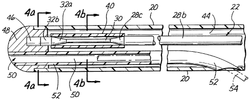
As shown in Figures 3-4, the catheter body comprises
a hollow tube having a longitudinal bore or lumen 44
extending therethrough. A rigid distal head or endcap 48
is inserted to the distal end of the catheter body 20. In
the embodiment shown, the distal head 40 has a generally
smooth rounded outer configuration so as to form a blunt
tip which is flush and continuous with the adjacent outer
2i 73718
WO 95/10233 PCT/US94/11550
11
surface of the catheter body 20. A blind cul de sac or
bore 46 is formed in the proximal side of the distal head
48 to receive the distal end of the ultrasound transmission
member 22 therein. As shown, the distal end of the
ultrasound transmission member 22 is inserted part way into
i
bore 46 and may be welded, adhered or mechanically engagecT
thereto so as to hold distal head 48 in its desired
longitudinal position within the distal end of the catheter
body 20 and also to form abutting contact between the
distal end of the ultrasound transmission member 22 and
the
distal head 48. As such, ultrasonic vibration which passes
distally through the ultrasound transmission member 22 will
be transmitted into the distal head 48, thereby causing
distal head 48 to vibrate in accordance with the energy
transmitted through ultrasound transmission member 22.
Also in the embodiment of the catheter shown in
Figures 3-4, a guidewire lumen 50 extends through the
distal head 48 and partially through a distal portion of
the catheter body. A guidewire (phantom lines) may be
passed through the guidewire lumen to facilitate insertion
and positioning of the catheter 10.
The guidewire lumen 50 is at least partially defined
by the inner lumen of a tube 52. The guidewire lumen 50
extends through a longitudinal bore formed in the distal
head 48 and through a distal portion of the lumen 44 of
the
catheter body 20. The proximal end of tube 52 is flush
with and may be bonded to the sidewall of the catheter 20,
thereby forming sidewall guidewire aperture 54 in catheter
body 20.
Also, in the embodiment shown, dual infusion apertures
56 extend longitudinally through distal head 48 in fluidic
communication with the hollow bore 44 of catheter 20. A
fluid infusion sidearm 58 is formed in the proximal end
connector assembly 24 to permit infusion of fluid through
the bore of the proximal connector assembly 24 and through
R'O 95/10233 PCT/US94/11550
21~~~~ 18
12
the hollow lumen 44 of the catheter 20 such that said fluid
will pass out of the dual infusion apertures 55 located in
the distal head 48 of the device. Such passage of fluid
through the catheter 20 may be for purposes of cooling or
controlling the temperature of the ultrasound transmission
member 22 and/or may also be for purposes of providing an
infusion of irrigation fluid, radiographic contrast~media,
oxygenated perfusate and/or medicaments.
One type of proximal connector assembly 24 which may
be utilized as part of the catheter device 10 is shown, in
detail, in Figure 7. The proximal connector assembly 24
shown in Figure 7 comprises an elongate, rigid body 55
defining a frontal portion 58, a mid-portion 60 and a rear
portion 62. The frontal portion 58 of the elongate body 56
is firmly connected to the proximal end of the catheter
body 20 by way of a threaded gripping member 64 engaged
thereto. In this respect, the proximal end of the catheter
portion 11 preferably has a flared configuration and
includes an annular flange formed on the outermost end
thereof which is brought into sealed engagement with the
connector assembly 12 when the gripping member 64 is
threadably engaged to the body 56. The proximal end of the
frontal portion 58 is connected to the distal end of the
mid-portion 60 of the elongate body 56 by way of a second
gripping member 66. As will be recognized, to facilitate
the aforementioned construction, threads are formed on the
distal ends of the frontal portion 58 and the mid-portion
60. Additionally, as seen in Figure 7, the proximal end of
the mid-portion 60 is non-threaded and is slideably
received into a corresponding bore formed in the distal end
of the rear portion 62 of the body 56. In this respect,
the mid-portion 60 is maintained in engagements to the rear
portion 62 via the utilization of an adhesive or other
suitable affixation method.
~ ~ l 3 ~ ~ g pCT/US94l11550
WO 95/10233
13
Referring further to Figure 7, the rear portion 62 of
the body 56 comprises a distal member 68, the distal end of
which is adapted to receive the proximal end of the mid
Y
portion 60, and a generally frusto-conical proximal member
70. The proximal end of the distal member 68 is formed of
a reduced diameter and is slideably inserted into a
complimentary recess defined in the distal end of the
proximal member 70. The proximal member 70 is maintained
in engagement to the distal member 68 via the utilization
of a threaded fastener 72 such as a screw which is extended
through the bore defining wall of the proximal member 70
and into a threaded aperture disposed within the reduced
diameter proximal end of the distal member 68. The
ultrasound transmission member 22 extend longitudinally
through the entire catheter portion 11 and through the
proximal end of the connector assembly 12. The ultrasound
transmission members 22 are then inserted into and engaged
by a threaded proximal connector 74 which is positioned
within a cylindrical recess formed in the proximal end of
the proximal member 70. The ultrasound transducer 18 is
cooperatively engaged to the proximal connector 74 in a
manner adapted to accomplish the passage of ultrasonic
energy through the ultrasound transmission member 22 in a
distal direction to the distal end of the catheter body 20.
The extreme proximal end of the proximal member 70 is
provided with a sonic connector assembly or apparatus
configured to effect operative attachment of the proximal
ends of the ultrasound transmission member 22 to the horn
of the ultrasound transducer 18. The sonic connector
assembly or apparatus is preferably configured and
constructed to permit passage of ultrasound energy through
the ultrasound transmission member 22 with minimal lateral
side-to-side movement of the ultrasound transmission
members 22 while, at the same time, permitting unrestricted
longitudinal forward/backward vibration or movement of the
WO 95/10233 PCT/US9~/11550
21-~ -~ -~ ~ g 14
1
ultrasound transmission member 22. Specifically, a distal
portion of the body of the threaded proximal connector 74
is configured to receive therein a compressible gripping
4
ferrule 76. The compressible gripping ferrule 76 has a
small central aperture formed therethrough through which
the ultrasound transmission member 22 passes, as shown. A
frontal member 78 is threadably tightened within the
frontal portion of the body of the proximal connector 74 so
as to compress the gripping ferrule 76, thereby causing the
gripping ferrule 76 to firmly grip and hold the ultrasound
transmission member 22 in place within the body of the
proximal connector 74. The proximal connector 74 may then
be compressed or crimped inwardly so as to be additionally
crimp connected or crimp fit to the proximal ends of the
ultrasound transmission member 22, thereby providing
further gripping and attachment of the sonic connector
assembly to the proximal ends of the ultrasound
transmission member 22. The proximal connector 74 is
further formed to permit the distal end of the ultrasound
transducer horn to be releasably engaged thereto and thus
releasably attached to the sonic connector assembly. Thus,
the frontal member 78, gripping ferrule 76, and proximal
connector 74 combine to form a sonic connector assembly to
which the horn of the ultrasound transducer 18 may be
attached and through which the ultrasonic energy may be
transmitted into the ultrasound transmission member 22. A
lumen 80 extending through the rear and mid-portions 62, 60
of the connector assembly 24 is specifically sized to be
large enough to permit the ultrasound transmission member
22 to pass therethrough with a small amount of space
remaining between the outer surfaces of the ultrasound
transmission member 24 and the innerlumenal surface of the
lumen. Also disposed within the mid-portion receiving bore
formed in the distal end of the distal member 68 is on O-
ring 82 which is used to prevent the passage of any fluid
2173718
WO 95/10233 PCT/US94/11550
along the outer surfaces of the lumen 80 into the proximal
member 70 of the rear portion 62.
B. Operation of the Preferred Embodiment
In operation, the catheter 20 described hereabove may
5 be inserted percutaneously, or otherwise, into a desired
anatomical structure such as a blood vessel. The proximal.
connector assembly 24 of the device will then be connected
to ultrasound transducer 18. Depression of on/off foot
pedal 59 will cause signal generator 14 to emit a desired
10 electrical signal through cable 16 to ultrasound transducer
18. Ultrasound transducer 18 will convert the received
electrical signal to ultrasonic vibration and such
ultrasonic vibration will be passed through ultrasound
transmission member 22 to the distal head 48 of the
15 catheter 10.
As the ultrasonic energy passes from the first region
26 of the ultrasound transmission member 22 into the second
region 28 thereof, the narrowing or taper of the second
region 28 will result in an increase in the amplitude of
the ultrasonic energy passing therethrough. Thereafter, as
the ultrasonic energy passes through the constant diameter
third region 30 of the ultrasound transmission member 22
the amplitude will remain substantially constant.
Thereafter, as the ultrasound energy passes from the third
region 30 to the outwardly tapered or enlarging fourth
region 32, the amplitude of the ultrasound will again
decrease in accordance with the change in outer diameter of
the ultrasound transmission member 22.
Although the invention has been described herein with
specific reference to presently preferred embodiments
thereof, it will be appreciated by those skilled in the art
that various additions, modifications, deletions and
alterations may be made to such preferred embodiments
without departing from the spirit and scope of the
invention. For example, the ultrsound transmission member
WO 95/10233 ~ ~ ~ PCT/US94/11550
16
of the present invention may be positioned within many
different catheters which differ in configuration and
construction from the preferred catheter shown in this
patent application or, the ultrasound transmission member
of the present invention may be positioned in, or
incorporated in, a guidewire or may be utilized independent
of any surrounding catheter sheath as described herein with
respect to the preferred embodiment. Accordingly, it is
intended that all reasonably foreseeable additions,
deletions, alterations and modifications be included within
the scope of the invention as defined in the following
claims.