Note: Descriptions are shown in the official language in which they were submitted.
~O 95110990 217 ~ ~ 9 2 PCIIGB941022~
BONE IMP LANT S
This invantlon to bone implants znd in par -~ul2r to
pros.heses fc. total join. replacement.
Total joint replacement is now becoming a c~mmonplace
method of t;~ating disorders, such 2s acute -~thrit~s,
whera the diseased joint is removed surgic-lly and
replaced with an artificial joint. As such cperatlons
hava become more common, problems arising from long-~e~m
wear of sucA ,oints have ~ecome apDare~t. In a number of
instances, it is found that bone resor2tion occ -s in the
region of the implant which leads to the loosenm.g of the
impl2nt in the bone canal, and the breakdown of zny cement
mantle between the implant and the bone canal.
Various suggestions have been made as to the cause of
such degeneration. It is believed that a primary reason
for this loosening is the ingress of wear particles,
generated by prolonged movement of the articulating
surfaces of the joint and that such particles migrate from
the area of articulation into the junction hetween the
implant and the bone. It is believed that wear particles
generated in this way at the articulating surfaces migrate
- along the cement/metal interface (or between the bone and
the implant), and cause endosteal erosion. Progressive
erosion ultimately causes breakdown of the cement mantle
or sufficient loosening of a cement-less stem that it can
become displaced from the bone canal or socket.
~095/10990 ~ 1 7 3 ~ ~2 PCT/GB9~/0228~
It is a primary object of the present in~sntion to
provide a msans for o~s-coming this problem and enable
total joint replacement prostheses to have a longer
effective life.
According to one ~spect of the present invention,
there is provided a method of sealing the interface
between a prosthesis and a bone, in which the prosthesis
is implanted, which method comprises applying a membrane
over the junction bet~een the prosthesis and the bone,
said membrane having a microporous structure whereby
liquids are able to p2SS through the membrane but wear
particles generated by articulation of the prosthesis are
excluded.
Preferably, the microporous membrane has openings
which are sized so as to exclude connecti~e tissue cells,
whereby bone regeneration under the membrane is
also encouraged.
Since the membrane is installed essentially
permanently in the area of the prosthesis, the membrane
should be one which is highly bio-compatible and
essentially inert to body fluids. Examples of suitable
polymer materials include silicone polymers, polyurethane,
polyethylene, polyesters, polypropylene, polyacrylates and
methacrylates and fluorinated olefins, especially
perfluorinated olefins, e.g. perfluorinated ethylene and
propylene. Polytetra 1uoroethylene is currently
217 3 9 9 2 pcTlGs9~lo~8~
_ 3
preferred. `~ethods of producing mic~~porous polvmer
membranes o~ this type a-e known and are cesc-:~ed, for
example, in US Patent Nos. 39535Oo & 4187390. ~- example
of one ccmme~cially available p-oduct is the ~-~~oporous
polytetrafluoroethylene materi21 manufacturec b~ W. L.
Gore & Associ~tes Limitec, under the t-ade mark "G~RE-TEX
ePTrr". Thi~ material h2s been used successful -J in the
past as a s~ture mate~ial cnd also fo- Fc~iodont21
material, to encourage bone grcwth in the area of tooth
roots where t~e gums are regressed. The ~ear ~articles
produced by 2-ticulation of the joint are generally small-
particles of metal or plastics material of micrc~ or sub-
micron size and the pore size in the membrane material
should be selected so as to exclude particles of such
sizes. The above cited US Patents give details of how
such microporous materials can be manufactured and its
disclosure is specifically incorporated herein. The
membrane may be a single sheet material or a laminate.
The invention will now be illustrated with reference
to the accompanying drawings, describing the application
of the invention to a total hip replacement prosthesis,
although it will be appreciated that the in~ention may
also be applied to other joints, including those in which
no articulation takes place. The method and procedure of
the present in~ention may be applied to both cemented and
cementless (press-fit) implants.
_ wo95/1osso ~1~ 7 39 g2 pcTlGBs~lo~28
In the accompanying arawings,
Figure ' is a diagrammatic view of a total hip
prosthesis illustrating the problem arising from long-term
wear in the prosthesis,
Figure 2 is a view similar to Figure l and
illustrates the solution provided by the present
invention,
Figure 3 is a further view similar to Figure 2 but
showing addi.ional details as to the manner in which the
membrane may be attached, and
Figure 4 is a partial view, slightly enlarged, of the
proximal part of the stem of the implant shown in Figure
3.
Referring to Figures l and 2, articulating movement
of the ball l in the acetabulum insert 2 causes wear
particles to be generated which are released into the
space within the joint and are pumped by such articulating
movement into fissures or openings between the
intramedulary canal and the stem of the femoral implant.
Similar ingress of wear particles takes place between the
acetabulum and the hemispherical socket implant 2. After
a period of time, such wear particles migrate along the
cement/metal interface and lodge in areas where they
initiate endosteal erosion of the bone. Ultimately, the
joint becomes so loose that a revision operation is
. .
necessary.
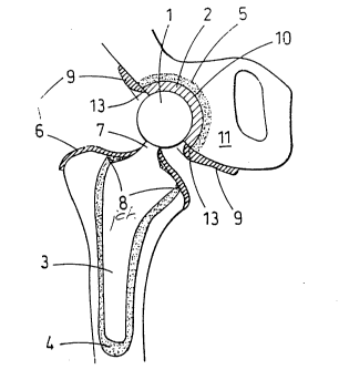
vo gS/log9O ~ ~ 7 3 ~ 9 2 pcTlGs94lo228~
The solution is illustrated in Figure Z, ~ which a
femoral implant having 2 stem 3 is installed i~. a femur
using a cement metal 1 and a socket 2 is similarly
installed using a man~le of bone c~ment in the
acetabulum. The proxi.~al end of the femur s covered
with a microporous memDr2ne 6, which is cut to â' ze to fit
snugly around the neck 7 of the femoral implant so that it
covers the junction 8, ~etween the bone cana: and the
implant, thus providing a primary seal against '~.e insress
of wear particles. Similarly, the soc~et 2 i~ provided
with a ring-like shaped me.~brane 9, covering th~ junction
10 between the socket 2 and the acetabulum 11. Obviously
a gap must be left so that free movement and a-~iculation
of the ball 1 in the soc.~et 2 is not interfered with but
some secure attachment of the edge 13 of the meIbrane to
the socket member 2 is highly desirable. Thls may be
achieved, for example, by welding e.g. by ultrasonic
welding of the membrane to the plastic material of the
socket 2. However, it may be possible to securely attach
the ring or collar 9 of membrane material to the
surro~ln~;ng area of the bone sufficiently securely by
-` suturing or stapling. Another method of attaching the
membrane to the socket m~mher is by means of an adhesive.
Suitable synthetic polymeric adhesives include
thermosetting adhesives such as acrylates, epoxy and
polyester resins, glass ionomer or polyurethanes.
~73~92 `
95/loggo PcTlGB91/0228~
Figures 3 and 4 show variations in the ,~ethod of
attachment OL the membrane over the proximal ~nd of the
femoral stem and femur. As can be seen in ~igure 3, the
membrane 6 is secured to the neck of the femoral implant
by trapping it between a separate neck component 20 and
the stem 21. In the arrangement illustrated in Figure 3,
the neck portion is a taper fitting to the femoral stem.
In another possible embodiment, the neck portion is
threaded and fits into a threaded socket within the stem,
screwing of the neck portion into the stem also trapping
the inner edge of the membrane and thereby securing the
membrane to the femoral implant. The periphery of the
membrane 6 extends past the junction between the canal and
the implant and is attached firmly to the bone by stapling
or suturing or by means of an adhesive, such as one of
those mentioned above. Photocurable resin adhesives such
as those used in dentistry may also be employed in order
to increase the speed of attachment to the bone.
Figure 4 illustrates an additional benefit of the
invention. By selecting a membrane 6, whose pore
openings are sized so as to exclude connective tissue
cells, the conditions necessary for bone growth are
encouraged beneath the membrane so that after a period of
time, bone will grow from the upper end of the femur in
the direction of the arrows 22 over the junction between
the stem of the femoral implant and the bone canal. This
~ ~ ~S 3 9 92
95/loggO pcTlGs94lo228l
will have the dual effect, of furtr.er locking t-s implant
fir~ly into positicn and, also, providing a per~-nent seal
pre~snting further ing-sss of wear particles ~used by
art~oulation of the jo nt components. It will be
app-eciated, therefore, that the invention is zpplicable
to cases whe e the pr~sthesis does not i.^lude any
articulating parts. In such cases, there is a ~ ~efit in
enc~uragins bone regene-ation. This is Fæ-ticul2rly
app~icable in the case o_ revision prostheses.
As explained above, the membranes employcr in the
present invention are preferably bio-compatible
microporous membranes which are manufacture~ by the
-ocess described in the above cited US Patents. The
mem~rane will preferably have a thic~ness bet-~een about
0.05 to 0.25 mm, especially about 0.08 to 0.2 mm, e.g. 0.1
to 0.18 mm.
The membrane may be a composite structure. For
example, the portion of the membrane in contact with the
prosthesis or overlapped cnto the surrounding bone may be
laminated to a plastics material which is more readily
bonded to the bone or the metal of the prosthesis. Such
plastic materials can be l~min~ted to the microporous
membrane by heat and pressure, with or without an
adhesive.
Although the invention as described above employs a
semi-permeable membrane, it is possible to use instead an
, ~ r ~ ~d~; -
~7~99~
WO95/10990 PCT/GB94/02284
impermeable membrane where the objective is solely to
exclude wear particles from the implant/bone interface.
In such cases, the membrane may be a continuous film of a
biocompatible sheet materialj preferably a luorinated
olefin, such as P.T.F.E.