Note: Descriptions are shown in the official language in which they were submitted.
~-- 21~0~S
DEVICE FOR INTRATRACHEAL VENTILATION AND INTRATRACHEAL
PU~MONARY VENTILATION INCLUDING REVERSE ~ u~I
Technical Field
This invention relates generally to medical devices, and
more particularly to devices for delivering fresh air, oxygen or
S an oxygen-containing ga~ to the trachea and lungs of a human or
veterinary patient.
Background of the Invent;on
Mechanical ventilation has long been used to support lung
function in a patient, and entails the use of warmed and moistened
fresh air, oxygen, or an oxygen-containing gas (such as an
anesthetic gas) supplied to the patient under pressure for
oxygenating the patient's lungs. One known method of mechanical
ventilation is intratracheal ventilation (ITV). ITV involves the
delivery of a warmed and well-humidified gas at or near the
patient's carina (the fork of the trachea leading to the bronchial
tubes). The gas is delivered through a ventilator tube positioned
in the trachea. The ventilator tube can be a catheter, a tracheal
tube, an endotracheal tube or the like, and the oxygen-containing
gas can be supplied through it on either a continuous or periodic
basis. In the former case, the ventilator tube can be supplied
with a positive end expiratory pressure (PEEP) valve, whose
periodic activation allows air from the lungs (enriched in carbon
dioxide) to exit the patient's body.
While they do provide some degree of oxygenation when
initially employed, the continued use of mechanical ventilators
for ITV is subject to signi~icant drawbacks. For example,
mechanical ventilators have traditionally been required to deliver
high gas pressures, in order to achieve adequate oxygenation.
~ ~ 1760~
There are several reasons for this. When a mechanical ventilator
is first installed, the interior of the ventilator tube usually
contains just air, rather than an oxygen-enriched gas; when
activated, this air is pushed into the lungs, while the oxygen-
enriched gas remains outside the lungs. The alveoli in the
patient's lungs expire carbon dioxide, and the expired carbon
dioxide builds up in the lungs after several ventilation cycles,
because the high pressure of the oxygen-enriched gas -- and any
straight air between the gas and the carbon dioxide in the lungs
-- prevents at least some of the carbon dioxide from escaping the
lungs. The elevated level of carbon dioxide leads the health
practitioner to supply even higher pressures of the oxygen-
enriched gas to the ventilator tube. After a time as short as
only a few hours, the patient's lungs may suffer damage from two
sources, specifically, the elevated ventilator pressure, and the
build-up of carbon dioxide. The result can be hypoxia,
respiratory acidosis, hypercarbia, iatrogenic lung damage,
pulmonary hypertension, overinflation, and/or pulmonary
parenchymal injury. These problems can even be severe enough to
result in the death of the patient. Further, while the~e problems
are more likely to occur and to be more severe in patients with
significantly impaired lung function (such as pediatric patients
and patients who have undergone a partial lung removal), these
problems can even occur in patients with healthy lung~.
Another known method of mechanical ventilation is
intratracheal pulmonary ventilation (ITPV). ITPV similarly
involves the delivery of an oxygen-containing gas at or near the
patient's carina. The gas is continuously supplied either at a
constant pressure, or by pressure pulses at a frequency of about
1 to 50 cycles per second. One drawback to this method is that
carbon dioxide outflow is periodic or intermittent, controlled by
relatively complex valve and timer mechanism~. As a re~ult, the
potential remains for an inadequate expiration of carbon dioxide
and a resultant progressive build up of the carbon dioxide level
-
5 ~
- 3 -
in the patient. Additionally, the cyclical peak pressures
typically employed in this method are high, often significantly
higher than the pressures employed in ITV. Accordingly, all of
the problems which may be encountered in the use of ITV may also
be faced during the use of ITPV.
One solution to these and other problems has been the
Kolobow reverse thrust catheter, such as disclosed in U.S. Patent
No. 5,186,167 (Feb. 16, 1993). The disclosure of that patent i8
expressly incorporated by reference herein. By way of summary,
the Kolobow device has a catheter preferably contained in a
tracheal or endotracheal tube. The catheter include~ a plurality
of ports through its distal end, the distal end of the catheter
being positioned at or near the patient's carina. Air supplied
through the catheter diffuses transversely through the ports,
creating zones of sub-atmospheric pressure which facilitate
removal of carbon dioxide-laden air from the patient's lungs. The
particular embodiment shown in FIG. 3C in the patent includes a
tubular portion 19 on the catheter tip 16 which defines an annular
exit port 17, directing the flow o~ air and oxygen in a direction
opposite the distal end 18 of the catheter. The patent notes at
column 8, lines 4 through 14, that while it is preferred that the
exit port is annular, it is not necessary to employ a port having
the specific shape of an annulus; rather, the essential feature
is to provide a means which directs the air and oxygen in a
direction opposed to the distal end 18 of the catheter tip 16.
Although not described in such terms in the patent, the low
pressure zones are produced by the well-known venturi effect. For
convenience, since the flow creating the venturi effect is
directed opposite to the incoming flow of air and oxygen, devices
of this type will be referred to herein as "reverse venturi
devices."
The Kolobow assists in the removal of carbon dioxide-laden
air out of the patient's lungs, but in accordance with the present
invention it has been discovered that when the arrangement is used
21760S~
- 4 -
in the presence of a tracheal or endotracheal tube, the
improvement is used in the presence of a tracheal or endotracheal
tube, the improvement in that removal is remarkable enhanced.
Oxygenation can be achieved at pressure levels significantly lower
than the pressures employed in Kolobow, thereby substantially
reducing the risk of trauma to the patient from elevated
ventilation pressures.
Unfortunately, the Kolobow or reverse venturi device (at
least, insofar as actually constructed in practice) potentially
presents a different risk to the patient. In the device, the
catheter tip 16 causing the venturi e~fect i5 generally of solid
material and is pressed over and attached to the distal end of a
hollow and flexible catheter. As a practical matter, such
attachment is problematic, due to the nature of the materials used
and the small dimensions encountered. The result is that the
catheter tip 16 may loosen and separate ~rom the distal end of the
catheter, and be deposited in one of the bronchi or lungs of the
patient. As a result, the catheter tip 16 may damage the tissue
of the lung and create complications such as ~luid pockets,
infections and patient discomfort. Furthermore, the separated
catheter tip 16 may require surgical removal, and the
complications attendant to lung surgery.
Sl]mm~r~ o the Invent;on
The foregoing problems are solved and a technical advance
is achieved in an illustrative reverse venturi device for creating
a sub-atmospheric pressure near the carina of a human or
veterinary patient, employed for intratracheal and/or
intratracheal pulmonary ventilation. The device is a reverse
venturi device, generally of the type disclosed by Kolobow, but
which in any of several ways prevents or avoids the complications
which might arise ~rom ~eparation o~ the catheter tip 16 of
Kolobow. More particularly, the present invention is directed to
~ ~1760~
- 5 -
a device for creating a sub-atmospheric pressure near the carina
of a human or veterinary patient, which includes a channel or
perforation for the passage of an oxygen-containing gas
therethrough, the channel or perforation being open in a direction
distal of the patient, so as to establish a zone of sub-
atmospheric pressure by reverse venturi effect during patient
exhalation. "Sub-atmospheric pressure" means a pressure below at
least the air pressure within the lungs and bronchi at the start
of patient exhalation, and is preferably a pressure below the
ambient atmospheric pressure.
The sub-atmospheric pressure created in the present
invention is advantageous in that it facilitates removal of carbon
dioxide from the lungs of the patient and permits intratracheal
and/or intratracheal pulmonary ventilation to be performed at
pressures less than those conventionally required for such
ventilation. The device of the present invention is also
advantageous in obviating the potential risks in prior reverse
venturi devices of the type disclosed in the Kolobow reference,
speci~ically, the pos~ibility o~ detachment of the tubular member
in those devices, and the possible complications of surgical
recovery o the tubular member from the bronchi or lungs of the
patient. The device of the present invention achieves this latter
advantage either by completely eliminating the tubular member 19
of Kolobow and similar reverse venturi devices, or by providing
a shoulder affirmatively preventing distal movement and loss of
it.
In a first aspect, then, the present invention is directed
to a device for creating a sub-atmospheric pressure near the
carina of a human or veterinary patient, which comprises a
tracheal or endotracheal tube having proximal and distal ends, a
wall connecting them, and a passageway for the flow of an oxygen-
containing gas; and which also comprises an insert received in and
circumferentially abutted by the distal end of the tracheal or
endotracheal tube, the insert having a surface defining at least
2~7~55
6 -
one channel in communication with the passageway of the tube, the
channel being open towards the proximal end of the tube; such that
a zone of sub-atmospheri.c pressure is established, inside the tube
and adjacent the distal end of the tube during exhalation of the
patient, upon passage of an oxygen-containing gas through the tube
passageway and the at least one channel of the insert.
Preferably, the tracheal or endotracheal tube is a
multiple lumen tube, in which the passageway is formed in the wall
o~ the tube and includes an outlet adjacent the distal end of the
tube. Also preferably, the insert i8 shaped as a hollow cylinder
having a plurality of axial channels and a circumferential channel
connecting the axial channels to the passageway outlet at the
distal end of the tube. Advantageously, the inner surface of the
wall of the tube and the outer surface of the insert together
define the at least one ch~nnel . The device can further comprise
a source o~ an oxygen-containing gas, preferably a continuous
source of such a gas, connected to the passageway of the tube.
This aspect of the invention is particularly advantageous
over the device disclosed by Kolobow in that it renders the
tubular portion 19 of Kolobow unnecessary, thereby affirmatively
preventing any risk of separation of that portion from the
catheter and its loss inside the patient, thus avoiding any
potential trauma or complications from procedures needed to
recover it.
Furthermore, the tracheal or endotracheal tube
advantageously includes a second passageway in the wall of the
tube, which has an outlet distal of the insert. This second
passageway is utilized for infusing drugs, but more importantly,
monitoring the pressure at the distal end of the tube and/or near
the carina of the patient.
In a second aspect, the present invention is directed to
a device having the same purpose, but which instead comprises a
catheter having proximal and distal ends, as well as a sidewall
extending between the catheter ends, the sidewall having an
~ 217605~
- 7 -
external surface; and which further comprises at least one
perforation through the catheter sidewall near the distal end of
the catheter; wherein the at least one perforation is acutely
angled with respect to the external surface of the catheter
sidewall, such that a zone of sub-atmospheric pressure is
established, outside the catheter adjacent its distal end during
patient exhalation, upon passage of an oxygen-containing gas
through the catheter and the at least one perforation.
Preferably, a plurality of the perforations are formed by
slits cut into the external surface of the catheter sidewall, the
slits being cut at an angle of less than 45 degrees with respect
to the external surface o~ the sidewall, more preferably at an
angle of no more than about 30 degrees. Also preferably, the
catheter sidewall is constructed of a nylon radiopaque material
tubing, or a material having an equivalent flexibility.
Advantageously, the device can further comprise a tracheal or
endotracheal tube in which the catheter is positioned, and/or a
source of an oxygen-containing gas connected to the proximal end
o~ the catheter.
This aspect of the invention is similarly advantageous
over the device disclosed by Kolobow in that it renders the
tubular portion 19 of Kolobow unnecessary, and similarly avoids
any potential complications from its separation or procedures
needed to recover it. Additionally, in the second aspect of the
invention, the catheter includes a passageway in the sidewall
thereof with an outlet distal of the at least one perforation for
advantageously monitoring the pressure near the carina of a
patient.
In a final aspect, the present invention is directed to
a device for creating a sub-atmospheric pressure near the carina
of a human or veterinary patient, which comprises a catheter
having proximal and distal ends, and an exterior surface extending
between them, the catheter having a defined diameter near its
distal end, and the distal catheter end having a shoulder
~ 217~05~
extending radially outward of the catheter a distance greater than
the defined diameter; which also comprises at least one radial
perforation through the catheter, located adjacent the distal end
of the catheter but between the proximal catheter end and the
shoulder on the distal catheter end; and which further comprises
a tubular member positioned about the exterior catheter surface
and extending over the at least one radial perforation, the
tubular member including a distal end abutting the shoulder on the
distal end of the catheter and which i8 substantially sealed to
the external surface o the catheter, and the tubular member also
including a proximal end opposite the distal end of the member
having an interior diameter greater than the defined diameter of
the catheter, 90 as to provide a gap between the tubular member
and the exterior surface of the catheter, such that a zone of sub-
atmospheric pressure is established, outside the catheter adjacent
its distal end during patient exhalation, upon passage of an
oxygen-containing gas through the catheter, the at least one
perforation, and the gap between the tubular member and the
exterior surface o~ the catheter.
Preferably, the shoulder on the distal end of the catheter
is annular in shape, and is formed by a plug received in and
sealed to the distal end of the catheter. Also preferably, the
tubular member is formed of shrink-wrap tubing, and is shrunk at
its distal end about the exterior surface of the catheter.
Advantageously, the device can further comprise a tracheal or
endotracheal tube in which the catheter is positioned, and/or a
source of an oxygen-containing gas connected to the proximal end
of the catheter.
This aspect of the invention is particularly advantageou~
over the device disclosed by Kolobow in that the shoulder
affirmatively prevents any risk of separation of the tubular
member from the catheter, and thus its 1088 inside the patient,
thereby avoiding any potential complications from such 1088 and
procedures needed to recover it. Furthermore, in the final aspect
~ 2~ 7~05~`
of the invention, the ca~heter includes a passageway with an
outlet distal of the at least one perforation for advantageously
monitoring pressure near the carina of the patient.
In Kolobow the catheters described in FIGs. 3A, 3B and 3C
positioned directly within the trachea of a patient without the
presence of a tracheal or endotracheal tube. It has now been
discovered that by intentionally locating such a tube around the
particular catheters of this invention, that the reverse venturi
ef~ect has been surpriæingly enhanced. No theory is postulated
at this time to explain the degree of enhancement achieved.
Rrle Descript;on of the Drawings
A better understanding of the present invention will now
be had upon reference to the following detailed description, when
read in conjunction with the accompanying drawing, wherein like
reference characters refer to like parts throughout the several
views, and in which:
FIG. 1 is a perspective view of a first preferred
embodiment of the present invention positioned in the trachea of
a patient;
FIG. 2 is a side exploded view of the embodiment shown in
FIG. l;
FIG. 3 is a cross-sectional view taken along line 3-3 of
FIG. 2;
FIG. 4 is an end view of the first preferred embodiment
of the present invention;
FIG. 5 is a partial view of a portion of the first
preferred embodiment of the present invention;
FIG. 6 is an end view taken rom line 6-6 of FIG. 5;
FIG. 7 is an end view taken from line 7-7 of FIG. 5;
FIG. 8 is a cross-sectional view taken along line 8-8 of
FIG. 5;
21~05~
-- 10
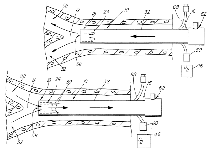
FIGs. 9 and 10 are schematic views of the use of the first
preferred embodiment of the present invention;
FIG. 11 is a side view of another preferred embodiment of
the present invention;
FIGs. 12 and 13 are partial views of the preferred
embodiment of the present invention shown in FIG. 11;
FIG. 14 is a partial view of another preferred embodiment
of the present invention; and
FIG. 15 is a partial view of another aspect of the
embodiment of FIG. 14.
Detailed Descript;on
With reference first to FIGs. 1 and 2, the first preferred
embodiment of the present invention for creating a sub-atmospheric
pressure near the carina 12 of a human or veterinary patient 48
i~ thereshown as a device 10, extending from outside the patient
48 to a location within the trachea 50 of the patient 48. The
carina 12 is, of course, the location at which the bronchi 52
split from the trachea 50, and lead towards the lungs 54 of the
patient 48.
More particularly, the device 10 first comprises a
tracheal or endotracheal tube 14 having a proximal tube end 16
positioned outside the trachea 50 of the patient 48, and a distal
tube end 18 spaced from the proximal tube end 16 and positioned
at or near the carina 12 of the patient 48. The tube 14 includes
a tube wall 20 connected to and extending between the proximal
tube end 16 and the distal tube end 18, as well as a tube
passageway 22 for the flow of an oxygen-containing gas
therethrough. The tube 14 is preferably a conventional multiple
lumen tracheal or endotracheal tube 32, in which the tube
passageway 22 (normally used for the application of suction to
remove fluids) is embedded in the tube wall 20 and is connected
by an appropriate valve and/or monitoring mechanism 60 to a source
~ ~176055
-- 11 --
46 of the oxygen-containing gas, such as warmed and moistened air,
oxygen, gaseous anaesthetic, or the like. The proximal tube end
16 is attached to a fitting or connector 62, and thence to valves,
regulators, monitors or controls (not shown) suitable to the
particular ventilation method to be employed. It is highly
desirable that tube 14 include a second passageway 64 in the wall
thereof, which has an outlet 66 distal of insert 24 for infusing
drugs and/or monitoring pressure near the carina of the patient.
A side arm 68 with a well-known Luer lock connector communicates
with second passageway 64 near the proximal end 16 of the tube 14.
Of course, it is highly desirable that the tube 14 also include
a line of radiopaque material 58 in the tube wall 20 and running
the entire length of the tube wall 20, to facilitate monitoring
of the position of the tube 14 in the trachea 50 of the patient
48. The positions of the second passageway 64, the line of
radiopaque material 58, and the tube passageway 22 are more
clearly shown in FIG. 3.
With continued reference to FIG. 2, but especially with
further reference to FIGs. 4 through 8, the device lo also
comprises an insert 24 received in and circumferentially abutted
by the distal tube end 18, for providing the reverse venturi flow
of the oxygen-containing gas through the tube 14. More
particularly, the insert 24 has a surface 26 defining at least one
insert channel 28 in fluid communication with the tube passageway
22, the at least one insert channel 28 being open towards the
proximal tube end 16. Preferably, when the tube passageway is
formed in the tube wall 20, the tube passageway 22 includes a
passageway outlet 34 located adjacent the distal tube end 18, and
the at least one insert channel 28 is in fluid communication with
the passageway outlet 34. Also preferably, the at least one
insert channel 28 comprises a plurality of channels, and more
particularly, at least one circumferential channel 36 in fluid
communication with the passageway outlet 34, and a plurality of
~ 217605!~
- 12 -
axial channels 38 connected to the tube passageway 34 by the at
least one circumferential channel 36.
The insert 24 is most preferably configured as a hollow
cylinder 40 having a central throughbore 56, although any of a
5 variety of other shapes for the insert 24 might be suitable, if
the material of the tube 14 is sufficiently flexible to ensure an
adequate seal to the insert 24, and if a central passage is
provided to allow the exit of carbon-dioxide enriched air from the
lungs 54, and out through the bronchi 52 and trachea 50. Also
preferably, it is the outer surface 42 of the cylinder 40 that is
the insert surface 26 on which the at least one insert channel 28
is formed. In such a case, the at least one insert channel 28 is
further defined by the inner surface 44 of the tube wall 20, at
the distal tube end 18.
The insert 24 may be composed of any suitable medical
grade material, such as a sterilizeable synthetic. However, as
the tube 14 will typically be composed of a material which is
somewhat flexible, it is highly desirable that the insert 24 be
composed of a material which i~ more rigid than the material
20 making up the tube 14; this ensures that the inner surface 44 of
the tube wall 20 does not collapse into the at least one insert
channel 28, and prevent the flow of the oxygen-containing ga~
through the at least one insert channel 28. It i8 therefore
advantageous to construct the insert 24 from a suitable rigid
25 material, such as Deltrin~ nylon material, into whose surface 26
the at least one insert ch~nnel 28 can be machined. The number
of insert channels 28 is not believed to be critical, so long as
one or more of them are open in the direction o~ the proximal tube
end 16, and can establish in the tracheal or endotracheal tube 14
a reverse venturi flow of the oxygen-containing gas away from the
lungs 54 and bronchi 52 of the patient 48.
With reference now to FIGs. 11 through 13, a second
embodiment of the present invention for creating a sub-atmospheric
pressure near the carina 12 of a human or veterinary patient 48
~ ~17~0~
is thereshown as a second device 80 which is positionable within
the trachea 50 of the patient 48 in the same manner as shown in
FIG. 1 with respect to the first device 10. The device 80 first
comprises a hollow catheter 82 having a proximal catheter end 84
positioned outside the trachea 50 of the patient 48, and a closed
distal catheter end 86 positioned within the trachea 50 of the
patient 48, at or near the carina 12 of the patient 48. The
device 80 also comprises a catheter sidewall 88 connected to and
extending between the proximal catheter end 84 and the distal
catheter end 86. The catheter sidewall 88 has an external surface
90 through which at least one perforation 92 is formed near the
distal catheter end 86. The catheter sidewall 88 also includes
a passageway 100 extending therethrough and has an outlet 102
distal of the at least one perforation 92 for infusing drugs and
monitoring pressure near the carina of the patient. A side or end
arm 104 with a well-known connector attached thereto communicates
with passageway 100 for infusing drugs and monitoring pressure.
The at least one perforation 92 is adapted for the passage of an
oxygen-containing ga~ therethrough, ~or example, from a source 46
20 of the gas connected through a suitable valve 60 to a fitting or
connector 62 at the proximal catheter end 84.
The at least one perforation 92 is acutely angled with
respect to the external surface 90 of the catheter sidewall 88 and
is open towards the proximal catheter end 84. The angle between
the at least one perforation 92 (that is, the direction of the
flow of the oxygen-containing gas through it) is advantageously
no more than 45 degreeg, is preferably less than 45 degrees, and
is most preferably about 30 degrees. The at least one perforation
92 is preferably several in number and can be most conveniently
formed as a plurality of slits 96 cut into the external surface
90 o the catheter sidewall 88, deep enough to allow the flow of
the oxygen-containing gas from the source 46, through the interior
of the catheter 82, and out the at least one perforation 92, that
is, out the plurality of slits 96.
~ 2~7~
- 14 -
The catheter 82 is composed of a suitably flexible,
medical grade material such as a nylon radiopaque material tubing,
or a similar material which gives the catheter sidewall 88 an
equivalent flexibility. The preferred plurality of slits 96
thereby form flexible flaps through which the oxygen-containing
gas can flow; such flaps would prevent or limit the backflow of
the gas, if it is supplied in a pulsed or periodically pressurized
fashion.
The device 80 preferably further comprises a tracheal or
endotracheal tube 98 in which the catheter 82 is positioned. The
tube 98 is preerably a single lumen tube with an open distal end
as shown to permit inhalation and exhalation, but can be a
multiple lumen tube (not shown) like the multiple lumen tube 32
of the first device 10. In the latter case, the passageway within
the wall of the multiple lumen tube can be used for monitoring
pressure or gas composition, infusing drugs as previously
described with respect to passageway 100 in catheter 82, or
removing fluid.
With re~erence now to FIGs. 14 and 15, a third embodiment
of the present invention for creating a sub-atmospheric pressure
near the carina 12 of a human or veterinary patient 48 is
thereshown as a third device 120 which is positionable within a
tracheal or endotracheal tube 160 in the same manner as shown in
FIG. 1 with respect to the first device 10. The device 120 iS the
embodiment of the present invention most closely related to the
Kolobow catheter, and first comprises a hollow catheter 122 having
a proximal catheter end 124, a distal catheter end 126 opposite
the proximal catheter end 124, and an exterior catheter surface
128 extending between the proximal catheter end 124 and the distal
catheter end 126. The catheter 122 has a defined catheter
diameter near the distal catheter end, and the di~tal catheter end
126 has a shoulder 130 ormed on it, extending radially outward
o the catheter 122 a distance greater than the catheter diameter.
In FIG. 14, the shoulder 130 is preferably annular in shape, and
~ ~1760~
- 15 -
is conveniently ormed by a plug 144 received in and sealed to,
and thereby closing, the distal catheter end 126. Advantageously,
the plug 144 includes a stem 146 received in the distal catheter
end 126, and a rounded semispherical or bullet-shaped head 148 on
the stem, the shoulder 130 then being formed as the base of the
semispherical or bullet-shaped head 148. In FIG. 15, catheter 122
includes multiple passageways 150 and 152. Main passageway 150
has a closed distal end for delivering an oxygen-containing gas
through the catheter wall, and second passageway 152 extends
10through the catheter with an outlet 154 at distal end 126 for
infusing drugs and monitoring pressure near the carina 12 of a
patient. The distal end of the catheter i~ heat tapered to form
shoulder 130 and close main passageway 150. Second passageway 152
is maintained opened when heat tapering distal end 126.
15The device 120 further comprises at least one radial
perforation 132 through the catheter 122. The at least one radial
perforation 132 is located adjacent to the distal catheter end
126, but is positioned between the proximal catheter end 124 and
the ~houlder 130 on the distal catheter end 126. The at least one
20radial perforation 132 is pre~erably several in number. The at
least one radial perforation 132 is adapted for the passage of an
oxygen-containing gas therethrough, for example, from a source 46
of the gas connected through a suitable valve 60 to a fitting or
connector 62 at the proximal catheter end 124. In FIG. 15, device
25120 further include~ an end arm 156 that is connected to connector
62 and communicates with second passageway 152.
The device 120 also comprises a tubular member 134
positioned about the exterior catheter surface 128, adjacent to
the distal catheter end 126 and extending over the at least one
30radial perforation 132. The tubular member 134 has a distal
member end 136 which abuts the shoulder 130 of the distal catheter
end 126 and which is ~ubstantially sealed to the exterior catheter
surface 128, but which does not cover or seal the at least one
radial perforation 132. "Substantially sealed" means merely that
2~60~
- 16 -
any flow of the oxygen-containing gas out the distal member end
136 is insufficient to prevent establishment of the desired
reverse venturi effect. The tubular member 134 also has a
proximal member end 138 opposite the distal member end 136.
Unlike the di~tal member end 136, the proximal member end
138 has an interior diameter greater than the defined catheter
diameter. A gap 140 is thus provided between the tubular member
134 and the exterior catheter surface 128, in fluid communication
with the at least one radial perforation 132 through the catheter
122. The flow of the oxygen-containing gas through the catheter
122, the at least one radial perforation 132 and the gap 140
provides the reverse venturi in this embodiment.
Manipulation of the tubular member 134 to give itæ
proximal and distal ends 138 and 136 different diameters can be
achieved in a remarkably elegant yet simple manner. Most easily,
the tubular member is composed of a shrink-wrap tubing having a
manufactured diameter greater than the diameter of the catheter
122, yet having a diameter after heat-shrinking no greater than,
and pre~erably less than, the diameter o~ the catheter 122. The
application of heat to only the distal member end 136 shrinks the
distal member end 136 closely about the exterior catheter surface
128, and preferably substantially seals the distal member end 136
to the exterior catheter surface 128; while the proximal member
end 138 is unheated and retains its original diameter, and is
thereby spaced from the exterior catheter surface 128 to create
the gap 140. Roughening or abrading the abutting portion of the
exterior catheter surface 128 prior to the introduction of the
tubular member 134 over the catheter 122 can facilitate such
sealing, as can the use of a suitable medical grade adhesive.
The device 120 can further comprise a tracheal or
endotracheal tube 160, open at the distal end, and preferably
close to the tracheal wall. The tube can be a single lumen tube,
or can be a multiple lumen tube like the multiple lumen tube 32
of the first device 10. In the latter case, the passageway within
~ ~17~05~
- 17 -
the wall of the multiple lumen tube can be used either for
monitoring pressure or gas composition, infusing drugs, or
removing fluid by suction.
It is important to recognize that the use of the shoulder
130 to secure the tubular member 134 on the catheter 122 is a
significant, practical improvement to the Kolobow reverse venturi
catheter. First, the shoulder 130 provides a physical barrier to
prevent the tubular member 134 from sliding off the catheter 122
in a distal direction, should the attachment or adhesion of the
tubular member 134 to the catheter 122 fail. Secondly, any
element forming the shoulder 130 (preferably, the plug 144 of
FIG. 14 or the tapered distal end of FIG. 15) can be more reliably
secured to the distal catheter end 126, than can the tubular
portion 19 of Kolobow be secured to the catheter tip 16 of
Kolobow. Again, the reason for this is a practical one; it is
easier to securely and reliably affix a rigid object (the stem 146
of the plug 144 of FIG. 14 or the heat tapered distal end of
FIG. 15) inside the end of a hollow tube of one or two millimeters
diameter (the distal catheter end 126) than it i9 to affix a rigid
object (the catheter tip 16 of Kolobow) over the end of a flexible
hollow tube of one or two millimeters diameter (the catheter of
Kolobow).
Use of the present invention in ventilation procedures
such as ITV and ITPV can now be easily understood. The passage
of an oxygen-containing gas from the source 46, through any of the
devices 10, 80 and 120, and out their respective channels 28,
perforations 92 or gap 140, creates during exhalation a respective
zone 30 (device 10, FIG. 10), 94 (device 80, FIG. 13) or 142
(device 120, FIG. 14 or 15) of sub-atmospheric pressure. Taking
the device 10 as an example, in operation the device 10 is
positioned with respect to the patient 48 so that the distal tube
end 18 lies at the level of the carina 12. (The distal catheter
end 86 of the device 80 or the distal catheter end 126 of the
device 120 would be positioned similarly, either with or without
~ 6~
- 18 -
a surrounding tracheal or endotracheal tube 98.) A moist, warmed
oxygen-containing gas is then introduced into the device 10 from
the source 46, through the valve 60, and allowed to flow out the
at least one insert channel 28, such as the circumferential
channel 36 and the plural axial channels 38. During the
inhalation phase of the respiratory cycle, as shown by the arrows
in FIG. 9 the gas passes into the bronchi 52 and the lungs 54,
oxygenating the lungs 54 in the desired manner. During the
exhalation phase of the respiratory cycle, as shown by the arrows
in FIG. 10, the oxygen-containing gas continues to flow out of the
channels 36 and 38 and creates the zones 30 of sub-atmospheric
pressure which aid removal of the carbon-dioxide laden air from
the lungs 54 and bronchi 52. It is critical to achieving this
reverse venturi that the oxygen-containing gas flow through the
device 10 at some time during the exhalation phase, preferably
continuously. As indicated, the devices 80 and 120 are used in
a similar manner.
The devices 10, 80 and 120 of the present invention may
be used either with or withou~ conventional mechanical ventilation
(MV). The details of use would be comparable to those disclosed
in the Kolobow patent at column 3, line 48, through column 4, line
54, which are expressly incorporated by herein. Indeed, any of
the devices 10, 80 and 120 of the present invention may be used
by substitution in any of the ventilation methods disclosed in the
Kolobow patent. More particularly, the devices 10, 80 and 120 may
be used in such methods in place of the catheter 1 shown in
FIGs. 1 through 3C of the specification of that patent. The use
of the devices 10, 80 and 120 is not limited to the constant-flow
methods of the patent specification, however, and the present
invention is expected to be useful in ventilation methods in which
the flow rate or pressure varies during the ventilation cycle, so
long as a sufficient flow rate through the devices 10, 80 and 120
is provided to create sub-atmospheric pressure adjacent to the
devices during patient exhalation, and aid removal of carbon-
~ ~1760~
-- 19 --
dioxide enriched air from the lungs o~ the patient. Constant-flow
methods are probably preerred, however.
The devices 10, 80 and 120 æhare the many advantages of
the Kolobow reverse venturi catheter. They reduce the size of the
dead space in the trachea which might trap carbon-dioxide laden
air in the lungs and bronchi, or which might block the
introduction of oxygen into the bronchi and lungs. They also
permit ventilation procedures to be successfully carried out with
lower gas flow rates and lower peak respiratory pressures,
reducing the risk of trauma to the lungs, and the other problems
mentioned above.
The other details of the construction or composition of
the various elements of the disclosed embodiments of the present
invention are not believed to be critical to the achievement of
the advantages of the present invention, so long as the elements
possess the strength or flexibility needed for them to perform as
disclosed. The selection of these and other details of
construction are believed to be well within the ability of one of
even rudimentary skills in this area, in view of the present
disclosure.
Industrial Applicability
The present invention is useful in the performance of
ventilation procedures, and therefore finds applicability in human
and veterinary medicine.
It is to be understood, however, that the above-described
device is merely an illustrative embodiment of the principles of
this invention, and that other devices and methods for using them
may be devised by those skilled in the art, without departing from
the spirit and scope of the invention. It is also to be
understood that the invention is directed to embodiments both
comprising and consisting of the disclosed parts.