Note: Descriptions are shown in the official language in which they were submitted.
WO95/15192 2177~L9 ~ pcTn~ss~ 986
~ARl~IOptlI,MoNARy BYPASS SYST~.M FOR CT,QSED-CHEST
INTERVENTION
FIELD OF THE INVENTION
This invention relates generally to devices and methods for ~ ru~
~udiuv~_ul~, pulmonary, and II~UIU~UI~;;CdI procedures in which the patient is
placed on carri~ l y bypass. More specifically, the invention relates to
less-invasive devices and methods for ,L~bli~llillg cardiu~,ullllul~uy bypass and
S 1~ r." .";"~ interventional procedures in the heart aDd great vessels.
BACKGROUND OF THE INVENTION
Various ~udiu~ ular, D~ U~UIYICaI~ pulmonary and other ;llt~ lLiu-
procedures, including repair or ~.1 ..,...,1 of aortic, mitral and other heart
valves, repair of septal defects, pulmonary Llllu~l~b~ullly, coronary artery bypass
grafting, dl~;iu~ y~ ,.c~,lullly~ treatment of aneurysms, c I~L u~ iolO~;i~l
mapping and ablation, and neurovascular procedures, are performed with the
patient connected to car~ -plllmcn~ry bypass (CPB) equipment to maintain
circulation of uisy~.ld~cd blood throughout the patient's circulatory system. Insome of these procedures, such as heart valve Ir~ and coronary artery
bypass grafting, cardiac function is arrested, and peripheral circulation of
Lcd blood is maintained completely by a CPB system. In other
procedures, such as angioplasty and ~L~ ~L~Illly, the heart remains beating, andCPB is used to assist the heart in ,,1~;,,l-;,,;. ~ circulation of U~y~Cll~t~ blood
during the procedure.
To establish cardiopulmonary bypass according to ~ullvcll~iullal tcf hniqllr~,
a vi~nous cannula is introduced into a major vein such as the inferior vena cava, or
into the heart itself, to withdraw d~Ay~;c~ t~ b~ood from the patient and deliver
the dcu~y~ t~d blood to a CPB system for oxygenation. An arterial cannula is
introduced into a major artery such as the aorta, an iliac artery, or a femoral
95Sl~TUlESH~d(RUU 26~
WO 95/15192 ; PCTIUS9-J/129~6
2~
artery, for delivering u~L ' blood from the CPB system to the patien~s
arterial system. ~
For endovascular procedures such as ~I~;iuplA~y and ~LII~ ul~ly in which
cardiac function need not be arrested, ill~.v~ iollal devices are introduced into an
5 artery such as a femoral artery, and the devices are L~ A11Y positioned at
the treatment site where the procedure is performed. For example, in ~IgiuplAa~yor d~ ~Lullly, a catheter is introduced into a femoral artery and advanced
through the aorta into a coronary artery to treat an occluded region therein. IfCPB is utilized during such procedures, the arterial and venous CPB cannulae are10 usually introduced into a femoral artery and femoral vein, ~ ly, by means
of a surgical cut-down in the groin area on one side of a patient's body.
Interventional devices may then be introduced into a femoral artery in the groinarea on the other side of the patient's body.
In those procedures in which cardiac function is arrested, on the other
15 hand, the heart and coronary arteries must be isolated from the remainder of thc
patient's arterial system. Using cullv~Liul,al techniques, the sternum is cut
rn~it~ltiir~lly (a median ~t~lllu~ullly), providing access between opposing halves of
the anterior portion of the rib cage to the heart and other thoracic vessels andorgans. Alternatively, a lateral thoracotomy is formed, wherein an incision,
20 typically 10 cm to 20 cm in length, is made between two ribs. A portion of one
or more ribs may be P~ ILIY removed to optimize access. Through this large
opening in the chest, a mechanical cross-clamp may be placed externally on the
ascending aorta duwll~ dlll of the ostia of the coronary arteries, but upstream of
the 1,1~ llinc~ Alir artery, so as to allow oxygenated blood from the CPB systen25 to reach the arms, neck, head, and remainder of the body. A catheter is then
introduced through the sternotomy or thoracotomy and inserted into the ascendingaorta between the cross-clamp and the aortic valve. Cardioplegic fluid is infused
through the catheter into the aortic root and coronary arteries to perfuse the
Illyu~udiulll. An additional catheter may be introduced into the coronary sinus for
30 retrograde perfusion of the myocardium with cardioplegic fluid. In addition, the
QI~SrlTUTE SHEET (P(ULE 26~
-
~ W095/151g2 2 17 7 4 91 PCTIUS9 11 ~86
~llyU~dlLliUIII is usually cooled by ir~igation with cold saline solution and/orapplication of ice or cold packs to the myocardial tissue. Cardiac L""~ wi
then cease.
While such open-chest techniques can produce significant benefits for some
5 patients, such techniques entiiil weeks of ~nqlif~li7~tinn and months of
, time, in addition to the pain and trauma suffered by the patient.
Moreover, application of an external cross-clamp to a calcified or d~ ulll~uu~
aorta may cause the release of emboli into the bPrhincPph~ , carotid or
subclavian arteries with serious CU--.~U~ such as strokes.
In response to these problems, new techniques have been developed
to facilitate the ~l r." ", - l~ r ûf cardiac procedures such as heart valve repair and
using endovascular ill~LIulne.l~s~ Plimin~tin~ the need for a
Jl~ ,y as well as the need for an external aortic cross-clamp. Such
procedures are described in co-pending application Serial No. 07/991,188 and
application Serial No. 07/730,559, which are assigned to the assignee of the
present invention and are illCul,UI ' herein by reference. Similarly, in
commonly-assigned U.S. patent application Serial No. 08/023,778, the complete
disclosure of which is il~,ul~u~L~d herein by reference, methods and devices aredescribed for ~I[UIIIIillg coronary artery bypass grafting and other procedures
20 through small incisions or cannulae positioned through the chest wall, obYiating
the need for a l~ y~ This new generation of minimally-invasive cardiac
procedures provides significant advantages over conventional open surgical
techniques, including reduced mortality and morbidity, decreased patient suffering,
reduced ho~rit~li7~tion and recovery time, and lowered medical costs relative to25 open-chest procedures.
In order to arrest cardiac function according to the techniques described in
the forPmPntinnP~1 patent applications, once cardiopulmonary bypass (CPB) is
established, an aortic occlusion catheter is ll~"~llllll,"~lly positioned, preferably
from a femoral artery, into the ascending aor~a so that an t:~,u~d~le member on
30 the distal end of the catheter is disposed between the coronary ostia and the
!3~JBSrltUTE SHEE~ iRUI 26~
O 95/15192 PCT/llS9~112986
., `
2~491
hrarhir,~phAIir artery. ~he 1~dllLIdble, member is expanded to occlude the
aseending aorta, stopping the flow of blood th~l~Lllluu~ ll. Cardiac function is then
arrested, usually by delivering LdULliU~ iC fluid through a lumen in the aortic
occlusion catheter into the ascending aorta upstream of the expandable member,
S and/or by delivering Ldldiu,ulc~ic fluid in a retrograde manner through a catheter
positioned in the coronary sinus.
Using the closed-chest techniques described in the rulr,~,. ..lirll-PA patent
Allylil -I;....c the arterial and venous CPB cannulae are preferably introduced into a
femoral artery and a femoral vein, IL~LiV~Iy, and IIAII~ 1AI1Y positioned in
10 the desired arterial and venous locations. The aortic occlusion catheter for
,UdU~iLior,;l,~ the ascending aorta and delivering cardioplegic fluid is preferably
introduced into a femoral artery as well. In addition, a cardiac venting catheter is
usually introduced through the internal jugular vein or femoral vein to remove
blood from within the heart, usually from the pulmonary artery. Further, where
15 retrograde infusion of cardioplegic fluid is utilized, a ~ lup~llbs;ull catheter is
introduced through the internal jugular vein or femoral vein into the coronary
sinus. Thus, for a closed-chest procedure utilizing ~diu~ulll.ol~ bypass,
uv~l~Lllld~ aortic occlusion, pulmonary arterial venting, and retrograde
perfusion of ~diu~l~id, thrce venous catheters and two arterial catheters are
20 utilized, requiring the use of both femoral arteries, at least one femoral vein, and
the internal jugular vein in the neck for introduction of these catheters.
In order to minimize trauma and the risk of ~ JI; I;.~ c such as
infection, it is generally desirable to minimize the number of vascular ~ ".I;.",c
or "sticks" which are made in a patient during a procedure. Such prnr~r~tir,nc are
25 a signific~mt cause of morbidity and mortality in cardiac procedures. The risks are
grcater where the p .Ir~ are either surgical cut-downs or large
1~ .IA~ IIC~ as are usually required for illLluduLLiull of the folr,-,r,.~;rl i aortic
occlusion catheter and venous and arterial CPB cannulae. The risks are
~uduLiuulduly high when such p~ . ~IAllUll~ are made on arterial vessels.
S~lBSrlTUTE SHEET ~JLE 2B~
WO 95/1!~192 2 1 7 7 4 ~ 1 Pf TlUS9.~ 8~
.
Moreover, in some cases, one or more of a patient's femoral arteries,
femoral veins, or other vessels for arterial and venous access may not be available
for i~lLiodu~ifJn of cannulae, due to inadequate vessel diameter, vessel stenosis,
vascular injury, or other conditions. In such cases, there may not be sufficient5 arterial and venous access to permit the use of femoral arterial and venous CPB
cannulae as well as other interventional devices, including an aortic occlusion
catheter, a cardiac venting catheter, an angioplasty or dll~ ull.y catheter, or
other device introduced through a femorai vein or artery, ~ t~ f .~ -J~ y as
part of a single surgical procedure. Therefore, unless alternate arterial or venous
10 access for one or more of these catheters can be found, the procedure cannot be
performed using minimally-invasive techniques.
Improved methods and devices are therefore needed for p.cf~l~ljchin~ CPB
and p~,lrullllill~ interventional procedures that reduce the number of arterial and
venous ~ re~uired for CPB cannulae and other endovascular devices.
15 The methods and devices will preferably facilitate isolating the heart and coronary
arteries from the remainder of the arterial system, arresting cardiac function, and
,I~I,I;~hi..~ cardiopulmonary bypass without the open-chest access provided by aful.. .y. The methods and devices should minimize the number of arterial
and venous ~ dliUlls required in such closed-chest procedures, and desirably,
20 should require no more than one femoral arterial penetration and one femoral
venous rpnptr~tifm In addition to procedures requiring arrest of cdrdiac function,
the methods and devices should be useful for a variety of closed-chest
illL~ iUlldl procedures that require the use of ~udiu~ullllull~uy bypass, evenwhere cardiac function is not arrested.
SUMI\IARY OF THE INVENTION
The present invention provides endovascular devices and methods for
pct~hljchin~ cardiopulmonary bypass and p~ru~ g i~Lt:lv~ iu~al procedures
within the heart and great vessels with a minimum of arterial and venous
p~ The system and method further facilitate partitioning a patient's
~UaST~TI~ SHET (RULE 26)
WO 9S/IS192 PCT/US9~112986
2177~
ascending aorta between the coronary ostia and the br:~nhir1eeph~lif artery to
isolate the heart and coronary arteries from the remainder of the arterial system,
and arresting cardiac funetion, by means of an endovascular device introduced
through a femoral or other artery.
Using the devices and methods of the invention, all blood flow through the
ascending aorta may be blockcd, cardioplegic fluid may be introduced through t-he
coronary arteries to perfuse the Illyuu~udiulll, and oxygenated blood from a CPBsystem may be infused into the arterial system :luwll~ al~l from the point of aortic
ocelusion, all through a single femoral arterial p~nf tr~ti()n Morcover, blood may
be vented from the heart to prevent distension of the Illyul,audi.,lll, and
dcu~y3, ' blood withdrawn from a venous loeation for w~y~lla~iull by the CPB
system, all through a single femoral or jugular venous pl~nrtr~tir)n
With the patient eonneeted to ear~ Flllml~n~ry bypass equipment to
maintain eirculation of oxygenated blood while the heart is stopped, surgieal
proeedures may be perfommed on the heart, coronary blood vessels and other body
structures using 11.-~ and/or endovascular tools, without the need for a
cul,~cl.Liùlldl gross Ll~OIa~ulul~y. Moreover, by p~Uliliullillg the aorta by
cllduv~,ulai occlusion rather than by extemal cross-elamping, the deviee of the
invention may substantially reduce the risk of embolus release assoeiated with such
eross-elamping.
For purposes of the present ~r~lin~ti(m "downstream" means in the
direction of normal blood flow through a blood vessel, i.e., further from the heart
in the arterial system, and closer to the heart in the venous system. "Upstream"mcans in the direetion opposite the du~ alll direction. With respect to
~5 deviees, "proximal" means in the direction toward the end of the deviee that is
closest to and held or IllalliJ!ulal~d by the user, while "distal" means in the
direction away from the user, opposite the proximal direction.
In a partieular aspect of the invention, an ~I~duvdsculau illlc;l v~ iUIIdl
device facilitating carlirlplllmon~ry bypass comprises a bypass eannula having adistal end eonfigurcd for introduetion into a blood vesse~, a proximal end, a blood
SllBST~TUTE SHEET (RULE 2~i)
Wo ss/lsl92 PCTlUSs~112986
2177~1
flow lumen L~ cb~L~ , and a port at the distal end in fluid c.."l."..,.~ with
the blood flow lumen. Means are provided at the proximal end of the bypass
cannula for fluidly connecting the blood flow lumen to a car iil~pl~ln~l~n~ry bypass
(CPB) system. An elongated catheter shaft is coupled to the bypass cannula so as5 to extend distally from the distal end thereof, and has a distal end configured for
po~;~iù~ g in the heart or in a great vessel near the heart, a proximal end, and an
inner lumen ~ ld~C~ t~ Liu--dl means are provided at the distal end of
the catheter shaft for 1~ r..~ an il,t~ ..Liu.lal procedure in the heart or in agreat vessel near the heart.
In a preferred c.llbo~illlcllt, utilized in the patient's arterial system, the
;llt.l~llLioll~l means comprises a device for partitioning the ascending aor~a
between the coronary ostia and the 1,,~ artery. In this e..ll,~dilll~ , an
; means such as an inflatable balloon is disposed at the distal end of the
catheter shaft for occluding the ascending aorta between the coronary ostia and the
15 1,..., ~ . artery so as to block substantially all blood flow IllCI~ UUEII-
Additionally, the device may include means at the proximal end of the catheter
shaft for delivering cardioplegic fluid through the inner lumen of the catheter shaft
into the patient's ascending aorta upstream of the occluding means. The bypass
cannula is configured for illLIudu.,Liull into an artery in the patient, and the blood
20 flow lumen in the bypass cannula is connected to a means for delivering
u~ G~cd blood into the patient's arterial system, such as a ~.~UdiUpUIlllUll~U.Ybypass system.
In another embodiment, utilized in the patient's venous system, the
interventional means comprises at least one inflow port at or near the distal end of
25 the catheter shaft for withdrawing blood from within the patient's heart or great
vessel. The inflow port is in fluid c~mmllnir~tion with the inner lumen of the
catheter shaft for receiving blood from the heart or great vessel. An inflatable balloon may also be provided near the distal end of the catheter shaft. In this
...Ill,Oll;lllr~l the bypass cannula will be positioned in a vein in the patient, and the
~31JBSl~TU~E SHEET (RULE 26)
WO 95115192 . PCTIUS9.1/12986 ~
217~4~1 ~
blood flow lumen in the bypass cannula will be connected to a means for receiving
dev~ ' blood from the patient's venous system, such as a CPB system.
In one c~ budil~ lL, the catheter shaft is fixed to the bypass cannula, and
may be an integral part thereof, i.e., an extension from the distal end of the
S bypass cannula. In this ~ ,Uif~l~, the bypass cannula has a lumen, which may
comprise the blood flow lumen, in fluid ~ ;..,. with the inner lumen in
the catheter shaft. Alternatively, the catheter shaft is slidably disposed in the
blood flow lumen of the bypass cannula, and may be removable from the bypass
cannula, and/or limited in its movement relative to the bypass cannula. The
10 bypass cannula may further be provided with a plurality of ports along a distal
portion of its length in fluid l (l, ,l"~ ;.", with the blood flow lumen to enhance
the flow of blood into or out of the blood flow lumen. In the arterial l ",l,o,i;,~. ..
the blood flow lumen is preferably configured to facilitate a fluid flow of at least
about 4 liters/minute at a pressure of less than about 250 mmHg.
The bypass cannula may further have an adaptor assembly mounted to its
proximal end. The adaptor assembly has first and second access ports in
~,..,.""";, ,~;nfl with the blood flow lumen, the first access port being configured
to receive the catheter shaft, and the second access port being configured for
connection to the oxygenated blood delivery means (in the arterial ~ o~ ) or
20 a means for receiving and w~ alillg d~ aL~d blood (in the venous
"I~o,l;".. ~). Usually, a hemostasis valve or other sealing means is mounted in
the first access port to prevent leakage of blood therefrom, both when the catheter
shaft is inserted through the first access port as well as when the catheter shaft is
removed from the first access port.
In a preferred ~.llb~lill,e.ll, the catheter shaft has a length of at least about
80 cm to facilitate 1l~,l~1l..l.,ll~1 positioning from a femoral vein or artery into the
heart or into a great vessel such as the ascending aorta or inferior vena cava near
the heart. The bypass cannula will usually have a somewhat shorter length. In
the arterial l "ll~o,l;",. ,l the bypass cannula has a length between about 10 cm and
60 cm, and preferably about 15 cm to 30 cm, such that the outflow port at the
9USSrlTUTE SHEET (RULE 26~
~ Wo g5/15192 217 ~ 4 ~1 PCTIUS9~ 986
distal end of the bypass cannula is disposed a substantial distance du....~ of
the occluding member on the catheter shaft. On the venous side, the bypass
cannula is preferably about 50 cm to 90 cm in length so as to extend from a
femoral vein to a point in the inferior vena cava near the heart, to a point within
5 the right atrium of the heart, or to a point in the superior vena cava near the heart.
AlL~ Li~ ~y, the venous bypass cannula may be configured for i~ udu~.Liol~ into
the internal jugular vein and positioning therefrom into the superior vena cava, the
right atrium, or the inferior vena cava. The catheter shaft in the venous
1 .,ll.o~1;" ..~ preferably has a length of between 50 cm and 70 cm so as to reach
10 from the distal end of the bypass cannula through the right atrium and right
ventricle, and into the pulmonary artery to withdraw blood therefrom.
According to the method of the invention, a distal end of a bypass cannula
is positioned in a blood vessel of a patient, and a proximal end of the bypass
cannula is connected to a CPB system to permit blood flow through a blood flow
15 lumen in the bypass cannula between the blood vessel and the CPB system. An
interventional device is then introduced through the blood flow lumen of the
bypass cannula into the blood vessel and advanced into the heart or into a greatvessel near the heart to perform an i1~ ;ullal procedure therein.
In a particular ~ o~ , the bypass cannula is introduced into an artery
20 du..l~ l of the patient's ascending aorta, and a distal end of a catheter shaft is
introduced into the artery through the blood flow lumen in the bypass cannula.
The catheter shaft is l~ ""~ lly positioned so that an -Yr~nrl~hlf- occluding
member attached to the catheter shaft near the distal end is disposed between the
patient's coronary ostia and the patient's b~ r~ artery. Oxygenated
25 blood is infused into the artery du~ Lleall~ of the occluding member through a
lumen in the bypass cannula. The occluding member is expanded within the
ascending aorta to completely block blood flow Ll-~ l~LII1~JU~,I1 for a plurality of
cardiac cycles. The patient's myocardium is then paralyzed.
~U8STiTU~ Si~EET (Ri~LE 2B~
WO 95/lS192 PCT/US9~112986
j . j~
2177491
When the occluding member is an inflatable balloon, the method further
includes the step of delivering an inflation fluid to the balloon through an inner
lumen in the catheter shaft of the device.
Usually the IllyUl dldiulll will be paralyzed by delivering cardioplegic fluid
5 through a lumen in the catheter shaft into the ascending aorta upstream of theoccluding means. Retrograde perfusion of cardioplegic fluid may also be providedby means of a catheter positioned in the coronarv sinus of the patient's heart.
In most ~ ~llb~dil..~ s, the bypass cannula will be connected to a CPB
system which withdraws blood from a venous location in the patient, oxygenates
10 the blood, and delivers the o~O ' blood to the blood flow lumen in the
bypass cannula on the arterial side. The dcw~y~,c~ d blood may be withdrawn
through a blood flow lumen in a venous cannula positioned in a vein such as a
femoral vein or internal jugular vein. Also, a cardiac venting catheter may be
positioned in the heart, usually in the pulmonary artery, to withdraw blood
15 therefrom and deliver it to the CPB system. In an exemplary ~ ,o~ the
cardiac venting catheter is introduced through the blood flow lumen in the venous
cannula. Preferably, the venous and arterial bypass cannulae are introduced into a
femoral vein and femoral artery, respectively, in the groin area on the same side
of the patient. In this way, both the venous and arterial bypass cannulae, as well
20 as the devices introduced l~ Lllluu~ may be introduced through a single
surgical cut-down or p~ ;u~ cuu~i punctures on a single side of the patient.
With the ~AU LiLiol~;llg device in position, the heart and coronar~v arteries
isolated from the remainder of the arterial system, and the heart stopped, various
diagnostic and interventional procedures may be performed. For example,
25 ~ ;C and/or endovascular ills~Ll~cll~ may be introduced into the thoracic
cavity, into the heart, or into great vessels for repairing or replacing the aortic,
mitral, or other heart valve, repairing septal defects, p~ lrc,l,l,i..~ coronary artery
bypass grafting, and the like.
Thus, using the system and method of the invention, a patient's heart can
30 be arrested and the patient placed on cardiopulmonary bypass without a
31Jl~STiTUTE SltEET (RUi E Z6)
Wo 95/15192 PCTn)S9~112s86
217~
11
CUII~ llLiUlldl gross llluldCulul~ly, thereby reducing mortality and morbidity,
decreasing patient suffering, reducing l)~ 1inn and recovery time, and
lowering medical costs relative to previous open-chest procedures. The
~;llduvd~ dl p~Li~iullillg device of the invention permits blood flow through the
5 ascending aorta to be completely blocked between the coronary ostia and the
l~lA. ~ llAli~' artery in order to isolate the heart and coronary arteries from the
remainder of the arterial system. This has significant advantages over the aortic
cross-clamps used in current cardiac procedures, not only obviating the need for a
gross ~ -AI ~ "'y, but providing the ability to stop blood flow through the aorta
10 even when nalrifil~a~inn or other cnmrliratinn~ would make the use of an e7~ternal
cross-clamp ,~ ;lAI~i;' Moreover, the device and method of the invention
arcnmrlich this with a minimum of arterial ~ IIA~ IC~ thereby ".;..;.
trauma and the risk Of ~ -"c such as infection.
The system and method of the invention may further be useful to provide
15 .dl.l;~ y bypass during endovascular il~lclvc~llLiul~dl procedures in which
cardiac function may or may not be arrested. Such procedures may include
dl~;u~la~Ly, dll1wc~1u111y, heart valve repair and 1~ septal defect repair,
treatment of aneurysms, myûcardial mapping and ablation, myocardial drilling,
and a variety of other procedures wherein endovascular illt~ ,.lLiu-~dl devices are
20 introduced through the bypass cannula of the invention and advanced into the heart
or great vessels. In this way, the invention facilitates ~u.ii..~ .y bypass
during such procedures without requiring additional arterial or venous
p. . ~ c
A further ~ AIIII;I~ of the nature and advantages of the invention may
25 be reaiized by reference to the remaining portions of the qlPrifitatinn and the
drawings.
BRIEF DESCRIPTION OF THE DRAWINGS
SIJBST1TUTE SHEEr (RULE 2~
WO 95/15192 PCT/US9--i/12986
2 ~ 7 r~
12
Figure I is a side elevational view of an endovascular device for
~uLiLiu~ the ascending aorta between the coronary ostia and IJI~
artery col.~LIu- t..l in accordance with the principles of the present invention.
Figure IA is an end view of a distal portion of the device of Figure I
5 illustrating the skew of the shaped distal portion.
Figures lB and lC are side elevational views showing alternative
. ."l,o(l;" .,~ of the shaped distal portion of the device of Figure 1.
Figure 2A is a IJcl~ iV~ view of a distal portion of the device of Figure
I in a first embodiment thereof.
Figure 2B is a ~,e.*,~Livc view of a distal portion of the device of Figure 1
in a second ~.Ilbudi~llcllL thereof.
Figures 3 and 4 are transverse cross-sections taken along lines 3-3 and 4-4
in Figures 2A and 2B, respectively.
Figures SA and 5B are transverse cross-sections taken along line 5-5 in
lS Figure ~A, showing alternative ~.lll.,Ailllrlll~ of the shaft of the device illustrateA
therein.
Figure 6 is a transverse cross section taken along line 6-6 in Figure 2B.
Figure 7 is a front view of a portion of a patient's arterial system
illustrating the introduction and advancement of the device of Figure I in the
20 femoral artery, iliac artery and aorta.
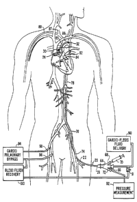
Figure 8 ~h~m~t~ y illustrates a system for arresting the heart
Llu~t~ in accordance with the principles of the present invention, wherein the
device of Figure 1 is positioned in the ascending aorta with c~udi~ ;ic fluid
delivery means connected to the proximal end and a u~u~ y bypass
25 system connected to the patient.
Figure 9 illustrates the distal portion of the device of Figure I positioned in
the ascending aorta with the occluding means expanded and a tissue cutting device
extended from the distal end.
3U~SrlTUTE SREET (RULE 26)
WO 95/15192 PCTII~S9~112986
217~9~
, .
13
Figures lOA-IOB are side and transverse cross-sections, ~*J~livc;ly, of an
alternative ~ o~ of an endovascular partitioning deYice Cu~Llu~ ,d in
accordance with the principles of the present invention.
Figures llA-llB are side elevational and transverse cross-sectional views,
5 respectively, of a further alternative r~ O~ of an endovascular IJ~u Li~iu~ -gdevice Cul. ,LI u~ed in accordance with the principles of the present invention.Figure 12A is a side elevational view of still another, lllllo.l;~l~. .1l of an
endovascular 1~ Li~iullil~, device constructed in accordance with the principles of
the invention.
Figure 12B is a transverse cross section taken along the line 12B-12B in
Figure 12A, showing a shaping element positioned in an inner lumen in the shaft.Figure 13A is a side elevational view of a further alternative c;llll~odil".,ll;of an ~l~duv~ ul~u ~ ~u LiLiullillg device constructed in accordance with the
principles of the present invention.
Figure 13B is a transverse cross-section taken through line 13B-13B in
Figure 13A.
Figure 13C is a transverse cross-section taken through line 13C-13C in
Figure 13A, showing a hemostasis valve with the aortic occlusion catheter
removed from the blood flow lumen in the bypass cannula in the device of Figure
13A.
Figure 13D is a pc~ live view of an obturator and guidewire for use
with the infusion tube in the device of Figure 13A.
Figure 13E is a side cross-sectional view of the partitioning device of
Figure 13A.
Figure 14A is a perspective view of a cardiac venting device ~ùll~LIuu~ in
accordance with the principles of the present invention
Figure 14B is a transverse cross-section taken through line 14B-14B in
Figure 14A.
!~3STlTUl~ S!tEET (RULE 26)
WO 95/15192 PCT/US9~112986
2 17 ~ 14
Figure 14C is a transverse cross-section taken through line 14C-14C in
Figure 14A, showing the hemostasis valve with the venting catheter removed from
blood flow lumen of the bypass cannula.
Figure 14D is a ~ ive view of an alternative ~onfie~ inn of a distal
5 portion of the device of Figure 14A.
Figure 14E is a perspective view of an obturator to facilitate i~ vdu-liol~ of
the device of Figure 14A.
Figure 14F is a side cross-sectional view of.the cardiac venting device of
Figure 14A.
Figure 15A is side elevational view of a further . .I.l.o~ of the cardiac
venting device of the present invention.
Figure 15B is a transverse cross-section taken through line ISB-ISB in
Figure ISA.
Figure ISC is a side elevational view of an alternative confiellr~inn of a
15 distal portion of the device of Figure ISA.
Figure ISD is transverse cross-section taken through line 15D-lSD in
Figure 15C.
Figure 16 is a front partial cut-away view of a patient's body showing the
positioning of the aortic l~au ~ilio~ lg device and cardiac venting device in
20 accordance with the method of the present invention.
l)ETAILED DESCRIPTION OF SPECI~C EMBODIl\~ENTS
The invention provides an cll~uvaa~ ul~u device for pa~ iull;,lg the
ascending aorta, as well as a system for selectively arresting the heart, which ar~
useful in p~lrullllillg a variety of ~udiuvaa~ular~ pulmonary, n. ~lluaul~ al~ and
25 other procedures. The invention is especially useful in conjunction with
minimally-invasive cardiac procedures such as those described in U.S. patent
application Serial No. 07/991,188, U.S. patent application Serial No. 07/730,559,
and U.S. patent application Serial No. 08/093,778, which are assigned to the
assignee of the present invention and have been incorporated herein by reference.
9UBST~lllTE SHEE~ (~lULE 26~
wo 95/1~192 2 ~ 7 7 4 9 1 PCTlUS91111s86
Proeedures with which the invention may find use include repair or ~ ofaortie, mitral, and other heart valves, repair of septal defects, pulmonary
Llllulllb~Lullly, el~-u,uhy~;ologieal mapping and ablation, eoronary artery bypass
grafting, ~ ,io,ulA~Ly, d~ vllly, treatment of aneurysms, as well as
5 neurovaseular and neurosurgical proeedures. The invention is partieularly
a Ivo~L~,cuu~ in that it allows the heart to be arrested and the patient to be plaeed
on ~ul~ u~ lAly bypass using only endovaseular deviees, obviating the need
for a Lllul_~ ulul~y or other large ineision. Moreover, even in cullvcllLiul,AI
open-ehest proeedures, the endovaseular aortie ,uAu~iLiu"i,lg deviee of the invention
10 will frequently find use where an extemal cross-clamp would raise substantial risks
of embolus release due to t~-Alrifir-Atinn or other aortic conditions.
Tuming now to the figures, a first preferred ~ .,lbûdinl~ .lL of an
~,lldUVd~ UIdl device for l ~ULiLiul~iilg the ascending aorta according to the invention
will be described. As illustrated in Figure 1, p~uli~iullillg device 20 includes a
shaft 22 having a distal end 24 and a proximal end 26. An expandable means 28
for occluding the ascending aorta is mounted to shaft 22 near distal end 24. In a
preferred ~ IllI,odilll~ , occluding means 28 comprises a polymerie balloon 30
(shown inflated) of a material, geometry, and 11iml~n~inn~ suitable for completely
oceluding the ascending aorta to bloek systolie and diastolie blood flow, as
20 deseribed more fully below.
Shaft 22 has a diameter suitable for illLIuduuLiul~ through a femoral or iliae
artery, usually less than about 9 mm. The length of shaft 22 is preferably greater
than about 80 em, usually about 9û-100 em, so as to position balloon 30 in the
aseending aorta between the eoronary ostia and the b!A~ ll;n- .~ ' artery with
25 proximal end 26 disposed outside of the body, preferably from the femoral or iliae
artery in the groin area. Altematively, the shaft may be eonfigured for
illLludu.,Lioll through the earotid artery, through the brachial artery, or through a
~r,~udLion in the aorta itself, wherein the shaft may have a length in the range of
20 to 60 em.
S~IBSTITUTE SHEET (RULE 26
WO95/lSlg2 ~ PCT/U591112986
2~749 1
16
Partitioning deYice 20 further includes a first inner lumen 29, shown in
Figures 2A-2B, extending between proximal end 26 aad distal end 24 with an
opening 31 at distal end 24. Additional openings in ~ with inner
lumen 29 may be provided on a lateral side of shaft 22 near distal end 24.
Shaft 22 has a shaped distal portion 32 configured to conform generally to
the curvature of the aortic arch such that opening 31 at distal end 24 is spacedapart from the interior wall of the aorta and is axially aligned with the center of
the aortic valve. Usually, shaped distal portion 32 will be generally U-shaped,
such that a distal segment 34 is disposed at aa aagle between 135- and 225, aad
preferably at a~u~uluA~ aL~ly 180- relative to aa axial direction defined by thegenerally straight proximal segment 36 of shaft 22. Shaped distal portion 32 will
usually have a radius of curvature in the raage of 20-80 mm (measured at the
radial center of shaft 22), depending upon the size of the aorta in which the device
is used. The ~ Iin-- of shaped distal portion 32 allows distal segment 34 to
be positioned centrally within the lumen of the ascending aorta aad distal end 24 to
be axially aligned with the center of the aortic valve, thereby facilitating infusion
or aspiration of fluids as well as illllu.lu~Liull of surgical tools through opening 31
without i~l~c~ru~ c with the wall of the aorta, as described more fully below.
In aa exemplary c ...l,o~li,a~nl, shaped distal portion 32 is preshaped so as tomaintain a perraaaent, generally U-shaped ~onh~llr~ti~n in an unstressed
condition. Such a preshaped configuration may be formed by positioning a
mandrel having the desired shape in first inner lumen 29, then baking or otherwise
heating shaft 22 aad the maadrel for a sufficient time and sufficient t~ lll,UCIa~UIC to
create a permaaent set therein, e.g., 1-3 hours at a Le~ laLulc in a raage of
120 C to 180 C, depending upon the material used for shaft 22.
Alteraative ~ o~illlplll~ of shaped distal portion 32 are illustrated in
Figures lB and IC. In the rllll)O.l;ll.P,~I of Figure lB, U-shaped distal portion 32,
rather than having a rnntinl~ou~, constant curvature, is preshaped in a more
angular fashion, with bends 33 of relatively small curvature separating segments35 which are either straight or of larger curvature. Bends 33 aad/or segments 35
~JBSTITUTE SHEET (RULE 26)
Wo 95/15192 2 1 7 7 4 9 1 pCTlU59~11298-
17
may further be configured to engage the inner wall of the aortic arch to deflectdistal end 24 into a desired position in the ascending aorta.
In the I.or~ of Figure lC, shaped distal portion 32 is configured in
a general "S" shape for introduction into the ascending aorta from a location
5 superior to the aortic arch. In this way, distal segment 34 may be positioned
within the ascending aorta, with proximal segment 36 extending from the aortic
arch through the hr~rhincPrh~lic artery to the carotid or brachial artery, or
through a penetration in the aorta itself, to a point outside of the thoracic cavity.
As shown in Figure IA, distal segment 34 may be skewed (non-coplanar)
10 relative to a central l.,l,r;ll..l;" ,l axis of proximal segment 36, in order to further
conform to the shape of the patient's aortic arch and align with the center of the
aortic valve. In an exemplary ,..~l~o~ . ..l distal segment 34 is disposed at anangle a relative to a plane containing the central axis of proximal portion 36,
wherein a is between 2- and 30, usually between 10 and 20-, and preferably
about 15-. The shape and rlimPncinnc of shaped distal portion 32 and angle a of
distal segment 34 may vary, however, according to the rrmfi~ur~ir~n of the aortic
arch in any individual patient.
In a preferred Clllb-.)dilllCII~, the device will include a soft tip 38 attached to
distal end 24 to reduce the risk of damaging cardiac tissue, IJ~uLicul~ly the leaflets
of the aortic valve, in the event the device contacts such tissue. Soft tip 38 may
be straight or tapered in the distal direction, with an axial passage aligned with
opening 31 at the distal end of shaft 22. Preferably, soft tip 38 will be a low
durometer polymer such as polyulcLl,~le or Pebax, with a durometer in the range
of 65 Shore A to 35 Shore D.
At least one radiopaque stripe or marker 39 is preferably provided on shaft
22 near distal end 24 to facilitate nuulu~ul~ic visualization for positioning balloon
30 in the ascending aorta. R~riinp~rll~P marker 39 may comprise a band of
platinum or other radiopaque material. Alternatively, a filler of barium or bismuth
salt may be added to the polymer used for shaft 22 or soft tip 38 to provide
l~diu~ c;~y.
SU~ST~TUTE SHEET (RULE 2
WO 9~/15192 PCT/I~S9 1/12986
~17~
18
As illustrated in Figures 1, 2A and 2B, a ~ e element 40 is
disposed in first inrler lumen 29 of shaft 22 so as to slide Inneitll~lin~lly relative to
the shaft. S~ rl.;l.~ element 40 may comprise a tubular stylet with a
IrmEitll~inl~l passage 44 for receiving a guidewire 42, as described below.
5 Alternatively, element 40 may comprise a relatively stiff portion of the guidewire
itself. S ,,' ~ element 40 may be a polymeric material or a l,;,~c~
metal such as stainless steel or nickel titanium alloy with a bending stiffness
greater than that of shaft 22. In this way, ~IIA;~ element 40 may be
advanced distally into preshaped distal portion 32 so as to straighten shaft 22,10 facilitating ,"l" "I ~r. ~ LIudu~,~iull of ~,~Lilio..;"~ device 20 into an artery and
advO.~ to the aortic arch. .~1l,~;~ lllrll;"~ element 40 may then be retracted
proximally relative to the shaft so that distal end 24 can be positioned in the
ascending aorta with preshaped distal portion 32 ~ ...r.., .";"~ to the shape of the
aortic arch.
A movable guidewire 42 is slidably disposed through first inner lumen 29,
either through l~meitl-~lin~l passage 44 in sL.d,~ c,lil~g element 40 (Figure 2B),
external and parallel to ~I-,I;gl.l~ element 40, or through a separate lumen (not
shown) in shaft 22. Guidewire 42 extends through opening 31 in distal end 24 of
shaft 22 and may be advanced into an artery distal to shaft 22, facilitating
advO.~ of shaft 22 through the artery to the ascending aorta by sliding the
shaft over the guidewire. In an exemplary ~ l,o~illl~llL, guidewire 42 is relatively
stiff so as to at least partially straighten shaft 22, so that ~ p element 40
is ~ II~C~ uy for introduction of shaft 22. In this, l.lbudi~ l-L7 guidewire 42 may
be, for example, stainless steel or a nickel titanium alloy with a diameter of abollt
l.Ommtol.6mm.
Shaft 22 may have any of a variety of configurations depending upon the
particular procedure to be performed. In one r~.l,bo~lill.~.lL, shaft 22 has a
multi-lumen . .",r~ l;.". with three non-coaxial parallel lumens in a single
extrusion, as illustrated in Figures 2A, 3 and ~A. The three lumens include first
inner lumen 29, which receives ~ element 40 and guidewire 42 and
~STITU~E SHEET (~U~E 2~
wo 95/15192 2 ~ 7 7 ~ ~ i PCTJUss~ 986
19
includes opening 31 at its distal end, an inflation lumen 46 which opens at an
inflation orifice 47 near the distal end of shaft 22 in .~ with the
interior of balloon 30, and a third lumen 48 which has an opening (not shown) at
distal end 24 of the shaft to sense pressure in the ascending aorta. In this
S ~ bl- ' t, the largest transverse dimension of first inner lumen 29 is preferably
about lmm-4mm. Adv~ Ju~ly, the distal opening in third lumen 48 is radially
offset from opening 31 in first inner lumen 29, so that infusion or aspiration of
fluid through first inner lumen 29 will not affect pressure I~ Ul~ lL~ taken
through third lumen 48.
In a second ~II.b~dil~ illustrated in Figure SB, shaft 22 has a dual
lumen inner member 50 and a coaxial outer member 52. Inner member 50
includes first inner lumen 29 which receives ~ element 40 and opens at
distal opening 31, and a third lumen 54 which has an opening (not shown) at its
distal end for measuring pressure in the ascending aorta. Outer member 52 defines
lS a coaxial inflation lumen 56 which, at its distal end, is in ~.,,,.".". i. -li.". with the
interior of balloon 30. Balloon 30 and outer member 52 may comprise a single
integrated extrusion, or balloon 30 may be bonded or otherwise attached to outermember 52 near the distal end of shaft 22 using well-known techniques. Outer
member 52 may have an open distal end which ~,..,.,....,i, ~llr~ with the interior of
20 balloon 30. Alternatively, the distal end of outer member 52 may be closed, for
example, by bonding to the exterior of inner member 50, with an inflation orifice
4~ provided as shown in Fig. 2A for .~ between lumen 56 and the
interior of the balloon.
In a third ~ IIlOl~ lr,ll illustrated in Figures 2B, 4 and 6, shaft 22 has a
25 first inner lumen 29 of large diameter configured to receive various types ofsurgical ill~LIulr~ , as well as to receive ~I"~ t~ element 40. An inflation
lumen 58 extends parallel to first inner lumen 29 and is in ~,..,.,...~..it,~lir)n with the
- interior of balloon 30 through an inflation orifice 61, shown in Figure 2B. In this
embodiment, shaft 22 may comprise a single extrusion containing inflation lumen
30 Sg and inner lumen 29, or two individual tubes bonded to one another, one tube
i~lBSTITUTE SltEET (RllLE 26~
, .. . ... ..... ........ . . ... .....
WO 95J15192 PCTIUS9~112986
21~7~91
containing lumen 29 and the other containing inflation lumen 58. With this
uu~ u~,~iu--, shaft profile can be minimized while making lumen 29 as large as
possible within the confines of the vessels in which the device is positioned. In
this ~ ",l,o~ first inner lumen 29 will have a diameter of at least about 5 mm
5 and preferably about 8 mm. Partitioning device 20 thereby provides a passage of
maximum diameter for endovascular introduction of surgical i~ lul-~ t~ such as
v;~ .., scopes, aspirators, irrigation tubes, cutting, stapling and suturing
devices, and the lil~e, as described in co-pending application Serial No.
07/991,188, which has been i..~u ~ herein by reference.
In some ~ o-~ , as shown in Figures 2B, 4 and 6, a wire braid or
coil 60 may be embedded in the wall of shaft 22 to enhance radial rigidity and to
maintain the transverse dimensions of first inner lumen 29. It is particularly
important to maintain the roundness of first inner lumen 29 where surgical toolsare to be introduced through the first inner lumen. If shaft 22 is made of
15 sufficient diameter to ~rCclmmo~l~t~ such tools through lumen 29, the shaft may
tend to flatten or kink when advanced into the curved region of the aortic arch.The use of wire braid or coil 60 to maintain lumen roundness allows tool profile to
be maximized and allows tools to be advanced through the lumen with minimum
f~,~t~ c. Wire braid or coil 60 may be formed of stainless steel or other
20 ~ C~"~Y I;II1C material such as nickel titanium alloy, aramid fibers such as
Kevlarn' (DuPont), or nylon.
Shaft 22 may be constructed of any of a variety of materials, including
l,i-,. ~,",~ e polymers such as polyu~ e, polyvinyl chloride, polyether block
amide, or l~oly~LI,yl~..e. In a preferred ~Illl)o~ of the device shown in
Figure 2A, shaft 22 is urethane with a shore durometer in the range of 50D-80D.
In the e.,lbo~i"u .,~ of Figure 2B, wherein shaft 22 may have a significantly larger
diameter as well as an embedded coil which both increase stiffness, a polyurethane
with shore durometer of 60A-IOOA may be used. Shaft 22 may have a bending
modulus in the range of 70 to 100 kpsi, preferably about 80-90 kpsi. A bending
modulus in this range provides sufficient stiffness to optimize pushability from a
S~STITU~E SHEET ~RULE 26~
Wo 95/15192 PcTluss~ll2ss6
21774qfl
21
femoral or iliac artery to the ascending aorta, while providing sufficient flexibility
to navigate the tortuous iliac artery and the aortic arch. Once partitioning device
20 has been positioned with distal end 24 in the ascending aorta, this bending
modulus also facilitates exertion of a distal~y-directed force on shaft 22 from
proximal end 26 to maintain the position of balloon 30 against the outflow of
blood from the left ventricle as the balloon is inflated In other embodiments, the
(1;,.,. ~lCi.~C geometry and/or materials of shaft 22, as well as coil 60, may be
varied over the length of the shaft so that the shaft exhibits variable bending
stiffness in vaIious regions For example, preshaped distal portion 32 may be
more flexible fo} tracking through the aortic arch, whereas proximaf portion 36
may be stiffer for pushability and resistance to ~ lr1,1
Balloon 30 may be co11~LI u~d of various materials and in various
gf.~nn~PtriPc In a preferred . I.o,~;.,,~,,l balloon 30 has a collapsed profile small
enough for U.lU~Liu1. into the femoral or iliac artery, e.g. 4-9 mm outside
diameoer, and an expanded (inflated) profile large enough to completely occlude
the ascending aorta, e g. 20-40 mm outside diameter. The ratio of expanded
profile diameter to collapsed profile diameter will thus be between 2 and 10, and
preferably between 5 and 10. The balloon is further configured to maximize
contact of the working surface of the balloon with the aortic wall to resist
,~ f ''I and to minimize leakage around the balloon, preferably having a
working surface with an axial length in the range of about 3 to about 7 cm when
the balloon is expanded. Textural features such as ribs, ridges or bumps may also
be provided on the balloon working surface for increased frictional effects to
further resist l);~ f.llf ll
Balloon 30 preferably has some degree of radial expansion or elongation so
that a single balloon size may be used for aortas of various diameoers. Materials
which may be used for balloon 30 include polyu1~Ll,~.~s, polyethylene
- oerephthalate ~PET), polyvinyl chloride ~PVC), latex, ethylene vinyl acetate (EVA)
and the like. However, balloon 30 must have sufficient structural integrity wheninflated to maintain its general shape and position relative to shaft 22 under the
S~SrITUl E SHEEI (RU~E 261f
_ _ _ _ _ _ _ _ _, . _ _ .. ...
TIIJS9-1/12986
WO 9!i/15192 PC
21~7~1
22
systolic pressure of blood flow through the ascending aorta. In an exemplary
balloon 30 is constructed of polyulcll~c or a blend of pOI~ulc~ ulC
and polyvinyl such as PVC or EVA. It has been found that such materials have
sufficient elastic elongation to A~C~ I IC~IA~I a range of vessel diameters, while
5 having sufficient structural integrity to maintain their shape and position in the
ascending aorta when subject to outflow of blood from the left ventricle.
In a preferred ~lllbodilll~.llL, balloon 30 is further provided with a pluralityof folds or pleats 62, shown in Figures 3 and ~, which allow the balloon to be
collapsed by evacuation to a small collapsed profile for il~Lludu~iù.- into a femoral
lû or iliac artery. In this ~l~lbU(~ , balloon 30 has a blow-up ratio, defined as the
ratio of the fully-inflated outside diameter to the deflated outside diameter (before
collapsing), of about 200%-400%, preferably 300%-400%. Pleats 62 are
preferably at least three in number and each have a width lclJlc~llLi.~,
~l~lwd~ cly 5-25% of the UilUUllll~ C of the balloon when deflated (but not
15 collapsed by subjecting the interior of the balloon to a vacuum). Pleats 62 may be
formed into the balloon during the balloon-making process by using a dipping
mandrel having lnn~irl~linAl flutes formed in its periphery. The mandrel is dipped
into a container of liquefied balloon material (e.g. polyurethane) so that a tubular
layer of material solidifies onto the mandrel, ~ullrullllil~g to the shape of the flutes.
20 The mandrel is then removed, producing a pleated balloon of substantially constant
thickness. Where a folded, rather than pleated, balloon is used, the folds may be
for--med after the balloon is made by vacuum collapsing the balloon onto a mandrel
into the desired collapsed profile and heating the balloon, or by expanding the
balloon under pressure and heat in a corrugated mold.
In alternative ( .. ,1)~.1;~.. ~1~ occluding means 28 may comprise any of a
variety of structures, including pivot, umbrella or fan-type occlusion ".~, IIAI~
actuated by pull wire, torque cable, or other type of ml'~hAni~A1, hydraulic,
electric, or shape-memory actuator. Further, occlusion means 28 may comprise
multiple occlusion devices arranged in tandem on shaft 22; for example, a pair of
30 balloons may be arranged one behind the other at the distal end of the shaft. In
S~113STITUl~ SHEET (RULE 26)
WO 95~15192 PCTIUS9~112986
~ 2177491
23
one ~ , an occluding balloon is disposed on the shaft to be pu~;Liu~
in the ascending aorta, while a seating balloon is disposed distal to the occluding
balloon so as to be pu~;~;u~lc in the left ventricle through the aortic valve, as
described in cûmmonly assigned co-pending application Serial No.
S attorney docket number 18409.003.01, entitled "System fûr r,,.[u~ .g a Cardiac
Prûcedure,'' filed , the complete disclosure of which is ;~ullJuldL~d herein by
reference. By inflating the seating balloon in the left ventricle, the position of the
occluding balloon in the ascending aorta may be maintained ag~unst the outflow of
blood from the left ventricle.
Referring again to Figure 1, a triple-arm adaptor 64 is attached to the
proximal end 26 of shaft æ. Triple-arm adaptor 64 includes a working port 66 in
.-~.,.".,1 ,. ~ with first inner lumen 29 through which ~ .,;"~ element 40,
guidewire 42, and in some r.~ o(li~ surgical or diagnostic ill ,tlulll~llL~ may
be introduced, as described below Working port 66 may also be adapted for
infusion of fluid such as ~ud;v~Jl~i~ fluid, saline or contrast solution, as well as
for aspiration of blood, fluids and debris through first inner lumen 29. Triple-arm
adaptor 64 further includes an inflation port 68 in c~lmml~nir~tinn with the
inflation lumen and configured for connection to an inflation fluid delivery device
such as a syringe 70. A pressure I-~u,~ .,L port 72 is in crlmmllnir~fi~n with
the third lumen (48 or 54) and is adapted for connection to a pressure
l device. Alternatively, where shaft 22 includes ûnly first inner lumen
29 and inflation lumen 58 as in Figures 2B, 4 and 6, port 72 may be in
~",..,..,..'. _~;n,~ with first inner lumen 29 and configured for pressure
--~u~ n~, fluid infusion or aspiration.
Referring now to Figures 7-9, a preferred rllll.O.l;".r,,l of the method of the
invention will be described. Initially, a partitioning device 20 of a size and
cU..rl~ul~ ", suitable fûr the particular patient must be selected. Usually, thepatient's aorta will be observed by means of a IIUUIU~UP;C imaging to determine
its size and shape, ~JdlLi~ul~uly in the region of the aortic arch. A pdu~iLio..iulg
30 device 20 will be selected having a length sufficient to allow occluding means 28
SJBSTITUTE SHEET ~RULE 26)
.. . . .. . . . ... . ..
WO 95/15192 PCT/US9 1/12986
217~
24
to be advanced into the ascending aorta from the point of introduction, which will
preferably be a femoral or iliac artery in the groin area. Further, a ~alLiLiullillg
device will be seleeted which has a preshaped distal portion 32 with ~lim~nci~n~and shape suitable for positioning the distal portion in the patient's aortie arch
5 such that distal end 24 is spaced apart from the inner wall of the aseending aortd,
preferably aligned with the center of the aortic arch. Usually, the preshaped distal
portion will have a radius of curvature d~Aul~ ~y equal to that of the aortic
arch as medsured to the center of the aorta, preferably within a tolerance of about
+/- 10 mm.
Referring to Figure 7, ~udlLi~iull;ng device 20 is preferdbly ~ ~,. .,IA". v~, 'y
inserted into a femoral or iliac artery 74 in the groin area using known teehniques
sueh as a cut-down or a l.IC;I~,UI~UI~VU~ technique such as the Seldinger technique.
Guidewire 42 is first introduced into femoral artery 74 and advanced toward the
heart through iliae artery 76 and aorta 78 so that the distal end of guidewire 42 is
in the aseending aortd (not shown in Figure 7). St~i~ht~ nin~ element 40 is
inserted into lumen 29 of shaft 22 and positioned in preshaped distal portion 32 so
as to strdighten the preshaped distal portion. With balloon 30 deflated, shaft 22 is
positioned over guidewire 42, introdueed into femoral artery 74 and advaneed over
guidewire 42 through iliae artery 76 and aorta 78. A lluu~u~u~e may be used for
vic~ li7~tiol- of radiopaque markers 39 on shaft 22 to faeilitate prlcit~ n~ As an
alternative or ~u~pl~ to lluu~u~c~ic imaging, ultrasonie c-~llo~dld;u~ l,y
may be used by, for example, positioning an ~I.uud..li~l~pllic transdueer in theesophagus.
As an alternative to femoral or iliae introduction, shaft 22 may be
25 introduced into carotid artery 87 or brachial artery 89. In such cases, distal
portion 32 of shaft 22 will usually have a generally S-shaped l nnfi~l~tion as
described above with reference to Figure lC. Such an S-shaped c-mh~l.nAtion
facilitates positioning balloon 30 in the ascending aorta with shaft 22 extending
superiorly from the aortic arch through bmrhi(!(~ph~lic artery 86.
S~)8STlllJI~ SHEET (RULE 26J
Wo 95/15192 PcTlus9~l29s6
2177~91
As illustrated in Figures 8 and 9, shaft 22 is advanced through aortic arch
80 until balloon 30 resides in ascending aorta 82 between coronary ostia 84 and
iC artery 86. As distal end 24 is advanced around the aortic arch,
'f' '';'~G element 40 is drawn proximally relative to shaft 22 so as to allow
5 preshaped distalA portion 32 to conform to the shape of the arch. In an alternative
bodill.~ llL, a relatively stiff guidewire may be used without a separate
~I...;~,l~t. .1;'1~ element, in which case the guidewire may remain in place as shaft 22
is advanced into the ascending aorta. !~lA~ element 40 and guidewire 42
may then be removed from shaft 22.
In an alternative technique, lu~uLiLiu-lil.~ device 20 may be introduced into
the aorta li..,... u~ uy;. ,lly In this ~ l~lbudi~ llL, distal end 24 of shaft 22 may be
introduced through a small incision or cannula into the chest cavity. A small
y.,~l~,ld~iUI~ is made in the aorta, either in the descending region or in the aortic
arch. Shaft 22 is then inserted into the aorta using forceps or other IllUld~,U~.Up;C
15 iUI~LIulll~,.lL~ introduced into the chest cavity through small incisions or cannulæ.
Such a technique may be useful where a patient's femoral or iliac arteries are
unsuitable for illllUU~..,il.~, pdlLiLiUllil.g device 20 p~:luui~AAI~uu~ly or by cut down
into those vessels.
As illustrated in Figure 8, once shaft 22 has been positioned so that balloon
30 is in ascending aorta 82 between coronary ostia 84 and 1,., I.;n~ lin artery
86, balloon 30 is expanded by injecting an inflauon fluid, usually a saline solution
with a .r.ll..~lAl)lli~ contrast agent, from syringe 70 through inflation port 68. In
an exemplary ~ û~ ..1 the balloon will be fully inflated in dy~lu 'y 5-15
seconds, depending upon the size of the inflation lumen and the viscosity of the25 inflation fluid used. In some ~IllI,odil~l~ llL~, blood may be allowed to flow through
inner lumen 29 and directed to cardiopulmonary bypass system 94 (described
below), thereby reducing the pressure of blood flow against balloon 30 during
inflation. When fully inflated, the exterior surface of balloon 30 contacts the inner
walls of the ascending aorta so as to fully occlude the vessel and block
30 s~lhct~nti~lly all systolic and diastolic blood flow past the balloon. While the heart
St.lBSr~TllTE SHE~ (RULE ~61
WO 95115192 PCTlUS9.i/12986
2~ 77~gi
26
remains beating, blood may flow from the left ventricle through the aortic valveand into the coronary ostia so as to perfuse the lllyù~diulll through the coronary
arteries. The heart and coronary arteries are thus isolated from the remainder of
the arterial system.
S In am alternative embodiment, a gaseous inflation fluid may be used in
order to increase inflation speed. In this way, balloon 30 cam be fully inflated in
less time than the period between systolic pulses, reducing the likelihood that the
outflow of blood from the left ventricle during systole will displace balloon 30from its position in the ascending aorta. Preferably, helium is used as the inflation
fluid, since helium, being highly soluble in blood, is unlikely to produce
potentially injurious gas emboli in the event of leakage from the balloon.
Alternatively, carbon dioxide may be used. A gas inflation pump and control
device similar to those described in U.S. Patent No. 4,771,765 and U.S. Patent
No. 4,902,272, which are hereby illcu~ L~ herein by reference, may be
utilized for delivery of p.~,~u,i~ helium through inflation port 68. The inflation
pump may be timed with the ,~."1l,., I;l.llc of the heart to facilitate inflation of the
balloon between systolic pulses. Using such a pump, balloon 30 may be fully
inflated in less than about 1 second, and preferably less than about O.S second. Figure 8 illustrates the ~ of a system for arresting the heart
con~Llu~ in accordance with the principles of the invention. A c~,l;u~ ;ic
fluid delivery device 90 is connected to working port 66. A pressure, -~ ,....,.device 92 may be connected to port 72 to monitor pressure in the ascending aortaupstream of balloon 30 through first inner lumen 29 (or through an in~Pr-nrl~n~
third lumen in shaft 2V. The patient is placcd on a ~u.1;1.~,1l1l,,..,.~,~ bypass
25 (CPB) system 94 to maintain circulation of oxygenated blood throughout the body.
Usually, a venous cannula 96 is positioned in a femoral vein for withdrawing
de-oxygenated blood. In addition, a pulmonary artery venting catheter (not
shown) may be positioned through the right internal jugular vein into the
pulmonary trunk to withdraw the blood contained therein, thereby d~u~
30 the left atrium. The withdrawn blood is delivered to CPB system 94 which
~'JBSr~TUTE SHEET (Rl~LE 26)
-
WO95/15192 2 1 7 7 4 ~ 1 Pcrlusslllw6
27
removes carbon dioxide and oxygenates the blood. The oxygenated blood is then
delivered to a femoral or iliac artery via an arterial cannula 98. A blood filter and
- recovery system 100 may also be connected to port 66 in ~Jdu~i~iU~ g device 20
via a routing switch 101 to receive blood and other fluids and debris from firstS irmer lumen 29 before or after delivery of ~dUViU~ I~giC fluid, filter the blood to
remove impurities, and deliver the blood to CPB system 94 for return to the
patient s circulatory system. Further aspects of a CPB system suitable for use in
the system of the invention are described in co-pending application Serial No.
07/991,188, which has previously been illcvllJuld~ed herein by reference, as well
10 as in F. Rossi et al., Long-Term Cardiopulmonary B~ass By Peripheral
'io~l ~n A Model of Total Heart Failure, Joumal of Thoracic and
C~u;vv~.uld, Surgery (1990), 100:914-921; U.S. Patent No. 4,540,399; and
U.S. Patent No. 5,011,469, which are all ;~ ul~!uld~d herein by reference.
With CPB established and balloûn 30 blocking blood flow through the
15 ascending aorta, the Illyv~uviulll may then be paralyzed. In a preferred
I ...I~rJ.~;". ..1 a cardioplegic fluid such as potassium chloride (~CCI) is delivered by
delivery device 90 through working port 66. Preferably, delivery device 90
includes a cooler (not shown) which cools the ~:dUUiV~ r,iC fluid so as to maintain
the heart at a low t~ d~ult;, e.g. 5-lO C, and to minimize demand for oxygen.
20 This is usually accoll.~ l,ed without applying extemal cooling to the heart as is
applied in ~vllv~ ivl~dl open cardiac procedures. The cardioplegic fluid is infused
into the ascending aorta through opening 31 at the distal end of partitioning device
20. The ~,du liùlJlcgiC fluid flows through coronary ostia 84 into the coronary
arteries so as to perfuse the lllyu~ Audiulll. Cardioplegic fluid may also be infused
25 in a retrograde manner through the coronary sinus, by means of a catheter (not
shown) positioned ll A"~1.l,";,lAIIy through the right interiûr jugular vein, asdescribed in co-pending application Serial No. 07/991,188. Heart contractions
will then cease, with circulation to the remainder of the patient s body maintained
by CPB system 94. Cardioplegic fluid flow to the patient s lllyuLdldiulll is
S~BS~iTUTE S~iEET (RULE 26)
WO95/15192 PCINS91112986
217~gl
28
maintdined on a periodic basis, e.g., about every 20 minutes, so long as the
yu~.~udiu-,l is to remain paraly~ed.
In addition to or instead of infusion of KCI, other teehniques may be used
to arrest heart uu~ d~,Liu~. The patient's body may be cooled in a
5 eold t~ cldLulc environment or by applieation of cold-paeks to the ehest to
reduee the h..ll~ d~u-e of the lllyu~udiulll sufficiently to induce fihrillAtil~n The
yuc~udiulll may be cooled directly by infusion of cold fluid such as saline
through the eoronary arteries. Alternatively, electrical fibrillation may be
A~.~U..,~ h. A by delivering electrical signals to the ll~yu~duJiulll by means of
10 electrodes plaeed on the exterior surfaee of the heart or externally on the chest.
However, cardiac arrest by means of fibrillation is generally less desirable than
chemical c~ud;u~ ic paralysis because there remains some degree of heart motion
which eould make surgieal intervention more difficult and because there is a
S;~ irl~u~Lly higher demand for oxygen, reducing the safety and duration of the
15 procedure.
Once the heart has been arrested and CPB P~tAhli~hP~ a surgical procedure
may be performed. The procedure will preferably be a less-invasive procedure
performed cllduvda~ ul~uly or ~ u~ lly~ as described in co-pending
application Serial Nos. 07/991,188 and 081023,778, which have been ill~VllJUl.~t~
20 herein by reference. Surgical procedures whieh may be performed using the
device and system of the invention include repair or Icl~la~ L of the aortic,
mitral and other heart valves, repair of ventrieular and atrial septal defects, septal
myotomy, cardiac mapping and ablation to correct ~ulllyLlllllids~ eoronary artery
bypass grafting, angioplasty, dll,e.~Lu-lly, as well as pulmonary, Il~llUaUl~ dl,
25 and other proeedures.
P~uLiLiulli~lg deviee 20 of the present inventiûn is p~i~LIlduly advantageous
for endovaseular introduetion of surgieal il~lulll.~ through the aorta for
proeedures sueh as heart valve repair and rPpl~PmPn~ As illustrated in Figure 9,preshaped distal portion 32 of shaft 22 eonforms to the shape of aortie areh 80 so
30 that opening 31 at the distal end is positioned eentrally within the aseending aorta
SlJ8STlTUTE SHEET ~RULE 2~
WO 9~/15192 PCTIus9~ ss6
217~91
29
and axially aligned with the center of aortic valve 104. This not only enhances
infusion of ~.liul,l~æic fluid through opening 31, but ensures that surgical
ill ~lUlllC;ll~:i such as valve cutter 106 introduced through first inner lumen 29 will
be aligned with aortic valve 104, either to remove the valve, or to pass through it
S for diac procedures. Ad~allL6~u~1y, soft tip 38 at the distal end of shaft
22 prevents damage to tissue, ~al~i~ulauly the fragile aortic valve leaflets, in the
event of contact therewith.
While being ~JauLh~ulauly useful in uUlljLI~ iUl~ with minimally-invasive
cardiac procedures performed ~l~duva~ul~uly and/or ~llola~os~u~icdlly, the
10 ~Jal~iLiull;.lg device and system for arresting the heart disclosed herein are also
useful in ~.UII~ .. iUII~I open procedures performed with a ~llOla~u~ullly.
PalLiLiù..ing device 20 may be used where an aortic cross-clamp would pose risksof embolus release due to . ~ or other aortic conditions. In open
procedures, ~JauLiLiulullg device 20 may be introduced through the femoral or iliac
15 arteries as described above, through the carotid artery 87, through the brachial
artery 89, or through a penetration in the aorta itself, which is accessible as a
result of the 11 .~, ~ ulu ly. In such cases, shaft 22 of ~JauLiLiullil16 device 20 may
be substantially shorter in length, for example, 20 to 60 cm.
When the procedure has been completed, the heart is restarted by
20 f' E any flow of,_aldiu~,lc6ic fluid through ~auLiLiu~ l6 device 20 or
retrogradely through the coronary sinus, ventilating the lungs, and perfusing the
coronary arteries with warm blood. The region upstream of balloon 30 may be
irrigated by infusing a saline solution through first inner lumen 29. Blood and
other fluids upstream of bal~oon 30 may then be aspirated through first inner
25 lumen 29 to remove thrombi or other emboli which may have been produced
during the procedure, preventing such emboli from entering the 1~
carotid, or subclavian arteries and greatly reducing the risk of romrlirofi-)nc such
as strokes. Balloon 30 is deflated to allow normal flow of warm blood through
the ascending aorta to the remainder of the arterial system. Normal heart
30 cùllLlà~Liol~s may resume promptly, or, if necessary, electrical ~fihrillo~ n may
gJ8ST~TUTE SHEET lRULE 26~
_ . . .... ... ... .. .. . .
WO95/15192 PCT/US91/12986
~ = 7
21774~
be - ' rd to correct heart rhythm. CPB is gradually ~ r~l, and CPB
venous cannula 96 and arterial cannula 98 are removed. Partitioning device 20 iswithdrawn from the body back through the site of entry, and the arterial
penetration is closed. If the patient has been put under general anesthesia, thepatient is then brought from anesthesia to consciousness.
It will be understood by those of skill in the art that various alternative
c~ of ~ iUv~s-,Uldl y.~uLi~iull;llg device 20 are possible without
departing from the scope of the present invention. One such a7,ternative
1,-1, ' is illustrated in Figures 10A-lOB. In this ~."bodi".~.,L, y~iLi~J-I;-l~;
device 20 has a pull wire 110 disposed in a lumen 112 in shaft 22. Pull wire 110is attached at its distal end to an anchor plate 114 at distal end 24 of shaft 22,
preferably offset from the central k~n~itll~lin~l axis of shaft 22. In one
cllll o.lil"...., pull wire 110 extends through a hole in anchor plate 114 and is
retained against the anchor plate by a ball 116 fixed to the distal end of pull wire
110. In other respects, device 20 is configured as described above in connectionwith Figures 1-9, including a balloon 30 mounted to shaft 22 near distal end 24,an inflation lumen 118 in ~.llllllll....i~AI;llll with the interior of balloon 30, a soft tip
38 attached to distal end 24 of shaft 22, and an inner lumen 29 in ~
with dista7, opening 31. Tension may be applied to the proximal end (not shown)
of pull wire 110 to deflect the distal portion 32 of shaft 22 into a shape suitable
for pu~ lillg distal portion 32 in the aortic arch (as shown in phantom in Pigure
10A). In an alternative embodiment, an axially rigid, laterally-deflectable rod may
be used in place of pull wire 110, whereby distal end 24 is deflected by applying a
compressive force to the rod.
In an - ,1~11.. LrA. configuration (with tension relaxed on pull wire 110),
distal portion 32 of the shaft is generally straight. Alternatively, all or part of
distal portion 32 may be curved in an "".1. ~l~C~-~A configuration to enhance
p~if~ ity in the aortic arch. Preferably, a mechanism (not shown) will be
provided at the proximal end of shaft 22 for applying tension to pull wire 110 and
30 for locking the pull wire to maintain distal portion 32 in a desired shape. Various
S~lBSTITllTE SHEET (R'JLE 2~
~ WO 95/15192 217 7 ~ ~ ~ PCTIUS91/12986
31
- may be used, such as those described in U.S. Patent No. 5,030,204,
the complete disclosure of which is il~ul~ui,,tcd herein by reference. Usually,
shaft 22 is introduced into an artery in a generally straight col~r.~.dliul" andtension is applied to pull wire 110 to deflect distal portiûn 32 as the shaft isadvanced into the aortic arch. Once distal portion 32 is positioned in the aortic
arch, tension on pull wire 110 is adjusted so as to position distal end 24 radially
within the ascending aorta so as to be spaced apart from the inner wall of the aorta
and axially aligned with the center of the aortic Yalve. Pull wire 110 is then
locked in tension to maintain distal portion 32 in its deflected cnnfi~ll~ti~n
A further alternative ~ bodi~ of partitioning device 20 is illustrated in
Figures llA-llB. In this ~ bodi~ llL, shaft 22 is positionable in an interior
lumen 120 ûf a guiding catheter 122. Device 20 may be configured as described
above with reference to Figures 1-6, including balloon 30 near distal end 24, inner
lumen 29, inflation lumen 46, pressure lumen 48, soft tip 38 attached to distal end
24, and triple-arm adaptor 64 attached to proximal end 26. Guiding catheter 122
has a proximal end 124 and a distal end 126, with axial lumen 120 extending
L~ . A soft tip (not shown) may be attached to distal end 126 to
minimize injury to the aorta or aortic valve in the event of contact therewith. A
proximal adaptor 128 is attached to proximal end 124, and has a first port 130 in
c~ ;oll with lumen 120 through which shaft 22 may be introduced, and a
second port 132 in 1^l ~ m with lumen 120 for infusing or aspirating fluid.
Port 130 may further include a hemostasis valve. Guiding catheter 122 also has adistal portion 134 which is either preshaped or deflectable into a shape generally
n-"~r(~ to the shape of the aortic arch. Techniques suitable for preshaping or
deflecting distal portion 134 of guiding catheter 122 are described above in
connection with Figures 1-6 and lOA-IOB. In an exemplary ~.,II,ùdil"e,lL, guiding
catheter 122 is preshaped in a generally U-shaped ~nnfi~l~tinn, with a radius ofcurvature in the range of 20-80 mm. In this r~ lol~ ,. ,1 a stylet (not shown) like
that described above in connection with Figures 1-6 is provided for ~ lg
S'J~STlTUTE SHEL7 (RULE 26~
WO 95/lS192 PCT/IJS9 1/12986
217~9~
32
distal portion 134 for purposes of s~lbcll~ pclllcly i.lL udu~i..g guiding catheter 122
into an artery.
In use, guiding catheter 122 is introduced into an artery, e.g. a femoral or
iliac artery, and advanced toward the heart until distal end 126 is in the ascending
5 aorta. A guidewire (not shown) may be used to enhance tracking. Where a styletis used to straighten a preshaped guiding catheter for ,"1" "~ U~U~Liù..,
the stylet is withdrawn as preshaped distal portion 134 is advanced through the
aortic arch. Once guiding catheter 122 is in position, shaft 22 may be introduced
through port 130 and lumen 120 and advanced toward the heart until balloon 30 is10 disposed between the coronary ostia and the 1~ artery, distal to the
distal end 126 of guiding catheter 122. The distal portion 32 of shaft 22 (Figure
1) is shaped to conform to the aortic arch by preshaped portion 134 of guiding
catheter 122. Balloon 30 is then inflated to fully occlude the ascending aorta and
block b~ood flow ~h~ LI.Iuu~
In yet another P . l,~.l;,~ .,1, shown in Figures 12A-12B, I~duLiLiu.. ;-~ device
20 includes a shaping element 140 ~iLiu..dblc in a lumen in shaft 22, such as
third inner lumen 48. Shaping element 140 has a proximal end 142, a distal end
144 and a preshaped distal portion 146. Preshaped distal portion 146 may be
generally U-shaped as illustrated, or may have an angular, "S"-shaped or other
20 c~ r~ r in an unstressed condition, which will shape distal portion 32 to
generally conform to at least a portion of the patient's aortic arch. Shaping
element 140 is preferably stainless steel, nickel titanium alloy, or other
l);~ cùl~ r material with a bending stiffness greater than that of shaft 22 so as
to deflect distal portion 32 into the desired shape. Shaping element 140 may be a
25 guidewire over which shaft 22 is advanced to the ascending aorta, or a styletwhich is inserted into third inner lumen 48 after shaft 22 is positioned with balloon
30 in the ascending aorta. In a preferred ~.lbodi,,l~.lL~ shaping element 140 isconfigured to position distal end 24 of shaft 22 in a radial position within theascending aorta to be spaced apart from the interior wall thereof, and in particular,
30 axially aligned with the center of the aortic valve.
SUBSl ~lUl E SHEET ~RULE 26)
Wo 95/~5192 217 7 ~ ~1 PCTIUS9~ 986
33
In a further aspect of the invention, illustrated in Figures 13A-13E,
~UIiLiu~ , device 20 is coupled to an arterial bypass cannula 150. Arterial
bypass cannula 150 is configured for connection to a c~u~ y bypass
system for delivering oxygenated blood to the patient's arterial system. Arterial
bypass eannula 150 has a distal end 152, a proximal end 154, a blood flow lumen
156 extending between proximal end 154 and distal end 152, and an outflow port
158 at distal end 152. A plurality of additional outflow ports 160 may be
provided along the length of arterial bypass cannula 150, IJAU~i~,ullly near distal
end 152. In a preferred 1 ...l,~.l;,,....1 arterial bypass cannula 150 has a length
between about 10 cm and 60 cm, and preferably between about 15 cm and 30 cm.
An adaptor 162 is connected to proximal end 154 of bypass cannula 150,
and includes a first access port 164 and a second access port 166, both in fluid~"".. " ~-l;.. , with blood flow lumen 156. Access port 166 is configured for
fluid connection to tubing from a C~ IIAIY bypass system, and preferably
has a barbed fitting 168. Aceess port 164 is configured to reeeive p~uLiLic"~ g
device 20 Ll.~ ..6ll. Preferably, a hemostasis valve 170, shown in Figures
13C and 13E, is mounted in access port 164 to prevent leakage of blood and otherfluids through access port 164 whether or not shaft 22 of ~ iLiul~illg deviee 20 is
positioned therein. TTf mllct~ valve 170 may have any number of well-known
Cor.,Llu~ Liu,l~, ineluding, for example, an l~ lrl ;~-. disk 169 having one or more
slits 172 through whieh shaft 22 may be positioned, and a diaphragm 171 adjaeentto the disk with a central hole 174 for sealing around the periphery of shaft 22. A
hemostasis valve of this type is described in U.S. Patent No. 4,000,739, whieh is
ill~,Ul~JUI_~I herein by reference. Other types of hemostasis valves may also beused, such as duek-bill valves, O-ring seals, and rotational or sliding mechanical
valves. In addition, a Touhy-Borst valve 173 including a threaded, rotatable eap175 may be provided on the proximal end of aeeess port 164 to facilitate clamping
and sealing around shaft 22 by tightening eap 175, whieh c~",~,c~ O-rings 177
about shaft 22.
SUBSTITU7E SHEET (RULE 2~)
WO g5/15192 - PCT/US9~/129~6
`
217 ~4~1
. ~
34
Shaft 22 of ~Lilioni,lg device 2Q and blood flow lumen 156 of bypass
cannula 150 are configured and ~ ~ to facilitate sufficient blood flow
through blood flow lumen 156 to support full cardiopulmonary bypass with
complete cessation of cardiac activity, vithout an Illl~rC;1~ 1` Ievel of hemolysis.
In a preferred . '-~-I;",. .,1 arterial bypass cannula 150 has an outer diameter of 6
mm to 10 mm, and blood flow lumen 156 has an inner diameter of 5 mm to 9
mm. Shaft 22 of ~U~u Li~iu~ device 20 has an outer diameter in the range of 2
mm to 5 mm. In this way, blood flow lumen 156, with shaft 22 positioned
therein, facilitates a blood flow rate of at least about 4 liters/minute at a pressure
of less than about 250 mmHg.
Arterial bypass cannula 150 is preferably introduced into an artery, usually
a femoral artery, with partitioning device 20 removed from blood flow lumen 156.An obturator 176, illustrated in Figure 13D, may be positioned in blood flow
lumen 156 such that the tapered distal end 178 of obturator 176 extends distallyfrom the distal end 152 of arterial bypass cannula 150. The arterial bypass
cannula 150 may be introduced into the artery by various techniques including
p~l~uL~u~ methods such as the Seldinger technique, but is usually of sufficient
size to require a surgical cutdown. A guidewire 180 may be slidably positioned
through a lumen 182 in obturator 176 to facilitate introduction of arterial bypass
cannula 150. Guidewire 180 is advanced into the artery through an ~ut~,iuLullly,and arterial bypass cannula 150 with obturator 176 positioned therein is advanced
into the artery over guidewire 18C. Obturator 176 may then be removed, allowing
Li~iu--i-l~ device 20 to be introduced into the artery through blood flow lumen
156, usually over guidewire 180. Guidewire 180 may be advanced toward the
heart and into the ascending aorta to facilitate positioning the distal end 24 of
u~u~iLiul.;.,~ device 20 therein.
In an altemative ~.I.bo li.ll~.lL, arterial bypass cannula 150 may be
configured so that ~LiLiulli-l~ device 20 is not removable from blood flow lumen156. In this ~".l~o~ , bypass cannula 150 is introduced into an artery with
IJ~uLiLiul.il~; device 20 positioned in blood flow lumen 156. Partitiûning device 20
S~JBSTITUTE SHEFr (RULE 26)
wo 95/15192 - PCT/US9~112986
~177~1
may be slidable within a limited range of movement within blood flow lumen 156.
Alt~ .'y, ~)dU~i~iUllillg device 20 may be fixed to arterial bypass cannula 150 to
prevent relative movement between the two. For example, shafl æ may be
extruded from the same tubing which is used to form arterial bypass cannula 150.5 Or, shaft 22 may be attached within the interior of blood flow lumen 156 or at the
distal end 152 of arterial bypass cannula 150. Additiûnally, distal end 152 of
bypass cannula 150 may be tapered to seal around shaft 22 and may or may not be
bonded to shaft 22. In this .ol~rl~ u-.lliùl)~ side ports 160 permit outflow of blood
from blood flow lumen 156.
A further . ,.l.f,.l;,~ of an interventional device ~;u~ lucL~d in accordance
with the principles of the invention is illustrated in Figures 14A-14F. In this
t, a cadrdiac venting device 180 is provided for withdrawing blood from
the interior of the heart to prevent distension of the Illyù~ dldiulll during
~u~ ,,ly bypass. A cardiac venting catheter for wiLlldla~ g blood from
the pulmonary artery in a patient's heart is described in co-pending applicationSerial No. 07/730,559 which has been ill~UI~ ' herein by reference. In the
present invention, cdrdiac venting device 180 includes a venous bypass cannula
182 having a distal end 184 and a proximal end 186. A blood flow lumen 188,
shown in Figures 14B and 14F, extends between distal end 184 and proximal end
186. An inflow port 190 in fluid cf~ ,."~ ;flll with blood flow lumen 188 is
disposed at distal end 184. A plurality of additional inflow ports 192 may be
provided in venous bypass cannula 182 near distal end 184. An adaptor 194 is
mounted to proximal end 186 and includes a first access port 196 and a second
access port 198 both in fluid f.."l."~l -1;l.,, with blood flow lumen 188. Access
port 198 is configured for connection to a tube from a dUdiU~lUIl lUlldUy bypasssystem, and preferably includes a barbed fitting 200. Access port 196 is
configured tû receive a venting catheter 202 ~ IUU~;II, and preferably includes
- a hemostasis valve 204, shown in Figure 14C. ~f-mf~f~ valve 204 may have a
construction like that of hemostasis valve 170 described above in connection with
Figure 13C.
S~ITUTE SHEET ~UlE 26)
WO 95/15192 PCT/US9~/12986 ~
217~
36
Venting catheter 202 includes an elongated flexible shaft 206 having a
distal end 208 and a proximal end 210. An inner lumen 212, shown in Figures
14B and 14F, extends from proximal end. 210 to distal end 208, and is in fluid
with an inflow port 214 in distal end 208. Additional side inflow
5 ports as shown in Figure 14F may also be provided near distal end 208. In one
': " t, an inflatable balloon 216 may be provided near distal end 208
proximal to distal port 214. An inflation lumen 218 extending through shaft 206
is in fluid c, with the interior of balloon 216 for delivering an
inflation fluid thereto. Balloon 216 may be used to facilitate placement in the
10 pulmonary artery, to facilitate ..~ ;.-L of wedge pressure in the pulmonary
artery, or for other purposes. Additionally, a pressure lumen 220 may be
provided in shaft 206, with a pressure port 222 at distal end 208 in fluid
~.~"..,- "", ~ ,n with pressure lumen 220. This facilitates pressure sensing at distal
end 208. A triple arm adaptor 224 is mounted to proximal end 210 of shaft 206.
Adaptor 224 has a first access port 226 in fluid .~.. """,.. ,. ~;.. with inner lumen
212, a second access port 228 in fluid cr..,l~ ;.," with balloon inflation lumen218, and a third access port 230 in fluid .~ with pressure lumen 220.
Blood flow lumen 188 and shaft 206 are .I;.rr ~ ~ .i and configured to
facilitate adequate blood flow through blood flow lumen 188 to support full
20 ç~rrlirlplllnlrm:~ry bypass with complete cessation of cardiac activity, without an
.,.1. ~:~.,1,1~ level of hemolysis. In a preferred emhorlimrn~, venous bypass cannula
182 has an outer diameter of 6 mm to 12 mm, while blood flow lumen 188 has an
inner diameter of 5 mm to 11.5 mm. Shaft 206 of venting catheter 202 preferably
has an outer diameter between about 3 mm and 4 mm. Such a configuration
25 facilitates a blood flow rate of at least about 4 liters/minute at a negative pressure
of less than about 150 mmHg.
The distal portion of venous bypass cannula 182 may be straight as shown
in Figure 14A, or, alternatively, may have a pre-shaped curvature as shown in
Figure 14D. Such a curved .. r.~ ,,.", may be ad~rllLa~ in order to guide
30 venting catheter 202 from the right atrium into the right ventricle through the
SUBSrlME SHEET (~ULE 26)
Wo 9S/IS192 PCTI'vss~112986
2177~91
37
tricuspid valve, as described more fully below. A variety of curves, from a
180 ' semi-circle, as shown in Figure 14D, to a curve of 90 or less may be
provided, according to the direction in which it is desired to guide venting catheter
202. An obturator 232 may be provided for ~ g the distal portion for
S Ul~lUU~vLiUI~ of venous bypass cannula 182. Obturator 232 has a stiffness which is
greater than that of the distal portion of venous bypass cannula 182 such that
~ ;~iU.lillg obturator 232 in blood flow lumen 188 straightens the distal portion of
bypass cannula 182. Obturator 232 may be provided with an inner lumen 234
through which a movable guidewire 236 may be positioned to facilitate
10 iulLIuvuu~iol~ into the patient's venous system.
Cardiac venting device 180 may be introduced using various i ' .
but, as with arterial bypass cannula 150 described above, will ordinarily require a
surgical cutdown. Usually, venous bypass cannula 182 is introduced into a vein,
preferably a femoral vein or internal jugular vein, without venting catheter 202lS positioned in blood flow lumen 188. Obturator 232 may be positioned within
blood flow lumen 188 to facilitate illilUdU~.~iUII. Preferably, venous bypass
camnula 182 has a length of at least about 75 cm to allow the distal end 184 to be
positioned near or within the right atrium of the heart via the inferior vena cava
from a femoral vein. Alt~ iv~:ly, venous bypass cannula 182 may have a length
20 of about 50 cm to 70 cm to facilitate il~llOdU~,~iUII through the internal jugular vein
in the patient's neck and positioning of distal end 184 in the superior vena cava
and/or right atrium Once venous bypass cannula 182 is in position, venting
catheter 202 may be introduced through access port 196 and blood flow lumen 188
until distal end 208 is within the patient's heart. Venting catheter 202 may then be
25 advanced until distal end 208 is in the desired portion of the heart to withdraw
blood therefrom. Venting catheter 202 preferably has a length of at least about
110 cm to reach from a femoral vein to the pulmonary artery, or a length of about
70 cm to 90 cm to reach from the internal jugular vein to the pulmonary artery.
Alternative ~"~ 1;"~ of cardiac venting device 180 are illustrated in
3~' Figures lSA-lSD. In the . . ,I,o.l;",. .,1 of Figure 15A, venous bypass cannula 182
SUBS~ SHEET ~RULE 26~
WO95/15192 PCTI[IS91/129~6
38
comprises a non-tapered proximal portion 240 and a tapered distal portion 242.
Blood flow lumen 188 extends from proximal end 186 to distal end 243. Inflow
ports 192 are in fluid 11~--111111..;~ -1;,-,l with b~ood flow Ibmen 188 as above.
Nv..; . c;d proximal portion 240 preferably has a length selected to allow inflow
ports 192 to be positioned within the right atrium of the heart or in the inferior
vena cava near the heart. A distal inflow port 244 and side inflow ports 246 areprovided at the distal end 243. Distal inflow port 244 and side inflow ports 246are also in fluid ~.~ ,.""";. -~ with blood flow lumen 188. Additional side
inflow ports may be provided over the entire length of tapered section 242. A
balloon (not shown) may also be provided a~ distal end 243, along with a pressure
port (not shown), and associated lumens, as provided in previous , I~o~
An adaptor 248 is attached to proximal end 186. Adaptor 248 may include an arm
250, preferably having a barbed fittin~ for connection to a tube from a
~u~ lllAly bypass system. Other access ports may be provided in adapter
248 for balloon inflation and pressure Ill~ UII.,~
The total length of venous bypass cannula 182, including proximal portion
240 and tapered distal portion 242, is preferably at least 110 cm to reach the
pulmonary artery from a femoral vein, or at least about 70 cm to 90 cm to reach
the pulmonary artery from the internal jugular vein.
Tapered portion 242 may be tapered from an outer diameter of 6 mm - 11
mm to an outer diameter of 3 mm - 5 mm at distal end 243, so as to provide the
flexibility and small profile necessary for positioning distal end 243 within the
pulmonary artery, while .,..;..~ a sufficiently large blood flow lumen 188 to
support full ~ ~u~ . y bypass with cardiac function arrested.
In yet another l l,l.~.l;,.. ~ 1l illustrated in Figures 15C and 15D, a shaft
206 of venting catheter 202 has a proximal end 252 which is attached to distal end
184 of venous bypass cannula 182. Shaft 206 has a distal end 254, an inner lumen256 (Figure 15D), and a distal port 258 in fluid ~ ;on with inner lumen
256 at distal end 254. A plurality of additional ports 260 may be provided alongshaft 206 near distal end 254. Proximal end 252 of shaft 206 is attached to venous
SU8Srll~ SHEFr ~RULE 2b')
Wo 9S/15192 2 1 7 7 ~ 9 1 PCr~S9~112g86
39
bypass cannula 182 by means of a frame 262, illustrated in Figure 15D. Shaft 206may be aligned coaxially with venous bypass cannula 182, or offset in an eccentric
lll Inner lumen 256 is in fluid commllnir~ti~n with blood flow lumen
188 in venous bypass cannula 182. In this way, blood withdrawn through distal
ports 258, 260 in venting catheter 202 flows into blood flow lumen 188, along
with blood withdrawn through inflow ports 190, 192. The proximal end of the
device has a cl~nfi~ ti~m suitable for connecting blood flow lumen 188 to a
Jullllull~uy bypass system, and may include an adaptor like adaptor 248
illustrated in Figure 15A.
Referring now to Figure 16, the use of the devices illustrated in Figures
13-15 will be described. Arterial bypass cannula 150 is positioned in femoral
artery 74, usually by cutdown, with obturator 176 positioned in blood flow lumen156. Guidewire 180 is first advanced through an ~U~l;Ulu-,ly into femoral artery74, amd arterial bypass cannula 150 along with obturator 176 are advanced over
guidewire 180 into the artery Obturator 176 may then be removed from blood
flow lumen 156. Access port 166 on adaptor 162 is connected to the u~
blood outlet of cartiinplllm~m ~ry bypass system 94
Venous bypass cannula 182 is introduced into femoral vein 270, usually on
the same side of the patient as femoral a}tery 74 in which arterial bypass cannula
150 is introduced. In this way, the same surgical cutdown may be used for
illLludu~Liul. of both devices. Venous bypass cannula 182 will usually be
introduced over a guidewire 236 as described above, and may have obturator 232
positioned in blood flow lumen 188 to facilitate illLIudu-,Liull If venous bypass
cannula 182 includes a shaped distal portion as shown in Figure 14D, obturator
232 may be used to straighten the distal portion for introduction Venous bypass
cannula 182 is advanced through the femoral vein, iliac vein and inferior vena
cava 274. Preferably, venous bypass cannula 182 is positioned so that the distalport 190 is within the right atrium 276 Inflow ports 192 will then be positionedwithin the right atrium 276 and/or within the inferior vena cava 274 near right
atrium 276.
SlJi~lTUTE S~IEET ~RULE 26)
wo 9~/15192 PCr/Uss~ll2s~6
--
21~7~1
C~udiu~ull~lull~uy bypass may then be initiated. Car ~inplllmrm~ry bypass
system 94 receives deo~yy~ t~d blood from the patient's venous system through
blood flow lumen 188 of venous bypass cannula 180, oxygenates the blood, and
returns the oxygenated blood to blood flow lumen 156 of arteAal bypass cannula
150.
Venting catheter 202 is then introduced through access port 196 into blood
flow lumen 188. Venting catheter 202 is advanced toward the heart through blood
flow lumen 188, and through distal port 190 into the right atrium 276. The
venting catheter may be positioned in various locations within the heart, however,
in a preferred ~ I,o~ , venting catheter 202 is positioned such that distal port214 is within the pulmonary artery 278. Usually, this will be . ~U,. ~ 1 by
positioning a Swan-Ganz catheter through blood flow lumen 188 and into right
atrium 276 before i l,lud~ lg venting catheter 202. Usually, a balloon on the
distal end of the Swan-Ganz catheter is inflated within the right atrium, and the
distal end of the Swan-Ganz catheter is advanced from the right atrium 276,
through the right ventricle 280, and into the pulmonary artery 278. Once the
Swan-Gan~ catheter has been positioned in the pulmonary artery, the balloon at its
distal end may be deflated, and venting catheter 202 is advanced over the
Swan-Ganz catheter until the distal end 208 of venting catheter 202 is within the
pulmonary artery. The Swan-Ganz catheter may then be removed from the
patient.
Access port 226 at the proximal end of venting catheter 202 is connected to
a d~ blood inlet of cardiopulmonary bypass system 94. Venting
catheter 202 withdraws blood from the pulmonary artery 278 and delivers the
blood to ~ -y bypass system 94. Alternatively, access port 226 may
be connected to a separate roller pump (not shown) which feeds the blood
withdrawn from the heart into filL~I/l~u.. .y reservoir 100, then returns the blood
to CPB system 94. If a balloon 216 is provided at the distal end of venting
catheter 202, a balloon inflation device, such as a syringe 282, is connected toaccess port 228, and inflation fluid is injected into balloon 216. A pressure
YJBSrlTU~ SHEET ~ 26)
wo 95/15192 2 1 7 7 ~ ~ 3. Pcrlus~ s86
41
Ill~au~ device 290 is connected to access port 230 for monitoring the
pressure within the pulmonary artery through pressure port 222.
Cardiac function may then be arrested. Guidewire 180 may be advanced
through arterial bypass cannula 150 until its distal end is in ascending aorta 80.
5 r~uLiLiu.,i"~ device 20 may then be introduced through blood flow lumen 156 into
femoral artery 74 and advanced toward the heart until balloon 30 is disposed in the
ascending aorta between l.,~ artery 86 and coronary ostia 84.
Guidewire 180 may then be removed. If ~JdULiLiOIlillg device 20 has a preshaped
distal portion 32, an obturator as described above may be used for ~ g
10 distal portion 32 during introduction. Occlusion balloon 30 of partitioning device
20 is expanded to occlude ascending aorta 82. Cardioplegic fluid is delivered
through inner lumen 29 of partitioning device 20 into ascending aorta 82 upstream
of occlusion balloon 30, from which the cardioplegic fluid flows into the coronary
arteries to perfuse the lllyu~,dldi..l~l. Cardioplegic fluid may also be infused in a
retrograde manner through the coronary sinus, as described above. The
IllyUCdUdiUIII is quickly paralyzed, and cardiac function ceases. Cdudiul~ulll~u-ldly
bypass system 94 maintains peripheral circulation of w~y~ d~d blood through
venous bypass cannula 182 and arterial bypass cannula 150.
It will be understood to those of skill in the art that a variety of devices
may be introduced through blood flow lumen 156 of arterial bypass cannula 150 orthrough blood flow lumen 188 of venous bypass cannula 182 in addition to aortic
~Jd~LiLiUllil~g device 22 and cardiac venting catheter 202. For example, coronary
angioplasty or d~ l~Lo.lly catheters may be introduced through arterial bypass
cannula 150 and advanced into the coronary arteries, facilitating CPB assist during
angioplasty and dLl,e~Lullly procedures through a single femoral arterial
pPnPt~ion A catheter for l~Llu~ ruah)ll of cardioplegic fluid from the coronary
sinus may be introduced through venous cannula 182 from either the internal
jugular vein or a femoral vein into the heart and into the coronarv sinus.
Electrophysiology catheters for myocardial mapping and ablation may be
introduced through arterial bypass cannula 150 or Yenous bypass cannula 182 and
SUBSrlTUT~ Sl !EET ~I~IILE 26~
WO 95115192 . PCTIUS9.1112986
2~7~ ~9~ 42
advanced into the heart or coronary arteries to facilitate CPB assist during such
procedures without an additional femoral arterial or venous pPnP~r~ n A variety
of h.lu~G~cu~ Llulllcll~ for inspecting and treating the heart and great vessels,
including ,.,.~ v~ ~, valve repair devices, valve removal devices, devices for
5 ill~luu~u~LiOll and attachment of valve prostheses, septal defect repair devices,
aneurysm treatment devices, and others may be introduced through arterial bypasscannula 150 or venous bypass cannula 182, facilitating CPB assist during such
Liull~ll procedures without requiring additional arterial or venous
The devices and methods disclosed herein offer significant advantages over
~u,.~.,Liu,,~I techniques. Important among these advantages is the ability to
establish cardiopulmonary bypass and perform interventional procedures within the
heart and great vessels with a minimum of venous and arterial pPnP~iOnC
thereby reducing c~bC~nh:~lly the morbidity and mortality of such procedures.
15 Further, the invention facilitates ~.rullllill~; such interventional procedures and
..g ~udiu~ullllolldly bypass through a single arterial penetration and a
single venous pPnPtr~ n In this way, the invention not only reduces the total
number of p. .. :.,,I;..l.i and the associated trauma and risks attendant such
p. . . ~ C but allows a greater number of patients to receive closed-chest
20 surgical treatment who, because of conditions in one or more femorai vessels, would otherwise be prevented from receiving such treatment.
The invention further facilitates arresting cardiac function and ~c~hljchjng
Y bypass by means of an endovascular device introduced through a
single femoral arterial penetration, eliminating the need for a conventional gross
25 Llluld~u~u,lly. By obviating the need to open the chest for external clamping of the
aorta, the invention facilitates the p~lrullll~ c of a new generation of
minimally-invasive cardiac and vascular procedures. Flimin~tinln of a gross
~llol~Vlu.,.y in such procedures produces lower mortality and morbidity, reducedpatient suffering, decreased h~q~it~li7~til r and recovery time, and reduced medical
30 costs. Moreover, the invention is useful even in open-chest procedures as a
SU~SrlTUrE SHEET (RULE 26~
wo 95/1'il92 2 1 ~ 7 ~ ~1 1 PCrn~S9~112986
.
43
substitute for the aortic cross-clamp where c: lrifir~ti~m or other conditions could
make external aortic clamping ulld~ able.
- While the above is a complete descnption of the preferred ~ bodi~ of
the invention, various alternatives, m~l~lifir~ mc and equivalents may be used.
5 Therefore, the above description should not be taken as limiting the scope of the
invention, which is defined by the appended claims.
SU8STITUTE SHEET (RULE 26)