Note: Descriptions are shown in the official language in which they were submitted.
995~:~57~LZt~;OmlDC ~ 1 7 7 7 8 ~L
REGISTRaTION OF NUCLE~R MFnIrT?~F IMAGES.
FIELD OF THE INVENTION
The present invention relates to the art of diagnostic
imaging. In particular, the invention relates to nuclear imaging
systems incorporating simultaneous transmission and emission
, L c~hy .
BACKGF~OUND OF THE INVENTION
SPECT (Single Photon Emission Computerized Tomography) is
used to study the three dimensional di3tribution o~ a
radionuclide in a patient. Typically one or more
radiopharmaceuticals are ingested or are in~ected into the
patient . When radiopharmaceuticals are in~ ected it is usually
into the patient ' s blood stream, to image the,,~ardio-vascular
system or to image specific organs which absorb the in~ected
radiopharmaceuticals. One or more gamma or scintlllation
detectors are positloned near the patlent to record emltted
radlation .
SPECT lmages' are generally produced by:
(a) rotating the aetectol(s) around the patient in order to
record emlssions from a plurallty of dlrectlons; and
(b) transformlng the recorded f.m~qq~nq, uslng methods well
known ln the art, lnto a I - ,L~hlcal multi-slice lmage, a three
dlmenslonal image or some other representation of the
dlstrlbutlon of the radlopharmaceutical in~ ected into the
patient ' s body.
One problem with SPECT is that the tissues surroundlng the
organs being imaged attenuate and scatter the radiation emltted
by the radlopharmaceutical, dlstortlng the resultlng SPECT
lmages. To solve thls problem, a SPTCT ( Slngle Photon
Transmlsslon Computerized Tomography ) image of the region being
lmaged, ls ac~ulred, slmultaneously wlth the SPECT lmage. The
SPTCT lmage provldes information regarding the attenuation and
scatteri~g characteristics of the region being imaged, so that
the multi-view ~m~q.q~n data can J~e corrected.
In order to acSIuire the simultaneous SPTCT image, a source
of radiation is placed opposite the patient ' s body from the
1, .
/
995XI~ ~Q~ 2~777~
detectors(3) and rotated with the detector(s). Preferably, but
not necessarily, the energy of the SPTCT source is different from
that of the radiorh;~rm=~-eutical so that the detector ls able to
easily differentiate the two radiations.
Since the ~m~ cy I nn image is ac~uired at the same time as the
transmission image, and the relative geometry of the SPTCT and
SPECT systems are known, the images are easily registered to one
another .
The diagnostic method that uses SPECT and SPTCT
simultaneously is known as STET ( Simultaneous Transmission and
n Tomography~. This method is ~ r;h~ in further detail
in US Patent 5,210,421, the disclosure of which is in-,oly~JL~ d
herein by reference.
One aspect of the present lnvention relates to the use of
STET imaging technigues for functional imaging. In this use, the
resultant STET image shows the metabolic activity of body tissue,
since dead or damaged body tissue absorbs the radioph~rr-^~utical
at a different rate (or not at all) from healthy tissue. When
used in this manner, the STET image shows the functional activity
of the body tissue, not its structural detail.
However, STET images have two drawbacks. First, as indicated
above, the STET image does not show much structural detail;
therefore, it is difficult to pinpoint where the imaged function
is occurring in the patient ' s body. Many diagnostic imaging
methods, in modalities other than nuclear ~-l~r~n~, reveal almost
exclusively structure and not function, therefore, it is hard to
compare STET images with other types of diagnostic images.
Second, a common me~hr ~ gy, especially in cardiac examination,
is to acguire a STET image shortly after in~ection of the radio
pharmaceutical and to ac auire another STET image of the same
region after a certain period of time. By comparing these two (or
more ) images, it is possible to learn still more about the
function of the tissue studied, such as the speed at which
different portions of tissue absorb and metaboli~e the
radiopharmaceutical. However, if the two STET images are too
different, it is not possible to losely compare them because the
3:1S ~. ~O~IZC
~ ``'~ 2177~84
operator can not match the dif ferent parts of the images to each
other .
3995 3:~5 11~55LIBX
.` ``~ ~ 21777~4
SUMMARY OF THE INVENTION
The present lnvention contemplates a method for registering
STET images and other functional images to images of other
modalities, and for matching two STET images taken at different
times of the same body region, thereby solving the above
mentioned problems.
In accordance with one preferred embodiment of the present
invention, a method for matching two STET images acquired at
different times uses the SPTCT data in order to identify
structure in the patient ' s body . When two STET images are to be
compared, the two respective SPTCT images are registered,
preferably, using a correlation method or another known image
matching method. Since the STET image is registered to its SPTCT
image, registering the two SPTCT images automatically registers
the two STET images.
In accordance with another preferred embodiment of the
present invention, a method for registering a STET image and a
structural diagnostic image ( such as an MRI, ultrasound or X-ray
CT image ) uses the SPTCT data in order to identify structure in
the patient ' s body . When the STET image is to be registered to
the structural diagnostic image, the structural SPTC~ image and
the structural diagnostic image are registered. This registration
is preferably ~ h.of~ through the choosing and comparing of
pLI 'n~nt body structures, such as the skeleton, organs or body
outlines. Once this matching is accomplished, a mapping between
the images can be defined, based on the mapping between the
prominent body structures chosen. This mapping is used to
transform one image so that it can be superimposed over the other
image .
Alternatively, prominent body markings on the SPTCT image
are saved as ~ u~ ~y marks with the STET image. These marks are
used to match the STET image to another ~u~.l,uLal image.
In accordance with yet another preferred embodiment of the
present invention, a method or registering a irst SPECT image
to a structural diagnostic image uses a second SPECT image to
serve as a structural image. Two SPECT images are acquired o the
"S~ 217778~
studied region, the first image is acquired using a first
radiopharmaceutical, which is selected so that the resultant
SPECT image shows the desired function, The second SPECT image is
acquired using a second radiopharmaceutical, which is selected so
that the resultant image shows some structure, such as outlines
of organs which can be used to register the second SPECT image to
another structural image. Alternatively, parameters other than
the radiopharmaceutical are varied in order to generate the
different SPECT images.
Matching between the second SPECT image and the structural
diagnostic image is accomplishea through the choosing and
eomparing of prominent body structure shown in both images.
Preferably, the two SPECT images are acquired simultaneously
using a dual isotope gamma camera, so that they are automatically
registered .
A mapping between the first SPECT image and the structural
diagnostic image is then created based on the inherent
registration between the two SPECT images and the matchlng
between the second SPECT image and the structural diagnostic
image. It should be noted that this preferred "o~l~mPnt does not
require a STET device, a SPECT device is sufficient.
In a simple situation, the sl~e and shape of the images is
not affected and only translation and/or rotation is required.
Where scaling is required, one of the images is scaled in
aceordanee with the eorrelation of a plurality of ehosen
struetural features or of the images as a whole. In one
embodiment of the invention, warping and other complex
eorreetions ean be applied to improve the match between the
images .
The term " structural image " as used herein means an image
that ls used to compare struGtures. The term "functional image"
as used herein means a functional image that is not used to
determine registration. As can be appreciated, functional images
may show structure and a substantial amount of structure in
struetural images may be eaused by funetionality.
Preferably for many types of studies, the aequisition of
784
SPECT, 6PTCT and STET lmages 18 synchronlzed to the cardlac
rhythm, the resplratory rhythm or other body motlons by
gatlng. In such gated lmages data acqulred durlng the lmaglng
process 18 blnned (or wlndowed) accordlng to a gatlng slgnal
derlved f rom the body rhythm.
Thus, ln a preferred ~ 1. of the lnventlon,
lmage acqulsltlon 18 gated to body rhythms and motlons.
Preferably, the structural lmages are also synchronlzed ln the
same manner. For example, gated CT lmages are used as
structural lmages lnstead of res~ular CT lmages when the STET
images are ~ated. An advantage o~ comblnlng STET lmaglng wlth
gatlng 18 the ablllty to correct blnned data for patlent
motlon durlng data acqulsltlon by reallgnment based on the
resJlstratlon of the lma51es. Thls corrects for smearlng
otherwlse produced by patlent motlon. Addltlonally, data from
separate blns 18 more easlly comblned.
Another advantage 18 the ablllty to correct organ
motlon caused by the gated rhythm, by applylng a geometrlc
~transformatlon to data acqulred based on the phase of the
gated~hythm. Yet another advantage 18 the ablllty to
reglster transmlsslon lmages to emlsslon lmages even when they
are not acqulred slmultaneously. A transmlsslon lmage of a
patlent whlch 18 gated to body rhythms can be automatlcally
reglstered to lts correspondlng gated emlsslon lmage, slnce
most of the mlsallgnment between the two lmages 18 caused by
body rhythms whlch are, ln general repet lt lve .
74210- 1 2
~199S3:1S~ 2 ~ 7 7 7 8 4
BRIEF DESCF~IPTION OF THE DE~AWINGS
Fig. 1 is a partial, s~mrl~fied schematic view of a slice of
the human body in the chest region, showing the heart, ribs and a
portion of functioning heart tissue;
Fig. 2A is a simplified schematic of a SPTCT scan of the
body slice from Fig. l;
Fig. 2B is a simplified schematic of a STET image of the
body slice shown in Fig. l;
Fig. 2C is a simplified schematic of a STET image of the
body slice shown in Fig. 1, acquired at a different time from
Fig. 2B;
Fig. 3 is a simplified schematic X-ray CT image of the body
slice shown in Fig. 1. ~
Fig. 4A is a simplified correlated STET image created by
al ;gn~n~ and superimposing the STET images from Fig. 2B and Fig.
2C;
Fig. 4B is a superposition image created from the functional
STET image in Fig. 2B and the structural image from Fig. 3;
Fig. 5 is a simplified schematic STET image with fiduciary
marks for aiding in correlation with structural images such as X-
ray CT scans; and
Fig. 6 is a simplified block diagram of a STET system
lncluding equipment for cardiac and respiratory gating.
~ 5. ~Im 3:15 ~i. I~K
~ `~ 217778~
DETAILED DESCKIPTION OF THE J~Kk-~hKKkl~ EMBODIMENT
The present invention does not require the use of any
specific STET device, and for most devices the invention can be
practiced by changes and/or additions in image processing and
registration. In addition, lt is possible to use the present
invention with NON-STET devices, provided that the SPECT and
SPTCT images can be registered to each other.
Fig. 1 in US patent 5,210,421 shows a typical STET camera
assembly which is used for acquiring STET images.
The process for acquiring these images typically lnrl-l~lPq
~ a) placing a patient on a couch, so that the part to
be studied will be in an examlnation area;
(b) injecting a radioph~ eutical into the patient;
( c ) acquiring pairs of SPTCT and SPECT images using one
or more detectors;
( d ) rotating the detector( s ) around- the examination
area, in order to acquire a plurality of image pairs;
(e) transforming the plurality of image pairs into a
multi-slice tomographical STET image, a three ~ nc~oni~l STET
image or another representation of STET data, the SPTCT images
being employed to correct the attenuation and scattering
artifacts in the SPECT images to produce the STET images;
(f) optionally, after an attending physician Px~m~nP~
this image, the patient is sent to rest and/or exercise and/or
rein~ ection;
( g ) after a period of rest or exercise, the image
acguisition process is typically repeated, with the patient
placed in as nearly as poss~ hl P the same position as during the
previous study, so as to facilitate comparing the new images with
the old ones.
Preferably for many types of studies, the acquisition of
SPECT, SPTCT and STET images is synchronized to the cardiac
rhythm, the respiratory rhythm or other body motions by gating.
In such gated images data acquired during the imaging process is
binned ( or windowed ) according to a gating signal derived from
the body rhythm.
jW5~:15~=L~ 21777~4
The following discussion refers to a section of the
patient ' s body being imaged, shown in Fig . 1. Fig. 1 is
simplified to include only a heart 1 including a functionally
active area 2 of the heart, ribs 8 and a backbone 3. In order to
simplify the discussion, only one slice is shown, even though the
STET image is three 1~ q~nAl Application of the invention to
three dimensions and choosing the correct slices is described
below .
Fig. 2B shows a STET image 6 of the body slice shown in Fig.
1, such as would be acquired in a heart study. In such studies,
most of the radiophArTn?c~utical is concentrated in the blood or
in soft tissues and specific organs such as the heart and liver,
so that the acquired STET image 6 shows mostly portions of target
organs and a fuzzy outline 9 of the patient ' s body. Fig . 2C shows
a later STET image 6 ' of the same region in the same patient.
With the passage of time, the radiorhAr~=~~tical is Ah5~rh~o~1 and
metabolized by the body tlssues, and the STET image changes, as
can be seen by oomparing image 6 with image 6 ' . In Fig. 2C a
functionally active area 2 ' is imaged which is larger than area
2.
Fig. 2B and Fig. 2C are STET images 6 and 6 ' o the region
shown in Fig. 1. The images 6 and 6 ' show functionally active
areas 2 and 2 ' respectively but not bones such as the ribs 8 or
even the non-active areas of heart 1. Fig. 2A shows a very
simplified SPTCT image 7 which is a structural image, much like a
standard X-ray CT, except for poorer resolution and lower organ
definition ability. The SPTCT image 7, shows heart 1, ribs 8 and
even baclcbone 3, but does not specifically differentiate the
functionally active areas of the heart.
In the later STET image 6 ', of Fig. 2C, there are
significant changes from the earlier STET image 6, of Fig. 2B,
making it difficult, if not impossible, to match correctly
functioning area 2 in image 6 with functioning area 2 ' in image
6 ' . In addition, it is difficult to identify correctly the
structural areas ~hich are functioning as revealed by the
radiorhAr~--Putical .
I
, Apdl 5~1~5 ~:15 rUI.. ZOri~
177784 v
~ second SPTCT image is acquirea simultaneously with image
6 ' . The SPTCT images acquired with images 6 and 6 ' are very
similar, since the patient ' s body structure does not change much
between the images, and the continuing diffusion of the
radiorh~rm~ t~tical which plays a crucial part in images 6 and 6 '
doeg not play a part in SPTCT imaging. Two types of differences
between the two SPTCT images are caused by:
(a) changes due to patient movement caused, for
example, by breathing; and
(~) changes due to different placement of the patient
on the e2~amination table.
Since the respective emission and transmission images are
acquired with the same known system geometry, the mapping o the
emission image to its respective tr~n -m~ ssl on image is also
known, so the two respective images can be r~r~ncr~r~red registered
to each other. The following discussion assumes that any
necessary registration between the two respective images has been
performed .
A preferred embodiment of the invention uses the following
process in order to transform a SPTCT structural image, which has
an associated registered STET image, so that it is registered to
a structural image:
(a) marking L~L~ 'n~nt body structures in the two structural
images;
( b ) correlating the prominent structures between the
structural images;
(c) det~rm1n~nJ a tranaLulll,Lion between the two structural
images, based on the correlation between the structures; and
( d ) transforming the SPTCT image in accordance with the
transformation found in (c).
The transformation will have a degree of complexity
c~L~",Llate to the images being aligned, and may include:
( i ) simple rr 1 ~ ~; t of the images;
( ii ) scaling of one o~ the images; and
( iii ) warping one of the images .
The functional STET image associated with the SPTCT image is
` ~ 2~77'7~4
transformed uslng the same transformatlon as that used for the
8PTCT lmage.
In a preferred embodlment of the lnventlon,
reglsterlng of two STET lmages 6 and 6' 18 achleved by
reglstering the two respectlve assoclated SPTCT lnages uslng
the above descrlbed method . The re~lst rat lon of STET lmages 6
and 6 ' f o l l ow8 aut omat l ca l l y .
In an addltlonal preferred ~ of the
lnvent lon a STET lmage 6 18 to be reglstered to a st ructural
lmage such as X-ray CT lmage, a MRI lmage or an ultrasound
lmage. Flg. 3 shows a CT lmage 70, such as 18 to be
reglstered to STET lmage 6. The reglstratlon 18 performed by
uslng the ~bove descrlbed process to reglster SPTCT lmage 7,
that 18 assoclated wlth STET lmage 6, to CT lmage 70. The
registratlon of STET lmage 6 to CT lmage 70 follows
automatlcally, uslng the same transformatlon used to reglster
the two structural lmages.
In yet another preferred . ~i t of the
lnventlon, a SPECT lmage 18 reglstered to a structural lmage,
~uch as an X-ray CT lmage, uslng a second SPECT lmage as a
structural lmage lnstead of uslng a SPTCT lmage. A SPECT
devlce 18 used to slmultaneously acgulre two lmages, wlth one
lmage showlng enough structure to be used as a structural
lmage. The two lmages are ac~ulred uslng a dual lsotope gamma
camera and a dlfferent radiopharmaceutlcal for each lmage.
Slnce the functlonal and the structural SPECT lmages are
automatlcally re~lstered, reglsterlng the structural SPECT
lmage wlth the X-ray CT lmage or other structural lmage
11
74210-12
21777~
automatically reglsters the functlonal 8PECT lmage wlth the X-
ray CT lmage or other structural lmage. Accordlngly, the
re~lst rat lon between the st ructural SPECT lmage and the
structural image i9 performed by uslng the &bove descrlbed
reglstration procesE. The registratlon of the functlonal
SPECT lmage to the structural lmage follows automatlcally,
uslng the same transformatlon used to reglster the two
structural lmages.
For example, to detect and locate malignant llver
lesions, two SPECT lmages and one CT lmage are acgulred o~ the
liver. A flrst SPECT lmage, which 18 acqulred uslng FDG,
highlights only malignant tumors and shows little body
structure. A second 8PECT image, acquired slmultaneously
using lntravenously in~ected Tc99m collold, clearly shows the
anatomlc boundaries of the liver and leslons. A CT lmage of
the llver and surroundlng tlssue also clearly shows the
anatomlc boundarles of the llver and leslons. Therefore, the
CT lmage (the structural lmage) 18 reglstered to the second
SPECT lmage (the structural SPECT lmage) uslng the
registratlon process descrlbed hereln. Consequently, the
~lrst SPECT image is reglstered to the CT lmage (because the
two SP3CT lmages are acqulred slmultaneously and, therefore,
automatlcally reglstered to each other) 80 that the mallgnant
leslons can be polnted out on the CT lmage.
Typlcally a three dlmenslonal lmage 18 acqulred and
processed as a serles of two dlmenslonal sllces. In order to
properly reglster sllces of three dlmenslonal lmages, as
12
74210-12
` 217778~
descrlbed above, sllce palrs that have the same locatlon along
the pat lent ' 8 longltudlnal ~ Z ) axls must be chosen .
In the case of matching two 6TET lmages,
COLL~ VI1r71n~ sllces from the two SPTCT lmages must be chosen.
Two preferred methods for matchlng sllces are:
(1) the operator chooses the approprlate sllces, based
on hls/her understandlng of the lmages and hls/her knowledge
of human anatomy; and
~ 11) slnce the lmage modallty 18 the same for both BPTCT
lmages, a computer can search for the closest matchlng sllce
palr uslng a correlatlon algorlthm.
Once the closest matchlng sllces are found, the
process contlnues as descrlbed above. Alternatlvely, uslng
lmage matchlng technlques known ln the art of lmage
processlng, the two SPTCT lmages can be matched ln the axlal
dlrection wlth a preclslon hlgher than the wldth of a sllce.
Slnce the STET lmage 1B a true three dlmenslonal lmage, one of
the two lmages can be "re-sllced", 80 that the lmage sllces of
one STET lmage are exactly allgned to the sllces of the other
STET lmage.
In the case of reglsterlng a STET lmage to a X-ray
CT lmage, the preferred way to flnd the correct matchlng CT
and SPTCT sllces 18 to have the physlclan choose the sllce
palr, based on hls understandlng of the lmages and hls
knowledge of human anatomy. Once the closest matchlng sllces
are found, the STET lmage can be re-sllced 80 that the ST~T
lmage sllces fall on boundarles of the CT sllces. For lmages
derlved from dlfferent modalltles, the Z scale may be
13
74210-12
217778~
different. A sllce scale factor may be derived belsed on
matchlng a plurality of structural features ln dlfferent
s 1 ices .
In an addltional preferred ~ 1. of the
invention, steps (a) and (b) of the reglstration process are
replaced by a slngle step of correlating the two images as a
whole. Additlonally, three dlmensional images may also be
correlated as wholes, without first slicing them and
correlating the slices.
In order to facilitate manual finding and matching
or marking of prominent body structures between images, it is
useful to dlsplay the images as three-dimensional images on a
computer screen and mark the pLI 1n~nt structure on the three-
dimensional lmages, 80 that the attending doctor will not have
to work directly with lmage slices.
Once the transformation between the two images is
known, many image processing techniques are applicable, for
example~ im~ge subtraction, rapid flipping of two or more
images, superpositioning of outlines of the active areas from
one 8TET image on another STET image or on a CT image and
pseudo coloring of different areas. Fig. gA shows the
superpositioning of the outline of an active area from the
STET image 6 on the STET image 6'. Fig. 4B shows the
superpositioning of the outline of the active area from the
8TET image 6 on the CT image 70.
In addition, the present invention enables
simultaneous processing and viewing of several images which
are registered to each other using the methods described
14
74210-12
21777~
hereln. For example, two lmages are dlsplayed slde by slde on
a computer screen, a portlon of one lmage 18 marked off and
radlatlon emltted by that portlon ls computed. The radlatlon
emltted by the matchlng portlon of the other lmage 18
calculated and dlsplayed automatlcally by the computer.
In general, the correlatlon algorlthms used for
matchlng lmages and sllces, between and wlthln modalltles and
the subsequently derlved transformatlons are any of a varlety
of methods known ln the art of lmage reglstratlon. The
followlng lmage reglstratlon methods are useful ln carrylng
out preferred ~ 1r- ' 8 of the lnventlon.
1. Landmark matchlng. CollP~I,ol ~1n~ anatomlcal or
external markers are ldentlfled ln the sets of data to be
matched. A mlnlmum root mean square all~nment transformatlon
18 then calculated to allgn one set of markers wlth the other
set. Preferably, the markers are ~dentlfled by an operator.
2. 8ur~ace matchlng. The surface representatlons
of two data sets are correlated by flndlng the transformatlon
whlch ylelds the minlmum root mean square dlstance between the
two surfaces. Thls method 18 descrlbed ln "Accurate Three-
Dlmenslonal Reglstratlon of CT, PET and/or MR Images of the
Braln", by Pellzzarl C . A., et al ., Journal of Computer
Asslsted Tl -"L~lly, volume 13, 1989.
3. Volume matchlng. The two data sets are
correlated by flndlng the transformatlon whlch ylelds the
maxlmum cross correlatlon value between the sets. Thls method
18 descrlbed ln "MRI-P~T Reglstratlon wlth Automated
14a
74210-12
21777~
Algorlthm", by Woods R.P. et al., Journal of Computer Asslsted
T~ yL~plly, volume 17, 1993.
4. 8patlal parameters matchlng. The two data sets
are correlated by matchlng spatlal parameters such as the
moments of the data sets. The moments can be matched by
flnding the prlnclple axls for whlch they attaln thelr mlnlmal
value. Thls method 18 descrlbed ln "The prlnclple Axes
Transformatlon - a Method for Image Reglstratlon", by Alpert
N.M., et al., Journal of Nuclear Medlclne, volume 31, 1990.
5. Invarlant geodesic llnes and polnts matchlng.
The data sets are analyzed uslng a dlfferentlal analysls of
thelr surfaces discrete representatlon, yleldlng llnes and
polnts whlch correspond to local maxlma and/or mlnlma of
surface curvature. A global afflne transformatlon 18 then
found that dellvers the best matchlng of the coLL~ n~lng
llnes and polnts from the two data sets. Thls method 18
descrlbed ln "The External Mesh and the Understandlng of 3D
6urfaces", research report number 1901 from
14b
74210- 1 2
~ 2~ 77~
the Institute National de Recherche en Informatique et en
Automatique ( INRIA), May 1993, and "New Feature Points Based on
Geometrical Invariants for 3D Image Registration", research
report number 2149 from the INRIA, both by Jean-Phillipe Thirion.
In an additional preferred embodiment of the invention,
f iduciary marks may be added to the STET image by f irst adding
f; d~Ct A~y marks to a structural image that is registered to the
STET image, and then transforming those marks to the STET image.
Additionally, these marks may be added from a template once the
tranxroL.Ilal,lon is known. Fig. 5 shows a STET image with f;~t~ y
marks thereon.
In a further preferred embodiment of the invention, image
acquisition is gated to body rhythms and motions. Preferably,
the xtructural images are also synchronized in the same manner.
For example, gated CT images are used as 3tructural images
instead of regular CT images when the STET images are gated. An
advantage of c~mhtntns STET imaging with gating is the ability to
correct binned data for patient motion during data acquisition by
realignment based on registration of the images. This corrects
for smearing otherwise produced by patient motion and enables the
use of longer acquisition times. Additionally, data from separate
bins is more easily combined.
Another advantage is the ability to correct organ motion
caused by the gated rhythm, by applying a geometric
transformation to data acquired based on the phase of the gated
rhythm. Yet another advantage is the ability to register
transmission images to emisgion images even when they are not
acquired simultaneously . A tr~nc m~ Sst on image of a patient which
is gated to body rhythms can be automatically registered to it
corresponding gated emisslon image, since most of the
misalignment between the two images is caused by body rhythms
which are, in general, repetitive. ~
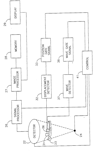
Fig. 6 indicates in simplified block diagram form a STET
system 21 equipped to accomplish either cardiac or respiratory
gating or both . System 21 generally comprises a detector 22 f or
detecting radiation. The radiation can be emanating from a
995 ~:15 ~ 1~
"~ ~ 2177~8~
patient 23 or from a raaiation source 24, typically comprising a
radioisotope material. When sourca 24 is a radioisotope,
detector 22 is preferably an Anger type camera.
The output of detector 22 is processed by a signal processor
26 . Processor 26 detPrmi n~c the location and energy of photons
striking detectors 22.
The output of signal processor 26 is further processed by
image procassor Z7 to provide lmage data using a memory 28. The
processed images are shown on display 29.
Gating controls are provided for system 21. More
particularly, respiratory gating uses a position sensor 31 which
senses the thorax position of patient 23 during the STET process.
The sensed ~; cpl ~ t is operated on to provide windows or bins
using a displacment detector 32. A position gate signal unit 33
provides gating signals to signal processor 26 based on the
thorax position determined by detector 32. The cardiac gating
system senses the heart beat with a sensor 3 6 . The R-wave is
detected by a wave detector 37. A cardiac gating signal is
provided to signal processor 26 by a wage gate slgnal unit 38
responsive to detection of the R-wave by detector 37 . U. S . Patent
4, 617, 938, the disclosure of which is incorporated herein by
ref erence, describes a gating system .
STET system 21 is shown to be under the control of a
controller 41 which supplies the appropriate control and timing
signals .
The present invention was described in the context of
nuclear medicine imaging ~ However~ the present invention is
applicable to other types of imaging systems, provided that
functional images (as described herein) have structural images
that are registered to them where needed. Additionally,
structural images of modalities other than X-~ay CT, MRI, ultra
sound and SPECT can be registered to nuclear ~fif -~ n~ images by
u1~ n~ the present invention.
It will ~e appreciated by persons skilled in the art that
the present invention is not limited by what has been
particularly shown and described herein. Rather, the scope of the
16
5' 1~5 J:15 p.t. 2~5~TI:X
~ 2~777~4
present invention is defined only by the claims which ~ollow: