Note: Descriptions are shown in the official language in which they were submitted.
Wo 95/15116 2 ~ 7 7 ~ 3 9 PCTIUS94/13736
m
SUDDEN CARDIAC DEATH PREDICTION
STATEMENT AS TO RIGHTS TO INVENTIONS MADE UNDER
FEDERALLY SPONSORED RESEARCH AND DEVELOPMENT
Patt of the work performed during ~ ' r ' of this imvention
utilized U.S. Government funds. The U.S. Government has certain rights in
this mvention.
BACKGROUND OF THE INVENTION
1. RELATED APPLICATION
This application is a; in-part of application serial number
07/948,529, filed September 22, 1992, now U.S. Pat. No. 5,265,617; which
is a ~ ;. "--m-part of application serial number 07/768,054, filed
September 30, 1991, now U.S. Pat. No. 5,148,812; which is a c,
in-part of application serial number 07/659,711, filed February 20, 1991, now
abandoned.
2. FIELD OF T~E INVENTION
The invention relates to cardiology. More specifically, the invention
relates to non-imvasive i~ , and ,, of individuals at risk for
sudden cardiac death. Cardiac vulu~,ldlJiliLy to ventricular fibrillation, the
mode of sudden death, is dynamically tracked by analysis of an
LIUL~I~I;O~
3. RELATED ART
Sudden cardiac death (SCD), which claims over 350,000 lives annually
in the United States, results from abrupt disruption of heart rhythm primarily
due ~o ventricular fibrillation. Fibrillation occurs when transient neural
triggers impmge upon an electrically unstable heart causing normally
W095115116 PCIIUS94/13736
2~ 77~
-2 -
organized electrical activity to become ~ and chaotic. Complete
cardiac dy~fi~ iull results.
The first step in preventing sudden cardiac death is identifying those
individuals whose hearts are electrically unstable. This is a major objective
in cardiology. If vulnerable individuals can be reliably identified non-
invasively, then preventiûn will be aided, mass screening will become
possible, and l)llA ~ gi~ l II of vulnerable individuals can be
tailored to prevent ventricular fibrillation.
~ O ' cardiac electrical stimulation has been used in patients to
provide ~luall~ila~iv~ r.-, . -~ ;- " on cl~crf rtihility and on the ~rf~,~,Li~ ,,,,. of
their 1' ' O therapy. ullru- 'y, this method requires cardiac
,AIh.,..;,~li.." and introduces the hazard of inadvertent induction of
ventricular fibrillation. Therefore, it is used only in severely ill patients and
is performed only in hospitals. It is unsuitable for mass screening.
A technique which has shown great promise is that of analyzing
alternans in the T-wave of an ~l~llu~aldiuOIalll (ECG). As used throughout
this disclosure, the term "T-wave" is defined to mean the portion of an ECG
which includes both the T-wave and the ST segment. Alternans in the T-wave
results from different rates of ~ of the muscle cells of the
ventricles. The extent to which these cells recover (or repolarize) non-
uniformly is the basis for electrical instability of the heart.
The consistent occurrence of alternans in the T-wave prior to
fibrillation is well ecf-lhlj~ Thus, detection of alternans promises to be a
useful tool in predicting vulnerability to fibrillation, if an accurate method of
quantifying the alternans can be developed. The following are examples of
cull~ ,iullal attempts to quantify alternation in an ECG signal: Dan R. Adam
et al., ~rlu~.ur~iull~ in T-Wave Morphology and S~Cr~rtihility to Ventricular
Fibrillation," Journal of Ele~lru~ ;y, vol 17 (3), 209-218 (1984);
Joseph M. Smith et al. "Electrical alternans and cardiac electrical instability,"
Circ~lation, vol. 77, No. 1, 110-121 (1988); U.S. Pat. No. 4,732,157 to
Kaplan et al.; and U.S. Pat. No. 4,802,491 to Cohen et al.
wo95llsll6 2 1 7 ~ 8 ~ 9 PCTIUS94113736
Smith et aL and Cohen et al, disclose methods for assessing myocardial
electrical instability by power spectrum analysis of the T-wave. These
methods derive an alternating ECG I.c,l~I-ol~,~y index from a series of
heartbeats. Sample point matrices are constructed and the alterrlating energy
at each of the sample points is computed using the analytical method of multi-
,1;"" ..c . -~ power spectral estimation which is calculated by ,U~DLlu~Lil.o the
discrete Fourier transform of the Hanning-windowed sample auto-correlation
function. Tbe alternating ene}gy over the entire set of sample points is
summed to generate the total alterrlating energy and then normalized with
lû respect to the average waveform to produce an ~alternating ECG Illul~llology
index (AEMI)."
While a powerful tool, Fourier power spectrum analysis averages time
functions over the entire time series so that rapid .~IIllyLlllll~ . changes,
such as those due to neural discbarge and I~ ,l r ' , are not detected because
data from these events are intrinsically non-stationary.
Kaplan et al. disclose a method for quantifying cycle-to-cycle variation
of a ~llya;~JlOgiC waveform such as the ~CG for the purpose of assessing
myocardial electrical stability. A pllyalOlogi~ waveform is digitized and
sampled and a scatter plot of the samples is created. Non-linear
1,,., r~" ;"" of the sample points determines a single parameter which
attempts to quantify the degree of alternation in the sampled waveform and
which is associated with the c~cr~rtihility of the ~ a;ulOgic waveform to enter
into an aperiodic or chaotic state. Kaplan et al. suggest that "1.,~ of
[this parameter] may provide an index of ECG waveform variability which
may provide an improved correlation with cll~rl~rtihility to ventricular
fibrillation thanpreviously available indices. " See col.3, lines 15-lg. Whetherventricular fibrillation is a chaotic state, however, is still very much in debate.
See D.T. Kaplan and ~. J. Cohen, "Searching for chaos in fibrillation, " Ann.
I~.Y. Acad. Sci., vol. 591, pp. 367-374, 1990.
Adam et al. disclose a non-invasive method which involves spectral
analysis of the alternation from beat-to-beat , ' ~' Oy of the ECG complex.
WO 95/15116 2 ~ 7 ~ 8 ~ q PCT/US94/13736
.
--4 -
The alternation of T-wave energy from beat-to-beat was measured to generate
a T-waYe alternation index ('I'WAI). This technique is unable to detect
alternation in waveform luul~ olu~;y which results in alternating wave shapes
of equal energy. In addition, the amount of alternation detected per this
method is dependent on the static portion of the wave shape. That is, the
same amount of alternation r~ 1 on a different amplitude signal will
result in different values for the T-wave alternation inde~ such that this
technique could completely obscure the presence of alternation in the original
waveform ,....,l.l,..l.~;f.c
In the absence of an effective method for dynamically 4u.~ iryill~ the
magnitude of alternation, i~ r; ;--l of alternans as a precursor of life-
threatening allhy ' and provision of a test for cardiac VUIll.,l~;liLy have
been 1 ~ lr In addition, the Wll~lliiUII~I attempts to quantify alternans
have employed inferior methods of alternans (i.e., ECG) sensing. The ECG
signals used for the Cohen et al. analysis were sensed via epicardial (i.e.,
heart surface) electrodes or via lateral limb, rostral-caudal, and 11nrr^~
leads. Smith et al. sensed via leads 1, aVF, and Vl 2. Adam et al. utilized
ECG lead I "because in this lead the ratio of the amplitude of the pacing
stimulus artifact to the amplitude of the QRS complex was usually smallest."
See Adam e~ al. at 210. Lead I, however, provides only limited ;.,ru, I.~ ;u~
regarding the f,lf~,ilu~hyalvlO~i~, processes occurring in the heart.
There have been occasional reports in the human literature noting the
presence of T-wave alternans in the precordial leads. However, there has
been no suggestion of a superior lead .,..,ri",..,.,;..,. from the body surface
which permits ~ lrll~ ~ of alternans as a uu~uliiLa~iv~ predictor of
sllc~rtihility to ventricular fibrillation and sudden death. For example,
alternans have been observed in precordial leads V~ and V5 during a PCTA
(r~ ,ui u.~,vua Tl, ~ --l Coronary Angioplasty) procedure on a fifty year-
old man. M. Joyal et al., "ST-segment alternans during p.,l~.UL~ll..,VUa
i ' ' coronary angioplasty," Am. J. Cardiol., vol. 54, pp. 915-916
(1984). Similarly, alternans were noted in precordial leads V~ through V6 on
wo 95/15116 PCrNS94113736
2 i 77~39
--5 -
a forty-four year-old man during and following a treadmill exercise. N. Belic,
et al., "ECG . - - ,; f ~ of myocardial ischemia, " Arch. Intern. Meevl., vol.
140, pp. 1162-1165 (1980).
Dispersion of ~ has also been integrally linked to cardiac
~ vlG~ y and has recently received ~-"~ attention as a potential
marker for vulnerability to ventricular fibrillation. The basis for this linkageis that the extent of llvvvluc_llv;Ly of recovery of action potentials is directly
related to the propensity of the heart to experience multiple re-entrant currents,
which initiate and maintain fibrillation and culminate in cardiac arrest. B.
Surawicz, "Ventricular fibrillation," vr. Am. cOn. Cardiol., vol. 5, pp. 43B-
54B (1985); and C. Kuo, et al., "(~1..,., .~ ;~1;. ~ and possible mechanism of
ventricular arrhythmia dependent on the dispersion of action potential
duration," Circ~lanon, vol. 67, pp. 1356-1367 (1983).
The most commonly employed non-invasive approach for measuring
dispersion is to obtain body surface maps to define the ~ . il,vti.. ,. of T-wave
.Jt~ and thus estimate the degree of unevenness of IcyulGli~GliOll and
y to ventricular fibrillation. F. Abildskov, et al., ~The expression
of normal ventricular Ir~ in the body surface ~ljctrihlltirn of T
potentials," Clrculation, vol. 54, pp. 901-906 (1976); J. Abildskov and L.
Green, "The recognition of arrhythmia vulnerability by body surface
~ ,vLIu~,Gld;u~lGyll;c mapping," Circv~lanon, vol.75 (suppl. 111), pp.79-83
(1987); and M. Gardner, et al., "Vulnerability to ventricular G llly '
assessment by mapping of body surface potential," C~rcv~la~ion, vol. 73, pp.
684-692 (1986). Although this approach has been in existence for over 15
years, it has received minimal usage in the clinical setting. The basis for thisis that the technique is, .,."1.. . ~u , as it requires over 100 leads on the chest
and extensive ~ ;- d analysis. Thus, it is used in only a few
specialized research centers.
Recently, these has been interest in analyzing QT interval dispersion
in the standard 12-lead ECG as a measure of vulnerability to life-threatening
allllyLlllll;Gs. The ' I l,."-~ ".lirl~ required is relatively
WO 95115116 ~ ~ 7 l~ dr 3 9 PCI[/US94/13736
-6-
'V~ r W~lld as it involves mainly subtraction of a minimum QT interval
from a maximum QT inoerval and 1~ the variance of the difference.
For example, it has been found that QT dispersion is an indicator of risk for
arrhythmia in patients with the long QT syndrome, who have greatly enhanced
~ y to ' ' released by the nervous sysoem. C. Day, et
al., "QT dispersion: an indication of arrhythmia risk in patients with long QT
intervals," l~r. Heart J., vol. 63, pp. 342-344 (1990). These ~.,~ Liull were
confirmed and exoended in C. Napolitano, et al., "Dispersion of
a marker of successful therapy in long QT syndrome patients
[abstract]," Eur. Heart J., vol. 13, p. 345 (1992).
The present inYentors' ~ 1 studies have ' ' that the
variance of T-wave dispersion in the epicardial ~ u~ exhibits a highly
significant predictive value in estimating risk for ventricular fibrillation during
acuoe myocardial ischemia. R. Verrier, e~ al., "Method of assessing
dispersion of ~ ; -, during acuoe myocardial ischemia without cardiac
electrical testing [abstract]," Circulanon, vol. 82, no. III, p.450 (1990).
Fu-;' , their data has ~ ' that a linear 1~ e~ists
between the epicardial and the precordial ECG. See U.S. Pat. No. 5,148,812.
This provides the scientific basis for utilizing precordial T-wave dispersion asa measure of the degree of ll~,t~,luc~ ,;.y of ~ , which occurs within
the heart.
Napolitano et al., supra, have shown in human subjects afflicoed with
the long QT syndrome that the variance of QT inoerval in the six standard
precordial leads of the ECG is more accuraoe than the limb leads in estimating
}isk of life-threatening ~ . These il. ~ tOI ~ have also
~' ' that dispersion of QT interval also provided a marker of
successful therapy in patients receiving beta-blockade therapy and those
undergoing cervical ~ "' y.
Within the last year, it has been ~ ' that QT interval
30 dispersion can predict the d~v~,lu~.. l.,.lt of Torsades de Poinoes, a precursor
arrhythmia to ventricular fibrillation in patients receiving ~IILidlllly~ , drug
WO 95115116 PCTIUS94113736
~ 21 77~39
--7-
therapy. T. Hii, ef al., "Precordial QT inoerval dispersion as a marker of
torsades de pointes: disparate effects of class la ~.,Li~llh~i' drugs arld
~I..;Od~lUIIC,'' Circulatfon, vol. 86, pp. 1376-1382 (1992).
Another method which has been explored to assess autonomic nervous
system activity, the neural basis for vulnerability to sudden cardiac death, is
analysis of heart rate variability (HRV). Heart rate variability, however, is
not an absolute predictor of SCD because there are major, non-neural factors
which contribuoe to sudden death. These include: coronary artery disease,
heart failure, myopathies, drugs, caffeine, smoke, ~.IIV;IUIIIII.~IIL~I factors, and
others. Accordingly, techniques which rely on heart rate variability to predict
cardiac electrical stability are not reliable.
Further, CUl~ iUllal techniques for analyzing heart rate variability
have relied on power spectrum analysis. See, for example, Glenn A. Myers
et al., "Power spectral analysis of heart raoe variability in sudden cardiac
death~ mrari~on to other methods," Ir~ Transactions on Biomedical
rngineering~ vol. BME-33, No. 12, December 1986, pp. 1149-1156. As
discussed above, however, power spectrum (Fourier) analysis averages time
functions over an entire time series so that rapid ~IIllyLlllllo~ , changes are
not detected.
Complex ~IPrnn~ n as a method for analyzing heart rate variability
is discussed in Shin ef al., "Assessment of autonomic regulation of heart rate
variability by the method of complex ~' "~ "" rEr~E Transactions on
Biomedical l~ngineering, vol. 36, No. 2, February 1989, which is ;III,UII~ '
herein by reference. Shin et al. teach a method of evaluating the influence of
autonomic nervous system activity during behavioral stress. A technique of
complex ~ ~ ' ' is used to analyze the patoern of beat-to-beat inoervals
to deoermine the relative activity of the ~yllllJa~ Li~. and I~lG~ylll~ai~ Li~
nervous sysoems. While Shin et al. exploited the dynamic analytical
.. 1,"".. ~ ;. c of complex .1. ~ -, they did not relate their results to
cardiac vulnerability.
WO 95/15116 2 i ~ 7~ 3 ~ PCI/[iS94/13736
Similarly, T. Kiauta et al. ~Complex ~ n~ -- of heart rate
changes during orthostatic testing," r~v~J;~ Computers in Cardiology,
(Cat. No. 90CH3011 'L), IEEE Computer Society Press, 1991, pp. 159-162,
discusses the use of complex ~ to assess heart rate variability
induced by the standing-up motion in young healthy subjects. Using the
technique of complex ~l, .,..vi.ll-l;..,, Kiauta et al. conclude that the complex
,iPm~-~ of the high frequency band probably refleets l~ala~ylll~aLII~
activity, but the complex ~' ' ' of the low frequeney band does not seem
to indicate by~ JaL}~,iic aetivity. Similar to Shin et al., Kiauta et al. do notrelate their results to cardiac ~ulll~,.ab;lily.
In summary, analysis of the IllUl~llUlo~y of an ECG (i.e., T-wave
alterrians and QT interval dispersion) has been recognized as a means for
assessing cardiac ~ u~ alJ;liLy . Similarly, analysis of heart rate variability has
been proposed as a means for assessing autonomie nervous system activity, the
neural basis for cardiac vulnerability. When ICD.,al111il~ vulnerability to
sudden cardiac death, researchers have cull~,.,iiullally relied on power
speetrum (Fourier) analysis. However, power spectrum analysis is not capable
of tracking many of the rapid allhy ' " changes which . l. --,.. ;.. T-
wave alternans and dispersion and heart rate variability. As a result, a non-
invasive diagnostic method of predicting vulnerability to sudden cardiae death
by analysis of an ECG has not aehieved elinical use.
What is needed is a non-invasive, dynamie method for completely
assessing vulnerability to ventrieular fibrillation under diverse pathologic
eonditions relevant to the problem of sudden cardiae death. Among the most
significant problems are enhanced discharge by the ~ylll~JaLll~,iic nervous
system, behavioral stress, aeute myoeardial isehemia, reperfusion, effeets of
r~ u~ agents on the autonomie nervous system, and intrinsie cardiac
effects of ~,l,,... - ul.~y,ir agents. To ' these conditions, the
method must not assume stationarity of data and must be sensitive to slowly
varying amplitude and phase over time. The diâgnostie system must be
sensitive to the faet that the area of injury to the heart ean vary j;6-.;rca~Lly,
WO 95/15116 PCI/US94J~3736
~ ~ J.~ 9
that extrinsic as well as intrinsic influences affect the electrical stability of the
heart, and that tbe elL~,LIu~ olv~ic end point to be detected must be
~Iy linked to cardiac vul~ L;liLy.
SUMMARY OF THE INVENTION
The present invention is a method and apparatus for non-invasive,
dynamic tracking and diagnosing of cardiac vulnerability to ventricular
fibrillation. lt is non-invasive as it detects vulnerability from leads placed on
the surface of the chest. Tracking and diagnosis of cardiac electrical stabilityare achieved through ~il""ll-,...,..- assessment of T-wave alternans, QT
interval dispersion, and heart rate variability. The method permits tracking
of transient but deadly ~Jallu~lly~;ùlO~ ;c events, such as enhanced discharge
by the ~y~ JaLh~,L;C nervous system, behavioral stress, acute myocardial
ischemia and reperfusion.
T-wave alternans, heart rate variability and QT interval dispersion are
' '!/ evaluated. T-wave alternation is an excellent predictor (high
sensitivity) of cardiac electrical instability but can be influenced by mechano-electrical coupling which does not influence cardiac v, ' ' li-y but reduces
the specificity of the measure. QT interval dispersion is a less accurate
predictor (lower sensitivity) of cardiac electrical instability but is not sensitive
to mechano-electrical coupling. However, potential artifacts may be generated
by eA~.c;,,;v~,ly low heart rate in QT interval dispersion or by its use of
multiple leads. Heart rate variability is a measure of autonomic influence, a
major factor in triggering cardiac _IIllyLlll.l;a~. By ~ u~ly analyzing
each ~1,~ .,....~..,~,-- (T-wave alternans, QT interval dispersion and heart rate
variability), the extent and cause of cardiac vulnerability can be assessed.
This has important IAI ;r~ ;-",~ for tailoring and assessing the efficacy of
drug therapy.
The method includes the following steps. A heart is monitored to sense
an ECG signal. The sensed ECG signal is then amplified and low-pass filtercd
before it is digitally sampled and stored. Estimation of alternans amplitude
W0 95/1~116 ~ 3 ~ PCT/US94/13736
-10-
and extent of dispersion and analysis of heart rate variability are then
separately performed.
Estimation of the amplitude of alternans is performed as follows. The
location of the T-wave in each R-R interval (heart beat) of the ECG is
estimated, and each T-wave is partitioned into a plurality of time divisions.
The sampled ECG signal in each of the time divisions is summed together and
a time series is formed for each of the time divisions such that each time series
includes f~",.~l..",.l;"~ time divisions from successive T-waves. The time
series are detrended before further processing in order to remove the effects
of drift and DC bias.
Dynamic estimation is performed on each time series to estimate the
amplitude of alternation for each time division. The preferred method of
dynamic estimation is Complex D- ~n~ " Other methods include
Estimation by S~lhtr~rtinn~ Least Squares F Auto Regressive
Estimation, and Auto Regressive Moving Average Estimation. The amplitude
of alternation is used as an indication of cardiac CllC~`~rtihility to ventricular
fibrillation (i.e., cardiac electrical instability).
Estimation of a measure of QT interval dispersion is performed by
analyzing ECG signals taken from a plurality of electrode sites. Dispersion
is determined by analyzing the ECG signals across the electrode sites. In the
preferred .. ,.1.~.1;,.. ~ one of five diffeRnt methods may be used to estimate
a dispersion measure. First, dispersion may be computed as a maximum
difference between QT intervals taken across the plurality of electrode sites.
Second, dispersion may be computed as a maximum difference between QT
intervals which have been corrected using Bazett's formula. Third, dispersion
may be estimated by a method which takes the standard deviation of a QT
interval ratio. Fourth, dispersion may be estimated by a method which takes
the standard deviation of the corrected QT interval ratio. Finally, dispersion
may be estimated by computing the maximum RMS (root mean square)
deviation of the ECG waveforms recorded from a plurality of sites.
wogs/lsllF 2 1 7 7 ~ 3 q PCTNS94113736
Analysis of heart rate variability is performed as follows. The apex of
each R-wave is l~'t~'nnim'rl, and the time between successive R-waves is
computed to deterrnine a magnitude (time) of each R-R interval. The
magnitude of each R-R interval is then compared to a L~lc '~ ' crioerion
S to eliminate premature beats. Ne~t, a time series of the ~ ' of the R-
R intervals is formed. Dynamic estimation is performed on the time series to
estimate the magnitude of a high frequency component of heart rate variability
and to estimate the magnitude of a low frequency component of heart rate
variability.
The magnitude of the high frequency component of heart rate
variability is indicative of ~ y~ Lll~ , activity. The magnitude of the low
frequency component of heart rate variability is indicative of combined
~yl~ ,.i., activity and ~ a,y~ Lh~, activity. A ratio of the low
frequency component and the high frequency component of heart rate
1~ variability is formed. The ratio is indicative of ~ylll~ .ic activity or vagal
withdrawal. In addition, recent studies have shown that particular emphasis
should be paid to the Very Low Frequency (VLF) (0.0033 to 0.04 Hz) and
Ultra Low Frequency (ULF) (<0.0033 Hz) spectral portions of heart rate
variability as a powerful predictor of arrhythmia in the first two years
following a myocardial infarction.
In the preferred I .,.1.-,.1;",. .1l of the invention, the ECG is sensed non-
invasively via the precordial or chest leads for optimal alternans detection.
Leads V5 and/or V6 detect the optimal alternans signal when the left side (the
most common site of injury for the ~JlU~ ,GLiUII of life-threatening ~IIIy '
of the heart is ischemic or injured. Leads Vl and/or V2 are optimal for
detecting obstruction of the right-sided coronary circulation. Additional
precordial leads, such as V9, may be useful for sensing alternans resulting
from remote posterior wall injury. A physician may use the complete
precordial lead system to obtain precise i ,. '~ .. ." -' i. ~,. non-invasively regarding
the locus of ischemia or injury.
WO 95/15116 ;~ 7 ~ 3 ~ PCT/US94113736
-12-
For the dispersion measure, a plurality of chest leads (e.g., the
standard precordial or some greater number) may be used to provide a
plurality of electrode sites across which dispersion may be measured. Heart
rate variability is easily sensed from any of the standard ECG leads.
The foregoing and other objects, features and advantages of the
invention will be apparent from the following, more particular description of
a preferred ~ ~ ~ ' to the invention, as illustrated in the ~U
drawings.
BRIEF DESCRIPTION OF THE DRAWINGS
FIG. lA is a typical ECG plot.
FrG. lB is a typical ECG plot and action potential plot illustrating the
correlation between dispersion of ~ and the QT interval.
FrG. lC shows a number of heart rate plots with c
spectral plots.
lS FrG. 2A is high-level block diagram illustrating the diagnostic
principles of the present invention.
FIG. 2B is a block diagram illustrating the diagnostic principles of the
present invention in a first example.
FrG. 2C is high-level block diagram illustrating the diagnostic
principles of the present invention in a second example.
FrG. 3 is a flow chart illustrating the method of the present invention.
FIG. 4 is a flow chart detailing the process of dynamically estimating
the amplitude of T-wave alternans (as performed in step 314 of FIG. 3).
FIG. SA is a flow chart detailing the process of dynamically analyzing
heart rate variability to determine the activity of the autonomic nervous system(as performed in step 314 of FIG. 3).
FIG. SB is a flow chart detailing the process of dynamically analyzing
heart rate variability to determine the ultra low and very low frequency
activity of the autonomic nervous system (as performed in step 314 of FIG.
3)-
Wo 95/15116 Pcr/uss4J~3736
2 ~ 778~i9
-13-
FIG. 6 is a flow chart illustrating a method for estimating first and
second measures of QT interval dispersion.
FIGS. 7A and 7B is a flow chart illustrating a method for estimating
third and fourth measures of QT interval dispersion.
FIG. 8 is a flow chart illustrating a method for estimating a fifth
measure of QT interval dispersion.
FIG. 9A is a high-level block diagram of the apparatus of the
invention.
FIG. 9B is a detailed block diagram of ECG detector and pre-processor
902.
FIG. 9C is a detailed block diagram of ECG processing system 904
comprising a ~ ,lu~,ul. ~
FIG. 10 is a detailed block diagram of the preferred ~ "l~ of the
heart monitoring unit (HMU) 900.
FIG. 1 lA is an ECG recorded within the left ventricle of a dog before
coronary artery occlusion as set forth in the animal study below.
FIG. llBshows~ of sixsuccessivebeatsfromFIG. llA
presented on an expanded time scale.
FIG. 12A is an ECG recorded within the left ventricle of a dog after
four minutes of coronary artery occlusion as set forth in the animal study
below.
FIG. 12B shows ~ of six successive beats from FIG. 12A
presented on an expanded time scale.
FIG. 13A is an ECG recorded within the left ventricle of a dog after
release of the coronary artery occlusion (during reperfusion) as set forth in the
animal study below.
FIG. 13B shows ~ iu.. of six successive beats from FIG. 13A
presented on an expanded time scale.
FIG. 14A is a surface plot of the T-wave oF the ECG for eight dogs
with intact cardiac innervation showing the effects of coronary artery occlusionand reperfusion.
WO 95115116 2 ~ 7 7 8 3 9 PCIIUS94/13736
-14-
FIG. 14B is a surface plot of the T-wave of the ECG for six dogs after
bilateral stellectomy showing the effects of coronary artery occlusion and
.cl,~,.r
FIG. 14C is a surface plot of the T-wave of the ECG for eleven dogs
during thirty seconds of stimulation of the ansa subclavia of the ~l . . .,1.,.1i ;1
left stellate ganglion showing the effects of coronary artery occlusion and
IC~
FIG. 15 shows the correlation between the occurrence of -r
ventricular fibrillation and T-wave alternans in ten dogs.
FIG. 16 is a graph showing the responses of the ~y . ' and
yl~ ih~,~ic nervous systems to a LAD coronary artery occlusion and
reperfusion as indicated by heart rate variability.
FIGS. 17A-17C illustrate the positioning of the precordial ECG leads
on the body.
FIG. 18 is a cross-section of the human body illustrating the positioning
of precordial ECG leads V,-V6 relative to the heart.
FIG. l9A is an ECG recorded from lead Il during coronary artery
occlusion in a dog.
FIG. 1 9B shows ~ of six successive beats from FlG . l 9A
presented on an expanded time scale.
FIG. 20A is an ECG from precordial lead V5 recorded ~ r ~ y
with the ECG of FIG. l9A.
FIG. 20B shows ~ of six successive beats from FIG. 20A
presented on an expanded time scale.
FIG. 21A is an ECG from a left ventricular illLI~c~lviLdly electrode
recorded cim~ / with the ECG of FIG. l9A.
FlG.21Bshows~ i-- ofsixsuccessivebeatsfromFlG.21A
presented on an expanded time scale.
FIG. 22 is a graph showing the relative magnitudes of alternans signals
sensed from lead 11, from precordial lead V5, and from a left ventricular
illLIcl~viLdly electrode.
WO95/15116 2 ~ 7 18 ~ 9 PCT/US94/I373Ij
-15-
FIG. 23 is a surface plot display obtained by the method of complex
' ' (as set forth above) of the T-wave of the V4 precordial lead
during ~ heart rhythm in a r~ , patient during ~ J.
FIG. 24 shows the level of T-wave alternans as a function of recording
S site in seven patients at three minutes of: ., .' !/-induced occlusion and
upon balloon deflation.
FIG. 25A and 25B illustrate an example positioning of a plurality of
ECG leads on the body for QT dispersion ~ t.
DETAILED DESCRIPTION OF THE PREFERRED EMBODIMENT
INTRODUCTION
The invention is directed to a method and apparatus for screening
individuals at risk for sudden cardiac death. In order to produce an optimal
testing .". :I..~nlnr,y, the invention takes a receiver operating ~1 - ", l. .;~1;,
(ROC) curve approach to cardiac risk ~ r~ The invention meets three
criteria required for successful risk !71.,liri -l;~.. and treatment:
(I) i.l..,liri.-li.... of subsets of patients at high risk for sudden
cardiac death;
(2) elucidation of specific ' by which sudden cardiac
death occurs; and
20 (3) i~lFntifir:~tif~n of ~,. l,.. ,: .. ~ at which treatment can be aimed.
The following terms are used herein:
Complex ;' ' A spectral analysis method which estimates the
amount of signal in a specified frequency band by frequency translation of the
signal and low-pass filtering.
Expert system: A domain-specific (e.e., medicine, F .. ~,;1.. ;.lp" ~rr-ol~ntin~)
- computer system built to emulate the reasoning process of the mind of an
expert in that domain.
WO 9S/15116 PCI/US94113736
2 ~ ~783~ --
-16-
neart rate ~.. ' ' ~.~. An estimate of the frequency content of variation inheart rate as a measure of automatic nervous system output.
~I~. .lidl infarction: Damage to or death of cardiac muscle, usually due
to coronary artery occlusion as a result of plaque rupture or formation of a
clot.
Negative I ~ . The probability that an individual is truly disease-free
given a negative screening test. It is calculated by dividing the number of truenegatives by the sum of false negatives and true negatives.
Neural net~ork: A computing model which emulates to some degree the
cll~,l.;k~ and function of a group of neurons. The network is trained to
interpret input data by adaptive adjustment of the strength of the
Positi~e y~ . The probability that a person actually has the disease
given that he or she tests positive. It is calculated by dividing the number of
1~ true positives by the sum of true positives and false positives.
1~ 1 '- . il~ . The probability that an individual actually has the disease, given
the results of the screening test.
S. ~ili . il~. The probability of testing positive if the disease is truly present.
It is calculated by dividing the number of true positives by the sum of true
positives and false negatives. True positives are the individuals for whom the
screening test is positive and the individual actually has the disease. False
negatives are the number for whom the screening test is negative but the
individual does have the disease.
WO95/15116 2 1 ~ 7 8 3 9 pcrAJss4ll3736
-17-
S~ . The probability of screening negative if the disease is truly
absent. It is calculated by dividing the number of true negatives by the sum
of false positives and true negatives. True negatives are individuals for whom
the screening test is negative and the individual does not have the disease.
False positives are the individuals for whom the screening test is positive but
the individual does not have the disease.
Sudden cardiac death: Natural death due to cardiæ causes, heralded by
abrupt loss of . within one hour of onset of acute symptoms, in
an individual with or without known preexisting heart disease, but in whom
the time and mode of death are llnp~rcrtp~ Sudden death is the leading form
of adult mortality in the industrially developed world, claiming one death per
minute in the United States alone. Coronary care unit and out-of-hospital
'-"`' .1..l;~.ll experience have shown that sudden death is due primarily to
ventricular fibrillation.
T-wave alternans: A regular beat-to-beat variation of the T-wave of an
uudldio~-all- which repeats itself every two beats and has been linked to
underlying cardiac electrical instability.
The preferred ' ' of the invention is discussed in detail below.
While specific cf~nfiellt~ti~n~ and ~ are discussed, it should be
understood that this is done for illustration purposes only. A person skilled
in the art will recognize that other ~ and ,.~ may be
used without departing from the spirit and scope of the invention.
The preferred ~..,1..,.1;1,,.: of the invention is now described with
reference to the figures where like reference numbers indicate like elements.
Also in the figures, the left most digit of each reference number CUII~,~U
- to the figure in which the reference number is first used.
Figure lA shows a l~ .lLa~ive human surface ECG 100. A
deflection 102 is known as the "P-wave" and is due to excitation of the atria.
WO 95/15116 PCTNS94/13736
2 ~ ~78~ --
-18-
Deflections 104, 106 and 108 are known as the "Q-wave, " "R-wave, r and "S-
wave, " respectively, and result from excitation (de-pol~ll ;~tiUI~) of the
ventricles. Deflection 110 is known as the "T-wave" and is due to recovery
(~r~ ) of the ventricles. One cycle (i.e., cardiac cycle or heart bcat)
of the ECG from the apex of a first R-wave to the apex of the next R-wave
is known as the R-R or interbeat interval. Heart rate variability (HR~) refers
to changes in the heart rate (HR) or length (time) of the interbcat interval from
one bcat to the next.
A portion 112 between S-wave 108 and T-wave 110 of ECG 100 is
known as the "ST segment". ST segment 112 includes the portion of the ECG
from the end of S-wave 108 to the beginning of the T-wave 110. Because this
invention is concerncd with alternans in the ST segment as well as in the T-
wave, the term rT-wave" in this disclosure, as noted above, includes both the
T-wave and the ST segment portions of the ECG. The inventors have found
that most alterrlation occurs in the first half of the T-wave, the period of
greatest vulnerability to ventricular fibrillation. See, Ncaring BD, Huang AH
and Verrier RL, "Dynamic Tracking of Cardiac Vulnerability by Complex
D ,~ ;u.. of the T Wave," Science 252:437-440, 1991.
This invention is also concerncd with the QT interval. The QT interval
is defined as the period between the beginning of the Q-wave and the end of
the T-wave. However, other definitions for the QT inoerval (e.g., from the
beginning of the Q-wave to the apex of the T-wave) may be used without
departing from the spirit and scope of the invention as defined in the claims.
Figure lB illustrates the concept of QT interval dispersion. A sample
ECG signal 150 and a cu,-c r ~ " cellular action potential 160 are shown.
Line 152 indicates the beginning of the Q-wave. Line 154 indicates the end
of the T-wave. Action potential 160 represents the cellular ~
occurring during the QT interval 156. Note that dispersion 158 occurs
primarily during the first half of the T-wave as illustrated between lines
162,164. This is the period in which the hcart is most vulnerable to cardiac
electrical instability.
WO 95/15116 2 1 ~ 7 ~ ~ q PCI~/US94/13736
-19-
A more detailed discussion of ECG sensing and analysis is provided in
Dale Dubin, Rapid l~.'LI~I ' ' '~n ~f EKG's, 4~ Edition, Cover Publishing
Company, 1990, which is i r ' ~ herein by reference.
Conventionally, autonomic nervous system activity, as indicated by
S heart rate variability, has been researched as an; ~ indicator of
cardiac VUIIl.,l~;lily (electrical stability). Autonomic nervous system activity,
however, is not an absolute predictor of cardiac vulll.,l~;liLy.
Further, LUllV.,~.~iUI~I research has evaiuated heart rate variability,
ECG , ' ~' Oy as indicated by T-wave alternans, and ECG l~lu,~llolv~;y as
indicated by QT interval dispersion as i,.. l.1,.. ,.1.. ,l variables indicative of
cardiac vulnerability. This also is an invalid ~Ccllmrrinn HRV and ECG
~"u~holuOy are linked, however, not invariably. Alternans, QT interval
dispersion and HRV can each change i~
Heart rate variability and ECG . ' ' "y measure different aspects
of ~,~ i;uv~ ,ulal control. Both must be assessed in order to fully diagnose
cardiac ~L Il~,l~ili~y. The inventors have discovered thaî
analysis of heart rate variability, T-wave alternans and dispersion yields
important diagnostic i.,r~.. -~;.". pertaining to cardiac VUII.. ,I~ili~y.
Heretofore, this i"r.", ;~ has not been available.
20 By "~i"",ll~,.. ~", it is meant that the analysis of T-wave alternans,
dispersion and heart rate variability is carried out on the same ECG data. It
is not necessary for this to be done at the same time. For example, the ECG
data may be stored and the individual analyses performed in sequence one
after the other.
2~i Cardiac vulnerability is affected by both intrinsic and extrinsic factors.
The intrinsic factors include coronary artery occlusion and l,dl iiUlll,yO~ ily.The extrinsic factors include the autonomic nervous system, ~I~,.""~ Oic
agents, body chemistry (e.g., el~ ul~), and other chemicals (e.g., from
- cigarette smoke, caffeine, etcetera).
An intrinsic factor can make a heart electrically unstabie and therefore
susceptible to SCD. T-wave alternans and dispersion are indicative of cardiac
WO95/15116 2 ~ PCTIUS94/13736
-20-
electrical instability caused by intrinsic factors. Without T-wave alternans, a
heart is not at risk of sudden cardiac death (~ ,uldl fibrillation). As the
magnitude of aloernans increases, so does the risk of sudden cardiac death.
T-wave aloernation is an excellent predictor of cardiac electrical
stability but can be influenced by mechano-electrical coupling. Alternans
measures both excitable stimulus and ll~ ob~ ;t~ of Ir~ ., of the
cardiac substraoe. It is an intrinsic property of an ischemic and reperfused
lll.v~/~ld;....l. However, mechano-electrical coupling (e.g., through pericardial
effusion and tamponade, abrupt changes in cycle length, drugs, and the like)
which does not have an influence on cardiac ~ / will influence
aloernation. Thus, a measure of alternation has a high degree of sensitivity buta low degree of specificity.
The inventors have discovered, however, that the low specificity of
aloernation can be addressed using a test which ' '~/ analyzes
another variable, QT interval dispersion. Dispersion is not a measure of
excitable stimulus and is not sensitive to mechano-electrical coupling.
However, its specificity is reduced in cases of low heart rate and due to its
l~iU,U;lt;ll.~ of multiple leads. The resulting cc." l ;~ - of aloernans and
dispersion yields an accuraoe predictor of cardiac electrical instability causedby intrinsic factors.
Extrinsic factors may also cause or increase the electrical instability of
the heart by causing or increasing aloernans and dispersion. The autonomic
nervous sysoem is a primary extrinsic factor which affects cardiac electrical
stability. Relative changes in actions of the ~ ylllpGLll~ sysoem versus the
,~ ,iic sySoem can increase the magnitude of alternans, resulting in an
increased vulnerability to SCD. However, a change in the autonomic nervous
system by itself is not an absolute cause or predictor of cardiac electrical
instability.
Heart rate variability is a measure of autonomic nervous system
function. Generally, decreased heart rate variability will tend to increase the
magnitude of aloernans. Further, as described in detail below, analysis of the
WO95/1~116 2 1 ~ PCrlUSs4/13736
-21-
spectral content of heart rate variability indicates that the high frequency (e.g.,
0.354 Hz) portion of the signal Wll~ Jlld~ to ~ Oylll~lLh~ (i.e., vagal)
activity while the low frequency (e.g., 0.08 Hz) portion of the signal
t ~ - . ' to combined ~y~ ;c and ~ y~ Ja~ Lt~ activity.
A detailed discussion of heart rate modulation by the autonomic
nenous system is provided in J. Philip Saul, "Beat-to-beat variations of heart
rate reflect modulation of cardiac autonomic outflow, " News in r~yA ;I7~0gi~ul
Sciences, vol. 5, February 1990, pp. 32-36.
Referring to Figure IC (reproduced from Id. at pûge 35), Saul shows
the heart rates and ~", ~ frequency spectra 120 for a patient with a
normal heart, 122 for a patient with congestive heart failure, 124 for a diabetic
patient with a peripheral neuropathy, 126 for a diabetic patient with a cardiac
autonomic neuropathy, 128 for a patient with a 1~.-"~ 1 heart pnor to re-
innervation, and 130 for a patient with a i , ' ' heart after re-
innervation. As can be seen from inspection of these data plots, the loss of
neural activity either due to diabetes or cardiac transplant is evident in the
absence of normal spectra. With return of normal innervation, the spectra at
least partially return.
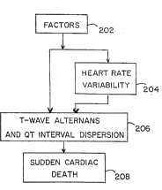
Figure 2A is a block diagram illustrating the diagnostic principles of
the present invention. Block 202 represents all factors which affect the
electrical function of the heart (e.g., drugs and/or diseases). Block 204
represents increased heart rate variability resulting from the factors of block
202. Block 206 represents alternation of the amplitude of the T-wave and
dispersion of the QT interval resulting from the factors of block 202. Block
208 represents sudden cardiac death resulting from ventricular fibrillation.
As shown, the factors of block 202 can lead to SCD in block 208 by
two major pathways. The first pathway is from block 202, through block 206,
to block 208. This results from a direct influence of the factors of block 202
- on the electrical stability of the heart, manifest in the form of T-wave alternans
and QT interval dispersion This mode of SCD would occur without a change
in heart rate variability because the nervous system is not involved A
. .
WO 95/15116 ~ 8 ~ ~ PCI/llS94/13736
-22-
corollary to this is that a sudden death prediction method which relies solely
on heart rate variability would not be adequate to detect SCD.
The second major pathway from the factors of block 202 to SCD in
block 208 is through blocks 204 and 206. This results from an influence of
the factors of block 202 on the autonomic nervous system. Drugs or heart
disease, for example, can ~;6llir~ 1y alter neural activity. This will be
expressed as changed heart rate variability. Certain changes in neural activity
which increase ~ylll,u~L~ , tone ~;6lliG~l-lly increase T-wave alternans and
QT interval dispersion and therefore could result in SCD.
The inventors have discovered that by combining an indication of heart
rate variability with an indication of either T-wave alternans or QT interval
dispersion, it is possible, not only to assess risk for SCD accurately, but alsoto determine whether a ~ in autonomic nenous system activity is
causal. This has important clinical c~ as it affects both diagnosis and
therapy. In the preferred ~ ' ~o~l; 1 ' both T-wave alternans and QT interval
dispersion are analyzed in C.~.-J.-II. 1;~.,1 with heart rate variability.
For example, terfenadine (Seldane) is a drug widely employed for the
treatment of sinus problems. It has recently been discovered that, when
terfenadine is used in ~ with antibiotics, SCD can result.
Terfenadine has no known effects on the autonomic nervous system and
~.,...I ...,lly does not affect heart rate variability. However, the drug can
result in alternans and torsades de pointes in isolated heart ~ICIU " and
is thus capable of directly de-stabilizing the electrical activity of the heart.The 1~ of T-wave alternans and/or QT interval dispersion is
therefore an essential approach to detect s~crf~rtihility to SCD induced by a
dill~ llLil,iu~;1 ,~,1l,1,;. -l;.." This is illustrated in Figure 2B.
For another example, digitalis drugs are the most commonly used agent
for increasing the strength of contraction of diseased hearts. The drugs
produce this effect by both direct influence on the heart and through alterations
in the autonomic nervous system. In the proper therapeutic range, there is no
significant negative effect on the electrical stability of the heart. However,
WO95/15116 ? ~ 7~ ~3 9 PCT~US94/13736
-23-
when the dose is either too high or the patient's health status changes due to
illness, the same dose of drug may become toxic. It is often difficult to
determine whether a patient is under-dosed or overdosed. By using a
combined alternans/dispersion/HRV analysis, it would be possible to determine
at what point a neurotoxic influence may lead to alternans and SCD. In
particular, high doses of digitalis decrease vagal tone and increase sy~ Lh,ii~,activity, effects which would be clearly detected in an heart rate variability
analysis. This is illustrated in Figure 2C. This ;,lr.,""-~i.", would be a
valuable asset in the therapeutic ~ of the patient.
As discussed above, traditional methods of quantifying hcart rate
variability or the magnitude of alternans have relied on power spectrum
(Fourier) analysis. However, power spectrum arlalysis is not capable of
tracking many of the rapid ~ lyilllllG~ ic changes which ~ . ;,. T-wave
alternans and heart rate variability. In the preferred '.IJ~I;lll. '~, the present
invention utilizes complex ~' ' to analyze heart rate variability and
T-wave alternans.
METHOD OF THE INVENTION
The method of the present invention for analyzing an ECG is now
discussed with reference to Figures 3-8.
An ECG signal containing a plurality N of R-R intenals is sensed from
a patient in real time at step 302. For alternans and heart rate variability
analysis, only a single ECG signal (i.e., an ECG signal sensed from a single
site) is required. For dispersion analysis, however, a plurality of ECG signals
(i.e., ECG signals sensed from a plurality of sites) are required. The
preferred method of non-invasively sensing the ECG signals is discussed in
detail below. Because the body is akin to a dipole, a large DC component will
be present in the sensed ECGs. This DC component is removed at step 304
- with a high-pass filter prior to ~mrlifir~ of the I~CG signals at step 306.
The amplified ECG signals are then low-pass filtered at step 308 to limit the
signal bandwidth before they are digitally sampled at step 310. The digitized
WO95/lS116 2~ 7~ PCT/US94/13736
-24-
data may then be stored on a magnetic or optical storage device at step 312.
Finally, the digitized ECG data is processed or analyzed at step 314.
Processing at step 314 involves: (1) producing an estimation of
alternans amplitude, (2) estimating the magnitude of discrete spectral
--- r of heart rate variability to determine the ,y~ "i., and
,ylll~clih~,(J~, influences on cardiac electrical stability, and (3)
the extend of QT interval dispersion.
As an alternative to this real-time signal pre-processing, the ECG
signals may be retrieved from the storage device (step 312) and processed
(step 314) at a later, more convenient time. Processing/analyzing step 314
involves three i~ u"~ alternans processing, heart rate
va}iability processing, and QT interval dispersion processing. Each is
discussed in detail below.
T-WAVE ALTERNANS
The analysis of alternans at step 314 is described in detail with
reference to Figure 4. At step 404, the apex of each R-wave in the signal data
for each of the N beats is located by finding the peak amplitudes in the
digitized signal. Premature beats are removed at step 406 by rJ '" 1"" ;`- "' ofeach R-R interval with fixed criteria. At step 408, a portion of the ECG
~1 ~l l r~ l(1; l Ig to an estimated location (with respect to R-wave 106) of T-wave
110 is identified.
At step 410~ the T-wave 110 and 112 portion of the ECG signal is
partitioned into "B~ time divisions, where "B" may include a single digital
sample or a plurality of samples. The area between the ECG and the
isoelectric baseline is computed for each time division, at step 412, by
summing the areas of all samples in the time division. Then at step 414, "N"
successive beats (e.g., from control through release in the animal ~ Al.. .; 111. ..`~
discussed below) are sequenced into a time series for each of the "B" time
divisions: (X(n), n = 1,2,...N).
WO 95/15116 2 1 7 7 ~ 3 9 PCrlUS94JI3736
-25-
A high-pass filter is used for detrending the time series at step 416 to
remove the effects of drift and DC bias (e.g., high-pass filtering removes the
large low-frequency variation in T-wave area that occurs during occlusion of
a coronary artery). A cleaner signal is then available for dynamic estimation,
which is performed at step 418 to estimate the amplitude of alternation for
each time series.
The estimation of step 418 may be performed via severai dynamic
methods. By "dynamic" method, it is meant any analytical process sufficientiy
rapid to track (i.e., estimate) transient changes such as those which occur in
alternans amplitude in response to ~ ya;Ol~ic and ~ Jpl~ya;~lu~i1 processes
triggering ~Illlly~ ..a. These include, for example, enhanced neural
discharge, acute myocardial ischemia and Ic~,l rl A "dynamic" method
should be able to track alternans from as few as d~ 'y ten heart beats
(or less). This precludes analytic processes (e.g., Fourier power spectrum
analysis) which require stationarity of data for several minutes. Specific, but
not exclusive, examples of methods for dynamic estimation include:
(a) Complex D~mr,~i lqti-.n
(b) Estimation by Sllhtrlrtinn
(c) Least Squares Estimation,
(d) Auto-Regressive (AR) F and
(e) Auto-Regressive Moving Average (ARMA) Fctin~ ir n
(A) COMPLEX DEMODULATION
Complex .i. ,..~.I..I-li.... is the preferred method of dynamic estimation
of the beat-to-beat alternation in the amplitude of each time series. Complex
.1.. ~ .. is a type of harmonic analysis which provides a continuous
measure of the amplitude and phase of an oscillation with slowly changing
amplitude and phase. It detects features that might be missed or
Ill;alc~ ll~d by standard Fourier spectral analysis methods which assume
stationarity of data.
By definition, alternans is a periodic alternation in the T-wave. The
magnitude of alternans, however, changes slowly during a coronary artery
W09511S116 ~ ~ 7 7 8 ~ ~ ~CTIUS94/13736
-26-
occlusion and more rapidly during release, making it quasi-periodic. As such,
it must be represented by a sinusoid with slowly varying amplitude, A(n), and
phase, ~(n):
X(n) = A(n) Cos[2~tf~LT + (p(n)] Eq. (1)
where: X(n)= the data sequence with alterrlation in its
amplitude
f~LT = ~ alternation frequency (E~z). It should be noted
that this frequency is half of the heart rate.
Using the identity
cos(x) = C ~ , Eq. (2)
the equation for X(n) can be rewritten as
X(n) = A(n) x (e ej~ + e I Df~ e j~n) Eq (3)
The method of complex ~ " requires ~ lyill~ this time
series X(n) by two times a complex eYr~nPnr~ at the alternans frequency [to
produce Y,(n)] and then filtering the result to retain only the low frequency
term Y2(n) as follows:
Yl(n) = ~(n) x 2e i2i'f~
= A(n) [el~n~ + ~ JA~a~ -1~1 Eq. (4)
Y2(n) = A(n) ~ ) Eq. (S)
The amplitude and phase of the alternans is then found from the filtered
signal, Y2(n), as follows:
where: Im and Re refer to the imaginary and real parts of Y~
WO95/15116 2 t ~ PCr/US94/13~36
-27-
A(n) = I Y2(n) 1
ç = magnihule of Y2(n) Eq. (6)
= JRerY2(n)]2 + Im[Y2(n)]2
~4(n) = p)u~se of Y2(n)
a ta~lm[Y2(n)]l ~q- (n
LRe[Y2(n)]~
For a more detailed discussion of complex f- ~~ ' ' see FoKrier
An~lysis of Time Series: An In~u.~iu,~, by Peter PIo- mfil-ltl John Wiley &
Sons: New York, pp. 118-150: which is illco~l~ul~l~cd herein by reference.
(B) ESTIMATION BY SUBT~ACTION
The subtraction method of dynamic estimation is an alternative which
may be substiwoed for complex ~l~mr~ llqri~n The subtraction method
involves subtracting the area of each time division (n) of an R-to-R interyal
from the area of the W~ p~Jlld;ll~ time division of a subsequent (n + 1), or
alternatively, a previous (n-l) R-to-R interval to form a new time series Y(n)
IC~ >CIILill~ Lhe magnitude of aloernans. Because this difference series Y(n)
may be positive or negat~ve, the absoluoe value or magnitude of Y(n) is used
for the magnitude A(n). That is:
Y(n) = X(n) - X(n - I) E~l. (8)
A(n) = ¦ Y(n)
= IX(n) - X(n-1)l Eq. (9)
= magnitude of al~rnans
Some errors may be introduced into this estimate due to the slowly
varying increase in magnitude of the T-wave size at the start of a coronary
occlusion and the reduction in size following the occlusion. Also, some T-
wave variation due to respiration is expected. Therefore detrending the
sequence X(n) using a high pass digital filoer, or equivalent, improves the
WO 95/15116 2 ~ 7 7 Q ;~; ~ PCTIUS94/13736
.
-28-
estimate by removing the effects of T-wave size changes. Also, averaging M
samples together, where M is the number of beats occurring during a single
respiratory cycle, aids in eliminating the respiratory effects on the estimate.
AlternatiYely, the digital filter may remove both trends and respiratory changesif tbe respiration frequency is sufficiently different from the heart rate, so that
the filtering does not alter tbe magnitude of the alternans estimate.
(c~ LEAST SQUARES EISTIMATION
The least squares estimation, which also turns out, in this c~se, to be
the maximum likelihood estimate for estimating sinusoid amplitude in white
noise, is a second alternatiYe which may be substituted for complex
~rm~~ inn to calculate a new sequence which is a dynamic estimate of the
amplitude of alternans. Least squares estimation of the amplitude of alternans
A(n) for the data sequence X(n) is derived as follows.
Assume for M points (e.g., 5 to 10 cardiac cycles) tbat:
X(n) = A cos(2-rf"Lrn) + N(n) Eq. (10)
where: N(n) represents additive noise
In order to minimize the noise term and estimate the alternans cnmrn- nt
create a new function T(A), where:
l~A) = ~ [X(~ - A Cos(2~fALr~]~ Eq. (11)
T(A) represents a measure of the difference between the model and the dat~.
The best alternans magnitude estimate results if T(A) (i.e., the noise term) is
minimiæd. To minimize T(A), take the derivative of T(A) with respect to A
and set it equal to zero:
Next, solve this equation for A(n) (shown simply as "A" above) and take the
absolute value of the result to yield the least squares estimate of the magnitude
W095/15116 ~ ~ ~783'~ PcrluS94113736
-29-
Eq. (12)
oT = -2 x jl+M-1 lcos(2~fALr~ [X(~ - A cos(2~fA~ ]} =
of the alternans:
Eq. (13)
A(n) = 1 ¦ ~j+M-I tX~ COS(21~fALJ~]¦
(D) AuTo-REG~EsslvE EST~ATION (AR)
Auto-Regressive (AR) Estimation is a third method of dynamic
estimation which may be substituted for complex ~ ;.," AR
5estimation models the alternans as follows:
Eq. (14)
X(n) = ~ ~ [a(k) x X(n - k)] + u(n)
In this model, "P" is the number of auto regressive L~Jrrr; ~ chosen for the
estimation. u(n) represents noise and accounts for the imperfect fit of the
estimation. The method of estimating the amplitude of alternans A(n) for the
data sequence X(n) first involves calculating a matrix of co-variance
10~ ffi~ n+c c(i,k) according to the following formula:
Eq. (10
c(i,~) = M p j~=+~+pl [X(J - ~) x X(l - k)]
where: â r the best estimate of the true value of "a"
P = the number of auto regressive ~ "â"
M = the number of cardiac cycles
The co-variance ~iue~;~ r~l~ are then used to form P" auto regressive
, ~,rrri, :. .. l~ "â" as follows:
The estimate of the alternans magnitude is then given by:
For a more detailed discussion of auto-regressive estimation, see
Modern Spectral Esh~nahon: Theory and Arrlirnrr~, by Steven Kay,
WO95115116 2 ~ 7~83~ PCTIUSg4/13736
-30-
Eq. (1
â(l) c(1,1) c(1,2) ... c(l,P)-I c(1,0)
â(2) c(2,1) c(2,2) .. c(2,1~) c(2,0)
:
â(P) c(P,I) c(P,2) ... c(P,~) c(P,O)
Eq. (17)
a2
2(n) e ~~
where: a2 = c(0,0) + ~" I d(n) c(O,n)
Prentice Hall, 1988, pp. 222-225; illl,UllJ~ ' ' herein by reference.
(E) AIJTo-REGREsslvE MOVING AVE~AG~ (ARMA) EsTn~ATIoN
Auto-Regressive Moving Average (ARMA) ~stimation is yet another
dynamic method which may be substituted for complex r' ' ' ARMA
estimation involves modeling the alternans with a data sequence X(n) as
follows:
Eq. (18)
X(n) = - ~ I [a(k) x X(n - k)] + ~po [b(k) x u(n - ~)]
Note that this equation is similar to the model of X(n) according to the AR
method, however, additional coPffiriPnf~ "b(k)" have been added to the model.
These .u r~; ~ are necessary when the spectrum of the data has contours
which are more complex than just spikes due to alternans and respiration
Jrl~ Let "â and "6~ be the best estimates of "a" and "b". The auto
regressive coefficient estimates are found by performing Newton Raphson
Iteration to find the æros of:
This minimiæs the error function:
WO95/15116 2 ~ ~ 7 ~ ~ 9 PCT/US94/13736
-31-
Eq. (19)
[( ~a ) ( ~b) ~
Eq. (20)
Q(a,b) = ¦ ~2 I(fl 1~1~ df
where~ ~-ol X(n) e~J2"f~¦2
A(f) = 1 - ~q, a(k) e -J2~k
B(f) = ~=o b(k)e -~2~
The estimate of the alternans magnitude is then giYen by:
Eq. al)
o2 ~I b(k) e~l2~fAa~
a(k)e
where: a2 = Q( d"6 )
For a more detailed discussion of auto-regressive moYing aYerage
estimation, see Modern Spectral F ` i~ Tfieory and ~pl;~r~` ~ns, by
Steven Kay, Prentice Hall, 1988, pp. 309-312; illLUl~ herein by
reference.
The resultant time series A(n), ~ of the magnitude of
alternans, which is produced in step 418 (by one of the dynamic methods set
forth aboYe), may then be anaiyæd for diagnostic purposes. This may include
producing a surface plot as shown in Figures 14A-C (described below).
lt will be understood by one skilled in the art that the Yarious steps of
filtering set forth aboYe may be performed by analog or digital means as
discussed below. It will further be understood that each of the Yarious
filtering steps may be modifled or eliminated from the method, if desired.
WO 95/15116
2 ~ 7 7 ~ ~ 9 PCTNS94113736
-32-
Note, however, that detrending is l~G~ U~ ly important for the Least Squares
Estimate Method.
Flimir~linn of the various filtering steps will, of course, lead to a
reduction in clarity and will add corruption to the sought after signals. The
amount of corruption will depend on the amount of noise present in the
specific data. The noise sources sought to be filtered include: white noise,
respiration induced electrical activity, premature beats, slowly varying trends
present in the area under the ECG waveforms, and other rnicrrl~ ollc noises.
HEART RATE VARIABILITY
The analysis of heart rate variability at step 314 is described in detail
with reference to Figures SA and 5B. Referring first to Figure 5A, a first
method of analysis is described. At step 504, the apex of each R-wave in the
signal data for each of the N beats is located by finding the peak amplitudes
in the digitized signal. At step 506, the R-R intervals (time) between
successive R-waves is computed. Premature beats are then removed at step
508 by comparing each R-R interval with fixed criteria.
At step 510, a time series of R-R interval data is formed by listing the
R-R interval times in order. At step 512, a second time series or sequence
(Rt), whose points are 100 msec apart and whose values are the R-R intervals
present at that time, is formed along the same time line. For example, if the
R-R interval data for a certain ECG signal has the values:
300 msec, 350 msec, 400 msec
then the series (Rt,t) would become:
(300,0), (300,100), (300,200), (350,300), (350,400), (350,500),
(350,600), (400,700), (400,800), ~400,900), (400,1000)
At step 514, the sequence (Rt) is filtered to remove any low frequency
trends. A cleaner signal is then available for dynamic estimation, which is
performed at steps 516 and 522 to estimate the magnitude of discrete spectral
of heart rate to determine the ayllllJa~ and l)~tl~ylll~clLh~,,iL
influences on cardiac electrical stability. This dynamic estimation at steps 516
W095115116 2 1 ~ ~ 8~ 9 PCT/IJS94/13736
.
-33 -
and 522 is performed using similar methods (except for Estimation by
S '.tra~ti~n) to those discussed above with respect to analysis of alternans at
step 418.
Specifically, the estimation at steps 516 and 522 may be performed Yia
S Complex L ~ .. ,.~.1 1.~;"", Auto-Regressive (AR) F.cti~ri~ln Auto-Regressive
Moving Average (ARMA) Fct;ln~ n, or other time domain methods.
Traditional power spectrum (Fourier) ana]ysis may be used, however, it is not
1 -1 because it will produce inferior results and some data (e.g.,
rapid changes in heart rate) may be lost.
Complex .I. ~ ;,-, is the preferred method of ~i "~ ;"~ heart
rate variability. Complex ,I..,...I.,~ -:;.." of heart rate variability is performed
as follows. At step 516, the sequence (R,) (from step 514) is multiplied by 2
e~J27r~, at f # 0.10 Hz to yield the low frequency component of heart rate
variability. "n" is the index of the data point in sequence (R~). In parallel
with the: . of the low frequency component of heart rate variability
at step 516, the high frequency component of heart rate variability is computed
at step 522 by IIlLlLilJly;llg the sequence (R,) by 2 e~2~), at f # 0.35 Hz
(i.e., a frequency close to the respiration frequency). The low frequency
component of heart rate variability is then low pass filtered (e.g., roll-off
frequency 0.10 Hz) at step 518. The high frequency component of heart
rate variability is low pass filtered (e.g., roll-off frequency # 0.15 Hz) at step
524. It should be noted that low pass filtering (steps 518 and 524) is part of
the method of complex .1.. ~ (steps 516 and 522).
The magnitnde of the high frequency (e.g., # 0.35 Hz) component of
heart rate is indicative of ~ ylll~JaLll~,ih, activity. The magnitude of the lowfrequency (e.g., ~ 0.10 Hz) component of heart rate, however, is affected by
both ~ylll~Jaill~.~;c, and p~ ylll~cL~ L;~ activity. Therefore, to discern the
influence of the ~y~ LII~.iC nervous system, the low frequency (LF)
component of heart rate (from step 518) is divided by the high frequency (HF)
component of heart rate (from step 524) at a step 520 to produce a ratio
(LF/HF). This ratio is indicative of the ratio of ~ylll~.,LII~LiC activity to
WO9S/15116 2~ ~$3~ P'`T/US94/13736
,y~ ,Li-, activity and can thus be used to assess ~ .,.iC activity.
Ratioing low and high frequency . of heart rate to estimate
ill.,,i., activity is further described in M. Pagani, et al., "Power spectral
analysis of heart rate and arterial pressure variabilities as a marker of
S sympatho-vagal interaction in man and conscious dog," C~rculanon Research,
vol. 59, No. 2, August 1986, pp. 178-193, i ~ d herein by reference.
Steps 516,518 and 522,524 of the method described above detect heart
rate variability using the method of complex ~ ;- Analysis of heart
rate variability using the method of complex f~ is further described
in Shin et al., discussed above.
Recently, there has been empirical evidence suggesting that particular
emphasis should be paid to the Very Low Frequency (VLF) (0.0033 to 0.04
Hz) and Ultra Low Frequency (ULF) ( < 0.0033 Hz) spectral portion of heart
raLe variability as a powerful predictor of arrhythmia in the first two years
IS following a myocardial infarction. The basis for Lhe predictive value of there
endpoints is uncertain, as VLF and ULF appear to reflect altered cardiac
sensory input, neural efferent activity, cardiac Ic~u...,;~ , renin-
angiotensin control, impaired baroreflex sensitivity and perhaps other factors.
See, for example, J. Bigger, et al., "Frequency Domain measures of heart
period variability to assess risk late after myocardial infarction," J. Am. cOn.Cardiol., vol. 21, pp. 729-731(1993).
Thus, it may be desirable to also analyze the very low frequency and
ultra low frequency ~- - 1-- , Il ~ of heart rate variability at least as an indicator
of h.llulGf,l,~)Lul sensitivity. The method for estimating the magnitude of the
VLF and ULF l .l ~ JI ~ of heart rate variability is described with reference
to Figure SB. Steps 504-514 are identical to steps 504-514 of Figure SA.
Steps 526 and 532 are substantially the same as steps 516 and 522,
IG~ ,ly, of Figure SB. That is, steps 526,532 estimate the amplitude of
cerLain spectral l,u r ' of heart rate variability. These steps may be
performed according to any of the methods previously described. However,
WO95115116 2 l 77~3~ Pcr/uS~4113736
-35-
for simplicity, the steps are described using complex !' ' ' '- which is
the preferred ~,111' ' '
At step 526, the sequence (R~ (from step 514) is multiplied by 2 e'~
~, at f ~ 0.00165 Hz to yield the ultra low frequency component of heart
rate variability. In parallel with this ~ . 1, the very low frequency
component of heart rate variability is computed at step 532 by multiplying the
sequence (R~) by 2 e(l2~), at f ~ 0.022 Hz. The ultra low frequency
component is low pass filtered (e.g., roll-off frequency ~ 0.00165 Hz) at step
528. The very low frequency component is low pass filtered (e.g., roll-off
frequency--0.018 Hz) at step 534. It should be noted that low pass filtering
(steps 528 and 534) is part of the method of complex d ~ ... (steps 526
and 532). Empirical evidence suggests that either the Very Low Frequency
or the Ultra Low Frequency spectral portions of heart rate variability may be
indicative of balule~ sensitivity, a powerful predictor of ;~ yi'
Moreover, baroreflex sensitivity (gain) may be analyzed directly as an
additional indicator of cardiac electrical stability The baroreflex sensitivity
may be non-invasively, 1 ~, " ;, ~ as follows. First, an ECG signal, a
signal indicative of arterial blood pressure, and a signal IClJll,~ll~ill~;
,.. v~ lung volume are digitized. The ECG signal may be processed
in accordance with the method of Figure 3 prior to ~ itiJ~tir,n In addition,
the peak amplitude for each R-R interval is determined to locate the apex of
each R-wave and premature beats are removed. The R-R intervals may then
be computed. Next, an ~ rv~ heart rate is computed for each R-R
interval.
An .. ~ vlcl lcaa;ve moving average model (discussed in detail above)
is used to ~1,,..~ ;~ the present heart rate as a function of past heart rate,
past lung volume, past arterial blood pressure plus a non-specific noise
component using the following formula:
where: N, M and P represent the number of previous beats; and a, b and c
represent the ARMA rol-ffiril-ntc The ARMA model is then used with the
WO95/15116 2 t 7~3~ PCrn7S94113736
-36-
Eq. (22)
N U
NRln) = [a(l`) x HR(n - I)] + ~, [b~j) x (b~ng vol~ime(n - ~)]
=l J l
+ ~, [c(k) x (BP(n - /c)] + noise
~-1
measured ECG, blood pressure and lung volume values to estimate values for
the çorMriPntc a,b and c. The ~ r~ `~ can then be used to determine the
baroreflex gain transfer function and the static and dynamic baroreflex gain.
(2T INTERVAL DISPE~SION
QT interval dispersion may be computed spatially (across a plurality
of ECG leads) or temporally (across plurality of beats from a single ECG
signal). In the preferred i ' ~ " t, QT interval dispersion is computed both
temporally and spatially. The dispersion is computed by analyzing the QT
interval across a series of electrode sites/signals. However, the beats from
each ECG site/signal may be averaged prior to measuring the dispersion across
several leads.
In the preferred ~ "~ a dispersion measure or estimation is
computed using one of five methods. These methods are illustrated in Figures
6, 7A, ~B and 8 and described below. Referring first to Figure 6,
a plurality N of ECG signals from N electrode sites are cim~ o-lcly
digitized in a step 602. This step represents steps 302-310 of Figure 3. In a
soep 604, the peak amplitude is determined for each R-R interval to locate the
apex of each R-wave. The apex of each R-wave is then used at step 606 to
determine the temporal location of the apex of each R-wave. Once the R-
wave in each R-R interval has been located, the temporal location of the
beginning of each Q-wave may be determined at step 608. Premature beats
are removed at step 610. At step 612, the temporal location for the end of
each T-wave is ~' ' The QT interval is then computed as a time
difference from the beginning of the Q-wave to the end of the T-wave at a step
614.
WO 95/15116 PCT~IJS94/13736
2J7~3:~
-37-
At step 616, each R-R interval is computed. The QT intervals from
step 614 and the R-R interval from step 616 may then be used at step 618 to
calculate a corrected QT interval QTc for each ECG signal (electrode site)
using Bazett's formula:
QT = QT inter~val
c ~R R t al
At step 620, the flrst measure of dispersion (Dispersionl) is computed as the
maximum difference between the QT intervals taken across the N electrode
sites. Similarly, at step 622, an estimate for the second measure of dispersion
(Dispersion2) is computed by taking the maximum difference between the
corrected QT intervals across N electrode sites. Essentially, in steps 620 and
622, the minimum QT interval is subtracted from the maximum QT interval
to yield a ma~imum difference. The maximum differences for the QT
intervals and the corrected QT intervals are then used as the first and second
measures of dispersion.
Figures 7A and 7B illustrate the method for computing the third and
fourth measures of dispersion. Steps 702-718 are substantially identical to
steps 602-618 of Figure 6. At step 720, an aYerage QT interval is computed
across the N electrode sites. At step 724, a ratio is computed for each QT
interval by dividing by the average QT interval computed at step 720. An
average QT ratio is then computed at step 728 by averaging the QT ratios of
step 724 across the N electrode sites. Finally, at step 732, a standard
deviation of the QT ratio is computed. This standard deviation is used as the
third measure of dispersion (Dispersion3).
Steps 722, 726, 730, and 734 are substantially identical to steps 720,
724, 728 and 732, rc~ ,Li~CIy. However, the corrected QT intervals from
step 718 are used in steps 722, 726, 730 and 734 to produce a fourth measure
of dispersion (Dispersio4)based on the standard deviation of the QTc ratio.
Figure 5 illustrates the fifth method of estimating a dispersion measure.
Steps 802-806 are substantially identical to steps 602-606 of Figure 6. At step
WO 9~/15116 2 1 7 7 8 ~ ~ PCrrUS94/13736
-38-
808, premature beats are removed from each EKG signal. At step 810, an
average ECG waveform is computed for each R-R interval using the N
electrode sites. At step 812, the RMS (root mean square) deviation of the N
ECG signals is computed from the average ECG waveform of step 810. At
step 814, the fifth measure of dispersion (Dispersion5) is taken as the
ma~imum RMS deviation for each beat.
ROC curves involving any two or all three of the parameters (i.e.,
alternans, dispersion and heart rate variability) may be constructed to increasethe specificity of the method of the invention.
APPA~ATUS OF T~ INVENTION
The preferred ~ .o~ l of the apparatus of the invention is
described with reference to Figures 8 and 9. Steps 304-308 of the method
may be performed using a ( Ullv~ iUllal ECG machine or may be performed
using dedicated hardware. Similarly, steps 312 and 314 may be performed on
a general purpose computer or may performed by dedicated hardware.
In the preferred rll,l.o.li", ,l the invention is carried out on a heart
monitoring unit (HMU) 900, shown in Figure 9A. HMU 900 includes ECG
sensing leads 901, an ECG detector and pre-processor 902 and an ECG
processing system 904. ECG detector and pre-processor 902, shown in
greater detail in Figure 9B, includes a high-pass filter 9022, a pre-amplifier
9024, and a low-pass filter 9026. ECG sensing leads (i.e., electrodes) 901
provide a signal from a patient directly to high-pass filter 9022.
In an alternate ,l,u ~ ECG detector and pre-processor 902 is a
,u~ lLiul~l ECG monitoring machine.
2~ Referring now to Figure 9C, ECG processing system 904 is described.
ECG processing sysoem 904 includes a ~", ' 111;1,11 , 9040
equipped with an analog-to-digital (A/D) conversion board 90~0. The steps
of the method are performed using a software program written in C
ianguage. The program follows the steps set forth above. It is
WO 95/15116 ~ 3 ~ "; 9 PCT/US94113736
-39-
believed that any skilled 1,l~ " would have no difficulty writing the code
necessary to perform the steps of this invention.
M . or computer platform 9040 includes a hardware unit
9041 which includes a ceMral processing unit (CPU) 9042, a random access
memory (RAM) 9043, and an input/output interface 9044. RAM 9043 is also
called a main memory. Computer platform 9040 also typically includes an
operating system 9045. In addition, a data storage device 9046 may be
included. Storage device 9046 may include an optical disk or a magnetic tape
drive or disk.
Various peripheral I r ' may be connected to computer platform
9040, such as a terminal 9047, a keyboard 9048, and a printer 9049. Analog-
to-digital (A/D) converter 9050 is used to sample an ECG signal. A/D
converter 9050 may also provide ~ ri~ of the ECG signal prior to
sampling.
Figure 10 shows the preferred ~ ' ' of HMU 900. The system
includes 16 channels to allow ~;,....Il- ,. - monitoring of a plurality of ECG
leads. High-pass filters 1004, pre-amplifiers 1006, and low-pass filters 1008
perform steps 304, 306 and 308, ~ .ly. High-pass filters 1004 have a
0.01 Hz roll-on. Low-pass filters 1008 have a 50 Hz bandwidth.
A personal computer 1010 includes an A/D converter (with
pl~l~ gain), a printer 1014, a re-writable optical disk 1016, and a
color monitor 1018. The program which runs on computer 1010 is preferably
menu-driven. A sample menu is shown on monitor 1018.
The menu-driven program may take, as input, ;"r."" -~;.... on a
patient's age, se~, medical history, and heart rate. This i"rl.",l-~;.", could
then be used to select a range of standard indices (discussed below) to be used
for ~ l The menu program would further allow the clinician/operator
to select the A/D sampling rate, the number of ECG channels to monitor, and
the gain of the A/D converter prior to c~ , data collection.
Thereafter, the clinician/operator could manually control removal of trends
~ ~ 7 7 ~ ~ ~ PCT/US94/13736
10-
and premature beats prior to performing the dynamic analysis of alternans,
heart rate Yariability, and QT interval dispersion.
Features of the menu-driven program may include selecting the method
of dynamic analysis to be used and selecting the results to be displayed. For
example, the clinician/operator may desire to view the ECG waveforms, the
time series data (e.g., for each bin of the T-wave both before and after
detrending for the alternans analysis; or for the R-R intervals in the HRV
analysis), or the actual estimate data (e.g., alternans magnitude, HRV high
frequency romrnnPnt HRV low/high frequency component ratio, dispersion
estimate result).
In the preferred ~ the heart monitoring unit may employ an
expert system or neural network for the data analysis. An expert system will
allow the monitûring unit perform complex diagnostic analyses. The program
may construct ROC curves based on any two or all three of the parameters
discussed above (i.e., alternans, dispersion and heart rate variability).
ANn~AL STUDY FOR ALTERNANS ANALYSIS
Animal studies were conducted by the inventors at Georgetown
University School of Medicine in V,l ,, D.C. Sixteen adult mongrel
dogs (20 to 30 kg) of both sexes were studied in accordance with the standards
of the scientific community. The animals were pre-medicated with morphine
sulfate (2 mg/kg, ~ - v ~ly) and Al- `Ih- `I'' ;I with alpha-chloralose (150
mg/kg, illL~ luu~ly), with ~ l doses of alpha-chloralose (600 mg
in 60 ml saline) as required. A left Lllul.l~,uLv..l~ was performed via the fourth
intercostal space.
A Doppler flow probe was placed around the left anterior descending
(LAD) coronary artery and occlusions were performed using a 2-0 silk snare.
Aortic blood pressure was measured with a Gould-Statham P50 pressure
transducer. The ECG was obtained using a 7 French USCI yu~llip(jlcll
catheter with an inter-electrode distance of 10 mm and an electrode width of
2 mm. The catheter was positioned in the apex of the left ventricle via a
WO 95~15116 PCT/I~S94/13736
2~7~J)9
-41-
carotid artery to coincide with the ischemia. This catheter placement was
found to produce optimal ECG sensing.
Bipolar ECG's were obtained with the negative pole being the second
electrode of the catheter and the positive pole being a ~ llc tl~,LIu~t placed
5 l ,,.. ~ .. l~ ,.. ,.. ~ly in the lower left hip region. A pigtail pressure catheter was
positioned to monitor left ventricular (LV) blood pressure. The area under the
LV pressure pulse of successive beats was analyzed using the technique of
complex ~ No evidence of mechanital alternans was found. The
tl~Llu~cld;o~l.l,ull;~, and h~ ludyl~l~ , data were . ly recorded on a
Thorn EMI FM tape recorder (45 to 50 db S/N ratio, bandwidth of each
channel 0 to 625 Hz). Arterial blood pH, pC02, and P02 were monitored using
an IllaLlu..~ll~Liull Laboratory 1304 blood gas analyzer and were maintained
within phy~;~lu~i~, ranges by adjusting ventilation parameters of the Harvard
respirator.
A bilateral stellectomy was performed to interrupt ay ~ '' ' ~ neural
input to the heart. This was ~ ~U.,.l.l;-h=(l by removal of the right stellate
ganglion via the right second interspace and by sectioning the ~
fibers and the caudal end of the left ganglion through the left Ll~ui~uLullly.
The ansae subclavia were left intact to permit pacing of the heart at a rate of
20 150 beats per minute. Pacing was d,~.. l.li~ll.~d by delivering electricalstimuli of 1.5 to 2 mA of 5 ms duration at a frequency of lOHz to the nerves
with a Grass S44 stimulator and an SIU7 stimulus isolation unit.
At the end of each ~rPrim~nr. the taped data was low-pass filtered to
limit the signal bandwidth to 50 Hz. The data was tben digitized at 500
samples per second, with a Compaq 386 computer equipped with a Metrabyte
DAS-20 A/D conversion board, and stored on an optical disk. The apex of
each R-wave for each of the N beats was then located by finding the peak
amplitudes in the digitized signal. Each beat was indexed by n from I to N.
The R-R interval was employed to sort out and remove premature beats which
could introduce artifactual spikes. The period from 60 to 290 ms following
the apex of each R-wave was determined to coincide with the location of the
WO95/15116 2 ~ 39 PCTIUS94/13736
~L2-
T-wave. This period wa~C divided into bins 10 ms wide for each successive
beat, and the area between the ECG and the isoelectric baseline was computed
for each 10 ms interval. N successive beats from control through release were
then sequenced into a time series for each of the 23 10-ms bins: (X(n), n =
1,2,.. N). A sixteenth order ~ .. JlLIl filter was used for both detrending
and ~IPmn~ in~ to remove the large low-frequency variation in T-wave area
that occurs during occlusion and to leave a cleaner signal for spectral
estimation.
Detrending was performed by low-pass filtering each time series with
the Butterworth filter and then subtracting the result from the original time
series to achieve a high-pass filtering function. To obtain estimates of the
magnitude of beat-to-beat alternation in the amplitude of each of these twenty-
three time series, complex ~' (as set forth above) was used.
The effects of LAD coronary artery occlusion and reperfusion on T-
wave alterrlans were tested before and after ~yll~ L~.. ,iiC .I~ V~.Liol~ and
ctim~ n Baseline data was obtained for four minutes, the artery was
occluded for eight minutes followed by abrupt release (reperfusion) and a 30-
minute rest period. As set forth above, heart rate was maintained constant by
atrial pacing at 150 bpm during assessment of the magnitude of alternans.
In eight dogs, a ~ occlusion was followed by a control
occlusion with nerves intact. The occlusion-release sequence was repeated
after stPllate ganglion ablation. Finally, the left stellate ganglion was
stimulated two to three minutes prior to occlusion, during the second and fifth
minutes of occlusion, and during reperfusion. In the second group of eight
dogs, the order of v.,~lliull~ was changed to rule out sequence-related error
by omitting the occlusion with nerves intact.
Figures 11A-13A show, l~,*,.,.,Liv.,'y, an cl~.~,LIu~.~ldio~ lll recorded
within the left ventricle before, during, and after coronary artery occlusion ina single lC~ v~ animal. Figures 11B-13B show ~ Of six
successive beats. Prior to occlusion (Flgure 11), the T-waves of each
succeeding beat are uniform. After four minutes of coronary artery occlusion
WO 95/15116 2 1, 7 ~ 3 3 ~ PCT/US94/13736
-43-
(Figure 12), there is marked alternation of the first half of the T-wave,
coinciding with the vulnerable period of the cardiac cycle. The second half
of the T-wave remains uniform. After release of the occlusion (Figure 13),
alternans is b;dilcuLiullal, with T-waves alternately inscribed above and below
the isoelectric line.
Coronary artery occlusion and reperfusion both resulted in significant
increases in the magnitude of beat-to-beat alternation in T-wave amplitude.
Figure 14 shows a surface plot display derived by complex i~ ';ùA. of
the T-wave of the Cl~,LIu~,aldiO~lalll before, during, and after coronary arteryocclusion in eight dogs with intact cardiac innervation (Figure 14A); after
bilateral stellectomy in six dogs (Figure 14B); and during 30 sec of stimulationof the ansa subclavia of the rir~ i Ieft stellate ganglion in eleven dogs
(Figure 14C).
The increase in alternans was evident within two to three minutes of
occlusion and progressed until the occlusion was terminated at eight minutes.
Upon reperfusion, there was an abrupt increase in alternans which lasted less
tban one minute. A remarkable feature is that the pattern of alternation during
reperfusion was bi-directionai, with T-waves occurring alternately above and
below the isoelectric line (Figure 13).
The time course of onset and offset of T-wave alternans during the
ûcclusion-release sequence coincides with the ~il appearance of
malignant ~a~ a lhylllll;a~ including ventricular fibrillation. Figure 15 shows
a correlation between the occurrence of ~r ventricular fibrillation and
T-wave alternans in ten dogs. Dogs which fibrillated exhibited a rapid rise in
aiternans within the first three or four minutes of occlusion and this change
was si~ll;Guall~ly more marked than that observed in animals which survived
the entire occlusion-release sequence (~=p<0.001. Values are means +
S.E.M.). The results were analyzed using a one-way ANOVA with Scheffé
correction for multiple r~ In both groups, the control values did
not differ ~ ll;rluall~ly from the normâl distribution by the Kolmogorov-
Smirnov test.
WO95/1~116 PCT/US94/13736
2~ ~83~ ~
44
It is noteworthy that aiternans is marked, though short lasting, during
This transient period of heightened vulnerability to fibrillation is
tilought to be due to liberation of washout products of cellular ischemia. The
differing, ' ~ responsible for vulnerability during occlusion and
reperfusion may account for the contrasting alternation pattern in T-wave
OY
The studies ~' that the ~y . ' nervous system exerts a
prominent effect on T-wave alternans, a finding which is consistent with its
established ~II~ J~ , influence. During coronary artery occlusion,
stellectomy (Figure 14B) reduced alternans during the early phase of occlusion
[from 15.8 i 6.6 at 4 minutes during control to 4.7 i 1.0 mV x ms (means
i S.E.M., p<0.05)], coinciding with the time when neural activity is high
in intact animals. However, later in the occlusion, extra-adrenergic factors
may play a role.
Sympathetic neural influences during the reperfusion phase also appear
to be tracked reliably by the present techniques. It was observed that stellate
ganglion ablation increased T-wave aiternans during reperfusion Ifrom 19.8
:t 3.0 to 29.8 i 3.3 mV x ms (p<0.02)]. This concurs with a previous
study indicating that stellectomy enhances reperfusion-induced vulnerability to
fibrillation. Stellate ganglion stimulation restored the magnitude of alterrlansto a value which was not statistically different from pre-uk~ lv~Liull levels.
The link between alternans and vulnerability is L ' c;d by the
finding that alternans coincides with the established timing of the vulnerable
period in the cardiac cycle. ~u~ J of successive beats indicates that
alternation is restricted to the first half of the T-wave (Figures 1 lB-13B). This
remained constant in all animals studied under the changing
conditions of ~ylll~ill.,.ic nervous system stimulation or ~ ,lva~iull.
ANMAL STUDY FOR HEART RATE VARL~BILITY ANALYSIS
An additional animal study conducted by the inventors was performed
to verify the correlation between heart rate variability and alternans. This
WO 95/15116 ~ ~ 7 ~ ~ 3 ~ PCT/US94/13736
-45-
additional study was performed substantially as set forth above. Six adult
mongrel dogs were used. LAD occlusion for ten minutes was followed by
abrupt release. T-wave alternans appeared within three minutes of occlusion
and increased to 8.97 ~t 1.58 mVolts-msec by the fourth minute coinciding
S with maximum changes in ~ala~y~ aih~t;1 (HF) activity and in the ratio of
ai}~",ic to flala~ ,aill.,Lic (LF:HF) activity. This is illustrated in Figure
16, where 1602 represents ~ala~ JaL~ , activity (HF Cull r t) and 1604
represents the ratio of ~ylll~aL~ , to IJala~ JaL}I~,iiC activity (LF:HF ratio).As can be seen from inspection, ~yllll~aill~,.ic activity increases during
occlusion while f/ala~ylll~aLl~ ic activiy decreases. At reperfusion, there is
no change in autonomic activity.
It is important to note that these ùh~ a~iu~ concur precisely with
previous studies in which nerve activity to the heart was measured using
recording electrodes and vulll~.ab;lily to ventricular fibrillation was assessedby ~.~ ., ' cardiac electrical sfirnlll7finn In these C~l,.,lill.. ,llL~, it was
shown that a major increase in ~ylll~,aLll.,Li~, activity ~,UIII ~ to increased
y to ventricular fibrillation. See F. Lombardi, R.L. Verrier, B.
Lown, ~r~ between ~lllpaih.,Lic neural activity, coronâry dynamics,
and vulnerability tû ventricular fibrillation during myocardial ischemia and
IC~ AmericanHear.fJournal,vol. 105,1983,pp.958-965. Amajor
advantage of the method of the invention is that i.,F~ derived in such
previous invasive studies can be obtained completely from the body surface
ECG by combining heart rate variability and T-wave alternans Illca~u
CLINICAL APPLICAB~TY
An ECG suitable for the analysis of heart rate variability is easily
measured using standard surface electrode çnnfi~ll~finnc However, alternans
and dispersion require more ~ sensing techniques.
With respect to alternans, the inventors have discovered that positioning
the ECG sensing erectrode into the apex of the left ventricle produces an
optimal ECG signal for sensing alternans. This illLIacaviLal~ electrode
WC 95/15116 2 ~ 7 7 8 ~ q PCT/US94113736
-46-
placement, however, requires invasive arld hazardous procedures such that its
clinicai, diagnostic applicability is limited. What is needed is a method fo}
sensing T-wave aiternans non-invasively on the surface of the body.
Before discussing sensing of the electrical activity of the heart, it is
helpful to understand a few basic principles. The electrical signals that are
sensed as an ECG include electrical currents that flow through the body as a
result of d ~ ;"" and lr~ ) of the myocardial cells. This
electrical activity may be sensed as a voltage between areas of the body (e.g.,
between the chest proximate the heart and an arm or leg).
Th~t r~ fi~lly, tbe voltage "V" at a position (xp,yp,zp) due to a charge
"q" at (xi,yj,z,) is given by the following equation:
V= q - V
4~e\/(Xp-X!)2+(y _y)2~(z _Z,~;)2 ECi. (24)
where: e = permitivily cons~
It is assumed that V~,f is æro for a unipolar electrode, as discussed below. If
the heart is modelled as a collection of charges then the equation directiy
below will ~pl~ the voltage VI~ sensed by an electrode located at a
point (xp,yp,zp).
E~i. (2~)
~ J ~ 4~Te~j(x -X)2 + (y _y)2 + (Z _z~)2
Under stable ~ the charges of the heart will
repeat almost identically to create a stable ECG signal. That is, the charge
distribution occurring x msec after the R-wave of one cardiac cycle will be
nearly identical to the charge ~ictrihl.tif)n occurring x msec after the R-wave
of the next cardiac cycle.
When alternans is present, however, the charge ~ ' will be
modulated such that the charge distribution occurring x msec after the R-wave
of successive cardiac cycles can be modeled as a static charge distribution pius
W095115116 ~ ~ 7~ PCT/IJS94113736
-47-
a time varying ~ ,, the source of the alternans. This time
var,ving charge .' ' resulting from alternans may be r~pre~n~rd by:
q~ "s = q cos(27r~fA~t)
where: 4 = the magnitude of the alterna~ing chargeE~I- (26)
fAL~r = alternahon frequency (Hz)
t = O, 1, 2, . . . number of beats
Locating the alternans charge at (0,0,0) produces an oscillating voltage
at (xp,yp,zp) as follows:
q cos(2~fOt)
Val~t~ ~ 1 2 2 2
4~ Xp +Yr +ZP
wh~re: V~ t~ = the magnitude of the alrernans voltage
measured at a point (xp,y
Eq. (27)
This results in a total voltage at point (xp,yp,zp) of:
V~oral = Vnont + V~trn~u Eq. (28)
V,~,~,, consists of an alternating component plus a constant ~ r ' To
maximize the amount of alternating component detected, (xp,yp,zp) must
approach (0,0,0). That is, the detecting electrode must be located as close as
possible to the portion of the heart that is generating the alternation signal.
For sensing a normal ECG, limb leads, such as lead 11 (left leg with
respect to right arm) can be used. Limb leads, however, are incapable of
detecting the small amplitudes of alternans. T I"Lill~ ly, the inventors have
discovered that alternans is a regional r~ that can be reliably
detected via the precordial ECG leads.
By regional, it is meant that the alternans emanate from the injured or
ischemic portion of the heart. For example, it was found that the alternation
wo 95/1511~ 2 1 7 7 ~ 3 ~ PCT/US94113736
~8-
signal is strongest in the left ventricle (LV) illLlawv;kuy ECG during a left
anterior descending (LAD) coronary artery occlusion. In fact, it was noted
that alternation is twelve times greater as recorded from a LV i~ a~viL~ly
catheter as compared with a right ventricle (RV) illLl~1~viLaly catheter.
Cu--cD~ulldill~ to this discovery, the inventors have found that alternans couldbe detected in the precordial surface ECG leads 1~ u~ lr, to the injured
portion of the heart. Note that the terms "lead" and "electrode" are used
,h~ ,~bly herein.
The precordial or chest leads are unipolar electrodes which sense the
ECG signal at the surface of the body. A unipolar electrode senses a positive
electrical current with respect to a neutral lead. The neutral lead is an average
of the voltage on the three standard limb leads: left leg, left arm, and right
arm. Ideally, the voltage on the neutral lead is zero.
The location of the precordial leads on the body surface is shown in
Figures 17A-17C. The precordial leads include leads Vl through V9 for the
left side of the body and leads VIR through V9R for the right side of the body.
Note that lead Vl is the same as lead V2R and that lead V~ is the same as lead
VIR.
The present invention is concerned primarily with precordial leads Vl
through V6 because they are closest to the heart and, therefore, yield the
strongest ECG signals. Figure 18 is a cross-sectional view of the human chest
area 1802 taken along a horizontal axis 1702 shown in Figures 17A and 17B.
Figure 18 illustrates the position of the heart 180'L in relation to front chestwall 1806. The relative positions of precordial leads Vl through V6 and the
~ u~ . r~ normal ECG signals present at each position are also shown.
Note that lead V5 resides directly over the left ventricular surface.
The inventors have discovered that leads V5 and/or V6 are optimal for
sensing alternans which result from injury to the left ventricle (e.g.,
obstruction of the left anterior descending artery), and leads V, and/or V2 are
optimal for sensing injuries such as obstruction of the right-side coronary
circulation. Additional precordial leads, such as Vg, may be useful for sensing
WO 95/15116 2 ~ 7 7 ~ ~ ~ PC'r/US94Q3736
-49-
alternans resulting from remote posterio} wall injury. Thus, a physician may
use the complete precordial lead system to obtain precise
regarding the locus of ischemia or injury.
In order to achieve the maximum sensitivity for alternans sensing,
attenuation by the skin and other body tissues must be reduced. Attenuation
by the relatively large impedance provided by the skin can be overcome by
proper skin abrasion, electrode jelly, or the use of needle electrodes. FurLher
reduction in attenuation can be achieved by selecting the path of least
resistance to the heart. This includes placing the electrodes between the ribs
rather than over them.
Figures l9A-21A show continuous ECG tracings obtained
~i,...,ll- ... v -ly from lead 11, lead V" and a left ventricular illLla1av;Laly lead,
,c".~ ,ly, during LAD coronary artery occlusion in a chloralose-
~nPcthPti7P~l dog. Figures 19B-ZlB show ~ of the successive
beats of Figures l9A-21A, I~,s~ ,Li~.ly. Note that the oU~ illlpVo~ll
waveform from lead 11 (Figure 19B) shows no ~:v..olo~ll~ly detectable
alternans. Lead V5 (Figure 20B), however, shows marked alternation in the
first half of the T-wave, ~ q.- l;l~ to the alternation observed in the
illLl~l~,aviialy lead (Figure 21B).
20 ~ ................................... of T-wave alternation from lead 11, lead V5,
and a left ventricular illLlal,aviLaly lead during LAD coronary artery occlusionin seven dogs was performed. The results are shown graphically in Figure æ
as a ~ of alternans energy from Leads 11 and V5 with reference to
the LV illLIacdv;Laly lead. E~xact correlation with the illLla1avi~ly lead will
produce a line with a 45~ angle. The significant linear l~,laLiu.. O~ (r2 =
0.86) between signals detected in V5 and the LV illLIacav;Laly lead indicated
that the precordial lead can be used as a surrogate, obviating the need to placea catheter in the heart. The slope in V5 (0.17 :t 0.05) was s;~;-lirl~,allLly
greater than in lead 11(0.08 ~ 0.02) (p<0.001). This finding is consistentwith Equation 22 with predicts a linear l~_laLiO.. olli~ between the detecting
electrode and the source. As shown, the signal frvm lead V5 is clearly laFer
WO 95/15116 2 ~ 7 ~ ~ 3 ~ PCTIUS94/13736
-50-
than that of lead II. The i.A~viialy lead provides a stronger signal than
both lead 11 and V5.
Under certain clinical conditions, it may be ad~ ~ to record
alternation from the right ventricle (RV) because of the nature of the cardiac
pathology. For example, under conditions of right heart llyp. l LIU~I-Y or otherpatbology, or right coronary artery disease, the maximum expression of
alternation may be detectable from a catheter positioned in the RV. Since a
catheter can be positioned from the venous side of the circulation, the RV
,"~" . ,;, lil,., is relatively low risk and routine.
In humans, coronary angioplasty was performed in seven patients with
greater than 70% stenosis of the LAD coronary artery. The angioplasty
induced a three minute occlusion and reperfusion. Significant increases in T-
wave alternans occurred within two minutes of occlusion and within ten
seconds of l~ ' Alternans occurred I ' 'S/ in leads V2,
V3 and V4, ~.UII~i:~Ul~lill2; to the sites overlying the ischemic zone. The
alternans level was si~llir.~.-~ly greater than that observed in leads 11, V" Vsand V6 and in the Frank leads (see E. Frank, "An accurate, clinically practical
system for spatial ~ Ul~ldiu~l~lly~ circr~lanon, vol. 13, 1956, pp. 737-
749). Alternation invariably occurred in the first half of the T-wave as
predicted above.
Figure 23 is a surface plot display obtained by the method of complex
'~ ' ' (as set forth above) of the T-wave of the V4 precordial lead
during ~ - U - heart rhythm in a l~ ~iiv~ patient during ~ 5/-
As can be seen, within two minutes of occlusion there was a significant
increase in T-wave alternans which persisted throughout the occlusion. A
marked surge in alternans upon reperfusion lasted less than one minute.
Figure 24 shows the level of T-wave alternans as a function of
recording site in seven patients at three minutes of angioplasty-induced
occlusion and upon balloon deflation. Alternans detected during occlusion in
leads V2, V3 and V4 (the sites overlying the ischemic zone) was S;~jllir~ ily
WO 95/15116 PCrr[TS94/13736
~ 2~7~39
-51-
greater than in leads 11, Vl, V5 and V6. Dunng l ' 1. alternans levels
in leads Vl-V4 were ~ lirl~ly greater than in leads 11, V5 and V6.
The precordial leads may also be used to sense a plurality of ECG
signals for the measure of dispersion. Alternatively and as a ~UIIIIJIU~ i to
body mapping, a plurality of electrodes may be placed across the chest and
back of a patient (e.g., 30 electrodes across the front and 30 electrodes acrosstbe back) to optimize the measure of dispersion. This electrode ~u~ r~
of illustrated in Figures 25A and 25B. Figure 25A illustrates a possible
electrode ~--r~ for the chest. Figure 25B illustrates a possible
electrode ~ .. ri~.,,AI;.. l~ for the back.
CONCLUSION
The ability to sense alternans non-invasively from a surface ECG via
the precordial leads and to track the alternans dynamically yields a major
advance in the quest for predicting SCD. Couple this with an analysis of heart
rate variability to determine the relative influence of the ~ylll~G~l~,Li~, and
ic nervous systems and with a measure of dispersion to improve
the specificity of the alternans measure, and a diagnostic tool of ~ ,l r~r .l. ., ~ .1
value in the field of cardiology results.
The inventors ~ . ' producing several indices for tne analysis
of the alternans, dispersion and heart rate varjability data. These include a T-wave alternans index, a heart rate variability index, a dispersion index and
several cross-correlation indices. The T-wave alternans index (expressed in
mV msec) may be norrnalized for age, gender, medical history, heart size,
heart rate, etcetera. Tables of normal data for the alternans index could be
established durjng exercise or behavioral stress tests. Monitored values of
alternans could then be compared to this standard index to yield diagnostic
i"r.." l;.... on cardiac health. This includes detecting and locating ischemic
or injured portions of the heart. Because of the regional nature of alternans,
c-.--~ ùll of the alternans from each precordial lead with a ~UII~
WO 95/15116 2 ~ ~ 7 ~ ~ 9 PCT/US94/13736
.
-52-
standard index value for that lead would allow an ischemic or injured site to
be located without the need for invasive ~
The alternans index may be developed along the lines of arterial blood
pressure indexes, for example, where pressure values in excess of
140mmHg/9OmmHg are deemed to be in the range where treatment is
indicated.
The heart rate variability index may be expressed as an HF amplitude
(in millicPr~m~lc) and a LF/HF ratio. Normative data may be established for
both endpoints. It will be important to establish when ~y~ .L~.~,.;c activity isexcessively high and/or when ~ LDylll~lLt~ activity is low. In addition, the
Very Low Frequency and Ultra Low Frequency spectral portions of heart rate
variability appear to be powerful predictors of arrhythmia which may be used
to provide additional diagnostic i ,r.-""-~;..., regarding myocardial infarction and SCD.
The cross-correlation index recognizes that a ~ ;.", of high
degree of alternans and low heart rate variability indicates a condition is which
the heart is ~ Li~,ulGlly prone to ventricular fibrillation. This is based on the
fact that lowered heart rate variability indicates high DyllllJdtil~, and low
~claDylll~Jdth~ activity. It is anticipated that a " -~h. .,~ l function (e.g.,
a product of the alternans and heart rate variability indices, a power function,etcetera) will be developed to produce the cross-correlation index from the
alternans index and the heart rate variability index. Empirical data will be
required to establish the precise I....llLiL~ Li~ Li~ J between the two. The
use of ROC curves will establish a result with the highest sensitivity and
specificity in the prediction of sudden cardiac death.
It is .' 1 that the invention will have great utility in the
d~ L of drugs, as their effects on autonomic activity and on the heart
itself can be closely monitored.
It is further ~ -r~ ~ that the heart monitoring unit could be
",;, ~ i and il-uu-L~ulG-~,d into an illl~ .llL~d~l~ ~ldi~ L~ldefibrillator
unit to sense alternans and heart rate variability, and then deliver drugs or
WO 95/15116 PCTIUS94/13736
2 1 77~39
-53-
electricity to prevent or abort life-threatening rhythms or to revert cardiac
arrest.
Although the invention has been described and illustrated with a certain
degree of ~ Lh~u~ it is understood that those skilled irl the art will
S recognize a variety of .. ' and ~ IU~ Lt~ within the
spirit of the invention and the scope of the claims.