Note: Descriptions are shown in the official language in which they were submitted.
WO 95116391
217 7 8 4 2 PCT/US94/146I2
S ~i.TH/lrl A N11 A DD A D ~ mrrc~ n~,W.~.. . ...-_....
TECHNICAL FIELD OF THE INVENTION
The present invention relates to a method for diagnosing, monitoring and
treating
cardiovascular pathologies, and more particularly to a method of determining
hemodynamic
parameters in a human cardiovascular system by analyzing arterial waveforms,
methods for
using the parameters so determined for diagnosing hypertension and other
cardiovascular
problems and diseases, and devices that incorporate the methods of the present
invention.
BACKGROUND OF THE INVENTION
Cardiovascular disease is a leading cause of death and disability. One
cardiovascular
disease that affects a large number of people is hypertension, which is
defined as abnormally
elevated blood pressure. Hypertension is quite common. It is estimated that
over 60,000,000
Americans suffer from hypertension.
To prevent cardiac disorders from causing death, serious illness and
disability, it is
important to monitor the condition of a person's cardiovascular system, and to
analyze the
data from the monitoring so performed to determine whether any pathologies
exist in the
person's cardiovascular system that should be treated to prevent further
degradation of the
patient's cardiovascular system.
The method used most often to monitor a cardiovascular condition is the
determination
of the blood pressure of the patient. Human blood pressure is normally
described by systolic
and diastolic pressure readings, which are usually given in millimeters of
mercury (mmFig).
The systolic pressure is the higher of the two values given, and the diastolic
pressure is the
lower of the two values given. From a physiologic standpoint, the systolic
pressure usually
represents that pressure at which blood begins flowing through an artery that
is compressed
by a blood pressure cuff during a blood pressure measurement. At pressures
above the
systolic pressure (supra-systolic pressures) the flow of blood through the
artery is blocked by
the blood pressure cuff' used to take the blood pressure reading. The
diastolic pressure is that
pressure below which the blood flow through the artery is unimpeded by the
blood pressure
cuff. A further explanation of the physiologic basis of the systolic and
diastolic blood
' pressure readings can be found in Chio, U.S. Patent Number 4,880,013, that
issued on
November 14, 1989, and Chio U.S. Patent Number 5,162,991, that issued on
November 10,
SURSTfnJTE SHEET (RULE 26~
W095116391 '~~~' ~ f, PC'll'1US94J14612
_a_
1992. The Chio '013 and '991 patents were invented by the Applicant, and are
assigned to
the assignee of this application.
It is generally accepted that a systolic blood pressure reading of greater
than 140
mmHg, and/or a diastolic blood pressure reading of greater than 90 mmHg is
indicative of
a hypertensive condition. These pressure readings are generally considered to
be indicative
of hypertension, regardless of whether these blood pressure readings are made
by non-invasive
or invasive blood pressure deterntination methods.
Although systolic blood pressure and diastolic blood pressure readings are
useful for
determining whether hypertension exists, they are not completely reliable. The
systolic/diastolic hypertension threshold (140 mmHg/90 mmHg) line of
demarcation does not
always provide a completely accurate guide for determining either which
patients are
hypertensive, or what factors caused the hypertension. In this regard, it is
believed that
approximately 80% of hypertension cases are categorized as "essential
hypertension." A
diagnosis of "essential hypertension" usually means that the causes of the
hypertension are
unlrnown. As such, these persons having "essential hypertension" may not be
diagnosed
accurately and reliably by only measuring the patient's systolic and diastolic
pressures. For
example, a patient may have a measured systolic and diastolic pressure of less
than 140
(systolic)/90 (diastolic), but still may be genetically hypertensive.
Conversely, a person may
have a measured systolic/diastolic blood pressure of greater than 140/90, but
may be not
hypertensive either through environment, or genetic causes. Most importantly,
it is difficult,
if not impossible for a physician to treat a patient's hypertension properly
if the physician
does not know the cause of the hypertension.
For more than twenty years, studies have been conducted to find other
physiological
hemodynamic parameters in addition to systolic and diastolic blood pressure
readings. For
example, in the mid-1970's, Watt performed studies that tried to evaluate the
"compliance"
or "elasticity" of an artery. Watt, T.B. at et al., Arterial Prec_sLm Contoi!r
An~lvsis for
Estimating HLman Vaccular Prop . i .c, J. Applied Physics, (1976); at pages
171-176. In
Watt's study, he used an electrical circuitry model, and a Windkessel model
that were
modified for a human arterial system to make his model for determining
physiological and
hemodynamic parameters. Watt's model defined two compliance components, C, and
Cz, a
Resistance, R and an Inductance, L. By using equations that had their genesis
in the electrical
circuitry art area, Watt further defined that C~ was the elastic compliance of
major or large
$[JBSITTtIfE SHEET (Rt)tE 26)
WO 95116391 217 7_8 4 2 - PCTYUS94114612
,.,
-3-
arteries. This factor (C,) was also called "proximal compliance." Watt found
that CZ is the
compliance of the smaller peripheral arteries, which is also referred to as
"distal compliance."
Watt reported that correlations existed between the value of the proximal
compliance
(C,) and the distal compliance (C~ and the existence of hypertension.
Primarily, Watt fotmd
~ 5 that hypertensive patients tended to have smaller compliance values (Ct
and C~. Since Watt's
study, many other studies have been conducted that were focused on the
arterial compliances
and their relations to various causes of hypertension. Many groups have
reported the
relationship between proximal compliance (C,) and hypertension. In U.S. Patent
Ntunber
5,054,493, which issued October 8, 1991, J.N. Cohn, et al. reported his
findings that distal
compliance (CZ) is more sensitive than proximal compliance (C,) for
determining
hypertension. Cohn therefore suggested that distal compliance (C~ was a better
parameter
for diagnosing hypertension than proximal compliance (C,). Cohn is also worth
reviewing
for its discussion of the Windkessel model, and its citation of a large
ntunber of references
dealing with studies relating to compliance. At coltunn 3, Cohn cites a larger
number of
studies conducted on the properties of the large proximal arteries, and the
relationship of the
properties of these arteries (in particular their compliance (Ct)) to
hypertension.
Since C2 is the distal compliance, and since distal compliance is strongly
influenced
by the reflection wave from the peripheral arteries in the arterial system,
its measurement may
need to be performed either by an invasive method, or alternately by a very
sensitive non-
invasive sensing device. An extremely sensitive non-invasive sensing device is
probably
necessary in order to obtain a near-perfect wave of the type that is typically
found when using
invasive techniques. This reflection phenomenon and its impact on its
measurement 'was
reported by Schwid, in Schwid, H.A., et al., C'omnuter Model nalvc;c ~f Raw t
a
_ __ _ ___... _...
Precctare Waveformc, J. Clinical Monitoring (198'n, Vol. 3, No. 4, at pages
220-228. ,
Additionally, the measurement of distal compliance (C~ may also be affected by
the
reflection wave. Further, the measurement of distal compliance may have
fluctuations caused
by other human factors, such as fluctuations in the arterial cross-section
area and arterial
' blockage at the measured limb. As such, distal compliance Cz is still not a
very reliable
parameter for determining the physical conditions of a human cardiovascular
system and other
hemodytlamic parameters. A recent study by Hayoz suggests that compliance may
not be a
valid indicia of hypertension, as Hayoz's study found that the elastic
behavior (compliance)
was not necessarily altered by an increase in blood pressure. ,fig, Hayoz, D.
et al., .ond pit
SUBSTITUTE SHEET (RULE 26)
WO 95116391 PC7f'/US94114612
-4-
A,~rPrv _Gompliance and Distensibilitv are Not Necessarily Reduced in H~,~err-
e~n_,
Hypertension 1992, Vol. 20, at pages 1-6.
Although the references cited above all relate to methods for determining
cardiac and
cardiovascular condition, and some of the methods discussed above relate to
hemodynamic
parameters other than the determination of systolic and diastolic pressure,
room for
improvement exists.
It is therefore one object of the present invention to provide an improved
method for
determining hemodynamic parameters in a human cardiovascular system.
SUMMARY OF THE~INVENTION
In accordance with the present invention, a method is provided for diagnosing
a
cardiovascular pathology in a patient. The method comprises the steps of (1)
gathering
cardiovascular condition information from the patient, and (2) determining the
patient's
systolic, diastolic and mean arterial pressures from the gathered
cardiovascular condition
information. At least one of the determined diastolic, systolic and mean
arterial pressures is
used to determine the patient's peripheral resistance. The determined
peripheral resistance is
then compared to a predetermined peripheral resistance threshold value. The
patient is then
diagnosed as having a cardiovascular pathology if the patient's determined
peripheral
resistance exceeds the predetermined peripheral resistance threshold value.
In a preferred embodiment of the present invention, the method further
comprises the
steps of using at least one of the determined diastolic, systolic and mean
arterial pressures to
determine the patient's cardiac output. The determined cardiac output is then
compared to a
predetermined threshold value. The patient is diagnosed as hypertensive if the
product of the
patient's cardiac output and peripheral resistance exceeds the predetermined
threshold value.
Preferably, the predetermined threshold value against which the determined
product of cardiac
output and peripheral resistance is compared is a predetermined mean arterial
pressure
threshold value, I.e. MAP = (CO)(PR).
Also in accordance with the present invention, a method is provided for
diagnosing
a patient as being at risk for having a cardiovascular pathology. This method
comprises the
steps of affixing a non-invasive pressure inducing means and transducer means
to a patient.
The pressure induced by the pressure inducing means is elevated to a supra-
systolic pressure,
and is then decreased over time to a sub-diastolic pressure. A data stream is
obtained from
SUBSTITUTE SHEET (RULE 26~
5
W095I16391 - . PGTlU594l14612
-5-
the transducer means. The data stream includes pressure data and pulsation
signal data, to
obtain a series of pulsation signal data waveforms. The wavefomls include at
least pulsation
signal data taken at a supra-systolic pressure, and pulsation signal data
taken at a sub-diastolic
pressure. A pseudo-aortic wave contour is created from the obtained supra-
systolic waueform
data and the sub-diastolic waveform data. The patient is then diagnosed as
having a
cardiovascular pathology by comparing the pseudo-aortic waue contour to
cardiovascular
contours exhibiting known cardiovascular pathologies.
Further in accordance with the present invention, a method is provided for
diagnosing
a patient as being at risk for having a cardiovascular pathology. This method
comprises the
steps of affixing a non-invasive pressure inducing means and transducer means
to the patient.
The pressure induced by the pressure inducing means is then elevated to a
supra-systolic
pressure. The pressure induced by the pressure inducing means is then
decreased over time
to sub-diastolic pressure. A data stream is obtained from the transducer
means. The data
stream includes pressure data and pulsation signal data, to obtain a series of
pulsation signal
I S data waveforms. The waveforms include at least pulsation signal data taken
at a supra-
systolic pressure, and pulsation signal data taken at a sub-diastolic
pressure. The peak cardiac
contractility is then determined from the data stream so obtained. The patient
can then be
diagnosed as having a cardiovascular pathology based on the determined peak
cardiac
contractility.
Additionally, in accordance with the invention, methods are disclosed for
determining
peripheral resistance, diastolic flow velocity, left ventricle contractility,
and the compliance
of the artery. Further, the invention comprises an apparatus for determining
these parameters.
One feature of the present invention is that a wide range of hemodynamic
parameters
can be determined through non-invasive means. Many of the parameters
discovered by the
Applicant, and disclosed in connection with this invention were not heretofore
either
obtainable, or recognized as being useful for diagnosing cardiovascular
pathologies. Further,
some of the parameters of the present invention were formerly obtainable only
through an
invasive procedure that usually involved catheterizing the patient. The
Applicant's invention
improves upon these prior invasive techniques, by enabling the practitio~gr to
have access to
a greater array of data without requiring the patient to go through the
discomfort and expense
associated with invasive procedures.
SIlBSTITtfTE SHEfT (R{1LE 26)
CA 02177842 2003-04-07
',2058-7
- 6 -
A further feature of the present invention is that
it provides a method for analyzing arterial pulse waveforms
which can be measured from non-invasive cuff pulse waves to
derive hemodynamic parameters, such as diastolic flow
velocity, peripheral resistance, compliance, or elastic
constant of an artery, a.nd cardiac (left ventricle (LV))
contractility.
Another feature of the present invention is that
the applicant has found that the peripheral resistance
derived from the diastolic flow velocity is a better method
for diagnosing hypertension than using compliance. The
cardiac (LV) contractility obtained by they applicants'
technique of using non-invasive means is useful for
determining not only hypertension, but certain other cardiac
problems and irregularities.
One broad aspect of the invention provides a
method for determining the cardiovascular condition of a
patient comprising the steps of: (1) deterwmining the
patient's systolic, diastolic, and mean arterial pressure
from cardiovascular condition information gathered non-
invasi.vely; (2) using the gathered cardiovascular condition
information to determine one of the peripheral resistance
and the diastolic flow velocity of the patient, and casing
the determined one of the peripheral resistance and
diastolic flow velocity to determine the other of the
peripheral resistance and diastolic velocity of the patient;
and (3) assessing the patient's cardiovascular condition
based on at least one of the determined peripheral
resistance and diastolic flow velocity of the patient.
3o These and other features will become apparent to
thase skilled in the art upon a review of the detailed
description of a preferred embodiment of the present
CA 02177842 2003-04-07
'72058-7
- 6a -
invention presented below, ire conjunction with the drawings
presented herewith.
BRIEF DESCRIPTION OF THE DRAWINGS
Fig. 1 is a graphical representation showing the
results of the study conducted of 14 subjects, wherein
derived distal peripheral resistance (PRA;) was plotted as a
function of mean arterial pressure (MAP),;
Fig. 2 is a schematic representation of a segment
of an artery;
l0 Fig. 3 is a schematic, sectional view of an
artery;
Fig. 4 is a schematic representation of an
arterial pulse wave showing the systolic cycle and diastolic
cycle; the peak positive slope and the peak negative slope
of the systolic cycle, and the diastolic slope of the
d.iastalic cycle;
Fig. 5 is a schematic representation of a simple
physiological model for the aorta, and tree large artery
system;
Fig. 6 is a schematic representation of a segment
of the aorta;
Fig. 7 is a graphical representation of an aortic
pulse pressure wave taken through catheterization, and a
supra-systolic and sub-diastolic pulse pressure wave form
taken through the non-invasive method of the present
invention;
Fig. 8 is a schematic representation of an
arterial system at a supra-systolic condition;
CA 02177842 2003-04-07
',2058-7
- 6b
Fig. 8A is a graphical representation of a.n
arterial pressure wave at a supra-systolic pressure;
Fig. 9 is a schematic representation of an
arterial system at a sub-diastolic pressu~°e;
WO 95116391 PGT/US94/14612
_7_
Fig. 1 I is a graphical representation of the relation of the various temporal
components
of the aortic wave (AO), and the left ventricle (LV) wave;
Fig. 12A is a graphical representation of a pseudo-aortic cuff wave plotted
against an
actual aortic pressure wave taken from an invasive catheter, illustrating an
aortic stenosis
cardiovascular pathology;
Fig. 12B is a graphical representation of a pseudo-aortic cuff wave determined
from
a non-invasive measurement according to the Applicant's invention which is
plotted against
an actual aortic wave taken from an invasive catheterization, illustrating an
aortic sclerosis
pathology cardiovascular condition;
Fig. 13 is a sample output taken from a DYNAPULSE blood pressure device
showing
a typical series of cardiovascular wave forms; and
Fig. 14 is a sample output taken from a DYNAPULSE blood pressure device
containing programming to display a first derivative curve (dP/dt) of a
portion of the pulse
pressure curve of a patient.
I. Overview
Analyzing an arterial pressure or pulse waveform to derive certain important
parameters, such as arterial compliance or elasticity constants, arterial
blood flow or velocity,
and peripheral resistance, as well as cardiac output and contractility are
used in the present
invention for diagnosing a wide array of cardiovascular pathologies, including
complications
and disorders, and especially for diagnosing essential hypertension.
One direct method for obtaining the above hemodynamic parameters, and in
particular
the arterial characteristics, is by using an ultrasonic echo technique. By the
use of an
ultrasonic echo technique, one may measure the pulse wave velocity and
diameter of an
artery. From these measured parameters, one can then use calculations to
determine the
compliance and blood flow. An indirect method for determining certain of these
parameters
is by fitting the arterial wave (an invasive catheterization wave) to an
electrical model known
as the Windkessel model. fo do this, one must assume that capacitance equals
compliance;
electrical resistance equals peripheral resistance; and conductance equals
inertia. However,
for an arterial pulse waveform obtained by~a non-invasive method (such as from
an inflated
cuff, pressure array and optical absorption, or reflection sensors) that
provide waveforms
SiiBSTiTIJTE SHEET (RULE 2B).
w0 95116391 PC'BYUS94114612
-g_
different firom those obtained from invasive catheterization wave, there is no
well-defined
method for obtaining the hemodynamic parameters. In this regard, it should be
noted that
cardiac parameters, cardiac output, and contractility are normally measured by
an invasive
catheterization method.
As described in greatei detail in Chio U.S. Patent #4,880,013, invasive
measurement
techniques are generally more disruptive and more expensive than non-invasive
techniques.
When possible, practitioners prefer to use non-invasive techniques to measure
blood pressure
parameters, as they cause less trauma to the patient. Therefore an ability to
measure cardiac
parameters by a non-invasive technique is preferred to an invasive technique.
The Chio '013 patent describes in detail the methodology for obtaining
arterial pulse
waveforms. The present invention analyzes these waveforms, to use these
waveforms to
derive hemodynamic parameters, such as diastolic flow velocity, peripheral
resistance, arterial
(distal) and aortic (proximal) compliances, and elastic constants.
Additionally, the present
invention uses the Chio waveforms to derive cardiac (left ventricle)
contractility. However,
the methods described in this application may also be used with waueforms
other than those
derived by the Chio method. For example, the analysis methods of the present
invention may
be useful when applied to waveforms measured by other invasive, or even other
non-invasive
sensing devices.
The applicant has discovered that both distal and proximal arterial
compliances are
dependent on the cross-sectional areas of the arteries. However, the diastolic
flow velocity
is independent of the size of the arteries. Hypertension is directly related
to the Peripheral
Resistance (PR) of a cardiovascular system, and blood flow velocity (V0) is
intrinsically
dependent upon the resistance in a manner described by the equation below.
V0 = (Pressure In - Pressure Out) = Peripheral Resistance
Therefore, two relative peripheral resistance parameters, PR, and PR2, may be
defined
as follows:
S stolic Pressure - Diastolic Pressure Eqn. 58
PRl = y
Vo
and
Mean Arterial Pressure - Diastolic Pressure Eqn. 59
PR2 = V
0
SUBSTITUTE SHEET (RULE 2B)
WO 95116391 s ~ ,, , PCT/US94/14612
-9-
where (Systolic Pressure - Diastolic Pressure) = Pulse Pressure (PP); and Va
equals the blood
flow velocity.
The applicant has found that a measurement of the diastolic flow velocity (as
defined
in this application), the defined relative peripheral resistances (PR, and
PRA, or some
combination of the flow velocity and peripheral resistance serve as better
indices or markers
for diagnosing hypertension than the use of a measurement of compliance. These
values are
believed to be especially useful in determining both essential hypertension
and in hypertension
cases in persons having a normal cardiac output. The instant invention's use
of parameters
other than compliance and distensibility to help diagnose hypertension are
believed to be most
useful, and represent a substantial leap forward in the art, especially in
view of recent studies
which indicate that compliance and distensibility may not be reduced in
hypertensive patients.
$~, Hayoz et al., ~yp~a.
The present invention also uses the relative peripheral resistance (as defined
above)
as a marker or index for diagnosing hypertension, and for providing guides for
methods for
treatment of hypertension.
Experimental studies undertaken by the applicant also support the applicant's
claim that
relative peripheral resistance can be used as a marker or index for diagnosing
hypertension,
and for providing methods for treahnent of hypertension.
Figure 1 is a graph showing the results of a study conducted of 14 subjects,
wherein
derived distal peripheral resistance (PRA was plotted as a function of mean
arterial pressure
(MAP). The graph is then divided into six sections, 10, 12, 14, 16, 18, 20.
These six
sections correspond to six different patient conditions. In reviewing these
sections, it should
be noted that these sections cannot only be related to mean arterial pressure
(MAP), and
peripheral resistance (PRA, but also to cardiac output. This chart can be used
as an indication
of cardiac output, as cardiac output is generally equal to mean arterial
pressure, divided by
peripheral resistance, as expressed by the equation
~ = CO
PR
Section 10 represents those patients having a normal peripheral resistance
(PR), and
a normal to low mean arterial pressure (MAP). Because of these conditions,
these patients
within Section 10 are those patients whose cardiac outputs (CO) are in the low-
normal range.
SIJBSTITU"fE SHEET (RULE 2&y .
WO 95116391 PC1YUS94114612
-10-
Section 12 represents those patients having a normal peripheral resistance,
and normal mean
arterial pressure. The patients of Section 12 also have a normal cardiac
output.
Section 14 represents those patients having a low peripheral resistance ~ and
a high ,
mean arterial pressure. As such, the patients of Section 14 have a high
cardiac output.
S Section 16 represents those patients having a high peripheral resistance and
a low mean '
arterial pressure. As such, these patients have a low cardiac output.
Section 18 represents those patients having a high peripheral resistance and a
normal
mean arterial pressure. As such, the patients of Section 18 have a low cardiac
output.
Section 20 represents those patients having a high peripheral resistance and a
high mean
arterial pressure. These patients may have a cardiac output that is either
normal or high.
As used in these discussions, a high peripheral resistance is generally one
above 0.6
(mmHg) (sec/cm). Further, a high mean arterial pressure is generally above 108
mmHg, a
low mean arterial pressure is generally below 80 mmHg, and a normal mean
arterial pressure
is between 80 and about 108 mmHg.
An analysis of Fig. 1 will reveal one of the applicant's novel methods for
determining
and diagnosing hypertension. Those patients who fall within Section 20 are
those persons
who are very likely to have a high peripheral resistance type hypertension.
Those patients
who fall within Section 14 are those persons who more likely have a high
cardiac output type
hypertension. Persons having high blood pressure hypertension, either caused
by high
peripheral resistance or by high cardiac output, are at a higher risk of
having a stroke or heart
attack than those not having hypertension. By determining the cause of the
hypertension
(either caused by high peripheral resistance or a high cardiac output), the
physician is better
able to plan an appropriate treatment to correct the patient's hypertensive
condition. For
example, a hypertensive person (or those with high blood pressure) who have a
high
peripheral resistance can usually be treated with a vaso-dilator. However,
those persons with
the high cardiac output are preferably treated with calcium channel blockers.
Substantiation of the invention is also provided in Fig. 1. It will be noted
that 8 of
the 14 catheterization patients whose results are represented in Fig. 1 were
confirmed to have
coronary artery disease (CAD). Fig. 1 also indicates that all 6 hypertensive
patients having
either a high peripheral resistance,or a high cardiac output (those in
Sections 14 and 20
respectively) have coronary artery disease. ~Howev~r, those normotensive
patients having a
normal mean arterial pressure, and a high peripheral resistance (those in
Section 18) also have
SUBSTffUIE SHEET (RULE 26)
WO 95/16391 PCT'l1T894/I4612
-I1-
a certain risk of having coronary artery disease. Persons with a normal mean
arterial pressure
and a low peripheral resistance (those iri Sections IO and 12) generally are
shown to have a
- lower risk of coronary artery disease, as indicated in this study. Thus, the
data of Fig. 1
supports the Applicant's thesis that certain parameters, such as peripheral
resistance, mean
arterial pressure, and cardiac output can be used as guidelines for diagnosing
hypertension and
other cardiac diseases. Additionally, as discussed above, peripheral
resistance, mean arterial
pressure and cardiac output measurements can be used to help diagnose the
source of the
person's hypertensive condition, and can thereby better facilitate treatment
of the patient's
condition.
The present invention also describes the method of deriving the cardiac (left
ventricle
(LV)) contractility. Normally, cardiac (LV) contractility is only obtainable
through an
invasive catheterization measurement. With the method of the present
invention, cardiac (LV)
contractility can be measured using non-invasive cuffpressure waveforms. This
cardiac (LV)
contractility parameter can be used for diagnosing certain other cardiac
problems. The present
invention's method for deriving cardiac (LV) contractility seems to be
relatively reliable. In
two clinical studies conducted by the applicant, cardiac contractilities of
968 and 1015
mmHg/second were obtained using the method of the present invention, which
compared
favorably, and generally similarly to measurements of I057, and 1000,
mmHg/second
(respectively) measured by invasive catheterization. In the present invention,
a Gaussian
curve is used for the calculation of cardiac contractility from a
reconstructed aortic wave or
supra-systolic cuff wave. However, other curve fitting methods may also be
used with this
concept, and are within the scope of the present invention.
II. The Phycicc and Phva~l_o, Tnd rlvin~~ the Present Inv noon
A. The Physics
An artery is a generally flexible tube whose interior is filled with blood.
The flexible
filled tube-like nature of an artery allows an artery to exhibit radial
motion, expansion, and
compression in a direction generally perpendicular to its wall. This radial
motion, expansion
and compression are generally in response to blood pressure (P(x,t)). The
blood pressure
within an artery is generally not constant, but rather changes constantly over
time.
To better understand the impact of a blood pressure on the motion, expansion
and
compression of an artery, your attention is now directed to Fig. 2. In Fig. 2,
(P,-P2) = dP,
5118~T1~UTE SHEET (Rll~ ~l
1
WO 95II6391 ~ ~ PC~YfJS94/14612
-I2-
which is the pressure difference inside the artery segment, which is itself a
function of both
time and space (dP(x,t). The volume of the arterial segment is defined as dV =
(A) (dx); and
"A" is the cross-section of the artery. As will be appreciated, both the
volume of the artery
(V) and the cross-sectional area of the artery (A) are functions of time and
space. One must
further assume an elastic constant (Ke) of the artery, so that its elasticity
characteristics can
be defined as follows:
dP(x,t) =[Ke] [dV(x,t)] =Ke[A(x,t)][dx] Eqn. 1
The compliance (C) of an artery can be defined as
Ke dP Eqn.2
When measurements are taken by a blood pressure cuff, the cuff will generally
have
a defined effective length (1~"ff), and a negligible constant of elasticity.
In such a case, the
pressure or volume variation curve is the integration of the elasticity
characteristic equation
(Eqn. 1) over the length of the cuff (1~"~. The result is the CuffPulse
Waveform of pressure,
as a function of the effective length of the cuff (1~), at a particular time,
t, set forth as
follows:
P~l~~,t~=Ke[A(t)]~1~~,~,~ Eqn. 3
The first derivative of the cuff pulse waveform is as follows:
dP(dt~t) -Ke[ dtt) ] l Eqn. 4
~.~1
In order to solve the above equation, and to derive the elasticity constant
(Ke), one
may assume that a simple sinusoidal pulse waue exists for both the pressure at
a particular
time (P~, and the cross-sectional area of the artery at a particular time
(At,~) in Eqn. 4, where
the maximum and minimum change in pressure over time (dP/dt) and change in
cross-
sectional area over time (dA/dt) should occur at the "zero" point, where the
arterial pressure
SUBSTITUTE SHEET (RtlLf 26J
W095116391 , PCTlUS94/14612
-13-
is at its mean value, or the mean arterial pressure (MAP), and the arterial's
cross-section is
at its mean value (Ao). By making these two assumptions, the following
equations become
true:
=Ke(( ~ ~ ~ [l~~ Eqn. 5
dt J~ L' dt
dp l . =Kef ( ~ ~ . ~ [l~u~] Eqn. 6
dt J~ 'l dt
In an ideal case, wherein there exists a sinusoidal pulse wave that is only
negligibly
influenced by external forces (such as when the cuff pressure is of a pressure
less than the
patient's diastolic pressure), the cross-sectional variation (dA~,~), can be
defined from a sketch
shown in Fig. 3. The sketch in Fig. 3 suggests that change in area equals
2~rtR times the
change in radius, as represented by the following equation:
dA=[2~tR] [dR] Eqn. 7
In this equation, R equals the radius of the artery. From this equation it
will be appreciated
that the change in the area of the artery over time can be represented by the
following
equation:
=(2~R)~~~ Eqn. 8
If one assumes that the radius of the artery at any particular time (R~t~) is
a function
Rt=Ro[sin(2~, ft)] Eqn. 9
of the mean radius and the frequency of the pulse, the following equation
applies:
I S where "f' equals the frequency of the pulse. Therefore, the, maximum
change in radius "R"
over time can be represented by the following equation:
~~TfftnE SHEET (RULE 26)
W095116391 ~~~~ PC1YUS94114612
-14-
=Ro[f] [cos(o)]=Ro(2~f] Eqn. 10
( dt,m~
Similarly, the minimum change in radius "R" over time can be represented by
the following
equation:
-Ro[2TC, f] Eqn. l l
( dt )~_
so that the following equation applies:
=2Ra[2zz,~]=4tt[Ro] [~] Eqn. 12
max min
and that therefore
~~-~ ~ ~~=(8~2] (R2o] ~ Eqn. 13
The pulse frequency, (~, equals 1/2TPP, where TPP may be defined as the peak-
to-
peak width, that is the time period between the peak positive slope (dP/dt)m~
and the peak
negative slope (dP/dt)~. Your attention is directed to Fig. 4 which shows an
arterial pulse
wave, wherein the peak positive slope (dP/dt)~ and the peak minimum slope
(dP/dt)~
are indicated. Therefore, the peak-to-peak value of the maximum, or positive
slope (or
change in pressure over time, (dP/dt)~) and the minimum, (or negative slope or
change
in pressure over time, (dP/dt)~) which equals the change in the pressure slope
pressure
over time, (dP/dt)~, can be obtained through the following equations.
_dP _ _dP _ _dP _
l l Eqn. 14
dt ~PP ~ dt /max ~ dt /min
SllBSTiTt>TE SHEET (RULE 2&)
w0 95116391 PGT/fTS94/146I2
-IS-
=KeLI ~ ~ - ~ ~ ~ . ~ ~lcu~] Eqn. 15
'dt~ dt~
' =Ke~8~2~ [Rzo, (l]~lc~"J [8~] [Ke] [AD] [lcuga [f] Eqn. 16
- 8t~[Ke] [Ao] [lcu~] _ Eqn. 17
2TPP
- 4~t [Ke] [Ao] [lc~,] Eqn. 18
T
PP
In the above equations
Ao=TC(R~2 Eqn. 19
which each equal the mean cross-sectional area "A" of the artery. The
elasticity constant, Ke
can be determined as follows:
Ke=~dPl = 8 7c ~ f ~Ao ~ lcu~ Eqn. 20
/PP
=(dPl [TPlr '' 4~ +Ao = lcu"~. Eqn. 21
l ~ JPP
Further, the compliance (C) can be obtained as follow:
1 _ (8~)(A~ (lc,~ (~
C=--
Ke (dpl ~ ~ Eqn. 22
I' ~ JPP
SUBSTITUTE SHEET tRULE 2b'j
W0 95116391 PCTlUS94/14612
-16-
=(4~) (A~ (l~~) = Tpp ~l Eqn. 23
dt lpP
In the case of a non-sinusoidal pulse waveform,
f=~ 1 ~ (Z'p~ Eqn. 24
2
Additionally, TPP should be used to calculate both the elasticity constant
(Ke) and the
compliance (C).
In a real human cardiovascular system, since the pulse waveform is not a
sinusoidal
fimction, the systolic cycle, Systolic Wave SW, and diastolic cycle, Diastolic
Wave (DW),
may be treated independently. As such, the cuff pulse waveform derived in Eqn.
4 for
determining the elasticity constant (Ke) and compliance (C) may only be valid
for the systolic
wave (SW). Therefore, the use of 1/2 the peak-to-peak width (1/2T~) as the
frequency of
the systolic wave (SW) is suggested.
The diastolic wave (DW) begins after the end of the systolic cycle (SW). The
diastolic wave (DW) begins at the "dicrotic notch" which is observed by an
invasive catheter
measured arterial pulse waveform as shown in Fig. 4. As shown in Fig. 4, the
dicrotic notch
(DN) is generally used as the line of demarcation between the systolic cycle
and the diastolic
cycle.
The equation of motion of this diastolic wave (DW) may be described by Eqn. 1,
above, so long as one assumes that at the location of the catheter tip (x,),
the pulse wavefonn
can be defined as follows:
dx
' Eqn. 25
dP x1 ~ =Ke~A~x~,t)~ [C-~~
dt ~ x~
=Ke[A(xht)~ ~VI] Eqn. 26
where ~ Eqn. 26A
v1=( at,~
SUaSPTUTE ShfEET (RULE 2Bj
~~ 7782
WO 95116391 ; r , $ '- PCT/US94/I46I2
-17-
One can also make two other assumptions. The first assumption is that an
almost
linear pressure decrease of a pulse pressure occurs from a point in the pulse
cycle beginning
at around the mean value (which is the mean arterial pressure (MAP) of the
pulse wave) to
the end of the diastolic cycle. Turning now to Fig. 4, this area is shown as
the section of the
pulse wave which begins at the dicrotic notch (DN) to the end diastolic point.
A second
assumption is that due to second and higher order harmonics in the diastolic
wave, DW
causing negligible oscillation, the slope (dP/dt)DW of this near-linear
diastolic wave (as
shown in Fig. 4) may be obtained from the diastolic wave (DW) at times t, and
t2 according
to the following equations:
~P~ DW= Pl~'~2-Ptzl,ul Eqn. 27
/ ( )
=Ke[Ao] [Vv] Eqn. 28
where Aa equals the mean cross-section area of the artery, and Va equals the
mean blood flow
velocity occurring during the diastolic wave DW.
From Eqn. 21, we have obtained the following relationships:
(Ke) (A dP ( pp] ~ 4~ ~ 1~~,. E n. 29
~ ~~ dt ~PP~ q
d lDW ~ ~ ~pp LTp_ Lv" y 47C l'u~,. Eqn. 30
(f~) (lcgf) ( d )Dw Eqn. 31
TPP , (dP)
PP
where the change in pressure over time of the pulse pressure
S13BST(TlffE SHEEP (RtJlE 26~
. . it ...
WO 95116391 PC1'IUS94114612
-18-
Eqn. 32
~PP
and the peak-to-peak width (T~,) are obtained from the first derivative curve
of the systolic
waue SW of a cuff pulse waveform, and the change in pressure over time of the
diastolic ,
wave
Eqn. 33
( dt )Dw
is obtained either (a) from the diastolic wave DW of an invasive catheter-
measured pulse
waveform, or (b) from a cuff pulse waveform that assumes a diastolic wave
having a linear
slope from the dicrotic notch to the diastolic end point.
B. A Simple Model of the Human Cardiovascular System
A simple physiological model for the aorta, and the large artery system is
schematically represented in Fig. 5. The primary cardiovascular components
shown in Fig.
5 include the heart 30, whose left ventricle (LV) pumps blood to the aorta 32.
The arteries
are represented only as the arterial branch 34 on which the cuff 36 exerts
pressure. Normally,
this will be the brachial artery. The remainder of the arterial system is
represented as artery
38.
Fig. 5 also displays three pressures, P" Pz, and P3. P, equals the blood
pressure from
the left ventricle of the heart 30 to the aorta 32. Pressure P2 represents the
pressure from the
aorta 32 to the artery 34 on which the cuff 36 is exerting pressure; and P3
equals the pressure
exerted on the rest of the arterial system 38.
When a catheter tip is placed inside the aorta, the pressure wave inside the
aorta
(Pa(~l), may be defined according to Eqn. 34 below, which is reproduced below.
It will be
appreciated that Eqn. 34 is generally equivalent to Eqn. 1, above.
~a(xr) ~xe(aurta>~dV(z,r)-IKe(aorra)~ I~au)~ I~~ Eqn. 34
Where, P" Ket,~",~ and A,t,~ are the pressure wave, elasticity constant, and
cross-
sectional area, respectively, of the aorta, as shown below in Fig. 6.
SUSSffTUTE SHEET (RULE 26)
W 0 95116391 PCTIUS94/14612
-19-
If one assumes that the "effective length" of the aorta is shown by "1e," by
integration
over the effective length (1~ the aortic pressure waveform (P~(,~t~) at aortic
location xa may
be defined as follows:
Pa(xa,0 yKe(aara), ~'4a(xa,rl] ~la] Eqn. 35
It is believed that a typical catheter-measured aortic pressure wave may have
an appearance
very similar to the wave described above, with the systolic wave SW and the
diastolic wave
DW separated from each other by a dicrotic notch.
Since a human cardiovascular system is a closed system with its base pressure
at
diastolic pressure (P~;~, the aortic pressure P~ can be described as
PQ(0 P (Ot+Pd~ Eqn. 35A
where Pe(~h is the pressure rise in the aortic pressure above the diastolic
point.
When a cuff is placed on the brachial artery of an arm, and the cuff is
inflated to a
pressure that exceeds the systolic point, a supra-systolic condition exists. A
supra-systolic
condition is schematically represented in Fig. 8, which illustrates that the
flow of blood
through the brachial artery 34 is blocked by the supra-systolic pressure
exerted on the brachial
artery 34 of the cuff 36. As will be appreciated, a cardiovascular system
wherein the brachial
artery 34 is at a supra-systolic condition will behave in a manner different
from one not at
a supra-systolic condition, due to the occlusion of one of the arterial
branches 34 (here shown
as the brachial artery).
When the pressure wave received by the cuff 36, is at a point where the cuff
pressure
exceeds the systolic point, a supra-systolic wave (P~t~l) exists, the pressure
of which is related
to the pressure at the aorta P$(~~ by a geometric transformation factor,
G~t~t, d a non-
geometric artery-cuff coupling factor (H~(t~) as set forth below:
Pss(t)-G'ss(OPa(~t+Pdia+H~0 Eqn. 36
The geometric factor and' non-geometric factor of a normalized supra-systolic
cuff
wave may be defined as follows: For the systolic cycle:
SUBSTITUTE SNEET (RULE 26~
WO 95/16391 t~ PC7C/US94/14612
-20-
G~~=1 H~~=0 Eqn.37
and for the diastolic cycle:
G~tl=1 H~~=Ft~ Eqn.38
where, F~,~ is an oscillation function of higher order harmonics.
Turning now to Fig. 8A, the pressure wave can be represented graphically as
follows.
When the blood pressure cuff is exerting less pressure on the arm than the
patient's diastolic
pressure (a sub-diastolic pressure), the patient's cardiovascular system, and
in particular his
aorta/artery system behaves differently than it behaves when the cuff is
exerting a supra-
systolic pressure. Turning now to Fig. 9; a cardiovascular system wherein the
cuff is exerting
a sub-diastolic pressure is illustrated. You will notice that the brachial
artery 34 is
unobstructed by the cuff 36. Because the blood flows through the brachial
artery 34
unobstructed by the cuff 36, the sub-diastolic cuff pressure wave (Psa~t~) may
be obtained, in
a manner similar to the supra-systolic wave through the following equation:
P~ltl=~G~t~ Pot~l]+Pd~+H~tl Eqn. 39
where, G~a~~~ and Fisaitl are the respective geometric factor and non-
geometric factor of the
artery/aorta system of the patient.
The geometric factor and non-geometric factor of a sub-diastolic wave may be
defined
as follows:
G~trl=1 H~~=0 Eqn.40
for the systolic cycle; and
G~~=1 H dl~ > O Eqn. 41
for the diastolic cycle
In order to obtain a pressure wave from a non-invasive cuff system that is
similar to
the pressure wave obtained from an invasive sensor system, it is important
that the non-
S11BS'flME SH~ET (RULE 26)
WO 95116391 PCT/U594II46I2
-21-
invasive sensors should be sensitive to the same frequency range as the
frequency range to
which the invasive catheter device sensors are sensitive. If one assumes that
a high frequency
Pa(~ Gn(r)LI'af0lh+Pa(t)II~+Pd~a+Ha(~ Eqn.42
sensor and a low frequency sensor are used, a non-invasive cuff pulse wave
(Po~,~), may be
redefined as
where "h" and "1" indicate the high and low frequency components,
respectively, and "n"
indicates the non-invasive cuff measurement of the pulse wave.
When a blood pressure cuff is inflated to a higher pressure, it generally
tends to be
more sensitive to higher frequency pulses. Conversely, when a cuff is inflated
to a lower cuff
pressure, it tends to be more sensitive to lower frequency signals. Therefore,
the supra-
systolic and sub-diastolic waves, Eqns. 36 and 39, may be redefined as
follows:
Pss(n-GsscyPaguF1+Pdia+Hss'(0~ Eqn. 43
~~cn-GSdcr~LPac~iL+P~+x~n~ Eqn. 44
Additionally, when one incorporates the assumptions discussed above for the
geometric
factors (G) and the non-geometric factors (H) as discussed above, from Eqns.
43 and 44, one
may reconstruct a pseudo-catheter (aorta/artery) invasive waveform (P~,~) from
the non-
invasive supra-systolic and sub-diastolic cuff waveforms. In this regard, it
is generally
preferred for reasons of clarity to discuss the formation of a pseudo-invasive
arterial wave
(P~,~) by considering the systolic curve, the systolic wave (SV~, and the
diastolic cycle and
diastolic wave (DG~ separately from each other. In this application, the term
"pseudo" when
placed in firont of a term designating a waveform (e.g. pseudo aortic wave,
pseudo aortic
wave contour, pseudo invasive arterial wave, etc.) is used to designate a
waveform which is
not measured directly but rather is created from the manipulation of data to
construct a model
that approximates the waveform of interest.
C. The Sxstolic Wave
SUBS'ffTUTE SHEET (RULE 26y
WO 95116391 S~ ' PC7C/US94/14612
-22-
Since G8S equals 0, Gsd = 1, H~ = 0, and H~ is very small, and may therefore
be neglected, Equations 43 and 44 can be transformed as follows:
Pss(r)-Pa(qlli+Pdia-Pa(t)x Eqn. 46
Psd(r)-Pa(qiL+Pdia-Pa(t)L Eqn. 47
where P~,1H and Pe(~1L are the high and low frequency components of the aortic
pressure at
a particular time (Pail), the pressure of a pseudo-invasive systolic wave
(P~~l), may be
obtained by assuming certain weights on the supra-systolic and sub-diastolic
wanes.
Assuming these certain weights leads to the following equation that describes
the pressure of
the systolic wave at a particular time.
(W~.~~1IP~(r~l+fw~l fP~(~l
~ +w Eqn. 48
ss sd
where WSS is the weight assigned to the supra-systolic wave component, and Wsd
is the
weight assigned to the sub-diastolic wave component. The weights that are
assigned to the
respective supra-systolic and sub-diastolic wave components (W~ and W~) can be
determined empirically. In order to determine these empirically, one does the
following:
First, one selects the values for the weight to be assigned to the respective
supra-
systolic and sub-diastolic wave components (W~ and W~, and uses these values
to
construct the pseudo-invasive systolic wave (P~,(t)), according to Equation
48. Next, one
compares the determined pseudo-invasive systolic wane to an invasive
catheterization, aortic
pressure wave (Pa(t)).
One then tries to fmd the best fit of the pseudo-invasive systolic wave
(PgW(t)) to the
invasive catheteri~ation aortic pressure wave (Pe(t)) and use these to
determine the weights
SUBSfIME SHEET (RUi.E 2fif
,. PCT/US94/14612
W O 95!16391
-
that are assigned to the respective supra-systolic and sub-diastolic wave
components (Wss
and w~.
The best fit wave factors of 17 patients are shown in Table 1, and an example
of the
fit is shown in Figs. 7, 12A and 12B.
Through the testing conducted on the 17 subjects reported above in Fig. 1, the
applicants found that the best fit yields the following mean weight factors: .
W~ = 1, W,d = 0.4, and Wd = 0.6. Eqn. 48A
The weight factors listed above represent "mean weight factors," and as such
are
generally applicable to most patients. As shown in Table 1, these weight
factors have a
standard deviation of 0, 0.29, and O.I9, respectively, which are the error
values of the weight
factors.
SUBSf(fUTE SHEET (RULE 26~
. , ~ ; '~;
WO 95116391 PCT/US94I14612
-24-
TABLE 1
WEIGHT FACTORS FOR THE BEST FIT OF NON-INVASIVE CUFF WAVES
TO CATHETERIZATION AORTIC PRESSURE WavF
Subject W~ W~a Wa
1 1 0.1 0.l
2 1 0.7 0.5
3 1 0.1 0.6
4 I 0.1 Ø6
5 1 0.1 0
5
6 1 0.9 .
0.55
7 1 0.5 0,7
8 1 0.6 O,g
9 I 0.5 0,g
10 1 0.1 0
6
IS 11 1 0.s ,
o.85
12 1 0.55 0.6
13 1 0.5 0.3
14 I 0.8 0.4
1 0.5 p.7
16 I 0.1 0.6
17 I 0.1 0,3
mean 1.00 0.41 0.56
std. deviation 0.00 0.29 0.19
weight factors usedweight factors
for used for
creating pseudo-systoliccreating pseudo-
wave, Eqn. 48 dyastolic wave
Eqn. 52
D. Diastolic Wave
For the diastolic wave (DW), since G~ = 0, Ga = 1, and since Hsslcl and H~a~c~
are
not equal to 0, Eqns. 43 and 44 can be transformed as follows to describe the
diastolic wave.
P~r~=P~+H~0 Eqn. 50
Psd(t)-Gad(0 Pa(t)(I)(L>+Pdia+Hsd(r)-Pa(A(L7+Hsd(O Eqn. 51
During the diastolic cycle, the aorta-arterial system is at a lower blood
pressure, and
the cardiovascular system is generally in a state of relaxation. Therefore,
the low frequency
wave component dominates over the high frequency wave component. As such, Pac
= PeIcIL,.
Further, if one assumes that H~alcl -_ (Wa) (H~td), a Pseudo-invasive
Diastolic Wave pressure
SUBSftTUTE SHEET (RULE 26)
2177842
.t.o-.>a _
W O 95116391 PCTIUS94/I46I2
-2s-
(Pd,~,(~)), which equals the aortic pressure Pe(~) may be derived as follows.
In the following
equation, "Wd' is a weight factor.
Pdw(0=Psd(t)+~wd~ ~Pdia-Pss(n) Eqn. s2
s Wd is a number which can be determined empirically, as discussed above in
connection with Table 1, and Eqns. 48 and 48A.
III. Left Ventricular Pressure Wave and Aortic or Pseudo Aortic Preaaure Waves
In the section above, the reconstruction of an aortic wave was set forth and
discussed.
Actually, a pseudo-aortic wave from an observation and mathematical
manipulation of values
was determined for cuff arterial waves. In an ideal case, since the systolic
cycle of the aortic
wave (the Systolic Wave (SW) as defined above) is a part of the left
ventricular wave, their
relation can be described by the graphical representation set forth in Fig.
11.
If one assumes simple Gaussian curves for both the aortic systolic wave, and
the left
ventricular systolic wave at a time "0" (which is the time of maximum pressure
or the systolic
is point), the equations for the aortic systolic wave SW may be defined as
follows:
P =P a z~°)' +p~ Eqn. s3
ao(~ P
In Eqn. s3, Pp = the pulse pressure; Ta = 1 /2 T~, of the aortic wave and Pd;a
= the diastolic
pressure.
Similarly, the equation for the pressure of the left ventricular (LV) systolic
wave (PLV~,~
can be defined as follows:
_t2
P =P a z(T'')2 Eqn. s4
Lv(~ sys
where PAS = the systolic pressure and T~ = 1 /2 T~ of the LV (left
ventricular) wave; where
TpP = the time between the maximum change of pressure over time, and minimum
pressure
over time of the left ventricular wave. In this regard, it should be noted
that T~ of the aortic
systolic wave is generally less than the TPp of the left ventricular wave.
SllBSTITIJTE SNEET (RULE 28)
WO 95!16391 ' ~ PCTlUS94114612
-26-
From the above equations, the maximum,
~~ ~ ~V~ Eqn. 55
or, since the Gaussian curve is symmetrical, the minimum
~~ ~ l LVl = - ~~ ~ 1 LVl Eqn. SSA
left ventricular contractility can be derived as follows:
aorta
dP LV ~ dt ~~ a 2(n2-1) Eqn. 56
[( dt ) ~ T
In Equation 55,
t _1
T = ~°=~1+~ Pdp ![e 2]l Z Eqn. 57
L J Jr
Ta . 1 ; and __1
ire Tr= a ~ Ta=Z~Tyn~aor~ T° 2~Tnn~cv
v
Other cardiac parameters may also be able to be obtained using the above model
1V. Operation of the Present Invention
A. =athering Cardiovascular Condition Information
SiJBSTffitTE SHEET {RULE 26)
W0 95116391 r PCTICTS94114612
-27-
The first step in either determining a particular hemodynanlic parameter (e.g.
peripheral resistance, cardiac output, blood pressure, etc.), or diagnosing a
cardiovascular
. pathology (e.g. hypertension, etc.), is to gather information about the
patient's cardiovascular
condition. The preferred method for gathering this information is that method
taught in
Chio's U.S. Patent Number 4,880,013.
The Chio method and apparatus relate to a non-invasive means and device for
gathering information about the cardiovascular condition of a patient. Chio's
method consists
of using an inflatable cuff and a means for picking up the pressure wave
signals made by a
cardiovascular system during the use of the cuff. The cardiovascular noises
are transmitted
by a transducer, which converts the pressure wave signals from audio signals
into electrical
signals. The analog-type electrical signals so obtained are converted by an
analog-to-digital
converter, and fed to a processing unit, such as a software containing
personal computer for
processing the information received. The data stream of information is
processed by the
computer into a usable graphic display. The "base" displays of the data stream
is a graphical
representation wherein pulsation signal data is displayed as a function of
either cuff pressure,
or time.
The computer can include software for manipulating the data stream to display
other
characteristics of the data stream. The display of pulse pressure as a
function of time (and
cuff pressure) will appear similar to the information shown in Fig. 2 of the
Chio '013 patent.
As the Chio method is described in greater detail in the Chio '013 patent, it
will not be
repeated here, but rather incorporated by reference into the instant patent
application.
The Chio method bears a similarity to most known methods for obtaining blood
pressure, in that the blood pressure cuff is first elevated to a pressure
above the patient's
expected systolic blood pressure. This "supra-systolic" blood pressure
comprises the starting
point for the acquisition of data. Over time, the cuff pressure is decreased,
past the systolic
pressure, into the range of pressure between the systolic and diastolic
pressures, and then
finally terminates at a point below the patient's determined diastolic
pressure. As will be
discussed in more detail below, the pulse waveforms that are obtained in the
supra-systolic
and sub-diastolic regions have been found by the Applicant to be especially
useful in
determining some of the hemodynamic parameters of interest of the present
invention.
From this data stream (and the waveforms so created), the patient's systolic
blood
pressure, diastolic blood pressure, and mean arterial pressure can be
detemlined.
St38STrfUT~ SHEET (RULE 2&~
i~
.:, t ,,.
W 0 95116391 PCT/US94114612
- 28 -
If one desires to use the Chio method, one can do so by purchase of the
DYNAPULSE
Blood Pressure Monitor Device manufactured by Pulse-Metric, Inc., the assignee
of the
present invention. As discussed above, a data stream of information should ~be
obtained
which includes both supra-systolic and sub-diastolic information, along with
information in
the area between the systolic pressure and the diastolic pressure.
Turning now to Fig. 13, a sample output is shown from the DYNAPULSE Blood
Pressure Monitor Device. Those waveforms, and that information in the range of
between
about 180 and 160 mmHg represents the supra-systolic information obtained from
the device.
In Fig. 22, the systolic pressure was determined to be 159 mmHg. The
information between
the determined systolic pressure (159 mmHg) and the determine diastolic
pressure (81 mmHg)
represents that information in the area between the determined systolic
pressure and the
determined diastolic pressure. As expected, the mean arterial pressure (109
mmHg), occurs
in this range. The material received between 81 mmHg and the end of the test
(approximately 45 mmHg) comprises the sub-diastolic information. Of this sub-
diastolic
information, the information of particular interest is that information at the
near-sub-diastolic
range between about 81 mmHg and about 65 mmHg.
In addition to the transducer disclosed in the Chio '013 patent, and used in
connection
with the DI'NAPULSE Blood Pressure Monitor, one may also use other pressure
sensing
devices, such as an ultrasound probe which are placed in the area of the
artery upstream from
the position of the pressure inducing cuff.
B. determining Perinheral Resistance, and Di~gnOy~ a Patient Llcin~_. the
Peripheral Resistance So DetermLed
As discussed above, one first deterniines the systolic, diastolic and mean
arterial
pressures from the data stream of information so taken. Along with other uses
(described
below) one use of the determined systolic and diastolic pressures is to define
the supra-
systolic and sub-diastolic regions. Pulse wave information taken from the
supra-systolic
region (supra-systolic pulse waves) is important because the arterial pulse
waves measured
at a supra-systolic pressure are measured at a pressure which exceeds the
systolic point. The
physiological importance of the supra-systolic segment of the pulse waves is
that above the
systolic point, no blood is flowing through the cuff area.
Similarly, the sub-diastolic waves are those arterial pulse waves which are
measured
at a cuff (or similar pressure sensing device) when the pressure inducing
device (e.g. the cuff)
SUSSTffUTE ShIEET (RULE 26~
2177842
W0 95116391 PC1'IUS941146I2
r' ~.
-29-
is inducing a pressure on the artery which is less than the diastolic
pressure. The sub-
diastolic pulse waves can be obtained by a pressure inducing cuff device, or
by other pressure
sensing means, such as an ultrasound probe. The physiological significance of
the diastolic
pressure is that the pulse waves detected at sub-diastolic blood pressures
represent a condition
of unimpeded blood flow through the artery.
Once the supra-systolic and sub-diastolic waveforms are obtained, along with
the
systolic, diastolic and mean arterial pressures, the next step is to normalize
the supra-systolic
and sub-diastolic waves to systolic and diastolic points. In order to
normalize these waves,
one first assumes that the maximum point of both the supra-systolic and sub-
diastolic waves
occur at the systolic pressure. Further, one should assume that the beginning
of the systolic
cycle of the supra-systolic wave, and the lowest point of the sub-diastolic
wave occur at the
diastolic pressure. Through these assumptions, both the supra-systolic and sub-
diastolic waves
can be normalized to systolic and diastolic pressures as shown in Fig. 7. In
Fig. 7, it will be
noted that the normalized supra-systolic and sub-diastolic waves are plotted
together with an
I S aortic pressure wave that was obtained by an invasive, catheterization
method. In the
example shown in Fig. 7, the normalized non-invasive waves and the invasive
catheterization
waves have their systolic pressures at 171 mmHg, their diastolic pressures at
96 mml3g, and
their mean arterial pressures at 116 mmHg. This "normalizing" procedure is
based on
Applicant's observations, and on an understanding of the theory underlying the
aortic cycle.
As described in Eqn. 46, the supra-systolic wave is dominated by the
"harmonic" (non-
geometric) component P ~~IH. Therefore, a supra-systolic wave should appear
similar to that
shown in Fig. 8A, with its starting point or a "mean line" at the diastolic
pressure, and its
peak at systolic pressure. It has been found by Applicant that both invasive
waves and non-
invasive sub-diastolic waves have their peak at systolic, and their nadir
points at diastolic.
The next step is to use Eqn. 31 to calculate the diastolic flow Va (which of
course is
related directly to the peripheral resistance and the pressure gradient). In
order to use Eqn.
31, one must also detemrine the effective length of the blood pressure cuff
(l~"ff) from which
the data stream is obtained; the time between peaks of adjacent waveforms
(T~); the change
in pressure over time (or the slope) of a diastolic wave
SUBSTITUTE SHEEN (RULE 26)
W0 95116391 ~~~ ' , > ~1~"~ _ PC1'IUS94114612
-30-
(aPl
l-J Eqn. 33
dt Dw
and the peak-to-peak change in the slope of a systolic wave
Eqn. 32
~~~PP .
With respect to the above-mentioned variables, the effective length of the
blood
pressure cuff (1~"~ can be measured. The typical effective length of the
standard adult size
blood pressure cuff is typically about 9 cm.
The time between peaks of adjacent waveforms can be determined empirically,
from
the pulse waveforms of the data stream. For example, in the data stream shown
in Fig. 12A,
the change in pressure over time of the diastolic wave can be determined from
the pseudo-
aortic wave by obtaining the pressure difference between the pressure at time
t, (0.5 seconds)
and time tz (0.6 seconds). In this case, the pressure at time t, is 122 mmHg,
the pressure at
time t2 is 11 I mmHg, and the difference therebetween is 122-11 I = I 1 mmHg.
Therefore,
the change in pressure over time (dP/dt) equals, 11 mmHg/0.1 sec, or 110
mmHg/sec.
Similarly, the maximum and minimum changes in pressure over time of the supra-
systolic
wave can be determined from the first derivative of the systolic wave with
respect to time.
Through the use of Eqn. 14, (dP/dt)PP Can be obtained. Further, the time
period between the
maximum and minimum changes in pressure can be determined from the first
derivative
curve.
The first derivative curve can be obtained through proper programming of the
central
processing unit that is used in conjunction with obtaining and processing the
patient's data
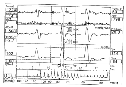
stream of information. Turning now to Fig. 14, a display from a DYNAPULSE
blood
pressure monitoring device is, shown. As the first derivative curve is a plot
of the change in
pressure over time (dP/dt), the maximum change in pressure over time (dP/dt)m~
and minimum
change in pressure over time, ((IP/dt)~ are readily apparent from the curve.
In a preferred embodiment, the
central processing unit can be programmed to (1) provide numerical values for
(dP/dt)m~ and (dP/dt)i";";
(2) determine Ty~, from determined values of t1 and tz; and (3) solve Eqn. 14
from the
determined values of (dP/dt)m~ and (dP/dt)m;".
SUBSTfME SHEET (RULE 28)
z1 ~7s~,~ . :.; ~ ,
W O 95!16391 PCT/US94/I46IZ
-31-
One uses Equation 48 to determine the pressure of a systolic wave at a given
time
T(~")(~ and to determine the change in pressure over time of the diastolic
wave (dP/dt)tdW~.
Once one determines the change in pressure over time of the diastolic wave and
the
maximum and minimum changes in pressure over time through the use of Eqn. 48,
one can
then determine the diastolic flow velocity (V~ according to Eqn. 31 reprinted
below
V = (4~) (l"',~') ( ~)v1v Eqn. 31
Tnn , ( dP)
dt pp
From the knowledge of the diastolic flow velocity, one can then calculate
either the distal
peripheral
resistance (PRA, or the systemic peripheral resistance (PR,) from diastolic
flow velocity
determined above, and the peripheral resistance parameter equations set forth
at page 8, supra.
More particularly, the systemic peripheral resistance can be obtained via the
following Eqn.
58.
PR! = Systolic Pressure - Diastolic Pressure
Is y
0
Similarly, one can determine the distal peripheral (PRA through Equation 59,
which
shows the results of the peripheral resistance that was measured and
detemlined by the
Applicant. In particular, Fig. 1 shows the determined distal peripheral
resistance (PRA of 14
patients. This charting of determined peripheral resistance of the patients as
a function of
mean arterial pressure is useful in diagnosing the cardiovascular pathologies
of the patients
whose peripheral resistance was determined.
In order to diagnose the patient, one first determines a clinically
significant threshold
value, for serving as a line of demarcation between those who are likely to be
at risk for
having a particular cardiovascular pathology, and those who are not. In the
instant case, the
Applicant has used the known "bench mark" threshold values for systolic,
diastolic and mean
SUBSfiTUTE SHEET (RULE 26)
W 0 95116391 ~ ~ ~ ~ PC1'IUS94114612
-32-
arterial pressures. The Applicant has also established a threshold value with
respect to distal
peripheral resistance also.
The standard threshold value for mean arterial pressure is 108 mmHg; for
systolic
pressure is 140 mmHg; and for diastolic is 90 mmHg. The threshold value
determined by
Applicant for peripheral resistance is 0.6 (mmHg) (sec/cm). -
Using these threshold values, the Applicant believes that a patient can be
diagnosed
as having a reasonable possibility of having a high peripheral resistance type
hypertension if
(1) the mean arterial pressure is greater than 108 mmHg; or the systolic blood
pressure is
greater than 140 mmHg; or the diastolic blood pressure is greater than 90
mmHg; and (2) the
distal peripheral resistance is greater than 0.6 (mmHg) (sec/cm).
It should be noted that the threshold value for distal peripheral resistance
of 0.6 is a
value that is likely to be modified, or better refined as further studies
become available.
If a particular patient satisfies the criteria discussed above, he would
generally be
diagnosed as having a high peripheral resistance type hypertension. In such a
case, the
indicated treatment would appear to be the use of a vaso-dilator.
Additionally, the peripheral resistance can be used to diagnose those high
cardiac
output (CO) type hypertensive patients. The Applicant has found that these
patients are those
who typically have a mean arterial pressure greater than 108 mmHg, or a
systolic pressure
greater than 140 mmHg, or a diastolic pressure greater than 90 mmHg. However,
the high
cardiac output-type hypertensive patients are distinguished from the high
peripheral resistance
type hypertensive patients in that the high cardiac output type hypertensive
patients typically
have a distal peripheral resistance of less than 0.6 (mmHg) (seclcm).
Although the Applicant has performed experiments using distal peripheral
resistance,
it will also be appreciated that systemic peripheral resistance can also be
used to determine
hypertension, and to diagnose the difference between high peripheral
resistance type
hypertensive patients and high cardiac output type hypertensive patients.
Although the steps set forth above for determining peripheral resistance are
described
as being performed manually (to a large extent), it will be appreciated that a
computer can
be programmed to perform the processes described above for both determining
peripheral
resistance and diagnosing a patient based on the peripheral resistance and
cardiac output so
determined.
SUBSTITUTE SHEET {RULE 26)
WO 95116391 PCT1US94/146I2
- 33 -
The peripheral resistance measurements discussed above can also be used to
determine
whether a patient has a high risk of having coronary artery disease. Once
again, the
peripheral resistance the mean arterial pressure that are determined from a
patient are
compared to threshold values. Based on the relation between the determined
peripheral
resistance and mean arterial pressure; and the threshold value, the patient
can be diagnosed
as either having a high, medium or low risk of having coronary artery disease.
It has been
found by the Applicant that generally patients have a high risk of having
coronary artery
disease if their mean arterial pressure is greater than 108 mmllg. Patients
have a medium risk
of having coronary artery disease if their mean arterial pressure is less than
108 mmHg, but
their distal peripheral resistance is greater than the threshold value of 0.6
(mmHg) (sec/cm).
Further, patients have a generally low risk of having coronary artery disease
if both their
mean arterial pressure and their distal peripheral resistance are Iower than
the threshold values
of 108 mmHg, and 0.6 (mmHg) (sec/cm), respectively.
C. TjetermilLtion ofa Pceudo-Aortic ~xlaW''Ori n ~r nr1 Trc TlcP for D
rmining,
('ar iov cc ,1 r Pa holoeiec
Another aspect of the present invention is the determination of a pseudo-
aortic
pressure wave contour, and the use of this contour to help diagnose patients
as having
cardiovascular pathologies. In particular, the method is suitable for
diagnosing cardiac aortic
disease conditions.
In order to determine the pseudo-aortic pressure wave contour of a human
cardiovascular system, a pressure inducing means and transducer is first
affixed to a patient.
A data stream is then obtained form the patient from the transducer means. The
data stream
includes pressure data and pulsation signal data. Preferably, the data stream
includes data
obtained at a supra-systolic pressure, at a sub-diastolic pressure, and at
pressures in the range
between the determined systolic pressure and diastolic pressure.
Using this pulsation signal data and pressure data, the pseudo-aortic pressure
wave
contour can then be determined.
In order to determine the pseudo-aortic pressure wave contour, one first uses
the
normalized supra-systolic wave and sub-diastolic wave data, as shown in Fig.
7, and as
discussed above. An integration of Eqn. 48 is used to determine this data,
including the
weight factors for yielding the best fit that are disclosed in Eqn. 48a, and
Table 1.
SUBSTiME SHEET (RULE 26)
WO 95116391 PC1'IUS94114612
-34-
Once the pseudo-aortic pressure wave contour (P~tl) is determined, one uses
this
normalized supra-systolic wave and sub-diastolic wave data in conjunction with
Eqn. 52 to
obtain the pseudo-diastolic wave contour (Pd,~,~~~). Additionally, the weight
(W~d~) to be used
in conjunction with Equation 52 is 0.6.
From this information, the pseudo-diastolic wave contour is obtained. The
pseudo-
systolic wave contour and the pseudo-diastolic wave contour are then combined
to form the
pseudo-aortic pressure wave as shown in Figs. 12A and 12B. The reconstructed
and
normalized pseudo-aortic pressure wave contour carries a large amount of
information about
a patient's cardiovascular system. In principal, it can be used for diagnosing
a large number
of characteristics of a cardiovascular system, and a wide range of human
hemodynamic
parameters, and cardiovascular pathologies.
For example, the pseudo-aortic pressure wave contour can be compared to a
chart of
known cardiovascular pathologies. For example, the pseudo-aortic contour can
be compared
to the variations in contour of the arterial pulse with correlated ECGs, such
as are shown at
Fig. 4.2 of K.G. Andreoli, et al., Ed., Comprehensive Cardiac Care, C.V. Mosby
Co., St.
Louis, 1983.
A comparison of the contours can determine things such as arterial sclerosis.
Turning
now to Fig. 12B, the "notch" in the pseudo-aortic pressure wave that occurs
between 101 and
l I5 mmHg, and 0.05 and 0.15 seconds is a notch that is very typical for
patients having
aortic sclerosis.
Turning now to Fig. 12A, a pseudo-aortic contour wave is shown that exhibits
aortic
stenosis. The stenosis in Fig. 12A is exhibited by the decrease in slope in
the area designated
110 in the ascending portion of the systolic cycle of the pulse pressure wave.
If one looks
at the comparable portion of the pulse pressure wave of Fig. 12, it will be
noted that the slope
does not decrease as quickly in the patient not having stenosis.
C.
In order to determine the peak left ventricular (cardiac) contractility of a
human
cardiovascular system, one first affixes a non-invasive pressure inducing
means and transducer
to a patient, to obtain a data stream from the transducer. The data stream
should include
pressure data and pulsation signal data. In these respects, the method for
determining peak
left ventricular (cardiac) contractility is generally similar to the methods
discussed above.
S~TtTIIIE SHEET (RULE 2Ba~
2.~ ~78~2
w0 95116391 PCT/IJS94/I46I2
-35-
The method for obtaining the data stream disclosed in the Chio '013 patent is
a preferred
method for obtaining this data stream.
Once the data stream is obtained, the pulsation signal data and pressure data
are used
to determine the peak cardiac (LV) contractility by the following steps. First
the supra-
systolic and sub-diastolic waves, along with Equation 48 (and the weight
factors) are used to
obtain a pseudo-systolic wave contour (P,~,~). The change in pressure over
time of the
diastolic wave is then determined, along with the peak-to-peak change in
pressure over time.
Using Equation 57, and the measured systolic (P~) and diastolic (P~ pressures,
and
peak pressure (PP = P~,S - P~;~, one can then calculate then Tr, which is the
ratio of Tppla~l
to T~O,~ as defined above in Section III, (Equations 53-57) and Fig. 11. Using
the above
derived parameters and Eqn. 56, one can then calculate the peak cardiac
contractility:
~~ d~~l~V~ Eqn. 55
The peak cardiac contractility may be used in the diagnosis of a
cardiovascular
pathology, as the peak cardiac contractility can be used as an index for
determining the
strength of the heart. As will be appreciated, the strength of the heart (or
lack thereof) can
be an important parameter for diagnosing a cardiac disease, or cardiac
failure.
D. Determining Arterial Comnlianca and Ftactioitv of a H ~
CardiovaccLlar ova m
The present invention can also be used for helping to determine arterial
compliance
(C~ and elasticity (K~ of a human cardiovascular system. In order to do this,
one uses Eqns.
27 and 28. In so doing, one can either measure the arterial cross-section, or
assume the
arterial cross-section (for a normal) to be Ao equals 0.67 cm2. One can
fiuther derive the
arterial elastic constant Ke, and the compliance, which equals the inverse of
the elasticity
constant (C = 1/Ke). Information about a person's arterial elasticity or a
compliance is also
a usefill parameter for diagnosing the condition of the patient's
cardiovascular system.
E. LTse in Comb~ation
Although the various factors and parameters have been discussed alone, it will
be
appreciated that the parameters so determined can be used in combination to
diagnose a wide
SUBSTfTUTE SHEET (RULE 26)
W 0 95116391 ~ PCT'/US94114612
-36-
variety of disease, and to provide either supporting or contradictory evidence
for other
parameters so determined, and diagnoses so made.
F. Devices for Determining the Above Parameters
The methods discussed above can be incorporated into a device for automating
the
processes discussed above. The primary requirement of such a device is that it
be capable
of obtaining the information needed. One such device that is capable of
obtaining the
information is the DYNAPULSE 200M blood pressure monitor manufactured by Pulse-
Metric,
Inc. of San Diego, California, the assignee of the present invention. The
DYNAPULSE
device is described in more detail in the Chio '013 patent. It includes a
blood pressure cuff
for exerting a pressure on the art of a patient. A transducer is provided for
capturing
cardiovascular signals, and converting those cardiovascular signals into an
electrical signal.
An analog-to-digital converter is provided for converting the analoged noises
to a digital
information stream. The digital information stream is then processed by a CPU
to create a
variety of information displays. The relationships and equations discussed
above in
connection with the determination of the various parameters can be programmed
into the
CPU, to enable the device to provide a display or printout of a type that can
be used by the
practitioner attempting to determine the various parameters. Some of the
parameters (e.g.
peripheral resistance) may be best displayed digitally, as a numerical value.
Others of the
displays (e.g. the created pseudo-aortic wave) may be better displayed
graphically. With
regard to the pseudo-aortic contour, the device can also include a program for
comparing an
obtained pseudo-aortic wave to aortic waves of known cardiovascular
pathologies, to suggest
probabilities to a practitioner of a particular patient possessing a
particular cardiovascular
pathology (e.g. aortic sclerosis or stenosis). Alternately, the display might
include a
suggestion of alternatives diagnoses that the practitioner may wish to explore
more fully
through a visual diagnosis. In this regard, a wave form display has been found
by Applicant
to be a useful tool, itself, in determining the parameters discussed above,
and for diagnosing
cardiovascular pathologies.
V. ~ORK1N(: FXAMPT_.ES. _ _
The first step in obtaining the various parameters necessary for making
the.diagnoses
of the instant application is to obtain a normalized supra-systolic and sub-
diastolic pressure
waves.
SUBSTITUTE SHEET (RULE 2B)
W 0 95116391 21 ~ 78 4 2 PCTIUS94I14612
-37-
One first obtains the systolic, diastolic and mean arterial pressures of the
patient. In
the instant example, this was performed by the use of the DYMAPI1LSE Model
200M blood
pressure device manufactured by Pulse-Metric, Inc., of San Diego, California,
the assignee
of the instant invention. The pressure wave so obtained from the patient is
shown in Fig. 13.
As discussed earlier, the supra-systolic wave forms are wave forms taken at a
pressure
wherein no blood is flowing through the cuff area. Therefore, the arterial
pulse waves
measured at a cuff, or similar at a device at a supra-systolic pressure
reflect this condition.
Taming now to Fig. 13, supra-systolic waves were obtained by using the
DYNAPLTLSE device. A standard blood pressure cuff was used to occlude blood
flow by
applying a pressure to the artery greater than the systolic pressure. The
supra-systolic waves
were then obtained by using a transducer, and an analog-to-digital converter,
as taught more
explicitly in the Chio '013 patent. Alternately, one can use an ultra-sound
probe to detect
cardiovascular sound at a supra-systolic pressure.
As part of the procedure, the pressure exerted by the cuff was initially a
pressure
greater than the patient's systolic pressure. Over time, cuff pressure was
reduced in a
generally linear manner to a point below the patient's diastolic pressure. All
during this time,
pulse pressure waves were being obtained from the patient. Pulse pressure
waves were
obtained from the patient at pressures below the patient's determined
diastolic pressure. The
sub-diastolic pulse pressure waves were obtained at this sub-diastolic
pressure. However, it
will be appreciated that in situations where only certain information is
needed (e.g. sub-
diastolic and supra-systolic only) the device can a used to obtain only the
information so
required.
The supra-systolic (SS) and sub-diastolic (SD) waves were then normalized to
the
systolic (Prys) and diastolic (P~;~ points. As discussed above, the systolic
and diastolic
pressures may be determined by use of the DYNAPIJLSE blood pressure monitor,
or by any
other method of determining blood pressure.
One then assumes that the maximum point of both the supra-systolic and sub-
diastolic
waves are the systolic pressure, and that the beginning of the systolic cycle
of the supra-
systolic wave, and the lowest point of the sub-diastolic waves are the
diastolic pressures.
Both the supra-systolic and sub-diastolic waves are then normalized to the
systolic and
diastolic pressures as shown in Fig. 7. Turning now to Fig. 7, the normalized
supra-systolic
and sub-diastolic waves are plotted together with an aortic pressure wave that
was obtained
SUBSTfTUTE SHEET (RULE 28)
~,~~$~'~ ' - r
WO 95116391 P(."TIUS94114612
-38-
by a catheterization method. In the example shown in Fig. 7, the normalized
non-invasive
waves and the invasive catheterization waves have their systolic pressure at
171 mm Hg, and
their diastolic pressure at 96 mm Hg.
Once one obtains the normalized supra-systolic and the sub-diastolic
pressures, one
can use Equation 48 and Equation 52, with the appropriate wave factors (W$5 l,
Wsd 0.4,
and Wd 0.6) to obtain the pseudo-aortic pressure waves, P~t~ and Pa~c
One then uses the normalized supra-systolic, sub-diastolic and pseudo-aortic
waves to
further calculate their slopes (dP/dt), at the maximum slope of the ascending
portion of the
pulse wave (dP/dt)~,~, and at the maximum negative slope of the descending
portion of the
pulse wave (dP/dt)~. As discussed above in connection with the discussion of
Fig. 12A,
this process can be performed through appropriate programming of the central
processing unit
used in connection with the gathering of the information stream from the
patient.
The peak values, ((dP/dt)m~, arid (dP/dt)~~ are then obtained. Using Eqn. 14,
(dP/dt)~ can then be obtained from the determined (dP/dt)m~, and (dP/dt)",;a.
In the example shown in Fig. 7, the variables necessary for using Equation 31
were
determined as follows:
L~"ff equals 9 cm, when using a standard adult size cuff having a width of 5
inches;
Tpp equals 0.24 seconds, the time between the peak maximum change in pressure
over
time (dP/dt)m~, and the peak minimum change in pressure over time (dP/dt)~;
(dP/dt)~ equals 1250 mm Hg/sec.; and
(dP/dt)Dq, equals 62.5 mm Hg/sec.
Using these values, and Equation 31, the diastolic flow velocity for the
patient whose
cardiovascular information was obtained in Fig. 7 is Ve 23.56 cm/sec.
One then uses Equation 22 and assumes that the arterial cross-sectional area
(Aa)
equals 0.67 cm'. From these values, one can further calculate the arterial
compliance (C) as
follows: C=0.26 cc/mm Hg.
One can then obtain the peripheral resistances (PR, and PRZ) using the
following
equations:
_ S stolic Pressure - Diastolic Pressure Eqn. 58
PRI
Vo
SUBSTITUTE SHEET (RULE 26~
2177842 - E ..,.
W095/16391 ~ ' PCT/US94/14612
-39-
and
Mean Arterial Pressure - Diastolic Pressure Eqn. 59
P~ -_ V
0
S Using Equations 58 and 59, one can calculate both the proximal peripheral
resistance
(PR,), and the distal peripheral resistance (PRA. The values of the peripheral
resistance for
the patient whose cardiac information is shown in Figs. 13 and 7 are:
PRl = 3.18 (mm Hg)(sec/cm); and
PRz = 0.85 (mm Hg)(sec/cm).
Left ventricular (cardiac) peak contractility was then obtained by using
pseudo-aortic
systolic wave (P,"t«) and its peak change in pressure over time (dP/dt), along
with Equations
56 and 57 to calculate the peak contractility of the left ventricle of the
patient whose cardiac
information is shown in Fig. 7.
V Eqn. 55
One example is illustrated. For a patient having a systolic pressure of 166 mm
Hg,
a determined diastolic pressure of 94 mm Hg, the pulse pressure (Pp) was
calculated as 72
mm Hg, according to the numerator of Equation 58. Using Equation 57, one can
calculate
Tn as being equal to 0.563.
The peak change in pressure over time was then obtained from the pseudo-aortic
wave
according to the following: ~(dP/dt)e~m~ = 804 mm Hg/sec. From this, one can
calculate,
using Equation 56, the peak left ventricular (cardiac) contractility. In the
instant example, it
was found that the peak cardiac contractility:
~~ ~ wVl Eqn. 55
equals 1015 mm Hg/sec.
The value so obtained by the applicant's non-invasive method was then compared
to
the value obtained from an invasive catheterization measurement. For the same
patient, using
the same information, the invasive catlleterization measured a peak cardiac
contractility of
1000 mm Hglsec.
SUBSTtTUTE SHEET (RULE 26)
WO 95J16391 PCTIUS94114612
-40-
The general similarity of the results are used persuasively for the
reliability of the
Applicant's invention.
VI.
A. ~yertension Diagno~jg
As part of the experimental procedure used to test the instant invention, the
Applicant
performed studies on various patients. Attached to this patent application,
and submitted
concurrently herewith, are several graphical representations showing
information received
during testing performed pursuant to this invention. These graphical plots
include figures
labeled Figs. 2-6, and 8-11.
In order to test the theory of using the peripheral resistance (PRA and PR2),
defined
above and derived from a measurement of the diastolic wave velocity, 14 human
subjects,
between the ages of 45 and 81 were studied. The invasive aortic pressure wave
forms and
non-invasive cuff pulse wave forms were obtained from these patients. Sub-
diastolic wave
forms were analyzed to obtain the parameters defined in Equation 31, . In
these studies,
the measured length of the cuff was 5 inches. This effective length was
calculated by
assuming that the effective length equaled the measured cuff length divided by
the square root
of 2. Thus, the effective length of the cuff having a measured length of 5
inches was equal
to 3.5 inches. Further, it was found that the change in pressure over time of
a diastolic wave
((dP/dt~DW) was calculated from the linear portion T, to TZ of the diastolic
wave (see, e.g.,
Fig. 11), and was confirmed by the invasive wave form. These values were then
used in
Equation 31.
Your attention is directed to Fig. 7, which illustrates the determination of
the diastolic
wave section, and its confirmation and comparison to the information derived
from the
invasive wave form.
Compliance was calculated through Equations 22 and 23 by assuming that a male
subject having a height of 60 inches (5 feet) has an arterial cross-section of
1 cm~, and that
a female of the same height has an arterial cross-section of 0.8 cm2. For
different heights,
the arterial cross-sections were adjusted accordingly by the following
equation:
Aot%~ _ (Ao)(HeighttX~/60).
Figs. 2 through 6 of the Appendix, and Fig. 1 (of the instant application)
display the
results for both systemic peripheral resistance (PR,) and distal peripheral
resistance (PR?), as
SUBSTITUTE SHEET (RULE 2&)
z1 ~~s~z
W095116391 .- - PCT/US94/14612
-41 -
both a function of the systolic, diastolic and mean arterial pressures of all
14 subjects. These
results substantiate the Applicant's fording that either systemic peripheral
resistance or distal
peripheral resistance can be used for diagnosing hypertension. The Applicant
also found that
peripheral resistance is preferably plotted against either mean arterial or
systolic pressure,
since, for elderly people, their diastolic pressure may decrease, and the
usual benchmarks for
determining hypertensive systolic and diastolic pressures (140/90 mm Hg) may
not be reliable
indicators.
In Figs. 8 and 9 of the Appendix are shown plots of the diastolic wave
velocity plotted
against systolic and diastolic readings taken from the 14 human subjects. The
data suggests
that people having a systolic blood pressure lower than 140 mm Hg, or
diastolic blood
pressure of less than 70 mm Hg tend to have lower diastolic wave velocity.
However, for
those patients having higher systolic and diastolic pressures, there appears
to be one group
of patients that has a higher diastolic wave velocity, whereas another group
has a lower
diastolic wave velocity. Since peripheral resistance equals mean arterial
pressure divided by
IS cardiac output, the greup having a lower peripheral resistance may have a
higher cardiac
output. Therefore, the treatments of the two groups of high blood pressure
should be
different. This is discussed above.
Figs. 10 and 11 of the Appendix are plots of the calculated compliances of the
14
subjects as a function of both systolic and diastolic pressures. Besides the
scattering of the
detertnined compliance values over a wide range of systolic and diastolic
pressures, the
Applicant has also noted that the trend of compliance compared to systolic
pressure is
opposite of the compliance when compared to diastolic pressure. This data
suggests that
compliance is not as valid and reliable of an indicator of hypertension as
determined
peripheral resistance determined in accordance with the present invention.
B. >Zeconstn!cHon of Aortic Wave Forma
In the clinical data and from the discussions above, it was stated that it is
possible to
create an empirically fitted pseudo-aortic wave, using Equations 51 and 52.
This pseudo-
aortic wave may then be used for diagnosing certain cardiovascular
pathologies, such as
arterial stenosis, sclerosis, etc. Furthermore, it may provide further
information on aortic
valve and left ventricular characteristics. By using the data of the above 14
subjects, and
appropriate wave factors, appropriate pseudo-aortic waves were able to be
constructed. Figs.
SUBSftTUTE SHEET (RULE 26)
W095/16391 ~ ~ PCT/US94114612
-42-
12A and 12B represent comparisons of pseudo-aortic waves constructed according
to the
present invention, and aortic waves measured by invasive catheterizarion.
C. I-eft Ventricular lCardiacl ContracHlitv From Cuff Pulce Wave Formc
Invasive catheterization left ventricular pressure waves and non-invasive cuff
pulse
wave forms of one human subject were recorded simultaneously. Using Equations
56 and 57,
P~;a and PP were determined using the non-invasive cuff. Additionally,
{dP/dt),~ was
obtained from the use of a 90% supra-systolic and 10% sub-diastolic wave
forms. Using this,
an averaged:
~~ ~ ~LV~~ Eqn. 55
was calculated to be 968 (~ 139) mm Hg/sec. , This compared favorably to the
cardiac
contractility of 1057 mm Hglsec calculated from the invasive catheter derived
left ventricular
pressure wave. Since the systolic wave of the pseudo-aortic wave is not
symmetric, the
change in pressure over time of the ascending part of the wave equals
approximately 675 mm
Hg/sec, whereas the change in pressure over time of the descending portion of
the wave
approximates 900 mm Hg/sec. If one uses the value 900 as the maximum change of
pressure
over time for the aorta (dP/dt)m"~, the left ventricular cardiac contractility
calculated therefrom:
~~ ~P~LV~ Eqn. 55
becomes 1106 mm Hg/sec. Therefore, for an asymmetric aortic systolic wave, the
Applicant
believes that it is better to use the larger value of its change in pressure
over time by
calculating the cardiac/left ventricular contractility. This has some
intuitive support since the
slope of the ascending aortic wave may be decreased by aortic stenosis or
sclerosis or other
unknown factors.
Although the invention has been described in detail with reference to the
illustrated
preferred embodiments, variations and modifications exist within the scope and
spirit of the
invention as described and as defined in the following claims.
SUBSTITUTE SHEET (RULE 2fi)