Note: Descriptions are shown in the official language in which they were submitted.
WO9~110777 2 1 7834 1 PCT/US94/1173
TITl.E: DETECTION AND 'I'R~ T OF 8REAST AND
C'.YN~t~Tl-~'TCAL CANCER
DESCRIPTI~N
.
1. Terhn;--~l Field
The present invention relates to newly
discovered antigens associated with breast and
gynecological cancers and to; ~A~say methods for
detecting these antigens in biological samples, as
well a~ immunotherapeutic methods for treating the3e
cancers with antibodies that bind to these antigens.
More specifically, the invention relates to the
discovery of immunological cross-reactivity between
antibodies to Human T o~Pfi~iency Virus (HIV-I)
envelope protein gpl20 and certain breast and
yynecological carcinoma cell surface and chromatin
antigens. This cros~-reactivity results in the
formation of new i lexes which are useful in
the i ~ qno5tic methodg of this invention.
2. Backcround
Breast carcinoma, together with carcinoma of
the ovary, account for one-third of all cancers
occurring in women, and together are responsible for
approximately one-quarter of cancer-related deaths
in f emales . Cancer of the f emale genital tract
accounts for almost 80,000 cases of invasive cancer
each year in the United States, with the majority of
these being one of three neoplasms; carcinoma of the
cervix, ~n~l~ LLial carcinoma, and celomic
epithelial carcinoma of the ovary . Except f or
cervical cancer, which is def initely linked with
Human Papilloma Virus infection, etiological agents
involved in malignant transformation of breast,
WO 95/10~77 ~ PCT/US94/11754
2 1 7834 ~ --
ovarian, or endometrial cells remain unclear. It
has b~en established that susceptibility to breast
and ovarian cancer is inherited in some families.
Between 5% and 10% of breast cancer and ovarian
cancer can be linked with inheritance of a gene
conferring high risk, followed by genetic changes in
epithelial cells. The gene which is believed to be
responsible f or inherited brcast-ovarian cancer has
been localized on the chromosome 17ql2-21 and named
locus 8RCA l; however, the sequences of the gene
located in this locus are completely unknown.
Approximately one in 200 women - 600,000 women in
the United States - have inherited susceptibility to
breast cancer which is not only associated with
BRCAl, but also with mutations in other genes like
P53, Her2/erbB2, estrogen receptor and others.
Genetic counseling for families with inherited
susceptibility to breast and ovarian cancer and
prophylactic mastectomy or oophorectomy represent a
widely discussed sub~ect.
Surgery, radiotherapy, and chemotherapy
represent three basic methods which are used in
management of breast cancer and gynecologic cancer.
High mortality of breast cancer and gynecologic
cancer patients indicates that the currently
available diagnostic and therapeutic methods are
unsatisf actory .
Immunotherapy and i r~ nosis with
monoclonal antibodies (MAb) represents another
approach which has been extensively developing and
improving during the past few years. In direct
approaches, MAb IgG2a and IgG3 mediate antibody-
dependent cellular cytotoxicit~r and/or exert
complement-dependent cytotoxicity . Most f requently
WO 95tlO777 PCT/I:S94¦11754
~ 2 1 783~ ~
used is radio-immunotherapy with radioactively labeled ~Lab.
Immunotoxins, which are the conjugates of MAb with the
subunit A of the ricin or diphtheria toxin, exhibit high
tumoricidal potential, however have a restricted application
due to high cytotoxicity.
Recently, a high number of monoclonal antibodies
directsd against br2ast, ovarian, and cervical cancer have
been developed and sfforts have been undertaken t~ use those
M~hs as i ~ gnostic and immun~therapeutic agents.
~owever, no significar.t progress ha~ been reported in
r~-n;~ ~ nt of rn~l i gn;~n~ies of the female reproductive tract
using these techniques.
A number of HIV-1 peptide3 and proteins have been
identif ied which elicit neutralizing antibodies in animals .
EP-A-339504 deacribes (-hP~;~l ly synthesized amino acid
peptides having the sequence of amillo acids from the EiIV-1
virus which m2y be used to induce the production of HIV
inhibiting ant i ho~ i e~ f or the treatment of AIDS and AIDS-
related complex. However, the prior art has not disclosed a
a means of syntll~i 7'. n~ ard using monoclonal antibodies for
p-arposes of detecting and treating breast and gynecological
cancer .
It i3 an object of this invention to provide
immunodiagnostic and immunotherapeutic method& which are
belieYed to be an innovative approach to managing breast and
gynecological cancers.
SUMMARY OF THE INVENTION
Brlefly stated, the present invention provides methods
for detecting the pre3ence of HIV-1-cros3reactive breast and
gynecological cancer-associated antigens (as hereinafter
defined) in bio ogical samples and to methods for treating
these cancers. In a preferred ' ~r~i t, th~se methods
utilize a monoclonal antibody developed against a synthetic
peptide corre3ponding to ' he variable domain of the Hum~n
T ~n~iciency Virus ~HIV-1) envelope protein gpl20 (amino
AMEN~D SHEET
IPEA/EP
W0 95/10777 PC~/US94/11754
2 1 7834 1
acid region 308-322), herein referred to as MAb 5023. This
MAb can be purchased from DuPont/NEN, 549 Albany Street,
Bo~ton, MA 02118, and is li3ted in the catalog of thi~
company entitled "DuPont/NEN Re~earch Products
AMEND~D SHEET
IPEA/EP
3/A
~ WO95/10777 -~ : I 2 1 7834 1 PCT/US94nl7~4
1992-1993" under Catalog Number, NEA-9305, the
Product description of which is "EiIV Monoclonal
Antibody gpl20-Neutralizing (sequence specific)
(mouse) 0.5mL". In accordance with this invention,
MAb 5023 has been found to be unique in its ability
to penetrate the cell and localize within the cell
nucleus and in its ability to recognize a family of
antigens expressed by breast cancer and
gynecological cancers.
Consequently, in accordance with this invention
there is provided a method for diagnosis of breast
cancer and gynecological cancer comprising exposing
a biological sample f rom a host suspected of having
said cancer to MAb 5023 and detecting the presence
of IlIV-crossreactive immunocomplexes. Fortuitously,
this invention makes possible a simple and direct in
vivo diagnosis of cervical cancer by administration
of MAb 5023 to the cervix and then detecting the
presence of i ~ lexes f ormed between said
antibody and cell surface proteins pl20 and p41 is
another embodiment.
In one aspect of the invention, the method
comprises detecting IlIV-I-crossreactive breast
carcinoma-associated antigens in a biological sample
comprising (a) exposing a biological sample,
suspected of containing the antigens, to an antibody
which recognizes the following cell me~brane
proteins: pl60, p80, p45, and p24 and the following
chromatin protein: p24, and (b) detecting the
presence of i n, _ _ l exes formed between said
antibody and said proteins.
Related, is a method for detecting EiIV-I-cross-
reactive, gynecological cancer-associated antigens
in a biological samp~e comprising (a) exposing a
~ WO95/10~ 2 1 7834 ~ PCT/US94/11754
biological sample, suspected of containing the
antigens, to an antibody which recognizes the
following cell membrane proteins, pl20, p41, and p24
and the chromatin protein, p24, and (b) detecting
the presence of immunocomplexes f ormed between said
antibody and said proteins.
The; , lexes thus formed constitute an
embodiment of this invention and comprise (a) at
least one of the following cell membrane proteins,
pl60, p80, p45, or p24, or the chromatin protein,
p24, and (b) an antibody which recognizes each of
said proteins. In a preferred embodiment the
antibody is MAb 5023. Generally the immunocomplexes
of this invention are in an; '; 1; 7~d form. In
this form the immunocomplexes are insolubilized, or
otherwise supported, on a variety of standard
'- ;1; 7~tion 8ubstrates . Examples of materials to
which the capturing agents can be attached are
glass, synthetic polymers, synthetic resins,
cellulose, nitrocellulose, and various metals.
Procedures for attaching these capturing agents will
vary rlPr~n-l; ng upon the agent and substrate
employed, but, in general, are well known in the
art . Some of the methods f or binding of antibodies
to a solid matrix are discussed in E Harlow and D
Lane, Antibodies: A LaboratorY Manual, New York:
Cold Spring Harbor Laboratory, pp. 511-552, 1988.
Another aspect of the present invention is a
method f or treating breast cancer and gynecological
cancer, selected from the group consisting of
ovarian, cervical, endometrial, and vulvar cancer,
comprising administering to a patient an antibody
which translocates and intprn~l; 7es to the nucleus
of the cell and which forms; -_ lexes with the
W0 9S/107?7 ~ ~ 2 1 7 8 3 4 ~ PCT/US94/11754
HIV crossreactive antigens of this invention, e.g.,
~b 5023. The antibody can be administered alone or
conjugated with a radioactive or cytotoxic ligand,
and is administered in a rhArr~ tically effective
dosage form, generally, for intravenous or
intraperitoneal in~ection.
The ~IV-I lus~Leactive cancer-associated
antigens of this invention are the pl60, p80, p45,
nnd p24 cell membrane proteins and the p24 chromatin
protein for breast cancer, and pl20, p41, and p24
cell membrane proteins and the p24 chromatin protein
for gynecological cancers (i.e., ovarian, cervical,
endometrial, and vulvar cancer ) .
In all of the methods brief ly described above,
the antibody may be labeled with an enzyme,
fluorochrome, radioisotope, or luminescer. In such
cases the step for detection would normally be by
enzyme reaction, f 1UUL~C(~ , radioactivity, or
1 llmi n~cr~n~~e emissions, respectively.
The methods described above may be applied to a
variety of biological samples in order to detect the
presence of the HIV-I-crossreactive cancer-
agsociated antigens of this invention or antibodies
thereto. Bodily secretions, bodily f luids, and
tissue specimens are all suitable samples in this
regard. r 1 ,~ of bodily secretions include
cervical secretions, vaginal secretions, human
breast milk, urine, and intraperitoneal ascitic
f luid . Suitable bodily f luids include blood .
Examples of tissue speri ~ include a variety of
biopsies, such as a biopsy of cervical dysplasia,
cervical cancer, ovarian cancer, vulvar cancer,
lymph nodes, and bone marrow. In addition tissue
fr~m all ~re~8 m~y be ol~A~nl n--l in connecti~ ~ith
~ WO 95/l0777 ` ~ 2 ~ 7 8 3 4 ~ PCr/Uss4/1l7s4
post mortem examination, including primary tumors
- and metastatic tumors, lymph nodes, bone marrow and
all organs.
In accordance with the present invention,
continuous hybrid cell lines can be estAhl i ~hf-rl that
produce monoclonal antibodies directed against an
antigenic det~rm; nAnt of the ~IV-I-crossreactive
cancer-associated antigens of this invention for u3e
in the methods described above. In a particularly
preferred ~ - lir--t, the cell line comprises the
hybridoma clone for MAb 5023.
BRIEF DESCRIPTION OF THE DRAWINGS
FIGURES lA-lE. Immunofluorescence staining of
SRBr 5
(A-D) and MCF7 (E) breast carcinoma cells with
fluorescein-conjugated sheep anti-mouse IgG after
incubation with MAb 5023 at 0C for 15 min (A), or
at 37C for l h (B), 3 h (C~, and 24 h (D, E).
FIGURE 2 . Autoradiographic detection of f ree
l2sI-MAb 5023 (lane l) and l25I-MAb 5023 accumulated
in the cytoplasm ( lane 2 ) and in the chromatin ( lane
3 ) after 24 h incubation with SRBr5 cells .
Cytopla3m and chromatin were prepared from 5 x 10
cells .
FIGURBS 3A and 3B. Western blotting of
different MAb against V3 loop of EIIV-I gpl20 with
membrane (M) and chromatin (Ch) proteins of SRBr5
cells separated in a 7.5% (A) or 1396 (B)
polyacrylamide gel.
Wo 95/10777 ~ ` 2 1 7 8 3 ~ 1 PCr/US94/11754
FIGURES 4A and 4B. Western blotting of MAb
5023 with membrane (M) and chromatin (Ch) protein~
of different cell lines 3eparated in 7.596 (A) or 1396
( B ) polyacrylamide gel .
FIGURE 5 . Electrophoretic analysis ( 7 . 5%
polyacrylamide gel ) and autoradiographic detection
of [35S]methionine-labeled plasma membrane protein,
cipitated by MAb 5023 from SRBr5 breast
carcinoma cells (lane l) and SW707 colorectal
carcinoma cells ( lane 2 ) .
FIGURE 6. Western blotting of human HIV-I-
neutralizing antibodies to env-2-3 with SKBr5 breast
carcinoma membrane proteins (lane l) and IIIV-I gpl20
( lane 2 ) . Proteins are separated in 7 . 596
polyacrylamide gel.
FIGURES 7A-7D. Western blotting of MAb 5023
(7A, 7B, 7C) and of MAb 5025 (7D) against HIV-I
gpl20 with pl;~ rane-containing cytopla3m (C)
or nuclear fraction (N), or pure chromatin (Ch).
Proteins were separated in 7.5-lOY6 polyacrylamide
gel . Dif f erent migration is due to the dif f erent
time of electrophoresis and dif f erent gel
concentrations . Prestained protein markers ( high
and low molecular weight f rom Bio-Rad ) were used in
each experiment.
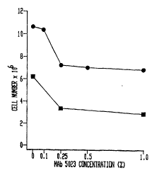
FIGURE 8. Effect of MAb 5023 on growth of
cervical cancer cell line SiHa. MAb was used as
ascites, the concentration i3 given as ascites
dilution in the medium l96=concentration of ascites
l:lO0 which corresponds to approximate MAb
WO 95/l0777 :~ PCr/USs4/~ 17.S4
2 ~ 7~34 1
concentration of 0.2~g/ml. The ;nh;hitnry
concentration 0 . 25 corresponds to 0 . 0511g/ml .
FIGURES 9A-9C. Electrophoretic analysis ( 10%
gel) and Western blotting of cytoplasm (C~ chromatin
(Ch), or total (C+N) proteins from cancer (A and B,
lane 2 ) and normal cells (B, lane 2 and C) with MAb
anti HIV-I gpl20 (Cells or sections of cancer or
normal tissues, obtained during standard surgical
plU- eduLt:s, were homogenized in 0.35 M sucrose/10 mM
RCl/1.5 mM MgCl2/10 mM Tris-HCl (pH 7.6)/0.12%
Triton X-100/12 mM 2 u~Loethanol ( 1 ml/3 X 106
cells or 0.25 - 1 ml/cm3 tissue), and centrifuged at
600 x g for 10 minutes. Supernatant was the crude,
membrane-containing cytoplasmic fraction. The
nuclear pellet was first washed with 0.2 M sucrose/3
mM CaCl2/50 mM Tris-HCl (pH 7 . 6 ), and then with 0 .14
M NaCl/lO mM Tris-HCl (pH 8.3) and centrifuged at
700 x g for 10 minutes. The pellet was swollen in
1 mM Tris-HCl (pEI 7 . 9 ) and centrifuged throughout
the 1.7 M sucrose 10 mM Tris HCl (pH 7) at 160,000 x
g for 80 minutes. Chromatin was pelleted at the
bottom of the tube.
Proteins were separated in 10% polyacrylamide
gel with 0.1% sodium dodecyl ~ulfate (SDS) in 250mM
Tris-HCl (pH 8.3), 195mM glycine, and 0.1% SDS,
according to Laemmli et al ( 76 ) . Each lane
corresponds to 3 X 10⁵, except lane 7 which
coll~a~.ds to 5 X 10⁴ cells. Blotting of proteins
from the polyacrylamide gel to the PVDF membrane was
performed in 25mM Tris-HC1 (pH 8 . 6 ), 192mM glycine
buffer, containing 10% methanol. Filters were
incubated with 1% BSA for 16 h, at 0C, then with
MAb anti-HIV-I gpl20 (DuPont)(511g/ml), washed with
Wo 9S/10777 ~ 2 1 7 8 3 4 1 PCTIUS94/11754
Tri6-glycine buf f er, and incubated with A 1 kA l; n~
phosphatase-conjugated goat anti-mouse IgG for 1 h.
After washing with TBST, membranes were incubated
with 0.196 1-naphthyl-phosphate and Fast Red, in
lOOmM Tris-HCl pH 9.5, lOOmM NaCl, 5mM MgCl2). A:
MCF7 breast carcinoma (lanes 1,2), SiHa cervical
carcinoma ~ lanes 3, 4 ), endometrial cancer ( lanes
5, 6 ) and cervical cancer ( lane 7 ) proteins . B:
ovarian cancer ( lane 1 ) and normal ovarian tissue
( lane 2 ) . C: normal skin, muscle and cervical
tissue and normal vaginal mucosal proteins.
FIGURE 10. Competitive inhibition of Mab anti
HIV-I gpl20 binding to the CytoplA~mi- proteins
extracted from mixed Mullerian tumor (A) by peptide
RIQRaPGRAFVTIGK SEQ ID NO: 1 (B), against which the
MAb was developed. The same amount of proteins were
separated in A and B, lane 1 (equivalent of 106
cells) and in A and B, lane 2 (equivalent of 0.5 X
106) (Peptide (18~g/ml) was incubated with MAb anti-
HIV-I gpl20 (511g/ml) for 1 hr on ice, next it was
incubated with the filter (B). In control (A), the
f ilter was incubated with Mab alone ) .
Reactivity of MAb anti HIV-I gpl20 with PVDF
strip containing HIV proteins extracted from AIDS
patients ' blood ( DuPont ) ( C ) . Cytopla3mic, chromatin
and total proteins were obtained as described in
(Cells or sections of cancer or normal tissues,
obtained during standard surgical procedures, were
homogenized in 0.35 M sucrose/10 mM KCl/1.5 mM
MgC12/10 mM Tris-HCl (pH 7.6)/0.1296 Triton X-100/12
mM 2-mercaptoethanol (1 ml/3 X 106 cells or 0.25 - 1
ml/cm3 tissue), and centrifuged at 600 x g for 10
minutes. Supernatant was the crude, membrane-
lU
WO 95~10777 ; PCT/U594/1 1754
2 1 7834 1
containing cytoplasmic fraction. The nuclear pelletwas firat washed with 0.2 M sucrose/3 mM CaC12/50 mM
Tris-HCl (pH 7.6), and then with 0.14 M NaCl/10 mM
Tris-HCl (pH 8.3) and centrifuged at 700 x g for 10
minutes. The pellet was swollen in 1 mM Tris-HCl
(pH 7.9) and centrifuged throughout the 1.7 M
sucrose 10 mM Tris HCl (pH 7) at 160,000 x g for 80
minutes. Chromatin was pelleted at the bottom of
the tube.
Proteins were separated in 10% polyacrylamide
gel with 0.1% sodium dodecyl sulfate (SDS) in 250mM
Tris-HCl (pH 8.3), 195mM glycine, and 0.1% SDS,
according to Laemmli et al ( 76 ) . Each lane
corresponds to 3 X 10⁵, except lane 7 which
corresponds to 5 X 10⁴ cells. Blotting of proteins
f rom the polyacrylamide gel to the PVDF membrane was
performed in 25mM Tris-HCl (pH 8.6), 192mM glycine
buffer, containing 10% methanol. Filters were
incubated with 1% BSA for 16 h, at 0C, then with
MAb anti-HIV-I gpl20 (DuPont)(5,ug/ml), washed with
Tris-glycine buffer, and incubated with A~ l in~
phosphatase-conjugated goat anti-mouse IgG for 1 h.
After washing with TBST, ~n~c were incubated
with 0.1% 1-naphthyl-phosphate and Fast Red, in
lOOmM Tri8-HCl pH 9.5, lOOmM NaCl, 5mM MgC12)-
FIGURES llA and llB. Representative DNAamplification analysis with primers SR 68/SK 69 (A)
and with P1/P2 (B) primers (Polymerase chain
reaction (PCR) OCS;ULlt:ll in the solution containing:
lOmM KCl, lOmM (NH4)S04, 200mM Tris-HCl, 20mM MgSO4,
196 Triton X-100 dNTP ( lOmM each of dATP, dTTP, dCTP,
and dGTP), 120-250ng of the primer, 2.5U Taq
polymerase, water up to 30111. DNA template was
ll
Wo 9S/10777 2 1 7 ~ 3 4 1 PCTrUS94111754
.
added at the amount of 0.2-l~Lg (20111), and paraffin
oil (70-1OO~L1). The reaction occurred in a thermal
cylinder (30-40 cycles). PCR products were
separated in 196 agarose gel with ethidium bromide ) .
A) lane 1: 123bp marker, lane 2: IIIV-I
infected T-cells, 3: SiEla, 4: MCF7, 5: mixed
Mullerian tumor, 6-8: normal skin (different
patients), 9: no template, 10: endometrial cancer
B) lane 1: HIV-I infected T-cells, 2-3: SiEia
(1 !lg and 0.1 119~ respectively)~ 4: mixed M~ riAn
tumor, 5-6: MCF7 (1 11 and 0.1 ,ug, respectively), 7:
endometrial cancer, 8: normal skin, 9: 123bp marker
FIGURE 12. Sequences of 140-150bp DNA
fragments of cervical cancer SiHa, endometrial
cancer obtained during standard surgery (EIV) and
breast cancer MCF7 ~1; f i ed by PCR with primers SK
68/SK 69 (see Fig. 3A). Nucleotides identical in at
least two or more cell types are boxed. 21bp of the
region identical or with high homology to E~IV-I are
marked with stars.
FIGURES 13A-13E. Transmission electron
micrograph of viral particles in SiEla (A, C ) and MCF7
(B,C) cells and of extr~ r vesicle obtained
from MCF7 cells (D). Viral particleG negatively
stained with uranyl acetate (E). In A, D and E,
samples were immuno-gold labeled with MAb anti-3IV-I
gpl20. The sizes of the immuno-gold particles were
15nm (A,D) and lOnm (E). V - viral particle~
FIGURES 14A and 14B. Effect of antisense
oligonucleotide RAK-I on growth of breast cancer
MCF7 cell line . Cells were grown f or 4 days in the
~bsence (A) or presence (B) of the antisense
12
WO 95110777 PCT~U594/117~i4
2~ 7~347
oligonucleotide RaR-} (5'-CCAGACTGTGAGTTGCAACAG-3' )
SEQ ID NO: 6 added daily at the concentration of 100
g/ml (day 1) and 50~Lg/ml (days 2 and 3).
Oligonucleotide 5 ' -TGTGACATCAGGCTCAAATC-3 ' was u3ed
as a negative control and did not affect cell growth
( not shown ) .
DETAII.ED DESCRIPTION OF THE lNV~.h.. IS~N
MAb 5023, developed against amino acid residues
308-322 of the variable V3 loop of HIV-I, reacted in
Western blotting with a 160,000 M2 (pl60) and 80,000
M2 (p80) cell 3urface antigens. Protein pl60 seems
to ~ sdllL an oligomeric form of p80. Another
MAb, 5025, directed again3t the same region 308-322,
and MAb against amino acid regions 307-328 and 308-
332, did not recognize pl60 or p80. Although MAb
5023 and MAb 5025 were developed against the same
synthetic peptide, there are minor differences in
the structure of the core epitope recognized by
these MAb, which account for the several-fold higher
affinity of MAb 5023 than that of MAb 5025. All of
the MAb tested recognized another cell membrane
antigen, p45. It is not established whether p45
IeplesellLs an in~ n~nt protein, a degradation, or
a processing product of pl60. Since MAb 5023 was
the only MAb which was able to enter breast
carcinoma cells and translocate to the nucleus, it
is likely that pl60 and (p80 ) expre~s a specific
epitope which is critical for MAb internatlization.
An llnRp".~ adsorption of the cell membrane-bound
125I-MAb 5023 to the chromatin during cell
fractionation may be eliminated, since pl60, p80,
13
o 95/10777 2 1 7 8 3 4 1 PCT/US94111754
: '
and p45 represent the specific markers of the cell
membrane f raction and are not f ound in the
chromatin. The fact that MAb 5023, but none of the
other MAb, was internalized suggests that an
antibody binding to a cell surf ace antigen is not
sufficient to induce the process of int~rnAl i 7~tion.
Instead, a specific epitope of the cell surface
antigen must be involved. The latest observation iu
consistent with previous studies, which proved that
only a few from many MAb developed against tumor-
associated antigens are internalized, while others
are unable to enter the cell.
Four antigens, pl60/p80, p45, and p24 have been
detected in breast cancer, and three antigens, pl20,
p4 1, and p24 are commonly expressed by cervical,
ovarian, endometrial, and vulvar cancer. All
antigens, except for p24 are selectively expressed
in the cytoplasmic/pl A ' ane fraction. Protein
p24 was detected in both E~1A~ ' rane and
chromatin f ractions .
Proteins pl60/p80, p45, pl20, and p41 were
undetectable in 1 An~ , colorectal carcinoma,
normal breast epithelial cells and, in non-infected
lymphocytes. The low molecular weight protein p24
is expressed in epithelial cells, lymphocytes, and
several cancer types.
Int~rnAl i 7Ation of MAb 5023 was also detected
in cervical cancer cells which express pl20. The
epitope recognized by MAb 5023, which may be
involved in internatlization, is well characterized.
The MAb 5023 was developed against amino acid
sequences 308-322 (RIQRGPGRAFVTTGK) of the variable
loop of EIIV-I gpl20, but this MAb binds to the
14
~ Wo 9~10777 2 1 7 8 3 4 ~ PCr~US94~11754
epitope GRAF. G preceding RAF is believed to be
critical f or internatlization .
Only MAb 5023, but none of other MAbs against
HIV crossreact with cancer antigens which suggests
that pl60 and pl20 are the proteins which mediate
int~rn~ ation. Lack of reactivity of pl60 and
pl20 with other Mabs directed against loop V3 of
HIV-I gpl20 suggests that holology of cancer
antigens and HIV antigen may be accidental and
restricted to a very short amio acid region. On the
other hand, the same molecular weights of
gynecological cancer and EIIV antigens (Mr 120,000~,
and correlation of the molecular weight of the
breast cancer antigen with the percursor f or HIV
gpl20 (Mr 160,000~ suggests that cancer proteins may
represent products of a retrovirus of homology to
HIV. This speculation is supported by homology of
the other cancer antigens (p45/41, p24) to HIV
antigens. Few other MAbs against the variable
region of HIV gpl20 also recognized p45 on the cell
membrane. Moreover, antibodies from AIDS-patients
which have been affinity-purified using HIV-I gpl20,
recognized p80 and p45 in breast cancer cells ); p41
in ovarian cancer.
MAb 5023, when int~rn~ ed and translocated to
the nucleus, stimulated RNA synthesis and promoted
growth of breast cancer cells. In contrast, growth
of cervical cancer cells was by 30-5096 inhibited by
MAb 5023.
Stimulatory effect of MAb 5023 on growth of
breast cancer cells, and inhibitory effect on growth
of cervical cancer cells suggests that pl60 and pl20
expressed by cancer cells may represent a growth
f actor receptor-like product ( s ) of a cellular proto-
WO 95/10777 ~ 8 3 4 1 PCT/US94/11754
oncogene. Retroviral origin of ElIV-crossreactive
antigens is also a possibility.
In addition to the potentially diagnostic and
prognostic value of ~IV-crossreactive cancer
antigens, an ability to internalize MAb 5023 makes
these antigens excellent targets f or immunotherapy .
Specificity of ~ab 5023 interaction with breast
cancer and gynecological cancer as well as
int~rn~ tion and chromatin binding, make MAb 5023
a potentially useful vehicle for different drugs
destined to the cytoplasm and/or nucleus. In
accordance with this invention, IL~b 5023 is also
used as a vehicle~for the radioactive ligand (l25I)
and f or the antisense oligonucleotide complementary
to proto vlluuq~lle Neu/ElER-2, also called erb~-2.
Neu-oncogene encodes cell surface receptor pl85Neu
which exhibits strong structural homology to EGF
receptor. The oncogenic potential of Neu is
released by multiple genetic ' -ni I , including
point mutation within the tr;~nl rane region,
truncations of noncatalytic se.lue1lces at both the
cytorlAfnni~ and the extr~ r domains or by
~Implification. Particularly, a strong association
between Neu amplification and oveIen~l~YYion and
rl in;r:~l outcome has been reported in breast and
ovarian cancer. It was shown that MAbs directed
against the product of the o11cogt:~-e Neu inhibit
growth of cancer. Dow~i regulation of the cell
surface receptor Neu expression by antisense
oligonucleotides will si~n;fir~ntly inhibit growth
of breast cancer and ovarian cancer. Since MAb 5023
is efficiently intF rn; l; ~ed, another nr~; L of
this invention is antisense therapy, in which an
16
WO9SI~0777 : 2 1 7834 1 PCT/US94/117~4
oligonucleotide complementary to specif ic regions of
the protooncogene Neu is conjugated with MAb 5023.
Antisense research has been ~rrAn~ rl during the
last five years. Antisense is the term coined to
describe the interaction between ol; q-~n~ l eotides
complementary to sense (~rc ~~~'~ or mRNA) molecules
that inhibit the production of the protein product.
It has been broadened to describe any therapeutic
oligonucleotide interaction with nucleic acids. The
h~n; r~ which are involved in antisense
oligonucleotides action involve: inhibition of
translation through the blocking protein binding to
specific regions of mRNA, formation of the triplex
between double stranded mRNA and the antisense which
i8 degraded by the RNase El, transcriptional arrests
af ter binding of the oligonucleotide to DNA which
prevents initiation or elongation of transcription,
inhibition of splicing, disruption of necessary RNA
structure, dest~h; 1; 7 ~tion, inhibition of
polyadenylation and others. Few major problem
uncovered in the application of the antisense
concept are: l ) extr~ r degradation of the
antisense oligonucleotide by nucleases present in
the blood, skin and other tissues at the site of
administration 2 ) uptake by the cells through
endocytosis and release into the appropriate
cellular compartment 3 ) kinetics of hybridization
with nucleic acids and af f inity to the garget
nucleic acid. Chemical ';f;cations at the 3' and
5 ' end3 of the oligonucleotides were developed to
decrease degradation of anti-sense oligonucleotides.
Two .l~L~a~:1.es have been tested in order to increase
antisense oligonucleotide delivery to the cell:
; L ~- mediated uptake and membrane-receptor-
17
WO9S~10777 ' 2 1 7 8 3 4 1 PCT/US94/11754
mediated transport systems . Cholesterol, poly ( L-
lysine ), interleukin, l, 2-di-O-hexadecyl-rac-
glycero-3-H-phosphate conjugated oligonucleotides
have been synthf--~i7~.1 for the purpo8e of utili7;n~
sp~ri f i r protein-mediated endocytic pathways .
Uptake of the oligonucleotides conjugated with the
specific ligand was si~n;f;r~ntly higher. MPrhAn;~
of release of oligonucleotides f rom endosomal
vesicles is not clear and it is speculated that it
may be induced by conformational changes of
endosomal membranes, transient membrane
dest~h; 1 i 7~tion or other ' -n;,
Another G ' i L of this invention is the
application of a MAb which is able to int~rn~l; 7e
and localize into the cell nucleus, e.g., MAb ~023,
to deliver an antisense oligonucleotide to the
target cells le.g., anti-Neu and other targets).
Compared to other delivery systems, use of MAb which
sr~ci~;r~lly interacts with breast cancer and
gynecological cancer cells provides a new
U~IIJVL Lullity to target antisense oligonucleotide into
a specific locus. In addition, since M~b is found
inside the cell in a nondegraded form, and it passes
the endocytic membrane very efficiently, immuno-
antisense therapy may represent an approach to more
general methods of genetic therapy.
In producing orl~n;~ 1 antibodies in
accordance with the present invention, continuous
hybridoma cell lines are established which
synthesize and secrete monoclonal antibodies which
bind to the EIIV-I Lus~L~active cancer-associated
antigens of this invention . In the pref erred
embodiment, the MA-h is sp~r; ~;r for a peptide
~:uLLa~"uo1~ding to the variable domain of the Eluman
18
~I Wo 9S/10777 , 2 1 7 8 3 4 I PCT/US94~11754
n~ firiF.nry Virus (HIV-I) envelope protein
gpl20 (amino acid region 308-322).
A3 a f irst step in the production of such
monoclonal antibodies, animal hosts are immunized
according to a conventional protocol in order to
induce the development of sp~rif;r~11y immune
lymphocytes ( known as plasma cells ) which produce
antibodies to the antigen. These lymphocytes are
lecuv~l d from the spleen of the immunized host and
are fused according to conventional experimental
protocols with myeloma tumor cells derived from the
same animal species to form giant somatic cell
hybrids. These hybrid fusion protocols, originally
reported by G Kohler and C Milstein (Nature 256:
495-497, 1975 ) are generally known by those skilled
in the art.
The cell-cell hybrid3 exhibit characteristics
of both parent cell types used in the fusion: like
the ~ n~nt myeloma parent, fused cell hybrids
have the capacity to grow rapidly and ; n~l~f; n; tely
in tissue culture; in addition, they have the
capacity to 3ecrete large amounts of the antibody
specif ied by the genes of the normal antibody-
secreting lymphocyte parent that participated in the
fusion. These hybrid cell lines are called
~'hybridomas. " After appropriate selection and
cloning, they are propagated in tissue culture or in
a genetically identical or i ~ ~omised animal
for an i nrl~f i ni te period in order to continuously
produce antibody to the antigen.
In order that they be easily detectable in
certain assays, the antibodies of the present
invention can be labeled with any of a variety of
standard substances which include radioactive,
19
W095/10777 ; 21 78341 pCr/US94111754 ~
fluorescence, or enzyme markers. Examples of such
standard markers are:
1. Radioactive: tritium carbon-14
phosphorus 32, iodiné-125;
2. Fluorescent: fluorescein, rhodamine,
phycoerythrin, Texas red;
3. Enzyme: Horseradish peroxida6e, A1 k;l1 i nf.
phosphatase, B-galactosidase.
Methods f or labeling antibodies with these markers,
and for detecting such markers, are generally well
known in the art, e.g., see Golub and Geen,
" Immunology: A Synthesis ", Second Edition, Sinauer
Associates, Inc., Sunderland, MA (1991), pages 167-
175 .
TT~'RT1~T..~ AND METHODS
Cell lines
SRBr5, SKBr3, MCF7, ~T12, and CAMA breast
carcinoma cell lines, SWl116 and SW707 colorectal
carcinoma cell lines, lAn~ 451 LU (The Wistar
Institute), and the T-cell lymphoma cell line SUPT1
were grown in Eagle ' 8 minimal essential
medium/Leibovitz ' 8 L15 ( 3: 4 ) medium supplemented with
10% fetal bovine serum. Cell line Silla (cervical
cancer) was obtained from the American tissue
Culture Collection.
Human Cancer: Gynecological cancer ( cervical,
ovarian, vulvar, and endometrial t was obtained
during normal surgical plo~e.lule~ (University of
Nebra~ka Medical Center ) .
~b and human antibo~ies
~ wo 95/10777 ~ 2 1 7 8 3 4 1 PCT~US94/11 7~i ~
MAb 5023 and MAb 5025 (against }IIV-I gpl20
amino acid region 308-322 ) are from the DuPont
Company . ( See AIDS Res and ~uman Retroviruses
1990;6:1115-1123. ~ MAb 0.5b against the region was
obtained from Dr. S. Mat3u3hita of the Kumamoto
University Medical School in Japan . ( See AIDS Res
and lluman Retroviruses 1988 j4:187-197.) VM77 (307-
328 ) was obtained from Dr. F. Veronese of Bionetics
Research, Inc. Human antibodies to env-2-3 were
provided by Dr . K . S . Steimer, Chiron Research
Laboratories, Emeryville, CA. Antibodie6 to env-2-3
represent a f raction of pooled sera f rom humans
identified and c~-nfi ' to be seropositive in HIV-I
3erological a33ay3, obtained by purification on the
af f inity column containing an unglycosylated f orm of
E~IV-I envelope protein gpl20 which was produced in
genetically engineered yeast . ( See Vaccines 1990 .
Cold Spring narbor Labs, Cold Spring Elarbor, NY,
1990, pp 313-320; and Science 1991;254:105-108. )
Iodination of MAb was routinely performed by the
IODOGEN method. (See Arch Biochem Biophys 1989;
271:366-379 . ) Specific activity of MAb was 10-20
cpm/pg, and of human antibodies, 3-6 cpm/pg IgG.
Intracellular locali~ation of ~b in intact cells
shown by indirect i~nunofluorescence staining
Cells grown as monolayers were replated at a
density of 5 x 10⁵ml in Nunc slide flasks (Denmark).
After 24 h, the te3ted MAb were added at
concentration3 from 10-100 ng/ml. After 30 min or
after 1 h, cell3 were wa3hed 3 times with phosphate-
buffered saline, fixed with 50 and 100% ethanol (10
min each), washed 3 times with P~3S, and incubated 1
h at 37C with fluoresceine-conjugated sheep anti-
21
WO 95/10777 ~ 2 l 7 8 3 4 1 PCT/US94/11754 ~,
mouse IgG 3erum (Collaborative Research). Afterwashing 3 times with PBS, cells were ~Y~-m; nF~t1 in a
f luorescence mi. L usCu~ .
Intracellular localization of ~I-MAb or ~I-
antibodies
Cells grown as c~nf 1~ nt monolayers were seeded
in a fresh medium at a density of 105 cells/per cm2
in a Nunclone (Denmark) flask. After 24 h, 12sI-
labeled mouse MAb or l2sI-labeled human IgG was added
at a concentration of 100-300 ng/ml and cells ( 10-20
x 106) were labeled for 24 h. Cell fractions were
obtained as described in Arch Biochem Biophy~;, 1989;
271:366-379 .
Cells were washed 3 to 5 times with PBS,
homogenized in 0.35 M sucrose/lOmM KCl/1.5 mM
MgCl2/10 mM Tris-ElC1 (pEI 7.6)/0.12% Triton X-100/12
mM 2-mercaptoethanol, and centrifuged at 600 g for
10 min. The supernatant, defined as the cytoplasmic
fraction (crude), was centrifuged for another 30 min
at 10,000 g to remove mitochondria, and then for 1 h
at 100,000 g to obtain the mi~:Lc_ 1 (plasma
membrane ) f raction . The nuclear pellet was f irst
washed with 0.2 M sucrose/3 mM CaCl2/50 mM Tris-PC1
(pE 7.6) and then with 0.14 M NaCl/10 mM Tris-HCl
(pEI 8.3) and centrifuged at 700 g for 10 min.
Nucleoplasmic proteins extracted with 0.14 M NaCl
were defined as the "sap protein" fraction. the
pellet was swollen in a small amount of 1 mM Tris-
EICl ( pll 7 . 9 ) and centrif uged throughout the 1. 7 M
sucrose, 10 mM Tris-ElCl (pl~ 7), at 160,000 g for 80
min. Chromatin was pelleted at the bottom of the
tube, and nuclear membranes were taken at the
interface. Nucleoplasm (the residual fraction after
22
~ Wo 95/10777 ~ 2 1 7 ~ 3 ~ I PCT/US91~11751
extraction with 0 .14 M NaCl ) was recovered from the
top, and mixed with the ' sap protein" fraction.
Radioactivity of 12sI-MAb bound to the particular
cell f ractions wa3 calculated using Avogadro ' 8
number and specific activity of the 125I-MAb.
Incubation of nuclei with 125I-MAb
Intact nuclei were isolated by L , i 7~tion
in 0.25 M sucro~e, 10 mM RCl, 1.5 mM MgCl2, 10 mM
Tris-ECl (pH 7.6), 12 mM 2 ~~~~ loethanol, 0.0296
Triton X-100, centrifugation at 600 g for 10 min,
and purification through 2.2 M sucrose, 10 mM Tris-
HCl (pE 7.9), 1.5 mM MgCl2 (90,000 g for 60 min).
Nuclei ( 2-3 x 10⁶ ) were incubated with 12sI-MAb
(10 ng/ml) in an incubation medium containing 0.25 M
sucro~e, 20 mM Tris-ECl (pH7.8), 10 mM MgCl2, and
500 ng/ml 1lnl~h.ol~1 bovine serum albumin. After
incubation, nuclei were centrifuged (600 g for 10
min), wa3hed 3 times with 50 mM Tris-ECl (pH 7.5),
12.5 mM NaCl, 12.5 mM MgCl2, homogenized in 1 mM
Tris-HCl (pH 7.6), and centrifuged through 1.7 M
sucrose and 10 mM Tris-ECl (pE 7.9). Nucleoplasm
was taken from the top, nuclear membranes from the
interface, and chromatin from the bottom of the
tube. Radioactivity of l2sI-MAb in the indicated
nuclear f ractions wa6 mea~ured and the number of
5I-MAb molecules was calculated as described
(Narod SA, et al. Lancet, 338, 82-83, 1991).
Specif icity of 125I-MAb uptake was estimated by
comparing the noncp~ci f i ~ adsorption level Of 125I-
BSA or 125I-non-int~rn~ ed MAb (Narod SA, et al.
Lancet, 338, 82-83, 1991).
The inhibitory ef f ect of wheat germ agglutinin
(WGA), which binds N-acetylglucosamine of the
23
Wo 95110~77 ~ 2 1 7 8 3 4 1 PCTIUS94/11754
nuclear pore protein and blocks intracellular uptake
of proteins (11, 12), was tested by incubatiny the
isolated nuclei with 12sI-M~b in the presence of
increasing concentrations of WGA (0.625 - 2.5
mg/ml ) -
Electrophoresis of proteins
Chromatin and membrane proteins were analyzedby electrophorenis in 7.5-15% polyacrylamide gel
with 0.1% sodium dodecyl sulfate (SDS) in buffer
containing 250 mM Tris-HCl (pEI 8 . 3 ), 195 mM glycine,
and 0.1% SDS, according to Laemmli et al. (Nat-~re
1971 j227:680-685. ) Gels were run at 100 V for 4 h,
stained with Coomassie blue, dried, and
autoradiographed. In an alternative approach, 12sI-
anti-human IgG was replaced by A 1 kA l; n~ phosphatase-
conjugated anti-mouse IgG.
Western blotting
Blottiny of proteins from the polyacrylamide
gel to the nitrocellulose or a PVDF membrane was
performed in the TBST buffer containing 10 mM Tris-
MCl (pH 8), 150 mM NaCl, and 0.05% Tween 20.
Transfer is performed at 50 V overnight.
Membranes were washed with water, followed by TBST
buffer. Filters were incubated with 1% BSA in TBST
for 16 h, at 0C, then incubated with a mouse i~Ab or
human anti-E~IV-I antibody for 1 h (2 mg/ml), washed
with TBST, and incubated with 12sI-sheep anti-mouse
IgG or 12sI-labeled anti-human IgG (1 mCi) for 1 hr.
After an extensive washing, the filter was dried and
autoradiographed. In some instances, l2sI-anti-human
IgG was replaced with Alk;-l ;nf~ phosphatase/conjugate
anti-mous~ IgG. 24
~ WO9S110777 ~ 21 78341 PCT/US94/11754
Immunopreclpitation of the eell I ' rAn- proteins
reeognized by monoclonal antibody MAb 5023
SKBr5 eell3 were ineubated for 18 h with
[35S]methionine (10 mCi/ml, specific aetivity lO00
Ci/mmol ) and fractionated into eytoplasm,
nucleoplasm, nuclear membrane3, and the chromatin
A~73r~rihf.A above. Cytoplagm wa3 centrifuged (105,000
9, l h), and a pellet containing microsomal fraction
was dissolved in 10 mM Tris (pEI 7.4), 0.5% Nonidet
NP40, 0.14 M NaCl, 5 mM EDTA, and l m.M
phenylmethylsulfonyl fluoride. The solubilized
mi.:L~,~ 1 fraction containing cyt~plA~mic membranes
was incubated with Ml~b 5023 against the E~IV-I gpl20
(2-5 mg/membranes from 5 x 10⁵ cells) for 1 h at 4C
with formalin-fixed Staphyloeeus aureus
(Calbioehem). After ineubation, S. aureus
containing the MAb-cell surf ace protein eomplexes
was washed with lO mM Tris-ElCl (pEI 7.4), 0.59
Nonidet NP40, 0.l96 SDS, and 0.14 M NaCl.
T oprecipated proteins were analyzed
electrophoretically according to Laemmli (Nature
1971;227:680-685) .
Effeet of NAb 5023 on RNA synthesis and eell
proliferation
SRBr5 cells were incubated l hr or 24 hr in the
cell culture media containing [5,6 3E~]uridine
(Amersham, sp act 48 Ci/mmol ) and 0 or 100 ng/ml of
MAb 5023. After the ineubation, cells wére
fractionated into the cytoplasm, nucleoplasm,
nuelear membranes and ehromatin as deseribed above.
Radioaetivity of chromatin-bound RNA, nUCl~Opl A~
RNA, and cytopl Af~mi ~ RNA was tested in the fraction
Wo9S/10777 i ~ 2 1 7834 1 PCT/US94/11754 ~
precipitated with 10 96 trichloroacetic acid and
f iltered on Whatman GF/C f ilters .
Effect of MAb 5023 on cell proliferation wa~
tested by counting the cells after 4 days of the
exposure to 0 or 100 ng/ml of MAb 5023.
RESULTS
Immunofluorescence detection of MAb 5023 inside the
breast carcinoma cel l s
Breast carcinoma SKBr5, MCF7, and colorectal
carcinoma SW1116 cells were incubated for different
periods with f ive MAb directed against the V3 loop
of }IIV-I gpl20, followed by incubation with
f luoresceine-labeled sheep anti-mouse IgG ( Fig . 1 ) .
Af ter 15 min of SKBr5 cell incubation with MAb
5023 at 0C, an imunofluuLt ~ e ring suLlvullded
breast carcinoma cells, which means that MAb 5023
bound to the cell surface receptor (Fig. lA). After
15 min to 1 h of incubation at 37 C, f luorescent
~pots were detected inside the cytoplasm ( Fig . lB ),
which suggests that the MAb was int~rn~ ed and
localized in the endosomal vesicles.
After longer t ~JO~5Ul~ (3-5 h), the fluorescence
of cytoplasm became more diffuse and seemed to be
distributed within the cytoplasm and the nucleus
(Fig. lC). After 24 h of incubation of breast
carcinoma cells~ the strong fluorescence of the
nucleus was easily distinguishable from the much
weaker flu~,lt ~c~llce of the cytoplasm (Fig. lD). The
pr~lt ; nAntly nuclear location of MAb 5023 was also
observed in MCF7 cells (Fig. lE). MAb 5023, after
24 h of incubation, was undetectable in control
colorectal carcinoma cells (not shown). MAb 5025,
26
~ WO95/10777 2 1 7 8 3 4 1 PCT~'US9t~17~
0.5b, and VM77 were undetectable in breast carcinoma
cells (not shown).
Internalization of 125I-M~b against ~IV-I gpl20 by
breat carcinoma cells
Intr.s~ r uptake of Ml~b 5023 by breast
carcinoma cells wa3 also observed by fractionation
of cells exposed to 12sI-labeled MAb (Table l).
12sI-MAb 5023 was int~rn;~1; 7~CI by the cells, and
localized in the cytoplasm and in the nucleus ( Table
1 ) . None of the other MAb tested again6t gpl20 was
int~rn~1; 7~d by breast carcinoma cells.
~ lectrophoretic analysis of the intf~rn;~1 i 7ed
12sI-MAb 5023 indicated that after 24 h of
incubation, 12sI-MAb 5023 extracted from the
cytoplasm and f rom the chromatin exhibited the same
molecular weight of both heavy and light chains as
did the native MAb ( Fig . 2 ) .
Identification of breast carcinoma antigens which
cross-react with the int~rn~ 7 i P i ng MAb against gpl20
To determine whether internalization of MAb
5023 is mediated by a specific antigen, MAb 5023 and
four other MAb were tested in Western blotting for
reactivity with plasma membrane proteins and
chromatin proteins of breast carcinoma SKBr5 ( Fig .
3). High molecular weight proteins (200,000 Mr and
45,000 Mr) were separated by electrophoresis in 7.596
polyacrylamide gel (Fig. 3A), and low molecular
weight proteins by electrophoresis in 13
polyacrylamide gel ( Fig . 3B ) . When the
electrophoresis of proteins was performed in 7.596
polyacrylamide gel, the MAb 5023 reacted with a
major 160,000 Mr (pl60) antigen of plasma membranes,
27
WO 95/10777 ; ~ 2 1 7 8 3 4 1 PCT/US94/1175
a minor band of the Mr 80,000 (p80), and a sharp
band of the Mr~ 45,000 (p45) (Fig. 3A, lane 2).
Other MAbs against gpl20 recognized p45, but not
pl60 and p80 (Fig. 3A, lanes 3-5). None of the
protein bands detected in a plasma membrane fraction
was detected in the chromatin ( Fig . 3A, lane 1 ) .
In a 13% polyacrylamide gel, a major protein-
band of the Mr 24,000 (p24) was detected in both the
cell membrane f raction and in the chromatin by MAb
5023 (Fig. 3B, lanes 3 and 4 ) . Other MAb te6ted did
not recognize the p24 (Fig. 3B, lanea 1, 2, and 5-
8) .
The test whether the breast carcinoma antigens
which cross-react with MAb against gpl20 of 3IV-I
are 3pecif ic f or the SKBr5 cell line, pla3ma
membranes and chromatins from other breast
carcinomas like SKBr3, MCF7, BT20, and CAMA were
tested for reaction with MAb 5023 (Fig. 4 ) . All
breast carcinomas expressed the antigen pl60, p80,
and p45 in the ' n~ fraction (Fig. 4A), and p24
in both the membrane and the chromatin f ractions
( Fig . 4B ) . In the chromatin, in addition to the
24,000 Mr protein, a 23,000 Mr minor band was
detected. Neither MAb 5023 (Fig. 4 ) nor any other
MAb ( not shown ) recognized any of the plasma
membrane antigens in colorectal carcinoma SW1116
(Fig. 4A, lane 8), lung r~rcin~ SW900 (Fig. 4A;
lane 6 ), 1 In~ 451 ~u (Fig. 4A, lane 7 ), or T
lymphocyte cell line STl (not shown). In l:~n~
cell line 451 Lu, but not in the other cell lines
tested, a low expression of p24/p23 was detected in
the chromatin (Fig. 4B, lane 5).
The typical prof ile of the Western reaction of
MAb 5023 with the membrane protein was the same in
28
~ WO95/10777 ~ ~ ~ 2 1 7~34 1 PCT~US9~/11754
the presence of the l~ 2-mercaptoethanol as in its
absence. Elowever, when the concentration of the 2-
mercaptoethanol increased to 5%, p80 was present in
higher concentrations than pl60 (not shown).
Immunoprecipitation of the plasma membrane fraction
from [35S]methionine-labeled SKBr5 cells with MAb
5023 revealed pl60, p45 (Fig. 5, lane l), and p24
(not shown). No proteins were i ~recipitated by
MAb 5023 from colorectal carcinoma SW707 cells (Fig.
5, lane 2), which confirms specificity of MAb 5023
reactivity with breast carcinoma antigens. The
protein p80 detected by Western blotting in the
plasma membrane fraction of breast carcinoma cells
(Fig. 4A) was not detected in the i nnrrecipitate.
We suggest that pl60 represents a dimeric form of
p80, where the monomer p80 in the native form i6 not
recognized by MAb 5023. We also cannot eliminate
the possibility that p45 and p24 in the cell
membrane represent degradation products of pl60.
Alternatively, all of the protein6 r~-oqni 7~ by MAb
5023 may originate from a one precursor protein.
The relative amount of pl60, p80, and p24 was
similar in samples obtained f rom independent
experiments. The relative amount of p45 varied from
experiment to ~-r~ri L.
Breast carcinoma cell cros~-reactivity and
internatlization o~ human HIv-T neutralizing sera
~ IV-I neutralizing human antibodies to env-2-3
( see Methods ) reacted in We~tern blotting with
80,000 Mr and 45,000 Mr breast carcinoma cell
membrane antigens (Fig. 6 ) . It was likely,
therefore, that a fraction of human antibodies to
/IIV-I recognized an epitope on breast carcinoma
29
Wo95110777 ~ 2 1 7834 1 PCTiUSg4/ll754 ~1~
.
cells also recognized by MAb 5023. To determine
whether human antibodies are internalized as the
mouse MAb 5023 is, antibodies to env-2-3 were
labeled with 12sI and incubated with SKBr5 cells. A
f raction of antibodies was f ound in the cytoplasm
and in the nucleus (Table l ) . The studies indicate
that a fraction of human anti-ElIV-I antibodies i8
able to penetrate breast carcinoma cells.
TABLE 1
Intf~rn~l; 7ation of MAb 5023 against EIIV-I gp120 and
of human neutralizing antibodies after 24 h
incubation with breast carcinoma cell line SRBr5
MAb Molecules per cella
CYtopla3m ~uçleus
5023 1,890 5,450
5025 350 200
VM77 60 20
0-5 35 60
antibodies to env-2-3 2,456 10,080
human IgG 50 45
a Mean from four experiments; SD=10% for MAb, 15%
for antibody env-2-3 and 2% for human IgG which
represents the control serum from ElIV-I-negative
people .
b Molecules bound to the chromatin, nucleoplasm,
and nuclear membranes. Chromatin bound 85-95% of
the nuclear 125I-MAb 5023.
Immunological crossreactivity of gynecological
cancer antigens anc~ HIV-I gpl20.
Gynecological cancer was obtained during
~tandard surgical procedures perf ormed in the Clinic
of the Department of Obstetrics and Gynecology,
University of Nebraska Medical Center. MAb 5023
~ WO95ll0M7 2 1 783~ ~ PCT/US94/11754
detected antigen pl20 in 5 of 6 tested ova}ian
cancer tissues, in 4 of 6 cervical cancer,
endometrial cancer, vulvar cancer, and pelvic cancer
of an unknown origin ( Fig . 7 ) . No proteins were
specif ically detected in rectal cancer .
In addition to pl20, protein p4 1 was detected in
several cancers. Most of gynecological cancer
tissues express also p24 in both cytoplasmic and
nuclear fractions, while other proteins were
detected only in the cytoplasm (Fig. 7 A, B, C).
Other MAb against EIIV-I, like MAb 5025 did not
detect any proteins.
Uptake of N~b 5023 by isolated nuclei
12sI-MAb 5023, when incubated with nuclei
isolated f rom Sl~sr5 breast carcinoma cells, was
f ound to enter the nucleus and bind to the chromatin
(Table 2). In control experiments, 12sI-BSA was used
instead of 125I-MAb. The 12sI-BSA did not enter the
nucleus. BSA (Mr 65,000) is a much smaller molecule
than immunoglobulin (Mr 155,000), and therefore a
passive diffusion of MAb due to nuclei damage during
preparation or a nonspecific adsorption of
n~-ql obulins to the chromatin during nucleus
f ractionation may be eliminated . To determine
whether MAb 5023 is taken up by the nucleus through
the mediation of the N-acetylglucosamine-bound
nuclear membrane receptor, which was found to
mediate nuclear translocation of SV40 large T
antigen and other proteins which contain the nuclear
localization signal, the ef f ect of WGA on nuclear
uptake of MAb 5023 was tested. Nuclear
translocation of MAb 5023 was ~;~n; ~ Antly blocked
by WGA ( Table 2 ), which suggests that the nuclear
31
Wo 95/10777 ~ ? 2 1 7 8 3 4 1 PCr/US941117~4
membrane receptor may be involved in intranuclear
translocation of t11is MAb.
Effect of MAb on ~NA synthesis and cell
prol if eration
RNA synthesis, measured as [ 3H ] uridine
incorporation into TCA-precipitable fraction,
increased by 25% after lhr, and by 40% after 24hr of
cell exposure, compared to cells not expofied to MAb
5023 (Table 2).
TAHLE 2
Effect of wheat genr~ agglutinin (WGA) on nuclear
uptake of MAb 5023 in a cell-free system
M~b uptakea ( cpm )
WGA concentra-
tion (mg/ml ) Nuclear Nucleoplasm Chromatin
membranes
011,700 1,530 lO0,000
0.625 lO,440 950 87,000
1 . 25 9, 950 860 75, 800
2.5 5,840 610 42,900
aFive x lCb nucleilml were incubated for l h at room
temperature with 125I-MAb. Data are shown as mean
from three experiments; SD=5-8%.
M~b 5023, when added to the cell culture medium
at the concentration lOOng/ml, stimulated cell
proliferation by 50% (Table 3). In contrast to the
growth promoting action of MAb 5023 in breast cancer
cells, growth of cervical cancer cell line SiEla
which ~ sse~ a moderate level of pl20 was by 30%-
50% inhibited by MAb 5023 at the concer.tration 0.2
ug/ml (Fig. 8). It is likely that different
~tructure of the cell surface antigen pl60 and pl20
may determine positive or negative growth-reaction
to the MAb.
3'
~ Wo 9Yl0777 2 1 7 8 3 4 ~ PCT~US94nl754
TABLE 3
Effect of MAb 5023 on RNA synthesis and cell
prolif eration
MAb 5023 Time of r3H]uridine Cell
concentration incubation incorporation number
(ng/ml) (cpm)a (millinn~)b
O1 hr 150,000
100 1 hr 188,000
024 hr 810,000
100 24 hr 1,250,000
04 days 12. 0
100 4 days 23 . 9
aInculuuLation per constant number of cells, data
are 3hown as mean from 2 ~r~ri Ls, SD=10%
b Means from 4 experiments, SD=10
DISCUSSION
MAb developed against IIIV-I gpl20 were found to
cross-react with antigens of breast carcinoma cell
lines and with antigens e~Le88ed in gynecological
cancer. MAb 5023 developed against amino acid
residues 308-322 of the variable V3 loop of E;IV-I
reacted in Western blotting with a 160,000 Mr (P160)
AND 80,000 Mr(p80) cell surface antigens. Protein
pl60 seems to represent an oligomeric form of p80.
In cervical, ovarian, endometrial and vulvar cancer
MAb 5023 detects a 120,00 Mr (pl20) and a 41~000 Mr
(p41) proteins. Another MAb, 502s, directed against
the same region 308-322, and MAb against amino acid
regions 307-328 and 308-332, did not recognize pl60
or p80. It was shown before that although MAb 5023
and MAb 5025 were developed against the same
synthetic peptide, there are minor diffelLLences in
33
wo g5,l0777 . ~ 2 1 7 8 3 4 ~ PCr/US941117~4 ~ ~
the structure of the core epitope recognized by
these MAb, which account f or the several-f old higher
affinity of MAb 5023 than that of MAb 5025. All of:
the MAb we tested recognized another cell membrane
antigen, p45 in breast cancer and p41 in
gynecological cancer. It is not estAhl; Rhe~l whether
p45 and p41 represent ;n~ n~ nt proteins, a
degradation, or prOn~'R,R; nq products of pl60 and pl20
respectively. Since ~L~b 5023 was the only MAb which
was able to enter breast carcinoma cells and
translocate to the nucleus, it is likely that pl60
(and p80) ~ L~sses a specific epitope which is
critical for MAb int~rnAl;7At;nn. An un3pecific
adsorption of the cell membrane-bound 125I-MAb 5023
to the chromatin during cell f ractionation may be
eliminated, since pl60, p80, and p45 and pl20 and
p41 L~:~Ie~ellL the specific markers of the cell
membrane f raction and are not f ound in the
chromatin. The fact that /qAb 5023, but none of the
other MAb, wa6 int~rnAl; ~ed suggests that an
antibody binding to a cell surface antigen is not
sufficient to induce the process of intf~rnA1; 7~tion.
InRtead, a Rr~; f i ~ epitope of the cell surface
antigen must be involved. The latest observation is
consistent with our previous studies, which proved
that only a few from many MAb developed against
tumor-associated antigens are int~rnAl i 7~1, while
others are unable to enter the cell.
MAb 5023, but none of the other MAb, recognized
p24 in the cell membrane fraction and in the
chromatin. In addition to the major band of p24,
chromatin also expre3ses a minor p23 band. The
chromatin did not show any trace of pl60, p80, and
p45, which eliminates the possibility that the
34
~ Wo 95/10777 ~ ! ~; 2 1 ~ 8 3 4 1 PCT/US94~11754
cytoplasmic protein p24 attached unspecifically to
the chromatin during cell fractionation. We suspect
that p24 may be involved in binding the translocated
MAb 5023 to the chromatin. It seems that p24
represent3 an antigen expressed on the cell surface,
as well as in the chromatin of breast carcinoma
cells and gynecological cancer cells. A weak
expression of p24 was also observed in the chromatin
of 451 in lAn, cells (Fig. 4B; lane 5).
Chromatin of T lymphocytes did not express p24/p23.
It is noteworthy that low expression of p24 was
detectable in the plasma membrane fraction of
several T lymphocyte cell lines. Binding of MAb
5023 to p24 was ~p~rif;-Ally inhibited by the
synthetic peptide RIQRGPGRAFVTIGR, towards which the
MAb 5023 was developed.
Breast carcinoma antigens pl60, p45 (Fig. 5),
and p24 (not shown) were effectively immunopre-
cipitated by MAb 5023, which suggests that the
native epitopes are also recognized by the MAb.
Protein p80 was not immunoprecipitated, which
suggest that the dimeric form (p160~ expresses the
epitope recognized by MAb 5023.
The results obtained show a def inite
immunological cross-reactivity of ~IV-I gpl20 and
breast carcinoma and gynecological cancer antigens.
Recently Khalife et al. reported an i nr~]o~icAl
cross-reactivity between the HIV-I virion
infectivity factor ~vif) and a 170 rlr surface
~ntigen of S. mansoni. E~owever, there iB no
antigenic cross-reactivity between HIV-I structural
proteins and s. mansoni antigens. Breast carcinoma
antigen, pl60 (and its ~ c form p80), deserves
special attention, since it seems to contain an
wo gs/lo777 ` ` ~ 2 1 7 8 3 4 1 PcTlus94lll7s4 !~
epitope whose recognition is critical for ~Ab
int~rn~1 i 7 1tion. The int~rnA1; 7ed MAb 5023 was
developed against a short region of E~IV-I gpl20,
covering amino acids 308-322 (RIQRGPGRaFVTIG~), but
this MAb binds to the much shorter amino acid region
GRAF. It is likely that this core epitope must also
be expre3sed in breast carcinoma pl60.
Alternatively, gpl60 may express a conformation
epitope homologous to that of HIV-I gpl20. Whether
the proteins of similar Mr in breast carcinoma and
HIV-I that cross-react with Ml~b against IIIV-I gpl20
~pIe~ l products of human or retrovirus genes is
currently unknown. We suspect that a retrovirus of
strong homology to HIV-I may be present in breast
carcinomas. Studies of breast carcinoma pl60 and
its immunological homology to gpl20 focuses our
~ttention on the possibility that antibodies able to
penetrate the infected cells may be expressed during
human HIV-infection. We have tested int~rn~1 i 7~qtion
of l25I antibodies to env-2-3, which represent a
fraction of the human HIV-I neutralizing antibodies
able to recognize unglycosylated form of gpl20. A
fraction of antibodies to env-2-3 was int~rnAl i ~ed,
and in Western blotting recognized p80 and p45 on
the cell membrane ( Fig . 6 ) . The results suggest
that intt~rnAli~ed antiho~ represent a fraction of
antibodies synthesized by AIDS patient~. Antibodies
i~ble to penetrate HIV-inf ected cells may play a
critical role in inhibition of syncytia f ormation
and in the process of virus neutralization.
Further studies conducted toward8 de~rm;ning
the origin of HIV-I crosareactive cancer antigens
suggest that a Female Cancer Virus, with genetic and
36
~ WO 95110777 2 1 7 8 3 4 I PCTiUSg4/117~4
immunologic homology to nIV-I, is specifically
expressed in breast and gynecological cancer.
Western blot hybridizatLons, of cytoplasm and
chromatin proteins from breast cancer MCF7 (Fig. 9A,
lanes 1,2) and cervical cancer SiHa (Fig. 9A, lanes
3,4) cell lines, as well as from fresh endometrial
(Fig. 9A, lane3 5,6), cervical (Fig. 9A, lane 7) and
ovarian (Fig. 9B) cancer, or mixed Mullerian tumor
(Fig. 10, lanes 1,2) (DeBraekeleer M et al., Cancer
Genet Cvtoqenet, 59(2), 135-137, 1992.; Rakowicz-
Szulczynska EM, et al., in Nuclear Localization of
Growth Factors and of Monoclonal Antibodies, ed.
E.M. Rakowicz-Szulczynska, CRC Press, pp.l80-197,
1993) with MAb anti-HIV-I envelope protein gpl20,
revealed antigens pl20, p42, p25 and pl7 which
correspond in size to the envelope proteins (pl20,
p42 ), ma jor structural protein (p24 ) and
myriGtylated Gag protein (pl7 ) of ~IV-I . Antigen
pl60, coLL~D~onding to the precursor of envelope
proteins in HIV-I, was also detectable in smaller
amounts in mixed Mullerian tumor (Fig. 10 ) as well
as in several breast cancer cell lines (Rakowicz-
Szulczynska EM, et al., in Nuclear Lo~l; 7~tion of
Growth Factors and of Monoclonal Antibodies, ed.
E.M. Rakowicz-Szulczynska, CRC Press, pp.l80-197,
1993; Rakowicz-Szulczynska EM, et al., Breast Cancer
Res Treatment, 1994 ) and gynecological cancer
(Rakowicz-Szulczynska EM et al., Breast Cancer Res
Treatment, 1994; Rakowicz-Szulczynska EM, et al.,
Breast Cancer Res Treatme~t, 1994). Antigens pl60
and pl20 were selectively detected in the
cytopl~mi-- fraction, while proteins p42 and p25
were detected in both cytopl ;l~rn; r and chromatin
fractions (Fig. 9 & 10). Normal ovarian tissue
37
WO 95/10777 ~ - 2 1 7 8 3 ~ 1 PCTIUS94/11754
(Fig. 9B), skin, musclea, normal cervical tissue and
normal vaginal mucosa tested negative ( Fig . 9C ),
which indicates that MAb anti-~IV-I reacted
selectively with cancer. Melanoma, lung carcinoma
and colorectal carcinoma also did not react wi~h MAb
anti-EiIV-I gpl20 (Rakowicz-Szulczynska EM, et al.,
in Nuclear T~Ol-A~ ation of Growth Factors and of
Monoclonal Antibodies, ed. E.M. Rakowicz-
Szulczynska, CRC Press, pp.I80-197, 1993; Rakowicz-
Szulczynska EM, et al., Breast Cancçr Res Treatmen~,-
1994 ), which proves that IlIV-cro3sreactive antigens
are selectively associated with breast and
gynecological cancer.
MAb, which recognized pl60, pl20, p42 and p24
in cancer cells, was developed against amino acid
sequences 308-322 (RIQRGPGRAFVTIGK) SEQ ID NO: 1 of
the variable loop of E~IV-I gpl20, and this MAb binds
to the epitope GRAF (Durda PJ, Bacheler L, Clapham
P, Jenowski AM, Leece B, Matthews TJ, McKnoght A,
Pomerantz R, Rayner M & Weinhold KJ. AIDS Res HllmAn
Retrgviruses, 6, 1115-1118, 1988; Langedijk JPM,
Back NKT, Durda PJ, Goudsmit J & Meloen RH. J Gen
Virol, 72, 2519-2526, 1991). G preceding RAF is
critical f or cancer antigen binding, since another
MAb, which recognizes RAF but forms weak
interactions with G, does not recognize cancer
antigens (Rakowicz-Szulczynska E, et al., AntibodY
ImmUnQÇQnj RA~;9nhArm, 6, 209-219, 1993). To assess
the specificity of MAb anti-hIV-I gpl20 binding to
the cancer cell epitopes, Western blots were
perf ormed in the presence and absence of the peptide
RIQRGPGRAFVTIGR SEQ ID NO: 1 towards which the MAb
was developed. Cytoplasm, isolated from mixed
llulleri~n tumor, _~ e1~ct u UOLe~iC~11Y ~ep:rated
~ Wo95/10777 - 2 1 7834 1 PCr/US94~1175~
.
in 10% polyacrylamide gel with SDS, blotted into the
PVDF membrane and expo3ed to anti-ElIV-I gpl20 MAb
which was preincubated (Fig. lOB) or not
preincubated (Fig. lOA) with the peptide. Antigens
pl20, p42 and p25 strongly reacted with MAb anti-
HIV-I gpl20 which was not preincubated (Fig. lOA),
but did not react with MAb which was preincubated
with the peptide (Fig. lOB). Thus, the peptide
RIQRGPGRAFVTIGR SEQ ID N0 :1 competitively blocked
binding of the MAb to the cancer antigens (Fig. lOB,
lanes 1,2) indicating, in HIV-I gpl20, that at least
the epitope GRAF, which is recognized by the MAb,
must also be present in cancer antigens. The
heterogeneity of cancer antigens recognized by MAb
anti-HIV-I gpl20 remains unclear since only gpl20
and its ~ ul~O pl60 were recognized by the same
MAb in the extract obtained from HIV-I infected
cells (Fig. lOC). Since the peptide blocked binding
of MAb anti-HIV-I gpl20 to all cancer antigens
(pl60, pl20, p42 and p25) nonspecific interactions
may be excluded.
To determine whether any extended genetic
homology between HIV-T genome and cancer antigens
can be anticipated, polymerase chain reaction ( PCR )
was perf ormed with DNA f rom cancer cells using HIV-
I-derived primers. Two sets of primers were derived
from HIV sequences located in different regions than
those encoding the variable region recognized by MAb
anti-HIV-I gpl20. The first set of primers (SR
68/SK 69 ):
SK 68 (7801-7820, region gp41 Env):
5 . . AA ~ r A ~ G--3 S EQ I D N0: 2
SK 69 (7922-7942, region gp41 Env):
5 ' -CCAGACTGTGAGTTGCAACAG-3 ' SEQ ID N0: 3
39
Wo 95/10777 2 1 7 8 3 4 1 PCT/IIS94/117S4
consisted of two 20-mers derived from the gene for
envelope protein gp41 of HIV-I (Laure F, Courgnaud
V, Rouzioux C, Blanche S, Veber~F, Burgard M,
Jacomet C, Griscelli C & Brechot C. Lancet, 2, 538,
1988). The aecond set of primers (P1/P2):
P1 (764-786, region Gag):
5 -TACATC~ r-TATCACCTAC-3 ' SEQ ID NO: 4
P2 (1041-1066, region Gag):
~ ; ' -T~ ~,T~f~Tr~T~ . C~ -3 ' SEQ ID N0: 5
wan derived from the Gag gene of HIV-I (Ou CY, Kwok
s, Mitchell SW, Mack DH, Sninsky JJ, Krebs JW,
Feorino P, Warfield D & Schochetman G. Science,
239, 295, 1988). Both sets of HIV-derived primers
initiated the PCR in the preYence of DNA isolated
either from HIV-I infected ly ~-_yLes, or from
breast cancer MCF7, cervical cancer SiHa cells,
endometrial cancer or mixed Mullerian tumor obtained
during surgery (Fig. 11). Negative reaction was
obtained with control DNA isolated from normal skin
(Fig. 11). PCR with DNA isolated from noninfected
lymphocytes is reportedly negative (Laure F, et al.,
Lancet, 2, 538, 1988) and was also negative in our
studies ( not shown ) .
The PCR products obtained with primer6 SK 68/SK
69 were of the same size (approximately 140bp) in
the case of MCF7, SiHa and gynecological cancer DNA,
~8 in the case of the DNA isolated from HIV-infected
cells which was used as the template (Fig. llA).
The sequential analysis of the amplified fragments,
revealed 140bp of cancer DNA with no si~n;f;~nt
homology to any known human gene. Of the 140bp,
105bp were identical in at least two of three
cancers tested and 53bp were identical in all three
cancers (Fig. 12). 21bp located at the 3 end of
o 95/10777 2 1 7 8 3 4 1 PCT/US94/117~4
the amplif ied region of MCF7 breast cancer DNA were
identical to the E~IV sequences (Fig. 12). In SiHa
cell DNA, 20 of 21bp were identical to HIV
sequences. In endometrial cancer DNA, l9bp were
identical to HIV-I sequences, 2bp were different and
2bp were inserted within the region of homoloqy with
HIV-I, MCF7 and SiHa.
The DNA f ragments amplif ied by PCR in the
presence of the second set of primers (P1/P2-Gag
derived) was only 130bp-long in breast, cervical and
endometrial cancer DNA, comparing to the 304bp-long
fragment obtained with DNA isolated from HIV-I
infected cells (Fig. llB~ and showed a limited
homology with HIV-I genome (not shown).
Amplification of breast, cervical, endometrial
and mixed Mullerian tumor DNA sequences, in the
presence of HIV-I gp41- and Gag- derived primers,
high homology of cancer and HIV-I sequences, in
addition to the immunological homology of cancer
antigens with the variable loop of HIV-I gpl20,
s~trongly suggested that retroviral sequences
homologous to EIIV-I genome might be integrated with
breast and gynecological cancer DNA. Presence of
HIV in cell cultures or in the fresh cancer tissue
was eliminated since HIV-I p24 was tested negative.
Since the DNA fragments, amplified in the presence
of ~IV-I Gag-derived sequences, were shorter in
cancer DNA than in HIV-I integrated DNA, HIV
infection of human cancer cells may be completely
eliminated. The fact that similar DNA sequences
were amplif ied in the presence of DNA isolated f rom
SiHa and MCF7 cell lines, as in the presence of DNA
isolated from endometrial cancer obtained during the
surgery, ruled out contamination of cell cultures
41
Wo 95/10777 ~ 2 ~ 7 8 3 4 ~ PCI/US94/117~4
with a virus as30ciated with several human cell
cultures ( Ilyin KV, Bykovsky AF & Zhdanov VM. CA,
32, 89-96, 1973; Porovic M, Kalyanaraman VS, Reitz
MS & Sarngadharan MG. Am J Cance~, 30, 93-99,
1982). To verify the existence of a female cancer
a~;sociated virus related to HIV-I, electron
microscopic studies were perf ormed with SiHa and
MCF7 cells.
Electron micro~copic analysis of the thin
sections of SiHa cervical cancer and MCF7 breast
cancer cells revealed large, membrane-coated
ve3icles (vacuoles) located in the cytoplasm (Fig.
12A), as well as on the edge of cells (Fig. 12B) and
outside the cells (Fig. 12C). Immuno-gold staining
with MAb anti-HIV-I gpl20 visualized the same large
vesicles localized in the cytoplasm (Fig. 12A,B).
Strong immuno-gold labeling of the villi-like
structures on the cell surface, as well as
intracellular and extracellular particles, was also
observed (Fig. 12s). Visualization of a cross-
section (Fig. 12D) of the large cytoplasmic vesicle,
located close to the cell surface, revealed several
oval-shaped, viral-like particles localized within
the vesicle. The viral-like particles were also
"budding from the extr:lr~ vacuoles (Fig.
12B ) . The size of the particles was approximately
120 - 150nm. T ~ guld staining of the membrane-
coated vesicles, obtained by ultracentrifugation
(lOO,OOOg x 1 hr) of the SiHa or MCF7 cell culture
medium, revealed similar viral-like particles
localized inside the membrane covered vesicles ( Fig .
12E). Negative staining of purified viral particles
iB shown in Fig. 12E. The size, 120nm, of the virus
might be 3uggested. Immuno-gold l ~hel i n~ of the
42
WO 95/10777 PCTJUS~4~117~J
-- 21 78341
isolated viral particles with MAb anti-gpl20
strongly supports the hypothesis about the viral
origin of HIV-crossreactive cancer antigens and is
consistent with genetic homology observed on the DNA
level. Analysis of the localization of the virus-
like particles strongly suggests that the virus is
either syn~hf ~; 7L~rl inside the membrane-coated
vesicles or, after synthesis, buds into the
intracytoplasmic vacuoles, which are then secreted
to the cell surface, fuse with the membrane and
release viral particles through exocytosis.
Exocytosis of the virus-containing vesicles might be
respon6ible for the formation of the characteristic
peninsula-like" surface of the cancer cells, with
very long villi-like structures. Alternatively,
intact vesicles might be removed from the cells and
the viral particles would, thus, be budding from the
surface. In contrast to HIV and several other
viruses, which usually bud from the cell surface
(Levy JA. The Retroviridae, Plenum Press, vol 1, 2
& 3 ), cancer associated viral particles were never
found to be budding directly from the cell surface.
An identical, spherical shape suggested that
these particles might represent a virus of the D
type. D retrovirus is associated with Simian AIDS
and was first isolated in 1970 from a naturally
occurring breast carcinoma in a captive rhesus
macaque (Chopra EIC & Mason MM. Cancer Res, 30,
2081-2086, 1970). Several laboratories demonstrated
D-type virus in established human cell lines ( Ilyin
RV, Bykovsky AF & 2hdanov VM. CA, 32, 89-96, 1973;
Porovic M, 3~alyanaraman VS, Reitz MS & ~Arn~A~lhAran
MG. Am J Cancer, 30, 93-99, 1982), however, origin
of these viruses was never es~Ahl i ~h~-l . Immuno-gold
43
2 1 7834 1
Wo 95/10777 PCT/US94/11754
labeling of the viral particleG, detected in
cervical cancer Sil~a and breast cancer MCF7 cells
with MAb anti EIIV-I gpl20, ~t~n~ir~ the above
described genetic and immunological homology of the
identified virus with IIIV. Since PCR analysis
revealed homologous sequences and Western blot
analysis revealed identical proteins in cervical,
breast and endometrial cancer cell lines or mixed
Mullerian tumors isolated from the patients, we
suggest that the retrovirus is of human origin.
Eiuman origin and cancer association of the ElIV-like
proteins gpl20, gp42 and p2~ is strongly supported
by the f act that identical proteins were f ound in
most ovarian, cervical, mixed Mullerian and vulvar
cancers obtained during surgery, as well as in five
different breast carcinoma cell lines (Rakowicz-
Szulczynska EM, Kaczmarski W, Steimer KS ~ Durda PJ.
IntPrnA 1; 70d ant i horl i ~ as a potential tool against
retroviral disease, in Nuclear Lo~li 7~tion of
Growth Factors and of Monoclonal Ant; hn~ ed.
E.M. Rakowicz-Szulczynska, CRC Press, pp.l80-197,
1993; Rakowicz-Szulczynska E, Raso V, Kaczmarski W &
Durda PJ. AntibodY T Coni Radior~h~rm, 6, 209-
219, 1993; Rakowicz-Szulczynska EM, McIntosh DG
Smith ML. Infect Dis Obstet GYnecol, 1994, in
press; Rakowicz-Szulczynska EM, McInto~h DG & S~mith
M~. MP~ h;~n;, of cancer growth promo~ion by E~IV-I
neutralizing antibodies; submitted to Breas~ Ca~cer
Res Treatment, 1994).
Labeling of breast cancer cells with MAb allti-
l~IV-I gpl20 was also observed when using
immunofluole~ e technique (Rakowicz-Szulczynska
EM, Kaczmarski W, Steimer KS ~ Durda PJ.
IntPrn~l; 7ed antibodies as a potential tool against
41
W0 95110777 PCT/US9 1~1~ 75 ~
2 1 7834 1
retroviral disease, in Nuclear Localization of
Growth Factors and of Monoclonal Antibodie~;, ed.
E.M. Rakowicz-Szulczynska, CRC Press, pp.180-197,
1993 ) . Fluorsceine staining of the cytoplasm and of
the nucleus was very strong, which confirmed the
observation that MAb anti-HIV-I gpl20 is recognized
and internalized as well by breast cancer cells as
by HIV-I inf ected T-lymphocytes ( Rakowicz-
Szulczynska E, Raso V, Raczmarski W ~ Durda PJ.
Antibody T ~c'oni Radiopharm, 6, 209-219, 1993).
To evaluate whether the identif ied HIV-
crossreactive cancer antigens play a role in
malignant growth of cells, antisense
oligonucleotides, complementary to the identif ied
sequences of cancer DNA, have been synthe~ized and
used to treat SiHa and MCF7 cells in vitro ( Fig .
13). Antisense oligonucleotides RAR-I (21-mer 5'-
CCAGACTGTGAGTTGCAACAG-3' ) SEQ ID N0:6, RAK--II (15-
mer 5 -rAArArrr~ArAArr-3~ ) SEQ ID N0:7 and RaR-III
(15-mer 5'~ AATCCCAAA-3' ) SEQ ID N0:8,
complementary to the cancer DNA sequences amplified
with HIV-I derived primers, inhibited growth of
cells by 50, 70 and 3096, respectively, within 4 days
( Fig . 13 ) . Control antisense oligonucleotides ( 5 ' -
TGTGACATCAGGCTcAaATC-3 ' ) did not inhibit cell
growth. The reç~ults indicate that expression of the
HIV-associated cancer antigens is critical for
growth of breast and cervical cancer cells. Since
the antisense oligonucleotides inhibited activity of
reverse transcriptase in cell culture by 95%, it may
be suggested that the identified retroviru~ plays a
~; ~n i f; C~nt role in f emale cancer growth promotion .
The cancer virus seems to contain a hybrid
genome with homology to different isolates of HIV,
Wo 95/10777 2 1 7 8 3 4 1 PCT/US941117S4
since the MAb anti-HIV-I gpl2~ was developed against
the gpl20 variable domain of HIV-IIIIb, and PCR
primers SK 68/SK 69 originated from the isolate HIV-
I~RV-2 The 21bp segment of breast, cervical and
endometrial cancer DNA, identical to HIV sequences,
is highly conserved in different strains of HIV-I.
The seguences preceding the conserved segment were,
to a certain degree, homologous to several HIV-I
strains .
The HIV-like antigens pl20, p42 and p25 were
detected selectively in breast and gynecological
cancer, but not in normal tissues and, therefore,
represent unique markers of female r~l i~n~nri~?c.
One of the HIV-crossreactive antigens is detectable
in the blood of breast cancer patients and it seems
to represent the novel marker of spontaneous female
r-l ign~qn~-ies. Since antisense oligonucleotide KAI~-
I, II and III inhibited growth of breast and
cervical cancer cells in vitro, the novel Female
Cancer Virus represent a promising target f or cancer
diagnosis, prognosis and therapy.
From the foregoing it will be appreciated that,
although specif ic ~; Ls of the invention have
been described herein for the purposes of
exemplification, various ~ifications may be made
without deviating f rom the spirit and scope of the
invention. Accordingly, the invention is not to be
limited except as set forth in the ~rp~nfl~cl claims.
46
21 78341
SEQUENCE LISTING
( 1 ) GENERAL INFORMATION: --
(i) APP~ICANT: Rakowicz-Szulczynska, Ewa M.
( ii ) TITLE OF lNV~ )N: Detection and Treatment of Breast
and Gynecological Cancer
( iii ) NUMBER OF ~ U~ N~ : 8
( iv ) CORRESPONDENCE ADDRESS:
A ADDRESSEE: Zarley, McKee, Thomte, & Sease
B STREET: 801 Grand Suite 3200
C CITY: Des Moines
D STATE: Iowa
E COUNTRY: United States
F) ZIP: 50309
( v ) COMPUTER R E l~ n ~RT ~ FORM:
(A) MEDIUM TYPE: Floppy disk
(B) COMPUTER: IBM PC compatible
(C) OPERATING SYSTEM: PC-DOS/MS-DOS
( D ) SOFTWARE: PatentIn Release # 1. 0, Version # 1. 25
(vi) CURRENT APPLIQTION DATA:
(A) APPLICATION NUMBER:
(B) FILING DATE:
(C) CLASSIFICATION:
(vii ) PRIOR APPLICATION DATA:
(A) APPLICATION NUMBER: US 08/138,141
(B) FILING DATE: 15-OCT--1993
( viii ) ATTORNEY/AGENT INFORMATION:
(A) NAME: Nebel, lleidi S.
(B) REGISTRATION NUMBER: 37,719
(ix) TEL~;CU.~.JNlCATION INFORMATION:
(A) TELEPHONE: 515--288--3667
(B) TELEFAX: 515--288-1338
(2) INFORMATION FOR SEQ ID NO:l:
;yu~;N~:~ CHARACTERISTICS:
(A) LENGTH: 15 amino acids
( B ) TYPE: amino acid
(D) TOPOLOGY: linear
( ii ) MOLECULE TYPE: protein
(iii) HY~ulH~llCAL: NO
47
~MEND~I~) SHEEr
IPEA/EP
wo gs/~o777 2 1 7 8 3 4 1 PCT/US94/1~7S4
( iv ) ANTI--SENSE: NO
( v ) FRAGMENT TYPE: internal
(xi) SEQUENCE DESCRIPTION: SEQ ID NO:1:
Arg Ile Gln Arg Gly Pro Gly Arg Ala Phe Val Thr Ile Gly Lys
(2) INFORMATION FOR SEQ ID NO:2:
( i ) SEQUENCE C8ARACTERISTICS:
(A) LENGTU: 20 base pairs
(B) TYPE: nucleic acid
(C) STRANDEDNESS: single
(D) TOPOLOGY: linear
( ii ) MOLECULE TYPE: DNA ( genomic )
( iii ) IlY~Olnk'l'lCAL: NO
( iv ) ANTI-SENSE: NO
`;`" (Xi) ::ik~"?UkN(.:k DESCRIPTION: SEQ ID NO:2:
'.C~ .GA AGCACTATGG 20
(2) INFORMATION FOR SEQ ID NO:3:
( i ) SEQUENCE CEIARACTERISTICS:
IA) LENGTEI: 21 base pai~s
( ~ ) TYPE: nucleic acid
(C) STRANDEDNESS: single
( D ) TOPOLOGY: linear
( ii ) MOl ~Er~TTT~F~ TYPE: DNA ( genomic )
( iii ) EIYPOTEIETICAL: NO
(iv) ANTI-SENSE: NO
(Xi) ~kl,~lUk.._k DESCRIPTION: SEQ ID NO:3:
CCAGACTGTG AGTTGCAACA G 21
(2) INFORMATION FOR SEQ ID NO:4:
48
4MENDED SHEET
I PF A/FP
WO95/10777 2178341 PCT/USg~ 7~
( i ) SEQUENCE CHARACTERISTICS:
(A) LENGTH: 22 base pairs
( B ) TYPE: nucleic acid
(C) S'rR~N~ )N~ : single
( D ) TOPOLOGY: linear
( ii ) MOLECULE TYPE: DNA ( genomic )
(iii) ~Y~u~ CAL: NO
(iv) ANTI-SENSE: NO
(xi) SEQUENCE DESCRIPTION: SEQ ID NO:4:
TACATCAGGC CATATCACCT AC 22
( 2 ) INFORMATION FOR SEQ ID NO: 5:
UkN~:k CE~ARACTERISTICS:
(A) LENGTH: 22 base pairs
(B) TYPE: nucleic acid
( C ) STRANDEDNESS: single
( D ) TOPOLOGY: linear
( ii ) MOLECULE TYPE: DNA ( genomic )
(iii) HYPOTHETICAL: NO
( iv ) ANTI-SENSE: NO
(xi) SEQUENCE DESCRIPTION: SEQ ID NO:5:
T~ T AGTACTTCCT GC 22
( 2 ) INFORMATION FOR SEQ ID NO: 6:
ik(,~Uk.._k CHARACTERISTICS:
(A LENGTH: 21 base pairs
(B TYPE: nucleic acid
(C STRANDEDNESS: single
( D TOPOLOGY: linear
( ii ) MOLECULE TYPE: DNA ( genomic )
(iii) HYPOTHETICAL: NO
( iv ) ANTI-SENSE: YES
49
4MENDED SHEET
IPEA/EP
WO 95110771 PCT/US94/11754
2 ~ 7834 ~
( xi ) SEQUENCE DESCRIPTION: SEQ ID NO: 6:
CCAGACTGTG AGTTGCAACA G 2 l
( 2 ) INFORMATION FOR SEQ ID NO: 7:
( i ) SEQUENCE CE~ CTERISTICS:
(A) LENGTEI: 15 base pair3
( B ) TYPE: nucleic acid
( C ) STR~NDEDNESS: 3ingle
( D ) TOPOLOGY: linear
( ii ) MOLECULE TYPE: DNA ( genomic )
( iii ) HYPOTE~ETICAL: NO
( iv ) ANTI-SENSE: YES
( xi ) SEQUENCE DESCRIPTION: SEQ ID NO: 7:
CAACAGCCTA CAACC 15
(2) INFORMATION FOR SEQ ID NO:8:
U~:N~:~ CE~A~ACTERISTICS:
(A LENGT~: 15 ba6e pairs
( B TYPE: nucleic acid
~,. (C STR~NL)~;NN~:~s: 3ingle
( D TOPOLOGY: linear
( ii ) MOL~CUT,~ TYPE: DNA ( genomic )
(iii) ~Y~Oln~llCAL: NO
( iv ) ANTI--SENSE: YES
(xi~ U~;N~:~: DESCRIPTION: SEQ ID NO:8:
TCTTCTAATC CCAAA 15
4MEN[~ED ~HE~ET
IPEA/EP