Note: Descriptions are shown in the official language in which they were submitted.
WO 95/lC409 2 ~ 7 9 2 6 2 PCr~US94/14699
--1--
TISSUE CUTTING DIE
Field of the Invention
This invention relates to cutting devices and is
particularly directed to tissue cutting dies used to precisely
and accurately cut various tissues to a particular
predetermined configuration, particularly in the fashioning of
an autologous tissue heart valve.
Backqround of the Invention
Heart valves are typically replaced due to birth deects,
stenosis (narrowing) of the valve (in which case the heart
must exert a great deal of force to pump the blood through the
valve) or insufficiency or incompetence of the valve, whereby
the heart is unable to prevent backf low of the blood . The
diseased or damaged heart valve is removed rom the patient
and replaced with some type of artificial or prosthetic valve.
The three main types of prosthetic heart valves are
mF.rh::n;r~l, biological and homograft. A detailed description
and background of these three types of prosthetic heart valves
may be found in U.S. Patent 5,163,955, assigned to Autogenics,
assignee of the present application and incorporated herein by
reference. These valves, however, have proven to be costly
and present an increased risk to the patient with respect to
durability and acceptability.
Most recently, ~ loro~~q tissue valves, i.e. valves
constructed with the patient' 8 own tissue, have been
investigated. However, since this type of valve utilizes the
patient~s own tissue, the valve must usually be assembled
during the same surgical, procedure in which the patient ' 8
diseased or damaged valve is removed. Therefore, valve
assembly must be completed in a rapid and efficient manner to
avoid urther risk to the patient.
- To construct an autologous heart valve, one typically
fits or mounts the patient's tissue onto a stent or some other
type of valve frame. This can be accomplished by several
methods. In one conventional method, the individual valve
lea1ets are cut rom a rouqhly sized piece of tissue and
~ndividually sewn or attached onto the rame. In another
Wo 95/16409 2 1 79~ 6 ~ PCT/US94114699 ~
_ z _
method, a single piece of roughly sized tis6ue is attached to
the valve frame and the excess tissue is trimmed away. soth
of these metho.ds, however, have proven to be time-con3uming
and unreliable .
The above referenced ' g55 patent ,1; ~1 n3~ a novel and
substantially improved cutting die for ~uickly and precisely
cutting autologous tissue into the desired configuration.
This die, however, does not provide the major 1 ~v~ ts and
advantages which have been incorporated into the cutting die
of the present invention. These advantages will become
apparent from the r\et~ Description of the Invention,
considered together with the drawings and claims.
Summarv of the Invention
The present invention provides a new tissue cutting
device for autologous tissue heart valves which is a
rnn~ ation of the die disclosed in the ' 955 patent.
The pref erred embodiment of the tissue cutting die of the
present invention comprises a cover, a base member, a slide or
actuator, and wing nuts.
The cover is comprised of a top member which is generally
rectangular i~ shape. Pour separate blades ~it within and
slightly extend above the top surf ace of the top member . The
blades are arranged in the top member 80 that they form a
precise outline of the desired cut piece of tissue. In
addition, the ends o~ two o~ the blades abut against the
r~-~; n; ng two blades, 80 that there are no gaps between the
blades. This advantageously ensures that the entire piece o~
tissue is cut in one step, including the four corners where
the blades meet. Thus, no ar~ t jnn~ manual cutting steps are
required to separate the two pieces of tissue.
The base member, similar to the cover, is comprised of a
base piece which i8 generally rectangular in shape, with a
tunnel running down its center length. Another component o~
the tissue cutting die is the slide or actuator. At the first
end of the slide there is a raised portion or "bump~. The
second end of the slide is flat and, therefore, does not have
a bump. The height of the bump is slightly greater than the
~ Wo95/16409 21 79262 PCr/US94/14699
--3 --
height of the tunnel in the base member
The tissue cutting die of the present invention is used
in the following manner. Pirst, a roughly sized piece o~
tissue is laid flat across the top surface of the base member
Next, the top surface of the cover is positioned onto the top
surface of the base member and the tissue. The cover and the
base member are aligned and secured together, thereby slightly
'^~l~lirl~ the blades into the piece of tissue
The actual cutting of the tissue occurs as the slide is
pulled through the tunnel of the base member. The second end
of the slide not having the bump is pushed through one end of
the tunnel until the second end of the slide emerges out the
other end of the tunnel. Next, the user of the tissue cutting
die pulls the second end of the slide until the first end of
the slide is pulled along the entire length of the tunnel and
emerges from the other end of the tunnel. During this
procedure, the bump, located on the first end of the slide,
pushes against the thin, flexible sheet. This, in turn,
causes the tissue to be pushed against the blades of the
cover. Thus, a uniform and consistent cutting force,
independent of the force applied by the operator, is applied
to the tissue so that the tissue is completely and
automatically cut along the perimeter of the desired
conf iguration of cut tissue .
After the tissue is cut, the cover is removed from the
base member Flnally, the resultant, pre-configured cut piece
of tissue is mounted onto the heart valve stent.
Brief De~cri~tion of the Drawinqs
FIGURE 1 is an exploded perspective view of the tissue
3 0 cutting die of the present invention
FIGIJRE 2 is an exploded perspective view of the cover of
the tissue cutting die of FIGURE 1
~IGURE 3 is a front view of an alternate embodiment of
the cover of the tissue cutting die with a one piece blade
FIGI~RE 4 is an exploded perspective view of the base
member of the tissue cutting die of ~IGURE 1
FIGURE 5 i8 a top view of the slide of the tissue cutting
21 7~6~
.
: ~ ~ 4
die of FIGURE 1.
FIGURE 6 i8 a side view of the partially assembled tissue
cutting die of PIGURE: 1, with one wing nut screwed onto a
threaded screw.
Detailed Descri~tion of the Invention
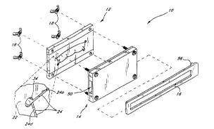
FIGURE 1 illustrates an exploded view of the disposable
tissue cutting die 10 of ~he present invention for precisely
and accurately cutting tissue to a predetermined configuration
for use in a medical prosthetic device such as an autologous
heart Yalve. As discussed in detail below, the tissue to be
precisely cut is placed onto the base member 14 and contacts
the cutting blade 24 supported by the cover 12 when the tissue
is locked between the cover 12 and the base member 14. The
slide 16 i8 then translated through a tunnel 50 of the base
member 14 to force the tissue between ridge 56 of slide 16 and
the cutting blade 24 SO a3 to precisely cut the tissue to
conform to the space defined by the cutting blade 24.
Ref erring to FIGURE 2, the cover 12 includes top member
20, insert 22, cutting blade 24 and four pairs of ferrules 26.
In the preferred embodiment, the top member 20 is made of a
translucent polycarbonate material, preferably of b;nm~
grade. This top member 20 is generally rectangular in shape
and has four through holes 28 located at the four corners of
the top member 20. The holes are used to align and attach the
cover 12 to the base member 14.
A channel 30 run9 the length of top member 20 and
projects into the channel 30 to form an island 32 ila the
middle of the top member 20. The outer periphery this island
32 is an outline of the desired cut tissue piece.
The preferred Amhn~limc.n~ of the heart valve disclosed and
claimed in the ' 955 patent provides an inner gtent having
tissue alignment members. Accordingly, the preferred
embodiment of this invention includes four pairs of ferrules
26 F-mh~ r1 within the polycarbonate material of the island
32, the location of these ferrules 26 corresponding to tL~e
location of the tissue alignment members of the heart valve
inner stent . Ferrules 26 typically extend approximately o . a89
A~.~,EN5E0 SltEEt
- : ; : . . .
' 792~2: : -
mm (35 mils) above the top surface of the island 32. In
addition, the diameter of the ferrules 26 corresponds to the
diameter of the tissue alignment members.
Cover 12 further includes an insert 22, made of semi-
translucent polycarbonate material, constructed 80 as to fit
within the channel 30 and completely surround the island 32 of
the top member 20. The gap between the edges of the island 32
and the insert 22 is large enough so as to accommodate and
retain the cutting blade 24. In one embodiment, the insert 22
i3 precisely configured 50 that a gap of 0.0508 mm ~0.002
inch) or les5 exists between the edges of the insert 22 and
the top member 20. In addition, the insert 22 has four
through holes 34 located at the corrl~cr~n~;n~ locations of the
f our corners of the island 3 2 .
- Cutting blade 24 is fixedly retained within the narrow
gap between the island 32 and the insert 22. Cutting blade 24
is advantageously formed from four discrete blades 24a, 24b,
24c, 24d. These blades are advantageously formed from thin
case hardened corrosion resistant steel having sufficient
~ ;h;l;ty to conform to the shape of the gap located hetween
the island 32 and the insert 22. The blade thickness should,
however, be s--f~ n~ to prevent deflection of the blade and,
consequently, an inadequately cut piece of tissue. In the
preferred '~o~ , the blades 24a-24d were made from a
strip of 0.1524 mm (0.006 inch) thick 5~;nl~qc steel,
supplied by American Safety Razor of West Virginia, having a
razor-sharpened edge. That razor-sharpened edge extends 0.889
mm (35 mils) above the top surfaces of the island 32 and the
insert 22.
A significant feature of the invention is that the ~our
corners of the space defined by the island 32 and the cutout
portion of insert 22 are relieved by ~our through holes 34 in
the insert 22. As shown in FIGUR~ 1, the end of the blade 24a
extends into a through hole 34 with the adjoining blade 24d
abutted up against. Without this relief of this cutout
portion, the precise juncture o~ the two blades 24a, 2~d would
involve such tight tolerances that manufacturing l?ractices
A~.',E!`~L,'-~ S~,EET
2 1 7 9 2 62
. .
typically dictate a slight gap between the blades. Die3
construCted with ga~s between adjoining blades leave a small
segment of uncut tissue at the corners of the opening,
requiring a manual cut af ter opening of the die to separate
the two pieces of tissue. In contrast, the present invention
merely involves making each blade 24a-24d slightly longer than
the actual perimeter of the i81and 32, with the excess blade
24a extending into one of the holes 34.
In an alternate embodiment of the present invention, a
flexible, one piece blade 25 is used to precisely cut the
piece of tissue. As shown in FIGUR13 3, the four through holes
34 in the insert 22 provide an additional advantage in that
they allow the cover 12 to e.~ cnmmn~te a flexible one piece
blade 25. Thus, the relief provided by the radius of the
through holes 34 allows a single continuous blade 25 to bend
around each of the four corners of the island 32. Without the
relief provided by these through holes 34, the otherwise close
tolerances at the corners of the island 32 would make it
difficult to ~ te a one piece blade 25. Thus, with the
relief, a one piece blade 25 would have to be precisely bent
to the angle formed at each of the four corners of the island
32 and still fit within the narrow gap between the island 32
and the insert 22. Therefore, the through holes 34 provide
substantial additional space to allow the one piece blade 25
to bend around each of the four corners of the island 32
FIGURE 4 shows an exploded perspective view of the base
member 14 of the tissue cutting die lO of the present
invention, in~ ;n~ a base piece 36, a thin flexible sheet
38, four threaded screws 40 and four washers 42. The base
piece 36 of the base member 14 is advantageously made of ~ a
translucent polycarbonate material, preferably biomedical
grade. The base piece 36 is generally rectangular in shape
and has four threaded through holes 44 located at the four
corners of the base piece 36.
A channel 46 is provided along the center length of the
base piece 3 6 of the base member 14 . The width of the channel
46 in the base piece 36 of the base member 14 accommodate the
A~ r ~3 5.~1E~T
, . , : ., .. , -, . - -
~ - 2 i 79262
.
width of the island 32 and, therefore, i8 slightly wider than
the width of the island 32.
The thin f lexible sheet 3 8 of the base member 14 provides
the cutting pad of the tissue cutting die 10. This thin
flexible sheet 38 is generally 0.8128 mm (0.032 inch) thick or
less. TEFIION is the preferred material for sheet 38 due to
its smooth surface r~r~tPristiC which pro~ides the correct
amount of adhesion for the tissue. The thin flexible sheet 38
is generally rectangular in shape and is similar in size (i.e.
length and width) to the top member 20 of=the cover 12 and the
base piece 36 of the base member 14. ~ocated at the four
corners of the thin flexible sheet 38 are four through holes
48. The location of the through holes 48 in the thin flexible
sheet 38 corresponds to the location of the through ho~es 44
in the base piece 36 of the base member 14.
Four threaded screws 4 0, as shown in FIGURE 4, are used
to attach the thin flexible sheet 38 to the surface of the
base piece 3 6 of the base member 14 . Each screw 4 0 enters
through the bottom surf ace of the base piece 3 6 of the base
member 14 and extends through the top surface of the thi~
flexible sheet 38. Due to the channel 46 provided along the
center length of the base piece 3 6 of the base member 14, the
thin flexible sheet 38 creates a tunnel 50 in the base member
14, as shown in FIG~RE 1
A washer 42 is positioned onto each of the screws 40 and
rests on the surface of the thin flexible sheet 38. The
th; ~-knPq~l of the washers 42 is determined by the height by
which the blades 24a-24d extend above the surfaces oi~ the
island 32 and the in~ert 22. Thus, by way of specific
example, if the blades 24a-24d extend 0 . 889 mm (35 mils) above
the surfaces of the i~land 32 and the insert 22, then the
washers 42 must be at least 0.889 mm (35 mils) in thickness.
This is to ensure that the blades 24a-24d are not damaged by
the thin flexible sheet 38 during shipment of the assembled
tissue cutting die 10. In addition, the washer~ enable the~
blades 24a-24d to securely hold the piece of tissue between
the cover 12 and the base member 14, without actually cutting
', ' ~ ': - . ' , , : - - - - . , :
2i79262
-- 8
the tissue. In thi8 specific example, the maximum thickness
of the washers 42 is calculated by adding the blade height (35
mils) and half of the nominal average tissue thickness (15-20
mils) .
FIG~RE 5 shows one ~mho~ of the slide 16 of the
tissue cutting die 10 of the present invention. The slide 16
is longer in length but slightly narrower in width than the
channel 46 in the ba8e piece 36 of the base member 14. The
slide 16 is advantageously made of thermoplastic a~d is
similar in thirkn~R~ to that of the thin flexible sheet 38.
A "bump" 56 or raised portion is located on the top 8urface of
the slide 16 near the end 52. The height of the bump 56 is
equal to or slightly greater than the height of the tunnel 50
in the base member 14. The opposite end 54 o the slide 16 is
generally flat. In the embodiment shown, slightly raised
~arrow" symbols 57 are located on the top surface of the slide
16 near its end 54. The middle or cut-out portion 58 of the
slide 16 is cut out, in the ~ shown, 80 that the
user' s finger can be inserted within the cutout portion
proximate to arrows 5 7 to obtain a firmer grasp of the slide
16, and thereby facilitate ~he pulling of the slide 16. The
use of the slide 16 will be described in detail below.
FIGU~B 6 illustrates a side view of the partially
assembled tissue cutting die 10 of the present invention, with
one o~ the four wing nuts 18 screwed onto one of the threaded
screws 40. The wing nuts 18 are advantageously made of
thermoplastic material and have a preformed in~ ni~l thread.
The wing nuts 18 are primarily used as a clamping means and,
therefore, are of adequate size to allow one to easily and
smoothly secure the wing nuts 18 onto=the screws 40.
The tissue cutting die 10 of the present invention is
used in the :Eollowing manner. First, a roughly sized piece oE
tissue is laid i~lat across the top sur~ace of the thin
~lexible sheet 38 o~ the base member 14. Due to the
slipperiness of the patient' s tissue and the a~hesion
characteristics of a material such as TEFLON (TM), the tissue
can be easily smoothed to lie flat~across the thin flexible
A~.~,-tN~D SHEET
.
.
~ . 21 79~2
.
... ., 9
sheet 3 8 . Next, the cover 12 i3 assembled onto the base
member 14 so that the blades 24a-24d can contact the thin
flexible sheet 38. This i5 accomplished by aligning the holes
28 located in the four corners of the cover 12 with the
threaded ends o~ the screws 40 protruding out the top surface
of the ba6e member 14. In addition, the cover 12 and tissue
are aligned so that the blades 24a-24d will cut the desired
portion of the tissue.
Once the cover 12 is ~CR~rnhl~l onto the base member 14,
the tissue is clamped between the two assemblies 12, 14 with
the wing nuts 18. During this step, the blades 24a-24d may
become slightly embedded into the tissue. However, due to the
strength or toughness of the tissue, the blades 24a-24d
normally do not completely penetrate the tissue.
The actual cutting of the tissue is accomplished by
pulling the slide 16 through the entire length of the tunnel
50 of the base member 14. The second end 54 of the slide is
pushed through one end of the tunnel 50 until it emerges out
the opposite end of the tunnel 50. Due to the symmetry o~ the
cover 12 and base member 14, the slide ~6 may be pulled
through either e~d of the tunnel 50, in the direction
indicated by the "arrow" symbols 57 located on the second end
54 of the slide 16. The user of the tissue cutting die 10
pulls the second end 54 of the slide 16 until the first end 52
of the slide 16 is pulled along the entire length o~ the
tunnel 50 and emerges out the other end of the tunnel 5D.
During this procedure, the bump 56, located on the firs~ end
52 of the slide 16, pushes against the bottom surface of the
thin flexible sheet 38. This, in turn, forces the tissue to
be pu~3hed against the blades 24a-24d and the ferrules 26 of
the cover 12.
The bump 56 located on the first end 52 of the slide 16
produces a ~wave" in the tissue and the thin flexible sheet
38. This ~wave~ allows one to sequentially oush portions of
the tissue into the blades 24a-24d, thereby enabling the
blades 24a-24d to work in shear. Due to the angle between the
5~D SHEET
:.. - ,- . . . .
- 2 1 79262
~ 1 o
four blades 24a-24d and the bump 56, all the blades 24a-24d
are able to cut the tissue in shear.
~fter the tissue is cut, the wing nuts 18 are unscrewed
from the threaded screws 40 so that the cover 12 may be
carefully removed from the base member 14. The resultant,
pre-configured cut piece of tissue is removed from the base
member 14 and mounted onto the valve stent. The remaining
excess tissue may be discarded.
Obviously, numerous variations and modif ications can be
made without departing from the spirit of the present
invention. Therefore, it should be clearly understood that the
f orms of the present invention described above and shown in
the figures of the At't_ , -nying drawings are illustrative only
and are not intended to limit the scope of the present
i~vention .
3 ',
- , .... : - -, .. . . . . -. . : . - . - . . - ~