Note: Descriptions are shown in the official language in which they were submitted.
2179304
1
DESCRIPTION
STENT FOR LIBERATING DRUG
Technical Field
This invention relates to a stent for liberating a drug
which is introduced into a vascular system such as blood
vessels, and more particularly to a stent used for a local
dosage o-f the drug.
Background Art
For instance, in angioplasties, vascular walls are likely
to be damaged by insertion of a catheter such as a balloon
catheter or an atheroma-resecting catheter thereinto so that
there occurs proliferation of the tunica intima due to a healing
reaction in the vascular walls, which frequently results in a
so-called restenosis_
Such a restenosis is caused by a hyperplasia of smooth
muscle cells and a majority of the recurrenceof the disease is
ascertained by an angiography, for example, 3 months after the
angioplasty operation.
The frequency of the restenosis sums to about 30 to about
40 % though it varies-depending upon facilities used in the
angioplasty operation. if any restenosis does not occur 3 months
:2179304
2
after the operation, it is suggested that the restenosis is no
longer caused subsequently. -
Meanwhile, any method for preventing the aforementioned
restenosis has not yet been established. However, attempts,
which has been made for this purpose until now, includes methods
in which an instrument such as a stent or an atheroma-resecting --
catheter is used, or other methods to which a genetic
engineering isapplied or in which drugs such as an
antimetabolite, e.g.; a carcinostatic agent, a fibroblast
hyperplasia-preventing agent, or the like are used.
However, in the event that the catheter, for example, the
atheroma-resecting catheter is used to prevent the restenosis of
blood vessels, patients suffer from a significant pain and such
an operation can be repeated only in a limited manner.
In addition, introduction of the stent into a portion
subjected-to the angioplasty provides some_effect to prevent
obliteration of blood vessels. However, since the stent itself
has no function for restricting a hyperplasia of smooth muscle
cells and preventing the restenosis, the essential problem still
remains unsolved. Moreover, upon the introduction of the stent
into a portion subjected to the angioplasty, there is a
possibility that a thrombus occurs. Under these circumstances,
in the event that the stent is used, in order to prevent
occurrence ofsuch a thrombus, there has been proposed a method
2179304
3
in which dosage of an antithrombotic agent-such as dextran,
aspirin, warfarin, or-the like is used.
On the other hand, it is considered that dosage of drugs
capable of restricting a hyperplasia of smooth muscle cells is
effective to prevent the restenosis without use of instruments
such as the stent, because such dosed drugs can function so as -
to prevent the sestenosis itself. However,-in this case, some
problem has been posed with respect to dosage method of these
drugs. - - -
Similarly, in the event that the stent is used together
with the antithrombotic agent to prevent the thrombus, some
problem has been also posed on the dosage of the antithrombotic
agent.
Inconseq~ence, a locally limited dosage is regarded as an
effective method for dosage of the drugs capable of restricting
a hyperplasia of smooth muscle cells or the antithrombotic
agent. The locally limited dosage is carried out by a method in
which a so-called dispatch catheter is used, a method in which a
sweat balloon catheter is used, a method in which a double
balloon catheter is used, a method in which the drugs are
selectively introduced through a catheter, or the like.
The_dispatch catheter is composed of a non-porous
polyurethane sheath and a spiral coil wound around the
polyurethane sheath. Drugs to be dosed are supplied into the
spiral coil so that the drugs can be brought into contact with -
2179304
4
walls of blood vessels. The sweat balloon catheter contains a
balloon having a microporous structure. When such a sweat
balloon catheter is used, drugs are gradually dosed through fine
pores of the balloon irto an interior of the blood vessels. The
double balloon catheter contains two balloons by which opposite
ends of the portion subjected to the angioplasty are closed such
that drugs are introduced through the catheter into a portion of
the blood vessel between these balloons.
The.aforementioned locally limited dosage methods can
advantageously increase a concentrationof the drug to be dosed,
because the dosage of the drug is carried out in the locally
limited region. To the contrary, since it is necessary to
continuously retain the catheter in the blood vessel and thereby
block a bloodstream, the locally limited dosage has such a
disadvantage that it cannot be used over a long period of.time.
For instance, in the event that the sweat balloon catheter or
the double ballooncatheter is used, the locally limited dosage
must be carried out within several minutes. whereas, even in the
event that the dispatch catheter is used or the drug is
selectively introduced through the catheter, the time required
for the dosage of the drug is limited to several hours. In
addition, these methods have a further problem that they can be
carried out only in an operating room.
Moreover, it is known that a whole-body dosage is made by a
peroral, transcutaneous or transluminal dosage of drugs so that
2179304
the drugs are circulated through the whole body and reaches
aimed cells. The whole-body dosage has an advantage that it can
be used over a long period of time.
However, in the avent of the whole-body dosage, a
concentration of the drug in the blood is undesirably raised so
that there is a possibility that unexpected side effects such as
hepatopathy, an aspiration accident, an excess or failed dosage
occur. In addition, when an antithrombotic agent is dosed by the
whole-body dosage method, fine arteries and veins in a brain are
damaged so that an intracerebral hemorrhage is likely to occur.
Moreover; in case that a long-term dosage is made, a large
amount of the-drug is dosed so that a huge medical expense is
required.
As described above, although many attempts has been made to
prevent the restenosis, for example, after an angioplasty
operation,-any effective method which makes the locally limited
and long-term dosage of drugs possible, has not yet been found
until now.
Disclosure of the Invention
The present invention has been accomplished to overcome the
aforementioned problems. It is therefore an object of the
present invention to provide a novel stent for liberating or
eluting a drug, which is capable of a locally limited and long-
term dosage of the drug.
2179304
6
As a result of long-term intense investigations and studies
made by the present inventors, the stent has been developed
based on a novel concept.
That is, in accordance with the present invention, there is
provided a stent which is adapted to be introduced into a
vascular system such as blood vessels. The stent is composed of
a stent body formed by weaving or knitting a fiber, which
contains a drug and is made of a biodegradable polymer having a
low-melting point at which pharmacological effects of the drug
are not damaged, into a tubular body.
In this case, the amount of the drug to be added to the
biodegradable polymer is determined depending upon a kind
thereof. When the amount of the drug in the biodegradable
polymer is too small, the drug released into the vascular system
decreases so that an effect by the dosage of the drugs cannot be
exhibited to a sufficient extent. On the other hand, when the
amount of the drugs in the biodegradable polymer is too large,
the healing process in walls of blood vessels is completely
restricted so that formation of fibers or coats becomes
difficult.
The kind of the drug added may be selected according to the
symptom or the aimed use. Examples of the drugs may include an
antimetabolite such as a carcinostatic, a fibroblast
hyperplasia-preventing agent, an antithrombotic agent or the
like. -
2179304
,
The-drugs as a solute are dissolved in the biodegradabl'e
polymer as a solvent to form a solution. The solution is then
hardened into a fiber from which the stent is prepared.
Alternatively, the solution may be coated on a rigid stent body
having an adequate mechanical strength, for example, a metal
stent body or a tubular woven or knitted stent body made of a
biodegradable polymer having a high melting point.
In this case, when heated to an elevated temperature, the
drug is susceptible to undesired change in its molecular
structure, which leads to loss of the aimed effect or conversion
to a toxic substance.
In general, the biodegradable polymer used as sutures, for
example, poly-lactic acid or poly-glycolic acid, has a melting
point ranging from about 2200 C to about 2400 C. Consequently,
there might occur an inconvenience that the drugs added thereto
is subjected to undesired chemical conversion, when heated to
such an elevated temperature.
Accordingly, it is required that the biodegradable polymer
has a low meting point at which the drug added can be present
without loss of the pharmacological effects. For example, it is
desirable that the melting point of the biodegradable polymer is
800 C or lower_ -
Examples of the suitable low-melting biodegradable polymers
may include poly-E-caprolactone, poly-D, L-deca-lactone, poly-
r
, = 2179304
8
di-oxanone or a copolymer of these compounds, which have a
melting point of about 63 C.
However, the aforementioned low-melting biodegradable
polymers cannot necessarily exhibit a sufficient mechanical
strength. In consequence,-it is suitable that the fiber composed
of the low melting biodegradable polymer containing the drug are
woven or knitted together with those made of a high-melting
biodegradable polymer to form the tubular stent body.
On the other hand, the drug added may include an
antimetabolite such as.a carcinostatic, a fibroblast
hyperplasia-preventing agent, or the like. For the purpose of
preventing the restenosis, TORANILAST is a preferred drug.
TORANILAST is an oral anti-allergic agent and widely used
as remedies for bronchial asthma, allergic rhinitis or atopic
dermatitis. It has been recently found that TORANILAST has an
effect of restricting a hyperplasia of smooth muscle cells. As a
result, the drug is expected to show an preventive effect
against the restenosis. Actually, the present inventors has
confirmed the preventive effect of TORANILAST against the
restenosis. -
The stent according to the present invention is adapted to
be introduced into a vascular system and retained in a
particular region of the vascular system. At this time, the drug
contained in the biodegradable polymer is released or eluted
into the vascular system over 3 months in association with
2179304
9
biodegradation of the stent. As a result, the drug contained in
the biodegradable polymer is allowed to be_continuously dosed
into a locally limited region of the vascular system over a long
period of timewhile maintaining its concentration in a constant
level.
In this case, such a locally limited dosage of the drug can
be carried out without any risk of causing adverse side effects
as observed in the case of the whole-body dosage. In addition,
this makes it possible to dose a relatively small amount of the
drugs over a long period of time.
Moreover, differing from the conventional locally limited
dosage, the present invention can provide a long-term dosage
without inflicting a serious pain on a patient.
Brief Description of the Drawings
Fig. 1 is a perspective view schematically showing one
embodiment of-a stent according to the present invention;
Fig. 2 is a perspective view schematically showing
essential parts of two folded yarn composed of a fiber made of a
high-melting biodegradable polymer and a fiber containing a drug
and made ofa low-melting biodegradable polymer;
Fig. 3 is a perspective view schematically showing another
embodiment of a stent according to the present invention;
Fig. 4 is a perspective view showing the condition in which
the fiber containing the drug and made of a low-melting
1. 0 2179304
biodegradable polymer is placed around a stent body formed from
a high-melting biodegradable polymer fibers and then melted so
as to adhere to an outer surface thereof.
Fig. 5 is a perspective view showing a high-melting
biodegradable polymer fiberwhich is knitted into a stent body
of a stent according to a further embodiment of the present
invention;
Fig. 6 is a perspective view showing the condition in which
the fiber shown in Fig. 5 is coated with a solution composed of
the low-melting biodegradable polymer containing the drug;
Fig. 7 is a perspective view showing a stent formed by
knitting the high-melting biodegradable fiber which is coated
with the solution composed of the low-melting biodegradable
polymer containing the drug;
Fig. 8 is a perspective view showing a stent body formed
from the fiber composed of the high-melting biodegradable
polymer;
Fig. 9 is-a perspective view showing a still further
embodiment of a stent according to the present invention in
which the stent body shown in Fig. 8 is coated with the dsug-
containing low-melting biodegradable polymer solution; and
Fig. 10 is a perspective view showing a still further
embodiment of a stent according to the present invention.
Best.Mode for Carrying Out theInvention
2179304
11
The presernt invention will be described in more detail by
way of specific examples by referring to the accompanying
drawings.
Examnle 1:
The present Example shows one example of a stent which is
effective for preventing a restenosis after an angioplasty
operation. In Example 1, a drug used there_is TRANIRAST (N-(3,
4-dimethoxy-cinnamoyl)-anthranilic acid) represented by the
following chemical formula:
O
CH30 / \ \ N
H Q
CH30 HOOC
TRANIRAST is one of fibroblast hyperplasia-preventing
agents. St has been reported by Tamai et al. of the present
inventors that clinical experiments, in which TRANIRAST was
continuously dosed for 3 months in a dosage amount of 600 mg per
day (one tablet after every meal), provided such a surprising
result that the restenosis rate is 15 % or lower. Consequently,
the drug has been expected to provide a remarkable preventive
effect against the restenosis. -
2179304
12
TRANIRAST was added to and dissolved in a biodegradable
polymer composed of poly-8-caprolactone having a melting point
of about63 C to prepare a polymer solution.
The thus-prepared-polymer solution was a mixture containing
TRANIRAST in an amount of 1 to 2 % by weight based on poly-8-
caprolactone.
The polymer solution was then subjected to a spinning
process to prepare a fiber composed of a TRANIRAST-containing
poly-s-caprolactone.
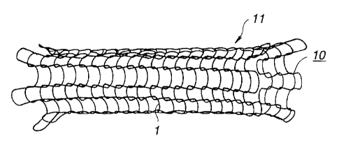
Next, as shown in Fig. 1, the fiber composed of a
TRANIRAST-containing poly-E-caprolactone was knitted into a
tubular shape to form a stent body 10. End portions of the fiber
constituting the stent body were treated to obtain a stent 11.
The thus-obtained stent 11 was produced by knitting the
poly-e-caprolactone fiber 1 having a diameter of about 0.05 mm
and a length of 90 crcti into a tubular shape having a diameter of
3 mm and a length of 20 mm.
Example 2:
The present Example shows another example of a stent which
is produced by knitting a drug-containing low-melting
biodegradable polymer fiber and a high-melting biodegradable
polymer fiber together.
In Example 2, as the drug-containing biodegradable polymer
fiber, there was used the TRANIRAST-containing poly-s-
caprolactone fiber 1 prepared in the same manner as described in
2179304
13
Example 1 above. The fiber was prepared in a similar manner to
that of Example-1 by subjecting the polymer solution containing
1 to 2 by weight of TRANIRAST based on poly-e-caprolactone to
a spinning process.
The-TRANIRAST-corntaining poly-s-caprolactone fiber 1 and
the high-melting biodegradable polymer fiber 2 was formed into a
two folded yarn 3 as shown in Fig. 2.'The two folded yarn 3 was
knitted into a stent body 10 to obtain a stent 11.
In this case; the high-melting biodegradable polymer fiber
2 constituting the two folded yarn 3 was ptoduced by subjecting
poly-lactic acid or poly-glycolic acid to a.spinning process.
In addition, the TRANIRAST-containing poly-8-caprolactone
fiber 1 constituting the two folded yarn 3 was a spun yarn
having a diameter of about 0.05 mm. The high-melting
biodegradable polymer fiber 2 was also a spun yarn having a
diameter.of about 0.05 mm. The stent body 10 was produced by
knitting the two folded yarn having a length of 90 cm to a
tubular body having a diameter of 3 mm and a length of 20 mm.
The size of the stent body 10 may be varied properly
depending upon the vascular system towhich the stent was
applied. -
Alternatively, the stent 11 can-be formed by first knitting
the stent body 10 and then coating the low-melting biodegradable
polymer solution composed of a mixture of a solvent and a drug
on the stent body 10, so that the amount of the drug contained
2179304
14
in the stent can be controlled properly. In this case, as the
low-melting biodegradable polymer solution, there is suitably
used a mixture solution prepared by mixing-70 cc of acetone, 1 g
of TRANIRAST and 1 g of poly-e-caprolactone together. In the
event that tha solution is coated, it is desirable that the
stent body 10 is subjected to a heat treatment to evaporate
acetone as the solvent component.
In the foregoing, the two folded yarn 3 composed of the
TRANIRAST-containing po1y-E-caprolactone fiber 1 and the high-
melting biodegradable polymer fiber 2 was used to obtain the
knitted stent body 10. However, a composite twisted yarn
composed of plural TRANIRAST-containing po1y-E-caprolactone
fibers l and plural the high-melting biodegradable polymer
fibers 2 may be used for the purpose.
Exa.mple 3:
In this Example, a high-melting biodegradable polymer fiber
2 was preliminarily knitted.into a tubular shape to prepare a
stent body 30. The TRANIRAST-containing poly-e-caprolactone
fiber 1 as the drug-containing low-melting biodegradable polymer
fiber was wound around the stent body 30 in an interlocking
relation to each other so as to form a stent 21, as shown in
Fig. 3. The fiber 1 was also produced by subjecting the polymer
solution containing 1 to 2 % by weight of TRANIRAST based on
poly-e-caprolactone to a spinning process.
2179304
In addition, the high-melting biodegradablepolymer fiber 2
used in this Example was also a poly-lactic acid fiber, a poly-
glycolic acid polymer fiber or a fiber composed of a copolymer
thereof.
In this Example, the stent body 30 may be also coated with
a polymer solution prepared by mixing l g of TRANIRAST as a drug
and 1 g of po1y-E-caprolactone.with 70 cc of acetone, so that
the amount of TRANIRAST to be contained in the stent 20 can be
controlled properly.
Examle 4:
In this Example, using the same procedure as described in
Example 3 above, the high-melting biodegradable polymer fiber 2
was preliminarily knitted into a tubular shape to form the stent
body 30. The TRANIRAST-containing poly-e-caprolactone fiber 1 as
the drug-containing Iow-melting biodegradable polymer fiber was
then wound around an outer circumferential surface of the stent
body 30 in an interlocking relation to each other so as to form
a stent 21, as shown in Fig. 3. The thus-prepared stent 20 was
heated by a heating means 35 as shown in Fig. 4 to smoothen an
outer surface of the stent. The heating means 35 usable here may
be a blower capable of blowing hot air.
Specifically, the stent 21 was heated to a temperature at
which the TRANIRAST-containing poly-E-caprolactone fiber 1 was
not completely molten, namely up to the melting point of poly-
s-caprolactone oY a temperature lowerthan the melting point,
2179304
- =
16
whereby an outer peripheral surface of the TRANIRAST-containing
poly-E-caprolactone fiber 1 was caused to melt so that the outer
surface of the stent 21 was smoothened.
The TRANIRAST-containing poly-s-caprolactone fiber 1 may be
also produced by subjecting the polymer solution containing 1 to
2 % by weight of TRANIRAST based on poly-E-caprolactone to a
spinning-process: inaddition, the high-melting biodegradable
polymer fiber 2 may be also a poly-lactic acid fiber, a poly-
glycolic acid polymer fiber or a fiber composed of a copolymer
thereof_
The smoothened outer surface of the stent 21 permits a
smooth insertion of the stent into a vascular system such as
blood vessels.
Examole 5-
In this Example, a high-melting biodegradable pnlymer fiber
42 was coated with a solution of a drug-containing low-melting
biodegradable polymer and then the coated fiber was knitted into
a stent 41.
In the production of the stent 41, a biodegradable polymer
material having a melting point higher than that of the drug-
containing low-melting biodegradable polymer was subjected to a
spinning process to obtain the biodegradable polymer fiber 42 as
shown in Fig. 5. At this time, the high-melting biodegradable
polymer fiber 42 used here may be fiber prepared by subjecting
~. 2179304
17
poly-lactic acid, poly-glycolic acid or a copolymer thereof to a
spinning process.
The biodegradable polymer fiber 42 was coated with a
solution 43 of drug-containing low-melting biodegradable polymer
as shownnin Fig. 6. The solution 43 of drug-containing low-
melting biodegradable polymer used here was a solution prepared
by mixing 1 g of TRANIRAST as a drug and 1 g of po1y-E-
caprolactone with 70 cc of acetone as a solvent.
Next, the high-melting biodegradable polymer fiber 42 on
which the drug-containing low-melting biodegradable polymer
solution 43 was coated, was knitted to form the stent 41.
Successively, the thus-knitted stent 40 was heated to
evaporate acetone. The stent 40 was preferably heated to a
temperature at which the drug-containing low-melting
biodegradable polymer 43 was still maintained in an unmolten
state. This was because melting of the drug-containing low-
melting biodegradable polymer 43 was to be prevented upon
heating.
Meanwhile, in the event that acetone as a solvent was
already evaporated during production of the knitted stent body
40, the heating step can be omitted.
Thereafter, the stent body 40 from which acetone as a
solvent was evaporated, was formed into the stent 41, as shown
in Fig. 7, by treating end portions of the fiber 42 constituting
the stent body 40.
~. ~ 2179304
18
In addition, the knitted stent body 40 may be further
coated with the low-melting biodegradable polymer solution 43
prepared by mixing 1 g of TRANIRAST as a drug and 1 g of poly-E-
caprolactone with-70 cc of acetone so that the amount of
TRANIRAST as a drug coated on the stent body 40, can be adjusted
to a proper level. in this case, it is preferred that the stent
body is heated to evaporate acetone as. a solvent.
Examx~l e 6 :
In the aforementioned Examples, the high-melting
biodegradable polymer fiber was first coated with the solution
of the-drug-containing low-melting biodegradable polymer and
then the fiber was knitted to form the stent. On the other hand,
in this Example, the high-melting biodegradable polymer fiber 42
prepared by subjecting_poly-lacitc acid, poly-glycolic acid or a
copolymer thereof to a spinning process was first knitted into a
stent boy 50 as shown in Fig. 8. Applied over the stent body 50
was a low-melting biodegradable polymer solution 53 containing a
drug as shown in Fig. 9 to form a stent 51 of this Example.
The drug-containing low-melting biodegradable polymer
solution 53 applied to the stentbody 50 was a polymer solution
containing 1 to 2$ by weight of TRANIRAST based onpo1y-E-
caprolactone in the solution. -
The application of the low-melting biodegradable polymer
solution 53 to the stent body 50 may be carried out by coating
the solution 53 over an outer circumferential surface thereof.
2179304
=
19
Alternatively, the low-melting biodegradable polymer solution 53
may be applied to the stent body 50 by immersing the stent body
50 therein.
Sxamule 7:
This Example shows a further example of a stent which is
produced by coating a drug-containing biodegradable polymer
solution over a stent body.
In this Example,=-=a stent 61 was produced by coating the
drug-containing biodegradable polymer solution on the stent body
made of metal to form a layer 62 composed of drug-containing
biodegradable polymerover an outer surface of the stent body
60, as shown in Fig. 10. The solution coated contained 1 to 2 %
by weight of the drug based on poly-E-caprolactone (having a
melting point of 63 C) in the solution.
The stent body 60 used above was made of a metal material
having a thickness of 0.05 mm to 0.1 mm and formed into a
cylindrical body having a diameter of 2.5 mm to 4 mm and a
length of 15 mm to 25 mm. Examples of the metal material may
include stainless steel, tantalum or the like.
The stent prepared in each of the aforementioned Examples
was introduced into the blood vessel after angioplasty operation
and held in place. As a result, it was confirmed that dosage of
TRANIRASTwas carried out in an adequate manner for a long
period of time, whereby occurrence of the restenosis was
considerably reduced.
2179304
ao
Industrial Applicability
As is apparently understood from the aforementioned
detailed description, the use of the stent according to the
present invention enables a continuous, locally limited and
long-term dosage of the drug.
In addition, such a dosage can-prevent occurrence of side
effects so EF.at pains inflicted on patients can be minimized.