Note: Descriptions are shown in the official language in which they were submitted.
W0 95/21377 217 9 710 PCTrt-S~Sl~13i3
IMPROVED REALrTINIE SCANNING FLUORESCENCE
ELECTROPHORESIS APPARATUS FOR TFIE ANALYSIS OF
POLYNUCLEOTTDE FRAGMENTS
FIELD OF THE I1VVENTION
This invention relates to improved apparatus for performing electrophoresis,
and
more particularly io an improved real-time scanning fluorescence
electrophoresis
apparatus for polynucleotide fragment analysis.
$ACKCiROUND OF THE INVENTION
Electrophoretic polynucleotide fragment analysis methods are used to
characterize
mixtures of poIynucleotide fi-agments based on their migration velocity
through a polymer
network under the influence of an electric field, i.e. their electrophoretic
mobility, in
combination with single or multi-color fluorescence detection. Typically these
methods
are applied subsequent to amplification of the target polynucleotide using a
method such
as PCR, e.g. Mullis, U.S. patent 4,683,202. Examples of such methods include
polynucleotide sequencing, e.g. Trainor, Anal.Chem., 62: 418-426 (I990),
restriction
fi-agment length polymorphisim (RFLP) analysis, e.g. Watkins, Biotechniques,
6: 310-319
(1988), and variable number of tandem repeat (VNT'R) or microsateilite
analysis, e.g_
Ziegle et ai., Genomics, 14: 1026-1031. Each of these methods can provide
valuable
genetic information about t:.~ target polynucleotide.
Current electrophoretic potynucleotide fiagment analysis systems are
characterized
by multiple electrophoresis lanes arranged in a planar array, e.g. a multi-
lane slab gel, in
combination with a real-time-scanning fluorescence detector, e.g. Hunkapiller
et al., U.S.
patent 4,8I 1,218. Multiple lanes are used to increase the overall throughput
of the
analyzer. In order to collect data during the electrophoresis from multiple
lanes, the
optical detector system is scanned across the w' : ofthe electrophoresis
chamber
perpendicular to the direction of migration of the labeled polynucleotides.
Preferably,
multi-color fluorescence detection is used to increase the information density
per lane, e.g.
for DNA sequencing, four Label colors are used, one color for each base. A
light source,
e.g. a laser, excites the fluorescent labels attached to the polvnucIeotide
fraQrnents, and
multiple emission filters discriminate between labels having different
spectral properties.
In addition, a computer is used to collect data consisting of time, lane
number, and
-1-
~
W'O 95/213 r' 217 9 710 PCT;2S95i0I353
fluorescence emission wavelength information, and transform it into useful
information,
e.g. DNA sequence.
A significant limitation on the speed and resolution of current polynucleotide
fragment analysis systems is the ability to dissipate the Joule heat that is
generated as a
result of the electric current passing through the electrophoresis medium.
Because of
problems caused by Joule heating, current systems are limited to low, e.g. 25
V/cm,
electrical fields, resulting in long analysis times, e.g. 8 hrs. Joule heating
and the resulting
temperature gradient across the gel can negatively impact the quality of the
separation in
two ways. Fuss, because heat is generated throughout the electrophoresis
medium but
only dissipated at its' outside surfaces, a parabolic temperature profile is
establish across
the depth of the channel. Since electrophoretic velocity is a strong function
of
temperature, approximately 2% per oC, this temperature profile leads to a
parabolic
velocity profile for the migrating analyzes. This spatial dependence of
velocity causes a
broadening of the ttligrating zones, leading to reduced separation
performance. The extent
of the temperature profile can be reduced by making the electrophoresis
channel thinner,
e.g. Bromley et al., Nucleic Acids Research, 19: 4121~I25 (1991); Stegettlann
et al.,
Methods in Molecular and Cellular Biology, 2: 182-184 ( 1991 ). Therefore, as
automated
system which incorporates thin electrophoresis channels would be desirable.
Second, if the average temperature of the electrophoresis medium becomes too
high, the structural integrity of the medium can be compromised. In the case
of polymer
gel media, e.g. crossIinked polyacrylamide gels, the elevated temperature can
lead to
complete destruction of the gel. The average temperature of the
electrophoresis medium
can be controlled by increasing the rate of heat transfer from the
electrophoresis channel
to the surrounding environment. Therefore, a system which more efficiently
transfers the
Joule heat generated as a result of the electrophoresis to the surrounding
environment
would be desirable.
A fiuther limitation on the speed and resolution of eiectrophoretic
separations is
the rate at which the detector can acquire data from fast moving analyte
bands. The most
desirable form of detection for polynucleotide fragment analysis would be
simultaneous
multi-color detection. However, current approaches, i.e. an indexable filter
wheel in
combination with a photomultiplier tube (PMT) detector, are not ideal because
the filter
- wheel must be,indexed rapidly enough to observe each color before it moves
out of the
detector region. This is problematic due to the high elecirophoretic velocity
of the
_2_
W0 95/2137% 217 9 710 PCTIU595/01353
polynucleotide fragments in high-speed systems. If a sufficient number of data
points are
not collected for each analyte band, e.g. 10 points per band, the ability to
discriminate
between adjacent bands is lost. One way to increase the rate of data
acquisition for a
multi-color system is to collect signals from all the colors simultaneously
rather than
serially. Therefore, a detection system which acquires all colors
simultaneously would
be desirable.
In light of the above, what was needed was an improved electrophoresis
apparatus
capable of accommodating high electric fields through enhanced heat
dissipation
characteristics and detector performance.
SUMMARY OF THE IrIVENTION
The present invention is directed to improvements to an apparatus for
electrophoretic polynucleotide analysis, said improvements leading to
increased
I S throughput of the system. The improvements include ~) incorporating a
spectral-array
detector to increase the rate of data acquisition, and (ii) incorporating an
improved
means to control the temperature of the electrophoresis medium. The analyzer
system of
the present invention is comprised o~ in combination,
An improved real-time scanning fluorescence electrophoresis apparazirs for the
electrophoretic analysis of ffuorescently-labeled polynucleotide fragments of
the type
having an electrophoresis chamber containing an electrophoretic separation
medium
capable of accommodating multiple electrophoresis lanes arranged in a planar
array, a
fluorescence detector mounted on a translatable stage, a light source for
exciting
fluorescent molecules, and a computer for collecting data consisting of time,
location,
fluorescence wavelength and fluorescent intensity information wherein the
improvement
comprises:
(a) a spectral-array detector for detecting the emission light from said
fluorescently-labeled p ~lynucleotide fragments including the simultaneous
detection of
multiple fluorescent labels,
(b) a temperature control means to control the temperature of the
electrophoretic
separation medium during electrophoresis.
g~F DESCRIPTION OF THE DRAWfir'GS
Figure I shows a vertically oriented slab gel.
_ 3 _
W'O 95/213'7 217 9 710 PCT'L'S95/013i3
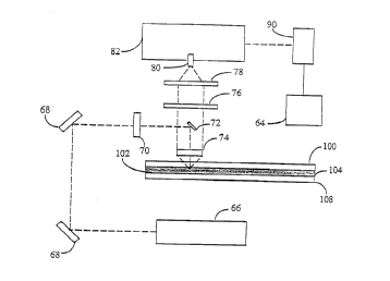
Figure Z shows a schematic diagram of the fight path in a preferred embodiment
of the
spectral-array detection system of the present invention.
Figures 3 shows a plate holder according to a prefered embodiment of the
invention.
Figure 4 shows a plate locating mechanism according to a prefered embodiment
of the
invention. .
Figure S shows a temperature control mechanism according to a prefered
embodiment of
the invention.
The term "polynucleotide" as used herein refers to linear polymers of natural
or
modified nucleoside monomers, including double and single stranded
deoxyribonucleosides,
I S n'bonucleosides, a-anomeric forms thereof, and the like. Usually the
nucleoside monomers
are linked by phosphodiester bonds or analogs thereof to form polynucleotides
ranging in
siae from a few monomeric units, e.g. 8-40, to several thousands of monomeric
units.
Whenever a polynucleotide is represented by a sequence of letters, such as
"ATGCCTG," it
will be understood that the nucleotides are in 5'-3' order from left to right
and that "A"
denotes deoxyadenosine, "C" denotes deoxycytidine, "G" denotes deoxyguanosine,
and "T"
denotes thymidine, unless otherwise noted. Analogs of phosphodiester linkages
include
phosphorothioate, phosphorodithioate, phosphoroseienoate,
phosphorodiselenoate,
phosphoroanilothioate, phosphotaniIidate, phosphoramidate, and the like.
As used herein, "nucleoside" includes the natural nucleosides, including 2'-
deoxy
and 2'-hydroxyl forms, e.g. as described in Kornberg and Baker, DNA
Replication, 2nd
Ed. (Freeman, San Francisco, 1992). "Analogs" in reference to nucleosides
includes
synthetic nucleosides having modified base moieties and/or modified sugar
moieties, e.g.
described generally by Scheit, Nucleotide Analogs (John Wiley, New York,
1980).
As used herein, the term "electrophoretic separation medium" refers to a
material
through which the polynucleotides are electrophoresed and which imparts a size-
dependent eIectrophoretic velocity to the polynucleotides. Typically, such
material is a
porous network formed by linear or branched polymer molecules, or she like,
e.g.
crossiinked polyacrylamide.
_.1 _
W095I213'i 217 9 710 PCTIhS9sID1353
As used herein, the term "elecuophoresis chamber" refers to the container in
which
the electrophorertic separation is contained. Typically, this container is
formed by two
rectangular glass plates which are separated by thin polymer sheets, spacers,
located
between the plates at the edge regions of the plates. This is traditionally
referred to as
slab electrophoresis. When the electrophoretic separation medium is a rigid
crosslinked
gel, this format is referred to as slab gel dearophoresis.
DESCRIPTION OF THE PREFERRED EMBODIIvtENTS
Figure 1 shows polynucieotide fragment samples (2) which have been labeled
with one of several ffuorophores loaded into loading wells {4) of vertically
oriented slab
gel (8), said gel motmted in the analyzer of the present invention. The
fragments are
electrophoresed through gel (8) where they are separated based on their
relative size.
Following separation, the fragments pass through laser excitation and
defection region
(12) where the fluorescently labeled polynucieotide fragments are detected.
The
fluorophores emit light at a specific wavelength based upon the particular dye
used,
thereby facilitating the identification of each fragment.
After the polynucleotide fragments have been separated, they are detected by a
simultaneous multi-color detection means. An important feattue of the
polynucleotide
analyzer of the present invention is the "spectral-array fluorescence
detector". As used
herein, the term "spectral- array fluorescence detector" refers to a detector
which employs
[) a means to spectrally separate the fluorescence emission light, such as a
diffraction
grating, or a prism, or the like, (ii) an array of detector elements sensitive
to light
radiation, such as a diode array, a charged coupled device (CCD) system, an
array of
photomultiplier tubes, or the like, (iii) an excitation light source, such as
an incandescent
bulb, an arc lamp, a laser, a laser diode, or the like, and (iv) associated
optics capable of
directing and conditioning both the excitation and emission light. The output
of a
spectral-array detector is light intensity as a function of array location,
wherein the array
location can be directly related to the wavelength of the light falling on
that location. One
example of such a detector is given by Karger et al., Nucleic Acids Research
19: 4955-
4962 {1991).
One preferred method of treating the output of a spectral-array detector is to
create a "virtual filter". As used herein, the term "virtual filter" refers to
a method of
manipulating data from a spectral-array detector such that a plurality of
discrete
-5-
WO 95/21377 ~ 17 9 710 pCTli'S95/013;3
wavelength ranges are sampled, wherein the location and bandwidth ofeach
wavelength
range can be dynamically changed using software. The virtual filter can mimic
a physical
interference or absorbence filter, however it has several important
advantages. First,
virtual-filters can be programmed to interrogate multiple emission wavelengths
simultaneously, malting possible the e~cient mufti-color detection of fast-
moving
analytes without the need to rapidly index a multiplicity of filters. Second,
virtual filters
can be programmed to detect a range of emission bandwidths. This is important
because
for any application, there exists an optimum bandwidth which results in an
optimum
combination of sensitivity and color discrimination: as the detection band
width is made
wider, the detector collects more light, hereby increasing sensitivity,
however, at the same
time, the broader bandwidth decreases the ability to discriminate between
closely related
colors. Third, virtual filters have essentially perfect transmission curves,
i.e. the filter ran
discriminate between very closely related colors. Forth, the selected
wavelength ranges
of the virtual filter can be easily adjusted using software to match the
characteristics of
various excitation light sources and dye sets. Therefore, changing dye
chemistries is a
simple matter of changing the virtual filter with software, whereas a
mechanical
modification of the system is required when physical filters are used.
Moreover, the
selected wavelength ranges and band widths of the virtual filter can be
changed
dynamically, i.e. doting the course of a rua, to compensate for any spectral
changes in the
dye labels which occur during a tun.
Figure 2 is a schematic diagram of the light path in a preferred embodiment of
the
spectral-array detection system of the present invention. Preferably, the
analyzer system
of the invention uses a laser as a fluorescence excitation light source, e.g.
an argon ion
laser that emits a 40 mW, 0.67 mm diameter polarized light beam having
intensity tttaxima
at wavelengths of 488 and 514 nm. Light from laser (66) is reflected off of
adjustabiy-
mounted turning mirrors (68) which direct the laser light to the desired
location.
Telescope lenses (70) then reduce the beam diameter to approximately 100 Elm,
and
bending mirror (72) directs the light into electrophoresis medium (104) at
right angles.
Light emitted from the laser-excited fluorescent label is collected by
aspheric
collection lens (74) which collimates the light in the direction of the
detector. The
emitted light then passes around bending mirror (72) and through laser
rejection filter
(76), thereby reducing the level of scattered laser fight entering the
detector. Because
the excitation laser light passes through the center of aspheric collection
lens (74), a
certain amount of laser light will be reflected directly back from the lens
surface in the
-6-
CA 02179710 1999-03-16
direction of the detection, causing unwanted background signal. Bending mirror
(72),
which is mounted in the center of laser rejection filter (76), acts to deflect
this
reflected light away from the colletion path thus reducing the amount of
reflected
light entering the detector. The collected emission light then passes through
plano-
convex lens (78) which focuses the emission light at slit (80) mounted on the
entrance
to spectrograph (82). (Spectrograph (82) utilizes a 405 g/mm, 450 nm blaze
grating
with a dispersion of 17 nm/mm.) After passing through spectrograph (82), the
light
then falls onto CCD (90). The output signal from CCD (90) is transmitted to
electronic computer (64) for subsequent data analysis and presentation.
To further increase the emission light signal and decrease background light
scatter, a nonconductive mirror coating is applied to the inside surface (102)
of front
gel plate (108). This surface reflects emission light back to the cllection
lenses rather
than allowing it to be lost to the surroundings through the front gel plate.
In addition,
when the primary laser light strikes this mirrored surface it is reflected
back through
the gel, thereby exciting additonal fluorophores resulting in more emission
light.
Furthermore, this mirrored surface decreases unwanted background light
generated by
the fluorescence of the front glass plate itself.
In order to interrogate all of the electrophoresis lanes on a real-time basis,
the
optical system described above, less turning mirrors (68) and computer (90),
is
scanned across the width of the electrophoresis chamber.
Another important feature of the present invention is the novel means used to
mount the electrophoresis chamber onto the analyzer. Preferably, the
electrophoresis
chamber is formed by two glass plates separated by two spacers located at the
left and
right edges of the plates. The glass plates are mounted into a plate holder
which acts
to support and secure the glass plates along with an upper buffer reservoir in
a
convenient manner. See Figure 3. The plate holder consists of rectangular
frame
(200) onto which is attached plurality of twist clamps (204). (Note that only
one twist
clamp is indicated in Figure 3, as (204), in order to retain the clarity of
the drawing.)
CA 02179710 1999-03-16
When twist clamps (204) are in the horizontal orientation, they service to
secure the
glass plates in the holder, and, when twist clamps (204) are in a vertical
orientation,
they allow the glass plates to be conveniently inserted or removed from the
plate
holder. The rectangular frame includes two locational registration notices
(208) to
insure the proper positioning of the plate holder in the analyzer. Beam stop
(212) is
15
25
-7a-
positioned so as to protect the user from direct
2179710
W'O 95121377 - - PCT/US95/01353
exposure to the excitation laser light. The frame also includes two handles
(202) to
facilitate transportation of the plate holder assembly. The plate holder
provides a means
for detachably mounting upper buffer reservoir (216). A protrusion (228) on
each side of
upper buffer reservoir (216) is positioned such that when the uppermost twist
clamps are
in the horizontal position, the upper buffer reservoir (216) is forced against
the front glass
plate, thereby creating a liquid-tight seal between the upper buffer chamber
and the front
glass plate. Upper buffer reservoir (216) contains electrode (220) and
electrical cable
(224) for connecting electrode (220) to an electrophoresis power supply. The
plate
holder is designed to secure glass plates of varying lengths. For applications
requiring less
separation and/or a shorter atlalysis time, a shorter length would be used,
and for
applications requiring more separation and for which longer analysis times can
be
tolerated, a longer length would be used.
A fiuther important aspect of the present invention is the plate locating
mechanism. In order to efficiently collect the fluorescence emission light,
the detection
region of the electrophoresis chamber must be properly positioned with respect
to the
collection optics. Specifically, the detection region must be aligned such
that the focal
point of the collection optics is Located within the separation medium, and
not in the wall
of the electrophoresis chamber. The plate locating mechanism insures that this
positioning
is reproducibly achieved. The mechanism will be described with reference to
Figure 4.
When a thin electrophoresis chamber is being used, i.e. less than 0.2 mm,
preadjusted
locating pins (300) fit through notches (304) in back glass plate (308) and
push front glass
plate (312) against front tip (324) oflocating pins (300). When a thick
electrophoresis
cfiamber is being used, i.e. greater than 0.2 mm, step-portion (320) of
locating pins (300)
is forced against back glass plate (312). Locating pins (300) are preadjusted
such that the
interior of the electrophoresis chamber is at the focal point ofthe collection
optics. Glass
plates (308 and 312) are forced against locating pins (300) by twist clamps
(330).
While increasing the electric freId across the electrophoresis chamber
increases the
speed of the electrophoretic separation, it also leads to increased Joule heat
generated
within the electrophoresis medium, which in turn can lead to destruction of
the
electrophoresis medium. To remove the heat generated by running "fast"
electrophoresis,
a temperature control mechanism (Figure 5 ) has been developed. The
temperature
control mechanism includes a back heat transfer plate (400) against which back
glass
plate (404) is mounted to the instrument. Preferably, heat transfer plate
(400) is made
from coated aluminum. The coating acts as an electrical insulator to inhibit
arcing
_g_
WO 95/213TT 21 l 9 710 PCT~2'S951DI353
between back glass plate (404) and the rest of the instrument. Within back
cooling plate
(400) are channels through which a flowable heat transfer medium can be
circulated.
Front heat transfer plate (408), also containing channels capable of being
&lled with a
flowable heat transfer medium, is contacted with front glass plate (412). Pump
(416)
circulates the flowable heat transfer medium from reservoir (420) through
front and back
heat transfer plates (400 and 408). Heat is removed from the circulating
flowable heat
transfer medium by passing it through heat exchanger (424), thereby cooling
the flowable
heat transfer medium to ambient temperature. If superambient heating or
subambient
cooling of the gels is desired for a specific application, the ffowable heat
transfer medium
passes through a heater or cooler (not shown) before flowing through the heat
transfer
plates. Active temperature control of the gel is effected by means of
temperature sensors
(430) mounted to the heat transfer plates in combination with computer (434)
which
regulates the temperature of the plates by controlling the flow rate of the
flowable heat
transfer medium through the heat transfer plates.
Although the invention has been illustrated by the foregoing description it is
not to
be construed as being limited to the materials employed therein but rather the
invention is
directed to the generic area as hereinbefore disclosed. Various modifications
and
embodiments thereof can be made without departing from the spirit or scope
thereof.
-9-