Note: Descriptions are shown in the official language in which they were submitted.
CA 02180266 2001-11-02
IMPROVED PULSED DISCHARGE SYSTEMS
FIELD OF THE INVENTION
The invention generally relates to the art of gas analysis and more
particularly to
the analysis of gas through chromatographic means.
BACKGROUND OF THE DISCLOSURE
The discharge systems of this disclosure utilize a pair of electrodes which,
in the
preferred embodiment, apply a transverse spark across a gap between the
electrodes, the spark
preferably being repetitively formed. Bipolar or monopolar discharge can be
used. An inert gas
flows between the spark electrodes. The spark creates photons of energy which
are emitted and
are used as described. In alternate aspects, particles are cleared or
energized in the spark gap and
energized particles subsequently surrender energy. 'the preferred inert gas is
helium with traces
of inert gases. The photon emission or loss of energy assists in
identification and measurement
of gas chromatographic column (GC hereinafter) eluted peaks from a typical GC
source.
SUMMARY OF THE INVENTION
Another aspect of the invention pertains to a method of analyzing a sample
compound comprising the steps of flowing a carrier gas through a chamber for
exposure to DC
current thereby energizing at least one component of the carrier gas to an
excited state as a result
of exposure to the DC current, commingling a gaseous sample with the carrier
gas within a
chamber wherein the carrier gas comprises at least one component in an excited
state, forming
2 0 one or more excited compounds in the gaseous sample resulting from photon
emission in the
decay of at least one excited component of the carrier gas wherein the
emissions involve an
energy exchange up to about 11.8 eV and determining the type and concentration
of one or more
compounds in the gaseous sample by measuring photon emission from the decay of
the excited
compounds in the gaseous sample.
2 5 Further the invention comprehends a gas detector for identification and
quantification of sample compounds, comprising an elongated chamber having a
first chamber
inlet at one end and an outlet at the other end and a gas flow path between
the first inlet and
outlet ends with two electrodes spaced apart and located to produce short,
repeated, high voltage,
pulsed DC current within the chamber across the gas flow path and wherein
spark duration
3 0 minimizes electrode erosion and permits observation of phenomena occurring
at and between the
1
CA 02180266 2001-11-02
DC current at and remote from the electrode legation. Means are provided for
introducing a
carrier gas into the chamber through the first chamber inlet and flowing the
carrier gas in the gas
flow path. Means are provided for introducing a sample gas into the chamber
through a second
inlet which is located downstream from the first chamber inlet and downstream
from the two
spaced electrodes and flowing the sample gas in the gas flow path whereby ions
are produced
by the spark or by metastable species of the carrier gas.
Still further the invention comprehends a method of testing an airborne sample
comprising the steps of providing an airborne sample flowing through a test
chamber,
simultaneously providing a carrier gas flowing through the test chamber,
within the test chamber,
forming with an electrical current a metastable species in the carrier gas
wherein the metastable
species is characterized by having a ground energy state and excited state of
sufficient time
duration to enable an energy transfer from the excited state of the metastable
species to the
airborne sample by the emission of photons and wherein the excited state
causes an energy
transfer to the airborne sample wherein the excitation energy range of the
metastable species is
selected to preclude energizing the constituents of air.
Further still, the invention comprehends a method of analyzing an eluted
sample
compound in a carrier gas comprising the steps of flowing the carrier gas
through a chamber for
exposure to a spark discharge across the chamber, the spark discharge being
formed across a pair
of spark forming electrodes cooperating with a pulsed DC current source and
introducing an
2 0 eluted sample into the carrier gas downstream from the spark discharge and
observing
downstream in the chamber spark caused current flow in the chamber to analyze
the
concentration of the eluted sample compound flowing through the chamber by
detecting changes
in the current flow.
Still further the invention pertains to a charged particle detector comprising
a
2 5 closed chamber having a helium gas flow inlet at a first end and spaced
outlet at a second end
to enable helium flow therethrough and spaced electrodes cooperating with a
pulsed DC power
supply responsive to DC current flow sufficient to enable an electrical spark
to be formed
between the electrodes, the electrodes being positioned to form a spark in the
helium flow into
the chamber to thereby create photon emission. Spaced detector means are
downstream in the
3 0 chamber for collection of charged particles downstream of the spark across
the gap wherein the
2
CA 02180266 2001-11-02
charged particles enable a current to be formed indicative of sample gas
concentration in the
chamber, an inlet downstream in the chamber controllably introduces a sample
and carrier gas
flow from a GC column at a selected location downstream from the spark forming
electrodes so
that the sample and carrier gas and the helium flow provide current for the
detector means.
Means are provided for optimizing the current by adjusting the selected
location for sample gas
and carrier gas introduction and by adjusting voltage applied to the detector
means.
Moreover the invention also comprehends a method for analyzing a sample
compound comprising the steps of flowing a carrier gas through a chamber
wherein the carrier
gas comprises an inert gas and a dopant gas wherein the dopant gas is selected
such that the
resonance energy of the dopant component is greater than the ionization
potential of the
compound to be measured in the gaseous sample, commingling a gaseous sample
with the carrier
gas within a chamber forming a composite gas, exposing the carrier gas to a
spark generated by
DC current and optically observing spark caused emissions in the chamber to
analyze the gaseous
sample component, wherein the emissions involve an energy exchange up to the
resonance energy
of the dopant.
The invention also pertains to a charged particle detector comprising a
circular
closed chamber having a gas flow inlet and spaced outlet positioned to direct
gas flow through
the chamber and the chamber directs the gas flow in a circle therein with
spaced electrodes
provided with a current sufficient to enable an electrical spark to be formed
in a gap between the
2 0 electrodes locating the spark thereacross, the electrodes being positioned
to form a spark in gas
in the chamber to create charged particles. A spaced detector electrode is in
the chamber for
collection of charged particles wherein the charged particles move to the
detector electrode to
form a current indicative of a sample gas concentration in the chamber.
Moreover the invention in another aspect pertains to a gas detector for
identification and quantification of sample compounds, comprising a circular
chamber having a
tangential chamber inlet and a tangential outlet and a circular gas flow path
between the inlet and
outlet ends, means for flowing an inert gas into the chamber and two spaced
electrodes located
in the chamber to produce repeated current sparks across the chamber wherein
gas interaction
forms energized particles in the chamber. A sample source is connected to
deliver gas into the
3 0 chamber and means is responsive to interacted sample gas and charged
particles to enable sample
3
CA 02180266 2001-11-02
gas detection in the chamber.
Yet further the invention comprehends an electron capture detector comprising
a
closed chamber having a helium flow inlet to enable helium flow therethrough,
spaced electrodes
forming a spark between the electrodes defining a spark thereacross, the
electrodes being
positioned in the chamber to form a spark through helium in the chamber. A
sample gas source
is connected to an inlet to the chamber to provide sample gas flowing in the
chamber and the
chamber and the inlet are constructed and arranged to flow gas in a circle in
the chamber. A
spaced detector in the chamber is provided for collection of current formed as
a result of the
spark across the gap wherein the spark irradiated helium enables a current to
be formed indicative
l0 of eluted gas sample concentration in the chamber and the detector measures
the gas sample in
the chamber by change in current flow.
Another aspect of the invention comprehends a method for analyzing a sample
gas
comprising the steps of exposing a first gas in a closed spark chamber to DC
current across the
chamber, energizing at least one component of the first gas to an excited
state as a result of
exposure to the DC current and permitting the excited gas to form ionizing
radiation by decay,
exposing a sample gas in a sample chamber to ionizing radiation resulting from
the decay of at
least one component of the first gas, forming charged particles in the sample
chamber as a result
of exposure to the ionizing radiation, measuring the charged particles wherein
the measurement
occurs in timed relationship to charged particle formation and selectively
determining
2 0 concentrations of compounds contained in the sample gas by utilizing the
measurements.
Still another aspect of the invention provides a method for selectively
analyzing
a sample of air for impurities comprising the steps of exposing a source gas
comprising krypton
in a closed spark chamber to DC current, energizing the krypton to an excited
metastable state
as a result of exposure to the current, exposing the sample of air to ionizing
radiation formed by
2 5 the decay of the metastable krypton within the closed spark chamber where
the ionizing radiation
passes from the closed spark chamber into an adjacent sample chamber through a
membrane
window, forming charged particles within the sample chamber by the selective
ionization of
impurities within the air sample while precluding the ionization of major
constituents of air and
determining concentrations of the impurities based upon measured magnitudes of
the charged
3 0 particles.
4
CA 02180266 2001-11-02
DESCRIPTION OF THE DRAWINGS
Fig. 1 is a sectional view through a spark operated system utilizing helium to
test
GC column peaks wherein an output signal is formed by ring shaped electrodes.
Fig. 2 is an alternate embodiment incorporating three ring shaped electrodes
with
a bias voltage and further including a trace gas input.
Fig. 3 is an alternate structure utilizing a sample input downstream of facing
electrodes and utilizing a set of spaced rings connected with selected
voltages.
Fig. 4 is a timing chart showing the timing sequence of coil charging
circuitry for
pulse formation.
Fig. 5 is an alternate embodiment in which helium is mixed with rare inert
gases.
Fig. 6 graphs emission radiation and ionization potential.
Fig. 7 shows several detector chambers provided with rare gases for analysis.
FIG. 8 is an alternate system showing dopant added to the helium.
Figs. 9A and 9B graph certain ratio measurements to determine sample
identification.
Fig. 10 is an alternate embodiment showing a round chamber utilizing circular
flow.
Fig. 11 is an exploded view of the round chamber in Fig. 10 and electrodes in
the
chamber.
2 0 Fig. 12 is a side view of the round chamber.
Fig. 13 shows in air analyzer.
DETAILED DESCRIPTION OF THE PREFERRED EMBODIMENTS
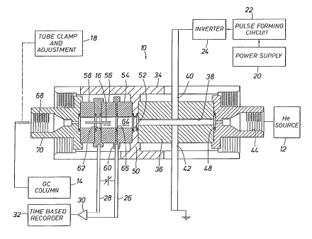
In Fig. l, a detector 10 uses helium from a helium source 12 regulated above
atmospheric pressure flowing from right to left. A GC column 14 provides flow
of solvent and
2 5 eluted sample. GC column 14 connects to a sample injection tube 16 moved
and clamped by an
adjustment mechanism 18 to a desired location. The power supply 20 provides
current for pulse
forming circuit 22. Inverter 24 forms alternating positive and negative
pulses. Conductors 26
and 28 are input to a differential amplifier 30 connected to time based
recorder 32.
The detector 10 has an elongate cylindrical shell 34 around an elongate
cylindrical
3 0 sleeve 36 about passage 38. 'fhe passage 38 is between electrodes 40 and
42.
The housing 34 supports fitting 44 connected with the helium source 12. Ring
48
4A
CA 02180266 2001-11-02
seals the body 36. Transverse web member 50 has a central opening 52 aligned
at cylindrical
spacers 54, 56 and 58. Circular electrode 60 forms a full circle around
passage 64. At the
surface of the passage 64, an exposed metal ring 66 connects to the circular
electrode 60. A
second circular electrode 62 is wider than the electrode 66. Sample tube 16 is
axially moved to
the left or right to vary current at electrometer 30. The sample tube 16 is
inserted through the
threaded detail 68 in the end fitting 70. The tube clamp and adjustment device
18 moves the
sample tube 16 in and out to vary sensitivity and performance. The terminals
62 and 66 have
an adjustable bias. Photon emission spectra through the passage 38 and 64
interact and charged
particles are either formed or neutralized depending on the sample material
creating current flow
at electrodes 62 and 66.
Helium (slightly above atmospheric pressure) flows at about 20 - 120
milliliters
per minute or between ten to thirty times larger than the flow from the tube
16. An elevated
temperature may keep samples in the volatile state. Spark duty cycle is in
Fig. 4. At 1000
pulses per second, a pulse is 10 microseconds or less.
FIGURE 2
An electron capture device (ECD) 110 has an elongate cylindrical housing 112
around cylindrical member 114 defining passage 116. Helium source 118 connects
to a fitting
detail 120 in a fitting 122. Spaced electrodes 124 and 126 terminate in
parallel end faces on
metal rods having a diameter of about 1/16" spaced approximately 1/16" across
the passage 116.
2 0 Smaller diameter of about 0.3 mm can be used. Larger electrodes having
sharpened points
transverse to the gas flow are permissible.
The passage 128 is defined by a spacer ring 130. Four similar rings are
separated
by three rings 132 with an exposed electrode ring 134. Rings 134 are first,
second and third
electrodes for operation of the ECD. The first ring has a negative 50 to 250
VDC and -100 VDC
2 5 is optimum. The next ring bias is about -5 VDC. The third ring is
permitted to float. The last
two rings input to an electrometer 136 to measure current output to a time
based recorder 138.
First and second injection tubes are concentric and move axially. Smaller tube
140
introduces a fixed flow of a trace gas 144. The second concentric tube 142
connects to the GC
column 148. The tubes I 40 and 142 are moved in LCD 110 and lock means 1 S0,
152 lock the
30 tubes at specified locations. Arrows indicate tube movement. Dopant gas and
GC gas effluent
are swept by the larger helium flow to the left past the electrometer
electrodes to form a signal.
4B
. __ _ 2180266
WO 95/18966 PCT/US95100046
FIGURE 3
A detector system 220 utilizes a carrier gas source 212
to provide helium and about 0.3% argon. The carrier gas inlet
opening 218 connects with right end cap 222 opposite the left end
cap 223. The end caps plug the tube 221.
Spark gap 230 is between opposing, parallel faces on two
electrodes 231 and 232 provided with a high voltage pulse. Sample
gas from a source 229 is injected into the tube 221 at a port 235
1 0 from a GC column or the like. Exposed metal rings 226 are spaced
along the tube 221 arranged serially downstream. Intermediate
rings 226 are tied to series resistors 233 for voltage drops. Ring
227 is connected to an electrometer 228.
Electrodes 226 are connected to series resistors 233. B+
supply 234 voltage (positive or negative) attracts the desired
charged particles. B+ voltage is pulsed and is controlled by a timer
216 and proportioned by resistors 233. The port 236 is aligned
with the port 218 which also is an observation port during the
spark. Photons impinge on an external spectrum analyzer 240
2 0 output to a recorder 241. Charging circuit 242 connects with a high
voltage discharge circuit 243 to provide a timed pulse for firing.
In FIG. 4, the top curve shows the charging pulse 244
for high voltage discharge circuit 243. That circuit forms an output
248, a pulse of short duration. Detection is delayed by a specified
2 5 time 252, and then a detection enable pulse 250 is formed.
Helium with a trace of argon flows into the spark gap
230 where ions and atoms are excited. Argon resonance lines are at
104.8 and 106.6 nm with corresponding energies of 11.62 and 11.83
eV. Excited argon (Ar*) from the spark gap 230 and sample
3 0 compound AB from the port 235 are mixed. Possible ionization
reactions are:
( 1 ) Ar* + AB = AB+ + e- + Ar
35 (2) Ar'~+AB=A+B++e-+Ar
5
WO 95118966 . ~ 218 0 2 6 6 pCTIU595100046
(3) Ar*+AB=AB*+Ar ,
where AB* = AB + h y
(4) Ar*+AB=A+B*+Ar ,
g where B * = B + h y
(5) Ar*--~ Ar + by (11.62,11.83 eV)
h y + AB --~ AB+ + e-
1 0 where e- denotes a free electron, * denotes an excited state, and h y
denotes spectral emission. Equation (3) and (4) reactions form
characteristic emission spectra signals for identification and
quantification. Equation (1) and (2) reactions produce free electrons
measured with electrometer 228, with the measured current
15 increasing with increasing concentration of compound AB.
Ar* radiation at 11.62 and 11.83 eV will not ionize any
compound with an ionization potential above 11.83 eV. Major
components of air are nitrogen (15.6 eV), oxygen, (12.08 eV), water
(12.6 eV), and carbon dioxide (13.8 eV). Air is not ionized and
20 impurities (pollutants) with ionization potentials below 11.83 eV are
ionized.
FIGURE 5
25 Ln monitoring for unwanted pollutants (BF3) in a plant
making N02, it is not possible to selectively ionize impurity BF3
without ionizing NOa. An atmospheric sample of air (nitrogen,
oxygen, water and carbon dioxide) may mask testing by emissions
from air constituents. Selective ionization of helium with less than
3 0 1.0% trace rare gas creates a relatively slow diffusing flux of
metastable helium which excites the dopant rare gases argon (Ar),
krypton (Kr), xenon (Xe), or neon (Ne). The helium-argon gas
emission has resonance lines at 104.8 and 106.6 nm. Argon emission '
therefore avoids ionizing air while ionizing impurities with ionization
3 5 potentials less than 11.8 eV. A helium-xenon gas has a resonance
6
WO 95I189fifi - ' - ' 218 0 2 6 6 p~~g95/00046
energy of 9.57 eV which selectively ionizes compounds with lower
ionization potential. Likewise, helium-krypton will produce
resonance energies of 10.64 and 10.03 eV. Helium-neon mixtures
will produce a resonance energy of 10.97. For a mixture of BF3 in
N O 2, helium-xenon gas is ideally suited in that the ionization
potential of NO~ is above the resonance of xenon yet the ionization
potential of BF3 is below. BF3 is selectively ionized while N02 is not
ionized.
Referring to FIG. 5, a pulsed capture detector (PCD) has
cylindrical housing 312 around cylindrical member 314. Passage
316 delivers helium from a source 318 through a valve 319 and
regulator 321 slightly above atmospheric pressure. The helium flow
is into manifold 323 threaded to a detail 320 in a fitting body 322.
Dopant Ne, Xe, Kr and Ar tanks 3 5 0 , 3 5 2 , 3 5 4 and 3 5 6 are
connected through valves 360, 362, 364 and 366 and pressure
regulators 370, 372, 374, and 376. Valve 319 and a selected
solenoid valve mix helium and rare gas Ne, Xe, Kr or Ar at the
manifold 323 which flows between the electrodes 324 and 326
across the gap 325 and exposed to the spark from the DC pulse
2 0 circuit 327.
The flow passage 316 connects downstream with a
larger axial hollow passage 328. Rings 334 and 335 are positioned
axially along passage 328. Ring 334 has a bias voltage and also
serves as a first terminal for the electrometer 336. The bias is
2 5 about -50 VDC to -400 VDC; and -200 VDC is illustrative. The ring
335 is the second terminal for the electrometer 336 to measure
current from the ionization of the trace compounds by the excited
dopant. Recorder 338 forms a record of the ionization current
measuring the trace compound. The injection tube 340 provides
3 0 sample gas supplied from the GC column 348. The injector tube 340
is coaxially centered within the exhaust passage 344 which connects
with passage 328 through a fitting 342 like the fitting 322. A
smaller fitting 346 is centered in the fitting 342.
Doped carrier gas flows from top to bottom while sample
3 5 gas from the GC column 348 enters through the injector tubes 340.
The sample and carrier gas (with dopant) commingle. Trace
7
CA 02180266 2001-02-15
compounds are ionized and electrometer 336 measures trace concentration. The
carrier gas flow
is substantially greater than the sample flow. The commingled and reacted
sample and carrier
gas is exhausted through the outlet 344.
Helium and the dopant flow into the PCD through fitting 320 into the spark gap
325 where ions and atoms in the excited state are formed. The dopant "D" is
energized and
excited to emit photons. Using argon as an example, emission forms resonance
lines at 104.8
and 106.6 nm with corresponding energies of 11.62 and 11.83 eV, respectively.
Helium
containing D* gas mixes with AB from the tube 340. D* emits the photon hyD in
proximity to
compound AB and reactions are:
(6) D* = D+hyD
(7) hyD + AB = AB+ + c_
(g) hYD+AB=A+B'+c-
(9) hyD + AB = AB'
where AB' = AB + by
(10) hyD+AB=A+B*
where B * = B + by
where hyD denotes photon emission of excited dopant D*. (9) and (10) reactions
form specific
and characteristic emission spectra, thereby enabling identification and
quantification. Equations
(7) and (8) describe reactions which produce free electrons measured with
electrometer 336
where electron current measures concentration of compound AB.
The present invention selects the dopant D thereby allowing selected
ionization of
components of the sample gas. If D = Ar and D* = Ar*, then Ar* radiation is h
y Ar = 11.62 and
11.83 eV and will not ionize any compound with an ionization potential above
11.83 eV. Air
is not ionized by the Ar* source while air pollutants with ionization
potentials below 11.83 eV
are ionized. One example comprises air with an impurity such as carbon
tetrachloride (CC14).
8
CA 02180266 2001-02-15
In another example, NOZ has impurity of BF;. If D = Xe, Xe exhibits a
resonance energy at 9.57
eV. The ionization potential of NOZ is 9.75 eV which is above the resonance
energy of Xe
while the ionization potential of BF3 is 9.25 eV which is below the resonance
of Xe. BF; in the
NOZ is selectively ionized while NO~ is not ionized. The electrometer 336
measures trace
concentrations of BF3. Ar, Kr and Ne are not suitable dopants since the
resonance energies are
greater than the ionization potential of NOz; therefore the NOZ as well as the
BF3 would be
ionized by these dopants.
In the passage 328, the radiation from the excited dopant is absorbed by the
analyte
and those components with ionization potentials less than the resonance energy
of the selected
dopant are current detected by the collecting electrode 335 and measured by
the electrometer 336.
Fig. 6 shows selected ionization concepts where the axis 380 represents dopant
emission radiation hyD in electron volts (eV). The line 382 locate the Ar
emissions at 11.62 and
11.83 eV. The line 386 represents the 10.97 eV emission from Ne and the line
388 represents
the 9.57 eV emission from Xe. Finally, emissions 384 are 10.03 and 10.64 from
Kr. Ionization
potentials are depicted on the axis 390. The line 392, 394, 396 and 398
represent the ionization
potentials of air constituents O, HZO, CO, and N, respectively. The ionization
potential 393 of
CCl4 is 11.47 eV. NO, and BF; potentials are 395 and 397, respectively.
For dopant emission photon hyD, any element or compound which is on the high
energy side of hyD (that is, to the right of the emission line in Fig. 6) is
ionized while any
element or compound which falls to the low energy side of hyD (that is, on the
left of the
emission line) will not be ionized. Dopant gases are selected based upon two
criteria which are
( 1 ) the ionization potential of the compound to be measured and (2) the
ionization potentials of
other constituents not measured which generate "noise" in the measure of the
compound of
interest.
c)
WO 95/18966 218 0 2 6 6 p~~g95ID0046
In operation, selected dopants are introduced into the
carrier gas by the solenoid valve from the reservoir of the selected
dopant gas. If Xe is the dopant, solenoid valve 362 allows Xenon
from the reservoir 352 to flow through the pressure regulator 372 _
to the manifold 323.
FIGURE 7
Four detector chambers 451, 453, 455 and 457 receive
GC column 448 flow from the GC conduit 472 to a valve 470 which
"splits" the flow into four parts. Conduits 440 connects to four
ionization detectors chambers 451, 453, 455 and 457. Four
different carrier gas sources 450, 452, 454 and 456 flow into the
detector chambers. Gas constituents are excited and commingled
with the sample gas splits. The excited carrier gases ionize the
sample, generating an ionization current. Mixtures of carrier and
sample gas are vented from each chamber through a port 444.
Ionization currents generated at chambers 451, 453, 455 and 457
are transferred to the computer 460. Measurements processed at
the computer 460 yield identity and concentrations of the sample
2 0 gas. Results from the computer go to a recorder 438. The number
of detectors can be varied. In analyzing a large number of different
compounds, accuracy and precision may be maximized by using
more detectors.
2 5 FIGURE 8
The pulsed discharge photoionization capture detector
(PDPID) has a long cylindrical housing 512 which contains a
cylindrical member 514 which is axially hollow at 516. The helium
source 518 flows through a valve 519 and regulator 521 to deliver
3 0 helium at a pressure slightly above atmospheric. Manifold 523 via
fitting 520 connects to a fitting 522 at the body 512 of the PDPID.
Reservoir 566 is connected through valve 564 and pressure
regulator 562 to the manifold 523. By opening valves 519 and ,
564, helium and dopant gas flow to the manifold 523 and into the
3 5 axial passage 516 and between the electrodes 524 and 526. ,
CA 02180266 2001-02-15
The electrodes 524 and 526 are about 1/16" with spaced end faces approximately
1/16" across passage 516. Electrodes 524 and 526 are electrically insulated
from the PDPID.
The electrode 526 is grounded while the electrode 524 is provided with a high
voltage pulse of
short duration by the DC source 527. The two terminals 524 and 526 form a
sharply fixed,
narrowly constrained spark so that the spark does not dance around the two
electrode faces and
remains a straight line.
Carrier gas is introduced into the PDPID from top to bottom. Sample gas from
the GC column 548 enters the passage 528 through the injector tube 540 so that
sample and
carrier gas excited by the spark commingle. Compounds are ionized producing a
response across
the exposed rings 534 and 535 input to the electrometer 536 indicative of the
sample and
concentration. After commingling and reacting, the mixture of sample and
carrier gas is swept
from the passage 528 of the PDPID and exhausted through the outlet 544. The
outlet is
supported in the fitting 546 in the end cap 542. The GC gas flow input is the
tube 535.
Helium and a dopant gas flows into the PDPID through fitting 520 and into the
spark gap 525 where ions and atoms are in the excited state. Dopant "D" is
energized and
excited. The excited dopant passes from the spark gap 525 through passage 516
into the passage
528 of the PDPID. Dopant D in the excited state emits photons. Using argon as
an example
dopant, emission resonance lines at 104.8 and 106.6 nm have energies of 11.83
and 11.62 eV,
respectively. By mixing dopant D with helium and exciting the gas at the gap
525, excited
i1
CA 02180266 2001-02-15
dopant D* is created. D* decays within approximately 5 microseconds after
excitation. Some
photons from decay pass through channel 516 into channel 528. Sample AB is
injected into the
channel 528 and exposed to photons hyD resulting from the decay of D*. Flow of
carrier and
sample gas is from top to bottom to the outlet 544. Reactions are exemplified
in Equations ( 1 )
to ( 10) above.
Table I summarizes emission spectra from helium, argon and krypton doped
helium. Other gas mixtures can be effectively used and the data primarily
support the examples
presented.
TABLE 1
EMISSION SPECTRA FROM HELIUM AND ARGON AND KRYPTON DOPED HELII1M
ACTIVE WAVELENGTH ENERGY
SPECIES (nm) (eV)
He 3gg
Hey 70 - 90 13.5 - 17.7
Ar 104.8 11.83
Ar 106.6 11.62
Kr I 16.5 10.64
123.6 10.03
Ar, 121 - 133.6 9.28 - 10.24
Kr~ 139.7 - 152.8 8.11 - 8.87
The sample gas may be split and passed through multiple detectors.
Electrometer output current with helium as a carrier gas, C,ie, is measured
and stored within the
computer 560. The electrometer outputs C,,~ + A~ and C,ie + k~. from the
second and third
detectors, respectively, are measured simultaneously and likewise stored
within the computer 560.
12
2180266
WO 95/18966 PCTIUS95/00046
The ratios
( 1 I ) R'Ar = CHe+Ar / CHe
and
( I 2 ) R'Kr = CHe+Kr / CHe
are computed. The system is first "calibrated" by measuring the
ratios R'Ar and R'Kr using a calibration gas comprising a known
amount of benzene. All other constituents exhibit ionization
potentials above the highest emission level of the carrier gas and,
therefore, do not contribute to the electrometer current readings of
the detectors. The ratios defined in equations (11) and (12) for
benzene gas are R"Ar and R"Kr, respectively. Ratios measured using
the unknown sample, normalized to a corresponding reading for
benzene of 100, are computed from the equations
( 13 ) RAr = 100 (R'Ar/R"Ar)
and
(I4) RKr = 100 (R'Kr/R"Kr)
Table 2 lists normalized ratios RKr and RAr for selected
2 5 compounds. The tabulation is presented for illustration only. If an
unknown sample gas RAr is measured at 77.8 +/- 0.8, the designated
uncertainty is attributed to random errors. In Table 2, the
compounds C3H7N02 (RAr = 78.3) and CH3CH0 (RAr = 77.9) and 1-
pentene (RAr = 77.6) all fall within the uncertainty of +/- 0.8. With
3 0 only two detectors, the unknown compound could not be uniquely
identified from ionization detection measurements. Assume that RKr
is 37.4 +/- 0.4. From Table 2, only 1-pentene is within the range of
values of RAr and RKr since the tabulated values of RKr for C3H~N02
and CH3CH0 are 0.74 and 43.4, respectively. The unknown
3 5 compound is, therefore, identified as I-pentene. The concentration
13
2180266
WO 95/18966 PCTJU595/00046
of 1-pentene is from CAr or Cgr standardized with a calibration gas
containing 1-pentene.
Computations are performed in real time with the
computer 560. The identification analysis is depicted graphically in ,
Fig. 9A. RAr is plotted on the axis 584 and Rgr is plotted on the axis
582. Corresponding "coordinates" for 1-pentene, C3H~N0~ and
CH3CH0, with expected systematic uncertainties for each value, are
taken from Table 2 and depicted as circles 572, 574 and 570,
respectively. Should RAr and Rgr plot within any circle of
uncertainty, the unknown compound is thereby identified. In the
previously discussed example, the measured values of RAr and Rgr
plot within the circle 572 and therefore the unknown compound is
identified as 1-pentene.
TABLE 2
NORMALI~D RESPONSE
RATIOS Rpr AND R~
FOR SELECTED
COMPOUNDS
COMPOUND RAr
CS2 204.0 38.3
1-hexene 81.7 41.8
C3H7N02 78.3 0.74
CH3CH0 77.9 43.4
1-pentene 77.6 37.4
2-methyl-1- 76.0 35.3
pentene
heptane 76.0 4.58
1-butene 70.5 24.3
butane 62.4 1.13
n-C3H70H 60.9 10.2
As a second example, assume that RAr is measured to be
2 0 76.8 +/- 1.0 and Rgr is measured to be 36.0 +/- 2Ø The illustrative
uncertainties are greater that usual. From Table 2, it is not possible
to define uniquely the unknown compound as 1-pentene or 2-
methyl-1-pentene since both fall within the uncertainty ranges. An
additional detector with gas dopant helps so that the normalized
14
CA 02180266 2001-02-15
ratio from this detector, denoted as "RX", delineates between the two
compounds in question.
The data using four detectors (which yields three ratios) is depicted
graphically in Fig. 9B
Coordinates representing 1-pentene and 2-methyl-1-pentene, with spheres
representing the
systematic uncertainty of the system, are depicted as 592 and 590,
respectively. Rh~ and RA~ are
plotted along the axes denoted by the numerals 582 and 584, respectively. The
ratio from the
additional detector, RX, is plotted along the axis denoted by 586 and is in
arbitrary units.
Hypothetical values for Rx 1-pentene and 2-methyl-1-pentene, (for purposes of
illustration), are
denoted by the numerals 596 and 595, respectively. Should values of RA~, R,~~
and RX for an
unknown plot within the sphere of uncertainty for either compound, the unknown
compound is
identified. The graphical interpretation is presented only for purposes of
illustration and is easily
adapted for computer interpretation.
FIGURES 10, 11 AND 12
The circular detection system 620 utilizes a carrier gas source 612 connected
to
the detector valve 613. The circular detector 620 in a representative GC
system utilizes a sample
source 611 connected with the loading valve 613. They provide a carrier gas
flow to a GC
column 615. System timer 616 controls operation. Compounds supplied with the
flowing carrier
gas flow through the valve 613 to the GC column 615. 'There is a tangential
inlet port 618 to
the detector interior to sustain rotational motion and discharge through a
vent port 619. The
collecting electrode terminal 621 is connected to the electrometer 628. The
terminal 621
connects with one ring electrode while the terminal 622 connects with a bias
electrode. A B+
supply 634 provides power. One output from the B+ supply 634 is to the timer
616 and then to
a charging circuit 642. The charging circuit operates with a high voltage
discharge circuit 643
to form an output pulse having a controlled polarity, controlled width and
specified current flow.
This is input at a first terminal 624 opposite a ground terminal 625. The
terminals 624 and 625
provide the DC spark in the detector 620. One of the two terminals is hollow
for delivery of
helium from a helium source 626.
~J
WO 95/18966 218 0 2 6 6 PC1'1US95/00046
A window 627 passes light to be emitted from the spark,
and observed by a spectrum analyzer 640. The analyzer 640
provides an output signal to the recorder 641. Helium is delivered
at the center of the detector 620 through the hollow electrode 624
from the reservoir 626. Dopant may be optionally introduced from
the reservoir 626' into the helium flow.
The detector housing 620 has two cylindrical shell
portions. One shell portion 629 incorporates a circular protruding
lip which enables the shell half 629 to join with a second shell
portion 631. The shell portions 629 and 631 join with an
overlapping lip arrangement so that a chamber 632 is formed. The
collecting electrode 621 is connected to a ring 633 while the similar
ring 635 is the bias electrode. The housing portions 629 and 631
are formed of a material which is not an electrical conductor. In Fig.
12, the shell portion 629 is provided with a tangentially located
inlet passage 618 to introduce gas flow at the interior tangential
edge of the cylindrical chamber. The port 619 is a vent located
radially inwardly.
2 0 FIGURE 13
The numeral 710 identifies the gas sampling apparatus
formed of an insulating material. The body 710 is divided into two
chambers by the partition or "window" 740 forming the upper spark
chamber 712 which is leak proof to the surrounding atmosphere
2 5 and a lower sample chamber. Two round and equal diameter
electrodes 714 and 716 protrude inwardly from the body 710 of
the detector. The spark gap 715 within the spark chamber 712 has
an insulating material at the faces of the electrodes 714 and 716
sufficiently thick to physically isolate the electrodes from the
3 0 environs of the interior of spark chamber 712 yet sufficiently thin
to allow the generation of a pulsed DC spark across the spark gap
715. Electrode 716 is electrically connected to B+voltage power
supply 720 while the electrode 714 is grounded at 722. The
voltage applied to the electrode pair is timed by a clock 738. The
3 5 spark chamber 712 is filled with helium and a trace of krypton.
16
2180266
WO 95118966 PCTIUS95100046
Sample gas enters the sample chamber through a port
726 and exits the chamber through the port 728. A small pump
delivers sample gas. The sample chamber contains circular
electrodes 730 and 732 recessed within the chamber walls and
exposed to the interior of the chamber. Electrode 732 is grounded
at 734. The electrode 730 is connected to an amplifier 737 and
then to the recording device 736. A clock 738 controls the applied
positive or negative voltage and times the recorder. The electrode
732 has the requisite voltage to attract desired charged particles
within sample chamber. The window 740 separating the spark
chamber 712 and the sample chamber is a thin membrane of
magnesium fluoride (MgF2) or lithium fluoride (LiF). The material
and dimensions are selected so that photoemissions at the desired
energy levels experience minimal absorption entering into the
sample chamber. The discharge heats the gas in the spark
gap 715. Heated relatively buoyant gas in the spark path rises in
the closed spark chamber 712 where it is cooled by mingling with
cooler gas. Simultaneously, cooler gas replaces the heated gas at the
spark gap 715. The net result is circulation within the closed spark
2 0 chamber 712 as depicted by the broken lines 718. Convective
circulation constantly supplies "fresh" gas to the spark gap 715.
Krypton in the excited state emits photons at 116.5 and
123.6 nanometers (nm) with corresponding energies of 10.03 and
10.64 electron volts (eV), respectively. This radiation passes
through the window membrane 740 and into the sample chamber
where it interacts with the sample gas. Each spark creates a fresh
supply of Kr* which, in turn, decays to the ground state by the
emission of 10.03 eV and 10.64 eV photons. The spark generation
system in cooperation with the helium-krypton gas mixture acts as a
3 0 self replenishing source of 10.03 eV and 10.64 eV radiation.
Sample flow is preferably continuous although discrete
samples may be taken. In air monitoring, small concentrations of
pollutant compounds AB and air are exposed to the photon flux of
energies 10.03 and 10.64 eV from the spark chamber 712 through
3 5 window membrane 740. This photon flux ionizes the compound AB.
Free electrons are collected at the electrode 730 which is at a
17
2180266
W0 95118966 PCT/US95100046
positive potential. Electrode 732 is at ground to retard ionic
recombination and to repel electrons. The free electron current from
the electrode 730 is recorded by the recorder 736 with the current
proportional to the concentration of AB. Electron current is,
S therefore, an analytical measure of concentration.
Recall that Kr* emits radiation at 10.03 and 10.64 eV.
This radiation will not ionize any compound with an ionization
potential above 10.64 eV. Major constituents of air are not ionized
by the emissions from Kr*, but impurities in the air sample
(pollutants with ionization potentials below 10.64 eV) will be
ionized.
While the foregoing describes the embodiments of the
present invention, the scope is determined by the claims.
18