Note: Descriptions are shown in the official language in which they were submitted.
CA 02180575 2004-08-06
1
BONE MARROW BIOPSY NEEDLE
FIELD OF THE INVENTION
This invention relates generally to a surgical instrument, known variously
as a biopsy needle or cannula that is used to gather tissue, such as bone
narrow, from living persons or animals for pathological study. More
specifically,
the invention relates to a biopsy needle having an improved structure for
severing a tissue sample and/or retaining the tissue sample within the needle.
BACKGROUND OF THE INVENTION
For various medical reasons, such as diagnostic tests or the
determination of suitability as a tissue donor, it is often necessary for a
physician
to obtain a sample of a patient's body tissue. In particular, bone marrow is
frequently retrieved for later pathological study. The current procedures and
instruments used for obtaining the samples, while not overly complex, almost
universally result in excessive patient discomfort and often overly extend the
patient's and operator's time, money and effort. In the standard bone marrow
procurement protocol, using currently standard instruments, (such as those
disclosed in U.S. Patent No. 4,262,676 to Khosrow Jamshidi), the patient is
prepared with a suitable local anesthetic at the appropriate marrow retrieval
site.
Then, a relatively narrow needle is inserted to obtain an aspirate of only
liquid
b.,r... ....,.........~. w,.,+.~ri.,l fr,r w,.,Linn ~IiiJoc fnr cv~min~~inn
~ffcr
WO 95/18568 ~~~~~ " PCT/US95/00662
2
staining. This portion of the procedure, referred to as the
bone marrow aspiration, is relatively leas painful than obtain-
ing a bone marrow biopsy.
After the aspirate is obtained and-the sl-ides and
specimens are prepared, if necessary, a biopsy of the fibrous
bone marrow is taken. A significantly wider bore needle having ,
an inner diameter that will house a suitable marrow sample is
first prepared with an inner stylet that extends beyond the
distal end of the outer needle. The atylet distal end may be
cut at an angle, with the leading edge sufficiently sharp to
pierce tissue and bone. With-the stylet in place within the
outer needle, the needle is pushed through the outer layers of
flesh until the bone is felt at the tip. The needle and stylet
are then pushed into the bone approximately 4 or 5 millimeters
until the needle appears to be solidly within the bone.
The stylet is then removed froiri the proximal end of
the needle, which opens up the core of the needle to the now-
surrounding marrow tissue. The outer needle is then usually
advanced another 1 to 2 centimeters at minimum with-a slight
twisting motion. Often, the distal end of the outer--needle
will also be provided with an angled cut and sharpened leading
edge to cut and core the tissue easily . By providing a alight
twisting motion as the needle is advanced, usually with no more
than quarter or half turns, an appropriate sample is cored from
the marrow tissue and enters the inner passage of the marrow
needle.
At this point, the marrow biopsy sample piece is
ready to be removed from the patient, although it is important
that the biopsy piece remain within the needle as the needle is
withdrawn. If the biopsy piece becomes dislodged and falls out
the distal end of the biopsy needle, the piece is irretrievably
lost. The procedure is then unsuccessful and must be repeated
from the beginning. -
various methods have been attempted by physicians to
prevent the biopsy piece from dislodging from the outer needle. '
~vo 9snss6s '~ I 8 d ~ 7
3
For example, some physicians, after the needle has entered the
bone fully and cored a sample from the marrow, will pull the
biopsy needle back a few millimeters and then forward a few
millimeters at a different angle than the first insertion.
This theoretically will "cut" the biopsy piece at the tip of
the needle. Other physicians attemptto dislodge or disrupt
the connection between the biopsy piece and the bone by making
multiple complete clockwise and counterclockwise rotations of
the biopsy needles while within the bone. Some physicians even
hit the proximal end of the biopsy needle at its handle in an
attempt to mechanically disrupt the connection between the core
biopsy specimen and the additional bone.
As can be plainly realized, these manipulations at
the end of the procedure, attempts at ensuring that the biopsy
piece remains within the needle, can often produce substantial
discomfort and anxiety. Sometimes when the bone marrow is very
soft, as in patients with osteoporosis, almost a11- of these at-
tempts are futile because the bone structure is so fragile.
Conversely, sometimes when the bone marrow is very fibrotic,
which occurs in patients with myelofibrotic diseases or in AIDS
patients, it is difficult to dislodge the core biopsy piece,
since the bone marrow itself is reinforced by surrounding
tissue. In those cases, the cored biopsy piece often remains
attached to the bone and is not removed in the biopsy process.
Other attempts at designing a more efficient and
successful biopsy needle have met with little or no success,
forvarious reasons, including the complexity of the devices.
For example, U.S. Patent No. 3,605,721 to Hallac, discloses a
biopsy needle in which an inner tube has a weakened portion -
represented by strips extending between distal and proximal
portions of the inner tube. The distal portion of the inner
tube is adhered to an outer tube and will not rotate. Once a
biopsy piece has entered the needle, the proximal portion of
the inner tube is rotated, causing the strips to twist together
~ 35 and eventually break off. This twisting motion tends to twist
W095I18568 ~~ ,'s ; PCT/US95I00662
4
the strips to the tube's center, thus hopefully keeping the
biopsy piece proximal of the twisted and broken strips for
later removal. This particular biopsy needle is only a dispos-
able device, since the strips are broken or irreversibly warped ,
by plastic deformation during the twisting process. Another
disadvantage is the lack of control over the twisting motions
or the breakage of the strips Essentially, the surgeon is-.
left to twist the inner tube until resistance to that twisting
is lost, indicating that the strips have severed. There is
,also no way of releasing the device's grip on tissue during
surgery, should any problems arise.
U.S. Patent No. 5,074,311 to Hasson discloses a
biopsy device that includes a pair of inner jaws that can be
actuated within the outer needle to "bite off" any biopsy piece
that has entered the outer needle. The disadvantages of this
device include multiple small-mechanical linkages and parts
including pivot pins, which are extremely difficult and expen-
sive to assemble and maintain, in-addition to the greatly in-
creased chance of mechanical failure which can be costly during
a surgical procedure.
crTMNLAR'l OF THE INVENTION
In view of the deficiencies noted in the known
devices and the current protocols, it is an object of the
present invention to provide an improved biopsy needle that
will sever-a tissue sample from surrounding tissue or hold it
with sufficient force such that the action of removing the
needle detaches the piece from the surrounding tissue.
It is another object of the invention to provide a
biopsy needle that reguires minimal manipulation of. the needle
at the end of the procedure, thus decreasing patientpain and
anxiety.
It is a further object of the invention to ensure ob-
taining a biopsy sample with each attempt, thus decreasing the '
number of necessary biopsy attempts, and the time, effort and
money expended on the overall procedure. '
CA 02180575 2004-08-06
It is yet another object of the invention to provide a biopsy needle
that is simple and inexpensive to manufacture, may be reusable, and is simple
to operate.
According to the present invention, there is provided a biopsy
needle for removal of tissue from a patient, comprising:
an outer tube having a proximal and a distal end;
an inner tube within said outer tube,
said inner tube having a proximal and a distal end;
a snare having two ends, one of said ends connected to said inner
tube and the other of said ends coupled to said outer tube, said snare has a
first
diameter and wherein in said second position, said snare has a second diameter
smaller than said first diameter, said snare being moved form said first
position
to said second position by rotation of said inner tube with respect to said
outer
tube in one direction and being moved from said second position to said first
position by rotation in an apposite direction.
According to the present invention, there is also provided a
reusable biopsy needle, comprising:
an outer tube having a distal end;
an inner tube extending within said outer tube, said inner tube
having at one end a portion which increases and decreases in diameter
substantially free of plastic deformation with rotation of said inner tube
relative to
said outer tube, said portion of said inner tube being coupled to said distal
end
of said outer tube.
According to the present invention, there is also provided a biopsy
needle, comprising:
an outer tube having a distal end;
an inner tube extending within said outer tube, said inner tube
having at one end a portion which increases and decreases in diameter
substantially free of plastic deformation with rotation of said inner tube
relative to
said outer tube, said portion of said inner tube being coupled to said distal
end
of said outer tube.
CA 02180575 2003-07-28
5a
Preferably, according to one embodiment of the
invention, an improved biopsy needle has an outer cannula,
an inner tube and a stylet. The distal end of the inner
tube is provided with a snare in the form of a coil
extending from the inner tube. The free end of the coil is
adhered to the inner surface of the outer cannula. Upon
rotation of the inner tube with respect to the outer
cannula, the coil will decrease in diameter to either sever
or hold the biopsy piece within the outer needle. After
removal of the needle from the patient, rotating the inner
tube in the opposite direction will cause the coil to
expand to its original diameter and allow the biopsy piece
to be removed from the needle.
Preferably, in another embodiment of the present
invention, a cylindrical member, integral with or securely
attached to the free end of the coil, is secured to the
inner surface of the outer cannula. Upon rotation of the
inner tube with respect to the outer cannula, the coil will
decrease in diameter with the cylindrical member providing
enhanced support for the coil.
BRIEF DESCRIPTION OF THE DRAWINGS
Other Qbjects, advantages and embodiments than those
described above will become apparent to those skilled in the
art upon reading the following detailed description of the
preferred embodiments in conjunction with a.review of the
appended drawings, in which:
Fig. 1 is a perspective view of a biopsy needle in accor-
dance with a first embodiment of the present inven-
tion;
Fig. 2 is an exploded view, with parts shown in section, of
the biopsy needle of Fig. 1;,
Figs. 3a-3e are detail perspective views on an enlarged
scale, of the distal end of the biopsy needle illustrating the
W0 95118568 ,,~ PCT/US95100662
218~~'~5
6
function of various components during operation of the biopsy
needle;
Fig. 4 is a cross-sectional view of the distal end of the
outer cannula;
Fig. 5 is a perspective view of the biopsy needle showing
operation by a physician;
Fig. 6 is a aide view of the inner tube of the present
invention;
Fig. 7 is a cross-section view through the handle piece of
the biopsy needle;
Fig. 8 is a perspective view of the distal end of a second
embodiment of the present invention;
Fig. 9 is an end view of the distal end of the second em-
bodiment;
Fig. 10 is a left side view of the outer cannula, the
right side being a mirror image thereof; and
Fig. 11 is an exploded view of the distal end of the second
embodiment.
DE'T'ArrFD DESCRTPTrpN pF THE PREFERRED EMBODIMENTS
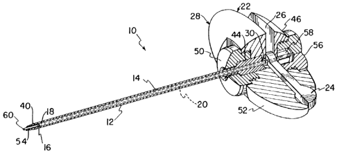
Referring now to Figs. 1 and 2, biopsy needle 10 has
an outercannula 12, an inner tube 14 with a cylindrical or
helical snare 16 at its distal-end 18, a stylet 20, and a
handle assembly 22. In Fig. 2, the assembly of thepreaent
biopsy needle 10 is shown in an exploded view.
As part of the handle assembly 22, a lever 24 is
mounted for rotation in a corresponding groove 26 within a
handle piece 28. Upon rotation, the lever 24 actuates the
snare 16 within the outer cannula 12 without any movement of
the outer cannula 12 relative to the patient (not shown). The
operation of this lever 24 is described more fully below. The
inner tube 14has a snare 16 at its distal end 18 and a gear 30
is mounted on its proximal end 32. The inner tube 14 is
inserted into the proximal end 34 of the outer cannula i2 so
that the gear 30 protrudes from the proximal end 34. AS can be
seen in Fig. 4, the interior of the outer cannula 12 has a
WO 95118568 PCT/U595/00662
P ~'ø
constant inner diameter A along-most of its length, with a
portion 38 having a smaller inner diameter B at its distal tip
40.
Preferably, the narrow inner diameter B at the distal
tip 40 is substantially equal to the inner diameter C (Fig. 3c)
of the inner tube i4 so that there will be no ridge or lip
within the instrument to impede tissue entering the instrument.
The inner tube 14 is inserted in cannula 12 until the snare 16
reaches the shoulder 42 provided on the interior of the outer
cannula 12 at the position where the diameter changes (see
Figs. 3b and 3c).
As best seen in Fig. 1, with the gear 30 extending
proximal of the cannula's anchor 44, the cannula and snare
assembly are attached to-the handle piece 28 at the pro:cimal
facing side 46 of the handle 22. The gear 30 of the snare 16
is inserted into a complementary hole 48 in the leverwhile the
anchor 44 of the outer cannula 12 mates with a complementary
hole 49 in the handle piece 28. Thus, when the lever 2~1 is
rotated within its groove 26 with respect to the handle piece
28, the inner tube 14 will rotate with respect to the outer
cannula 12. A cannula cap-50 is assembled onto the distal tip
40 of the cannula and threadedly engaged to the forward facing
end 52 of the handle piece 28. 'I'he stylet 20 is inserted into
the proximal end 32 of the inner tube until a distal tip
portion54 of the stylet extends beyond the distal tip 40 of -
the cannula. A stylet cap 56 can then be threadedly engaged to
the proximal facing side 46 of the handle piece, covering the
proximal end 58 of -the stylet to prevent it from moving proxi-
mally within the inner tube 14.
As can be seen in Fig. 3a, both the distal ends 40,
54 of the stylet 20 and the outer cannula preferably have
sloped end faces 60, 62 although it is not necessary. This
improves the cutting action of the both the stylet and the
outer cannula by providing sharp leading edges 64. In this
position, the stop 66 at the proximal end 58 of the stylet
W095/18568 , PCTIUS95/00662
8
preferably mates with a complementary indent 68 in the handle
piece 28 to maintain the rotational orientation of the stylet
20 with respect to the outer cannula i2 such that the slopes of
the two distal ends 40, 54 are approximately parallel. This is
the configuration that would be used for inserting the biopsy
needle 10 into the patient and through the bone into the softer
bone marrow tissue within.
Aa can be seen in Fig_ 3b, which is a partial cutaway
view, the free end 70 of the coil-snare 16 includes a tab 72
that engages or isattached to a hole 74 (Fig. 4) on-the
interior surface of the outer cannula 12. This hole 74 prefer-
ably extends through the entire wall of the outer cannula. If
desired, the tab 72 can be adhered to the hole 74 in the outer
cannula through the use of adhesives, welding, or any known
attachment process. After the needle 10 is inserted into the
marrow, the stylet 20 is withdrawn proximally without any move-
ment of the outer cannula 12 with respect to the patient,
minimizing discomfort. As can be seen iri Fig. 3c, marrow
tissue may now enter the passageway within the outer cannula 12
through the distal end 40 of the outer cannula and can enter
the inner passageway of the inner tube 14, preferably to a
position proximal ofthe snare 16.
To operate the snare- 16, i.e. to cause cutting and/or -
holding of the biopsy piece 76 within the inner tube 14, the
lever 24 attached to the proximal end 32 of the inner tube is
rotated in the direction of arrow D as seen in .Figs: 3d-3e. Of
course, the snare 16 can be designed such that rotation in the
opposite direction causes the same effect. With full rotation
(180°) of the lever 24, the inner tube 14 and snare 16 achieve
a position similar to that shown in Fig. 3e, in which the inner
tube 14 has been rotated approximately 180°. Since the free
end 70 of the snare is fixed--to the outer cannula 12, the
result of the rotation is that the coil of the snare ~.6 will
tighten so that the cross-sectional area through the snare 16
is approximately lees than-a third of the area when in the open
fl'O 95!18568 ~ 8 ~ ~ ~ ~ PCTIUS95/00662
9
configuration. It is also contemplated that any decrease, even
a slight decrease, in the cross-sectional area of the snare
will cause pressure on the biopsy-piece 76. Therefore, while
the current amount of rotation is preferred, it is not neces-
sary for the proper functioning of the present invention.
As seen in Fig. 5, movement of the lever 24 can be
independent of any movement of the handle piece 28 or the outer
cannula 12. Therefore, the outer cannula 12, which is in
direct contact with the patient while the sample is taken, can
remain substantially stationary. There is little or no discom-
fort at this step of the procedure, where previously this had
been one of the more uncomfortable steps.
With the tightening of the snare 16, there is a high
probability that the biopsy piece 76 will remain in the needle
10 as the needle is removed. If the tightening of the snare 16
does not immediately cause the biopsy piece 76 to be cut, it
will be significantly squeezed and/or notched, such that
rearward motion of the needle 10, which causes rearward prea-
aure on any biopsy piece 76 proximal of the snare 16, will
cause material proximal of the snare 16 to detach from material
that is distal ofthe 'snare.
As can be seen in Fig. 7, the handle 22 includes
several features designed for ease of use of the physician and
ease of manufacture and construction. The handle piece 28 in-
eludes a groove 26 that receives the lever 24 while permitting
its rotation. The groove 26 has two notches 78 that generally
protect the lever 24 from any accidental contact with the
physician when in either the full-open or full-closed posi-
tions, but allow access to the lever. Further, the holes in
the handle piece 28 that receive the anchor 44 of the outer
cannula and the atop 66 of the atylet have shapes that are
complementary to the anchor or stop, in order to prevent
rotation of those two components with respect to the handle, as
previously discussed. The proximal and distal facing sides 46,
WO 95118568 ~ PCTIUS95100662
52 of the handle piece are also provided with threaded regions
for receiving the cannula and atylet cape 50, 56.
Once the biopsy needle 10 has been used and the cap
tured material has been ejected through either the proximal or
5 distal ends of the inner tube, the biopsy needle 10 is then
ready to be sterilized for=its next use. 1f necessary, the
entire biopsy needle can be disassembled, although the tab 72
at the free end of the snare must be disengaged from the hole
74 in the outer cannula. This can be accomplished With any
10 small tool pushed through hole 74. If the free end 70 of the
snare is permanently adhered to the outer cannula 12, it then
may be necessary to sterilize the outer-cannula and inner tube
as a single unit. However; due to the small number of parts
and relative ease and low cost construction of the present
needle, it is also contemplated that such a device is easily
disposable.
Thus, it can be seen that a low cost, simply-manu-
factured biopsy needle will attain improved results-over known
devices, not only in the success rate of the marrow extraction
procedures, but also -a marked increase in patient comfort
throughout the procedure. One desirable side benefit of this
increased comfort might be increased participation in bone
marrow donor programs for transplant candidates.
Figure 8 illustrates another embodiment of the
present invention. Referring to Figs. 8 and 11, the coil snare
16 is provided with a cylindrical member 80 at its distal end.
Although it is preferred that the cylindrical member be inte-
gral with the free end of the coil snare 16; the cylindrical
member 80 may alternatively be securely attached to the free
end of the coil snare 16.
The cylindrical-member 80 is provided with two
equidistantly spaced raised rectangular members 82 positioned
about the circumference of .the cylindrical member 80. As more
clearly shown inFig. 9, each rectangular member 82 includes a
tab 84 which engages or is secured in a hole 86 on the interior
'W095118568 ~ PCTlUS95I00662
11
surface of the outer cannula 88 (Fig. 10). Each hole 86 is
sized and positioned to accept a tab 84 that preferably extends
through the entire.wall-of the outer cannula 12. Each tab 84
may be secured to outer cannula 88 in the same manner as
described in connection with tab 72 of Fig. 3b. The cylindri-
cal member 80 provides increased strength to the free end of -
the coil snare 16 without changing the manner in which the
present invention operates.
Referring again to Figs. 8 and 11, the-coil snare 16
is illustrated as a member separate from, but connected to the
inner tube 14 rather than integral with the inner tube 14 as
shown in Figa. 3a-3e. Fig. 11 shows an exploded view of the
distal end of the inner tube 14 and coil snare 16 clearly
illustrating the manner in which they are connected. The
distal end of inner tube 14 includes three equidistantly spaced -
curved recesses 110 for matingly receiving three equidistantly
spaced curved male portions 112 integral with the coil snare
16. Formed in this manner, the inner tube 14 and the coil -
snare 16 may be made of two different materials, with the inner
tube 14 preferably-made of a rigid material while the coil
snare 16 is preferably made of a flexible plastic material and
then the coil snare 16 may be permanently adhered to the inner
tube 14 using an adhesive.
Also 111ustrated in Fig. 11 is an exploded view of
the coil snare 16. The coil snare 16 comprises three prefers-
bly integral portions, the cylindrical portion 80, a helical
portion 114 (shown in its actuated or reduced diameter condi-
tion) and a base portion 116 which carries the male portions
112. As is evident from the figure, the helical portion 114
operates in the same manner as the coil snare described-in
connection with Figs. 3a-3e.
While the embodiments shown and described above are
' fully capable of achieving the objects and advantages of the
present invention, it is to be understood that these embodi
' 35 menta are shown and described solely for the purposes of
W0 95118568 ~ ~~ PCf/US95100662
12
illustration and-not for limitation. Accordingly, many addi-
tions, modifications, and substitutions are possible without
departing from the scope and spirit of the invention as defined
in the accompanying claims-.