Note: Descriptions are shown in the official language in which they were submitted.
CA 02180659 2005-08-04
20208-1627
- 1 -
USE OF A COLLAGEN MEMBRANE IN PREPARATION OF AN IMPLANT FOR
GUIDED TISSUE REGENERATION
The present invention is concerned with wound
healing and in particular with the use of a collagen-
containing membrane in guided tissue regeneration.
In any situation, such as following surgery
especially oral or dental surgery, where wound healing
is desirable, it has proved to be difficult to provide
conditions which prevent ingrowth of other tissues into
the area where regeneration is required. Thus, for
example, where a substantial portion of a tooth root is
removed due to decay or disease, it is desirable that
healthy bone regeneration occurs to replace the bone
tissue removed. However, it has been found that the
cavity left by removal of the bone is quickly filled by
connective tissue and that this ingrowth of connective
tissue effectively prevents bone regeneration.
In order to overcome such difficulties the
technique known as "guided tissue regeneration" has been
developed. In this method a membrane is surgically
inserted around the periphery of the wound cavity. The
membrane prevents or hinders the invasion of the wound
cavity by unwanted cell types and thus allows the
preferred cells to grow into the cavity, thereby healing
the wound.
Two membrane types are currently used in guided
tissue regeneration:
1) Synthetic, non-resorbable PTFE membranes, such
as Goretex (trade mark); and
2) Synthetic resorbable membranes formed from
glycolide and lactide copolymers.
However, both of these membrane types suffer from
serious disadvantages. The PTFE membrane, although
exhibiting suitable characteristics of porosity,
strength and flexibility, remains non-resorbable and
therefore a second surgical operation is required to
WO 95/18638 PCT/GB95/00008
- 218Q65~
remove the membrane. The requirement for further
surgical procedures may be traumatic for the patient and
may also damage the new tissue regenerated thus
extending the treatment period.
The second membrane type is woven from glycolide
and lactide copolymer fibres. Whilst this membrane is
resorbable the breakdown products are irritant and this
irritance may have undesirable effects on the patient.
Both prior art membranes act as filters, allowing
liquids to pass freely and forming a barrier to cells.
However, the membrane surface is not "cell-friendly",
ie. it does not stabilise blood clots or support cell
growth. Consequently, neither of the prior art
membranes provide optimal conditions for cell growth and
wound healing.
We have now found a membrane with ideal
characteristics for guided tissue regeneration.
The present invention provides a resorbable
collagen membrane for use in guided tissue regeneration
wherein one face of said membrane is fibrous thereby
allowing cell growth thereon and the opposite face of
said membrane is~smooth, thereby inhibiting cell
adhesion thereon.
The two opposing sides or faces of the membrane
thus have different textures which affect cell growth in
different ways.
The smooth side acts as a barrier or filter to
hinder cell ingrowth and will prevent undesirable cell
types from colonising the cavity described by the
membrane through physical separation. By contrast, the
fibrous side of the membrane is haemostatic (stabilises
blood clots) and aids cell growth by providing a
suitable support for the new cells. In use, therefore,
the membrane should be inserted with the smooth side
outermost and the fibrous side facing the cells where
regeneration is desired.
CA 02180659 2005-08-04
20208-1627
- 2a -
According to one aspect of the present invention,
there is provided use of a physiologically acceptable and
resorbable collagen membrane which is obtained by
purification of a natural collagen membrane so as
substantially to retain its natural collagen structure and
which has opposing fibrous and smooth sides in manufacture
of a guided tissue regeneration implant for implantation in
a human or non-human animal body, wherein said membrane is
to be oriented upon implantation so that said fibrous side
will face an area of said body where tissue regeneration is
required and allow cell growth thereon, and said opposing
smooth side will inhibit cell adhesion thereon and act as a
barrier to prevent passage of cells through the membrane.
WO 95/18638 PCT/GB95/00008
2180559
The membrane for use in guided tissue regeneration
according to the present invention may be derived from a
natural collagen membrane. Being derived from a natural
source, the membrane for use in the present invention is
totally resorbable in the body and does not form toxic
degradation products.
Further the membrane has a tear strength and tear
propagation resistance comparable to that of textile
material in both wet and dry states. The membrane can
therefore be surgically stitched if required. the
membrane material is strongly hydrophilic and has good
adherence when wet allowing several layers to be stacked
together. When moist the material is very elastic which
allows irregularly shaped or uneven wounds to be
properly covered.
In both humans and animals, certain membranes
surrounding important organs and separating different
tissues and cells are made up of collagen. Examples of
such membranes include the pericardium arid placental
membranes on the macroscale and basal membranes on the
microscale.
Collagen products are now used widely in medicine.
A variety of collagen materials are available including
soluble collagen, collagen fibres, sponges, membranes
and bone implants allowing diverse usage of this
material, for example, collagen fibres and sponges for
haemostasis, collagen membranes for wound covering or
implantation, and injections of soluble collagen in
plastic surgery. Nonetheless, it has not previously
been recognised that a collagen membrane would be
suitable for guided tissue regeneration.
Various artificial collagen-containing membranes
have been described in the prior cut and proposed for
the dressing or coverage of wounds. Thus, in WO-A-
88/08305 (The Regents of The University of California)
there is described a ccnnposite skin replacement which
consists of a layer of human epidermal cells together
CA 02180659 2005-08-04
20208-1627
- 4 -
with a layer of a biosynthetic membrane which may be formed
of collagen and mucopolysaccharides. However, the
collagen/mucopolysaccharide portion is of uniform texture
throughout and does not exhibit the properties of the
membrane proposed herein for guided tissue regeneration.
Furthermore, such membranes are strongly immuno-reactive and
can only be used on the donor of the cells. Another
artificial collagen-containing membrane is described in
DE-A-2631909 (Massachusetts Institute of Technology). This
membrane consists of a minimum of two layers, the first
layer being a combination of collagen and
mucopolysaccharides and the second layer being a synthetic
polymer such as a polyacrylate. However, this membrane is
totally non-resorbable, the collagenous layer being so
tightly cross-linked internally that resorption cannot
occur.
The membrane for use in the present invention may
be derived directly from naturally occurring membranes
which, as far as possible, retain their natural collagen
structure. The membrane sources include sections of hide
with a grain side, tendons, various animal membranes etc.
The membrane may be derived from mammalian peritoneum or
pericardium membrane, placenta membrane or basal membrane.
A preferred source of membrane is the naturally occurring
peritoneum membrane, especially taken from calves or
piglets. Peritoneum membranes from young pigs aged 6-7
weeks old (weighing 60-80 kg) are especially preferred.
The membrane material for use in the present
invention should preferably consist of pure, native (not
denatured), insoluble collagen. However, in an animal's
body, collagen is accompanied by a number of substances
CA 02180659 2005-08-04
20208-1627
- 4a -
which have undesirable chemical, physical and/or
physiological properties. The collagen therefore has to be
freed from these substances by purification. Since the
nature of such substances varies considerably, enzymatic
purification is virtually impossible. It is thus preferable
to carry out purification chemically, taking care to
minimise any alternation to the chemical
WO 95/18638
pCT/GB95/00008
- 5
structure of the collagen and thus to maintain its
original native properties.
According to a further aspect of the present
invention we provide a method of preparing a membrane as
described above in which a mammalian collagen membrane
having a smooth face and a fibrous face is subjected to
treatment with alkali to saponify fats and degrade
alkali sensitive substances and then acidified to
degrade acid sensitive substances, followed by washing,
drying, degreasing and optional cross~linking.
During purification, the following changes occur:
- non-collagenous proteins are eliminated
- glycosaminoglycans and proteoglycans are dissolved
and eliminated
the fats are partially saponified, and totally
eliminated.
During such treatment, the following undesirable changes
may also occur:
- hydrolysis of the amide groups of asparagine and
glutamine
- a shift in the isoelectric point
- cleavage of crosslinking bonds
- transamidation, with the formation of isopeptides
- racemisation of amino acids
- cleavage of peptide bonds.
The level of amide nitrogen in the membrane serves
as an indicator of these changes. For example, it has
been found that if the amide nitrogen content falls by
about half tie. from 0.7 mmol/g to 0.35 mmol/g) then
more than 95~ of the collagen is still present in its
native state. The basis of this measurement is the
hydrolysis of amide groups in the amino acids asparagine
and glutamine:
WO 95/18638 PCTIGB95/00008
6
218Q659
NH NH
CH - CH2 - CHZ - CONHZ NaOH CH - CH2 - CHz - COONa + NH3
CO CO
The degree of purification of the collagen can be
determined by amino acid analysis. Collagen is
hydrolysed to form amino acids, which means that this
analysis indicates pure collagen and elimination of non-
collagenous proteins but not the denaturing of collagen.
Together with amino acid analysis of the collagen,
the glycosamine and proteoglycans content can also be
analysed. These contaminants are hydrolysed and the
monomeric glycosamine and hexosamine content of the
membrane is determined by chromatography. It has been
found that the quantity of glycosamine and galactosamine
after purification is approximately 1 molecule to 10,000
molecules of amino acids.
In one method of preparing the membranes, the raw
materials are first treated with alkali. For this step,
solutions of NaOH are used in concentrations from
0.2 - 4% by weight. The fats are saponified, and any
accompanying proteins sensitive to alkalis are
eliminated together with any other substances sensitive
to alkalis, such as glycosaminoglycans, proteoglycans,
etc. The process is controlled by determining the amide
nitrogen. At the end of the alkaline treatment the
level of amide nitrogen should be between 0.3 and
0.5 mmol/g.
The second step is the treatment of the material
with inorganic acid, usually HC1. Acid-sensitive
contaminants are eliminated, the fibres are greatly
swollen and in this way the fibrous structure is
loosened. Acidification is continued until the material
218~6~9
is homogeneously acidified.
After this, the material is washed. It has proved
useful to wash the material until the pH has changed
from 0.5 - 1.5 (during acidification with HCl) to
between 2.5 - 3.5. The washing is preferably carried
out with distilled water.
The swollen material can now be levelled out
(split), to achieve a uniform thickness.. Further steps
include a de-swelling operation, neutra7_isation and
thorough washing of the material. For this, the
material is first treated with an acidic: (pH 2.8 - 3.5)
common salt solution (concentration 5 - 10% by weight).
The material is thus completely de-swollen. It is then
washed with excess of slightly alkaline distilled water ,
until the pH of the material reaches 5.F3 - 6.5. The
material is then thoroughly washed with distilled water
(pH 6.0). This brings to an end the first phase of
production, namely purification. This is followed by
drying and degreasing.
The material is dried by repeated washing with
acetone. This causes shrinkage of the collagen fibres
and, as a result, an open structure remains. The
degreasing is carried out with n-hexane. This
eliminates the last traces of hydrophobic substances
from the material.
The dry thickness of the membrane for use in guided
tissue regeneration according to the present invention
should ideally be between 0.1 and 1.0 mm but can be
influenced by swelling of the material
The membrane may thus be split or sectioned to
achieve the required thickness, provided that the dual
textures of the membrane are maintained..
The membrane may further be treated to adapt its
properties to suit a particular wound type. Thus, the
AMENDED Shy
~18~65~
_8_
collagen of the membrane may be cross-linked to
stabilise the membrane and reduce the rate of absorption
by the body.
All the crosslinking agents known l:~itherto and used
for medical products can be used for the=_ membranes (e. g.
formaldehyde, glutardialdehyde,
hexamethylenediisocyanate, dicyclohexylcarbodiimide,
etc.). Physically, crosslinking may be carried out by
the application of heat. In this case 1=he crosslinking
effect is admittedly smaller but is sufficient for most
applications. Conveniently the collagen of the membrane
is physically crosslinked by heating to 100-120°C (for
30 minutes to 5 hours), thereby extending the
degradation time.
Conveniently the degree of cross-linking introduced
will be such that the rate of reabsorption of the
membrane correlates with the growth of t:he new tissue
and healing of the wound. For example, osteocytes take
approximately 6 weeks to regenerate a tooth cavity and
thus a membrane which is absorbed in a period of 8-12
weeks would be suitable for lining that wound type.
Clearly the membrane should not be heavily cross-linked
otherwise the rate of absorption would be too slow and
in extreme cases the membrane becomes non-absorbable.
One other modification which may be made to the
membrane is to coat or impregnate the fibrous side with
a glycosaminoglycan (GAG) such as hyaluronic acid,
chondroitin sulphate, dermatan sulphate or keratan
sulphate.
hMENDE~ Sl-~~ET
~l~OfiS~
_ g _
- Glycosaminoglycans such as hyaluronic: acid are
important as regulatory molecules which affect tissue
structure. They have a favourable influence on:
- cell infiltration
- the formation and degradation of the fibrin
matrix
- swelling of the matrix
- phagocytosis
- vascularisation
Shortly after injury the content of GAG in a wound
increases. Hyaluronic acid and related GAGs bind to
fibrin and form a three dimensional matrix (clot) which ,
is interwoven within the fibrin matrix. The original
fibrin matrix is thereby deformed, swells and becomes
more porous. This permits better and faster
infiltration and migration of the cells into the matrix.
Hyaluronic acid and fibrinogen react specifically
with one another, even if one or other molecule is in a
solid state.
In the inflammatory stage of injury hyaluronic acid
stimulates granulocyte function, alters the properties
of the surface of polymorphonuclear leukocytes and
regulates the phagocytosis activity of cells.
During the conversion and breakdown of the
hyaluronic acid fibrin matrix, smaller fragments of
hyaluronic acid are produced. Small fragments of
hyaluronic acid stimulate the construction of new blood
vessels.
Additionally, GAGs such a hyaluronic acid make
collagen incapable of provoking an immune reaction in a
host animal. In order to achieve this the collagen must
be reacted with at least one weight percent of GAG acid.
~t~FN~'1Fp ~f~~T
~'_18065'~
-lo-
GAGS are carrier for structural and biologically
active proteins. It has been found that: GAG protein
complex plays a very important part in scar-free wound
healing in the fetus.
For these reasons impregnation of t:he collagen
membrane with GAGS such as hyaluronic acid causes
improved tissue regeneration within a we>und or bone
lesion.
In a further aspect, the present invention provides
a membrane for use in guided tissue regeneration, one
side of said membrane having a smooth texture, the
opposite side having a fibrous texture, said membrane
being impregnated with one or more GAGS.
Preferably, the GAG concentration increases through.
the thickness of the membrane, with the highest
concentration of GAG being on the fibrous side of the
membrane.
The GAG material may be introduced into the
membrane as a gel which is spread onto the fibrous side
of the membrane and then allowed to dry. This approach
achieves a decreasing concentration gradient down into
the membrane whilst the GAG does not completely
penetrate through the membrane.
Whilst we do not wish to be bound by theoretical
considerations, it is believed that the chains of the
high MW GAGS act to guide the new cells down onto the
membrane surface which can then act as a. support for
cell growth.
It is thus particularly beneficial that the fibrous
side of the membrane is in the form of a. composite
matrix including GAGS.
Hyaluronic acid and other GAGs naturally in the
body with the skin containing 19% and the peritoneum 13%
(by weight) hyaluronic acid. As naturally occurring
substances GAGS do not cause any problems regarding
toxicity or resorption, but rather are believed to act
as a natural nutritional substance for the cells.
RMENDED SH~Ft
~180~59
- 11 -
Hyaluronic acid and other GAGs are produced industrially
and are thus readily available in commercial quantities.
Conveniently the membrane according to the present
invention contains 0.1 to 30o by weight of a GAG, for
example hyaluronic acid, for example 2-l.Oo by weight.
If required other pharmaceuticals such as
antibiotics (eg. tetracycline), chemothe~rapeutics (eg.
taurolidine) and other drugs may also beg incorporated
into the membrane.
The present invention also provides the use of the
resorbable membrane described above, optionally
including one or more GAGS such as hyaluronic acid as
additive in the manufacture of a component matrix for
use as a guided tissue regeneration implant.
One particularly beneficial application of the
membranes in guided tissue regeneration is after
orafacial or dental surgery. Here it is often important
for bone regeneration to take place, for' example after
partial removal of a tooth root or section of jaw. The
constricted orofacial area makes surgery difficult and
thus a non-toxic fully resorbable implant for guided
tissue regeneration is highly advantageous. In
addition, the nature of the membrane is particularly
suited to encouraging the growth of osteocytes (bone
tissue) .
The present invention further provides a method of
creating wounds or lesions of the human or non-human
animal (preferably mammalian) body, said. method
comprising application of a membrane as described above
to the wound or lesion, said membrane being orientated
so that the fibrous side faces the area where tissue
regeneration is required. The method is particularly
suitable for the treatment of orofacial wounds or
lesions.
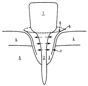
Figure 1 shows a membrane according to the present
At,~FNDED SHEET
WO 95/18638 PCTlGB95/00008
- 12 -
invention in use for bone regeneration of a tooth (1)
which has suffered heavy bone loss around the root (2)
resulting in a cavity (3) normally filled by healthy
root. The root (2) protrudes through a layer ~f
connective tissue or gum (4) into a bone ;socket (5). To
prevent ingrowth of connective tissue (4) into cavity
(3) a membrane cover (6) is placed around the outermost
edge of the wound. The membrane (6) extends all round
the wound cavity (3). The membrane (3) h<~s a smooth
side ( 7 ) which faces ' away from cavity . ( 3 ) and a fibrous
side (8) which faces into cavity(3). The fibrous side
(8) provides a supporting surface for new cells growing
outward from root (2), whereas the smooth side (7) of
the membrane (6) prevents cells of connective tissue (4)
invading the wound cavity. Membrane (6) :is resorbed
slowly back into the body, optimally membrane absorption
correlates to the time taken for wound healing.
The present invention may be further illustrated by
means of the following non-limiting Example:
The peritoneal membranes from young calves are
completely freed from flesh and grease by mechanical
means, washed under running water and treated with 2%
NaOH solution for 12 hours. The membranes are then
washed under running water and acidified with 0.5% HC1.
After the material has been acidified through its entire
thickness (about 3 hours) the material is washed until a
pH of 3.5 is obtained. The material is then shrunk with
7% saline solution, neutralised with 1% NaHC03 solution
and washed under running water. The material is then
dehydrated with acetone and degreased wit3z N-hexane.
The amide nitrogen content of the material is x.47
mMole/g.
CA 02180659 2005-08-04
20208-1627
- 13 -
(Eastoe, E; Courts, A; Practical Analytical Methods for
Connective Tissue Proteins (1963)).
Reaaents
1. 2 N HC1 (160 ml of conc. HC1 made up to l litre)
TM
2. 0.05 M Borax - 0.15 N NaOH solution (19.1 g
Na2B40~. 10 H20) + 6 g NaOH in 1 litre, topped up
with cold distilled HZO.
3. Indicator - mixed in ethanol (0.33% methylene blue
+ 0.050 methylene red)
4. 1% boric acid with the indicator
g boric acid in 1 litre distilled cold H20
8 ml indicator
0.7 ml 0.1 N - NaOH
5. 0.01 N HC1
Method
1. 1 g of dried collagen mass is dispersed in 50 ml of
2N HC1 and boiled for 1 hour. The volume is made
up to 50 ml at 20°C.
2. 5 ml of this dispersion are placed in a
micro kjeldahl flask, together with 20 ml of
solution 2; the mixture is distilled in 20 ml of
solution 4. Distillation takes 6 minutes.
3. The solution is titrated with reagent 5.
Calculation
ml of acid consumption x 20 = mmol % amide N
1.36 ml of O.O1N HCl x 20 = 27.2 mmol o
- 0.27 mmol/g amide N