Note: Descriptions are shown in the official language in which they were submitted.
WO95119803 2 1 8 0 7 4 l PCT~S95/00872
TEMPORARY MEDICAL ELECTRICAL LEAD
REFERENCE TO RELATED APPLICATIONS
This is a continuation-in-part of co-pending
application serial number 08/184,712 entitled "TEMPORARY
MEDICAL ELECTRICAL LEAD" of Mehmanesh et al. filed
January 21, 1994.
FIELD OF THE INVENTION
The present invention relates to the field of
cardiac stimulation and specifically to the field of
temporary stimulation of cardiac tissue through a medical
electrical lead.
BACKGROUND OF THE INVENTION
Atrial arrhythmias and supra ventricular
tachycardias, such as atrial fibrillation, atrial flutter
and atrio-ventricular reentries, are a common postoperative
complication among patients who have had heart surgery.
See, for example, Cardiac Surg. Kirklin JW, Barrat-Boyes BC
(Eds.): NY 1993, pg. 210. During the first 10 days after
heart surgery it is estimated postoperative supra
ventricular tachycardia occurs in up to 63 percent of
patients. See, for example, "The Importance of Age as a
Predicator of Atrial Fibrillation and Flutter After
Coronary Artery Bypass Grafting", Leitch et al., J. Thorac.
Cardiovasc. Surg., 1990:100:338-42; "Atrial Activity During
Cardioplegia and Postoperative Arrhythmias", Mullen et al.,
J. Thorac. Cardiovasc. Surg., 1987:94:558-65.
The presence of these arrhythmias, which in an
otherwise healthy patient may not be unduly serious, may be
especially harmful to heart surgery patients. The
hemodynamic condition of these patients is often already
compromised by either the surgery itself or the effects of
prolonged anaesthesia or both. Supra ventricular
tachycardias may further cause a very irregular ventricular
rate which may even further deteriorate their hemodynamic
condition. Such further deterioration is especially
serious for patients with a compromised left ventricular
WO95/19803 2 1 8 0 7 4 l ~CT~S95/00872 ^--
function. These complications may present a serious
impediment to the recovery of the patient. See, for
example, "Maintenance of Exercise Stroke Volume During
Ventricular Versus Atrial Synchronous Pacing: Role of
Contractility", Ausubel et al., Circ., 1985:72(5):1037-43;
"Basic Physiological Studies on Cardiac Pacing with Special
Reference to the Optimal Mode and Rate After Cardiac
Surgery", Baller et al., Thorac. Cardiovasc. Surg.,
1981:29:168-73.
Due to the serious and potentially life
threatening nature of these conditions, postoperative
treatment is often aimed at preventing arrhythmias, such as
through drugs. Drugs, however, have been found to not
always be effective at preventing arrhythmias. Thus it is
often necessary to provide a means for terminating any
arrhythmias which may occur. One common method used has
been through over-pacing.
For example Waldo et al. in "Use of Temporarily
Placed Epicardial Atrial Wire Electrodes For The Diagnosis
and Treatment of Cardiac Arrhythmias Following Open-Heart
Surgery," J. Thorac. Cardiovasc. Surg., 1978, vol. 76, no.
4, pgs. 558-65 discloses the use of a pair of temporary
heart wires placed on the atrium to diagnose and treat
arrhythmias by antitachy overdrive pacing. Specifically
the temporary heart wires were sutured to the atrial wall
at the time of the heart surgery. Once the patient was
ready to be released the wires were removed by traction or
pulling upon their external end.
Temporary postoperative atrial and ventricular
pacing with temporary heart wires has been found to
successfully treat many of the potential post-operative
arrhythmias. As such the procedure has become widespread -
at least 100,000 procedures per year. Several problems,
however, were encountered with the system disclosed by
Waldo et al., referred to above. One problem was the
stability of the heart wire within the atrial wall.
Because the wall undergoes constant motion, the temporary
heart wire lead was found to dislodge an unacceptable
amount. Secondly, the relatively thin atrial wall,
- WO95/19803 2 1 8 0 7 4 1 PCT~Sg5/00872
especially on elderly patients, was sometimes torn by
traction upon the lead for removal.
An improved method of temporarily affixing heart
wires onto the atrium was achieved with the introduction of
the Medtronic Model 6500 Temporary Myocardial Pacing Lead
System. That lead system featured a silicone atrial
fixation disk to fasten the lead to the atrium.
Specifically the silicone atrial fixation disk was
permanently sutured to the atrium. The lead was positioned
so that it was trapped between the disk and the atrial
tissue. The lead could thereby be removed by simply
pulling it from between the disk and the tissue. The
rubber disk remained in the body after removal of the
electrodes. The advantages offered by such a fixation
system included more reliable lead fixation along with
protecting the relatively thin atrial walls from tearing
during lead removal. Thus the Medtronic Model 6500
Temporary Myocardial Pacing Lead permitted post-surgical
temporary antitachy over-drive pacing to be performed more
safely.
In spite of the improved systems or methods to
achieve antitachy overdrive pacing it is not, however,
always effective in terminating postoperative atrial
arrhythmias or supra ventricular tachycardias. When drugs
and over-pacing are not effective in the prevention or
termination of postoperative supra ventricular
tachycardias, or because of main negative inotropic side
effects relatively contraindicated, it may become necessary
to perform atrial defibrillation, synchronized to the R-
wave of the electrogram, to terminate these potentiallylife-threatening arrythmia. Because of the large energies
involved for defibrillation, however, the temporary heart
wires could not be used.
External atrial defibrillation, although an
effective treatment, has profound side effects. First it
should be noted that in contrast to ventricular
defibrillation, where conversion to normal sinus rhythm is
required at the first shock, atrial defibrillation may be
obtained after several shocks because ventricular
WO95/19803 - 2 1 8 0 7 4 1 PCT~S95/00872
contraction continues during supra ventricular tachycardia.
In addition, due to the high energy required (40 to 360
Joules), the application of shocks, besides their number,
is not tolerated well by a conscious patient. Therefore
external defibrillation is preferably performed under
general anaesthesia or at least sedation. Of course the
use of anesthesia gives rise to another risk to the
patient.
External defibrillation requires relatively high
energy because the electrical source is not positioned
directly upon the cardiac tissue but rather must pass
through the thorax, which tends to dissipate the energy. In
contrast, internally applied atrial defibrillation, such as
may occur during surgery through defibrillation paddles
placed directly on the heart, requires considerably less
energy because the defibrillation electrical energy is
applied only to the tissue that needs to be defibrillated.
In fact, direct atrial defibrillation may be accomplished
with only l.0 Joule pulses in contrast to the 40 Joule and
greater pulses for external defibrillation. See, for
example, Kean D., NASPE abs. 246, PACE, April 1992, pt. II,
pg. 570.
It should be understood the defibrillation
success rate is dependent on the delivered energy. The
lower the energy, the lower the success rate and the higher
the number of shocks to be applied to obtain defibrillation
success. With direct atrial defibrillation, because the
energy may be applied directly to the heart, the energy
level can be chosen such that both the shock level as well
as the number of shocks required may be tolerated by the
patient.
SUMMARY OF THE INVENTION
It is thus an object of the present invention to
provide a temporary atrial defibrillation lead which is
capable of providing electrical stimulation pulses of
sufficient energy to result in defibrillation at a
tolerable level.
-_ WO9S/19803 2 t 8 0 7 4 1 PCT~SgS/00872
It is a further object of the invention to
provide a temporary atrial defibrillation lead which may
provide sufficient energy to the atrium so as to be
tolerated by the patient and therefore delivered without
the necessity of general anaesthesia.
It is a further object of the invention to
provide a temporary atrial defibrillation lead which may be
reliably fixed to the atrium through a fixation pad.
It is a further object of the invention to
provide a temporary atrial defibrillation lead which may be
safely and reliably removed from the atrium.
It is a further object of the invention to
provide a temporary atrial defibrillation lead which may be
safely and reliably removed from the atrium without the
necessity of a surgical intervention.
In accordance with the above objects there is
provided a temporary atrial defibrillation lead featuring a
PTFE felt pad in which three parallel stainless steel
defibrillation wire electrodes are mounted. The pad
contains holes which expose the electrode wires in a
discontinuous fashion. The three electrode wires are
merged into one polyurethane insulated lead body, proximal
to the pad. At the proximal end of the lead body a
stainless steel connector pin with break away needle are
mounted for the percutaneous exteriorization of the lead
pin in an area separated from the surgical incision. The
break away needle can be broken off to make the connector
pin suitable for connection to a therapeutic device, such
as a defibrillator. The PTFE pad is permanently implanted
on the atria and remains implanted after removal of the
temporary electrode sections. The temporary electrode
sections may be removed by gently pulling them at their
proximal end.
BRIEF DESCRIPTION OF THE DRAWINGS
Other objects, advantages and features of the
present invention will become apparent from the following
specification when taken in conjunction with the
WO95/19803 2 1 8074 1 PCT~S95/00872 --
accompanying drawings in which like elements are commonly
enumerated and in which:
FIG. 1 is a plan view of a lead according to the
present invention used to connect a pulse generator to a
heart.
FIG. 2 details the connector assembly used in a
lead according to the present invention having the break-
away needle attached.
FIG. 3 details the connector assembly used in a
lead according to the present invention having the break-
away needle broken away.
FIGS. 4 and 5 detail the lead body used in a lead
according to the present invention.
FIG. 6 is a plan view of a lead according to the
present invention.
FIG. 7 is a sectional view of the lead shown in
FIG. 6 taken along line 7-7.
FIG. 8 is a sectional detail of the stranded
conductor and bus within the mounting pad.
FIG. 9 is a graph illustrating the force required
to remove a lead according to the present invention.
FIG. 10 is a perspective view of an alternate
embodiment of the present invention.
FIG. 11 is a cross sectional view of the pad of
2 5 FIG. 10 showing a conductor woven through pad.
The drawings are not necessarily to scale.
DETAILED DESCRIPTION OF THE PREFERRED EMBODIMENT
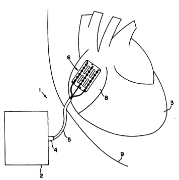
FIG. 1 is a plan view of a lead l according to
the present invention used to connect pulse generator 2 to
heart 3. As seen lead l has essentially three sections:
connector assembly 4, lead body 5 and electrode assembly 6.
Connector assembly 4 connects lead l to pulse
generator 2. Details of connector assembly 4 may be seen
in FIGS. 2 and 3. As seen connector assembly 4 features a
break-away needle ll which mates with pin assembly 12.
Specifically break-away needle ll has recess 17 which mates
with finger 18 of pin assembly 12. In the preferred
embodiment pin assembly 12 is stainless steel. Break-away
21 80741
-- WO95/19803 PCT~S95/00872
needle 11 is provided on pin assembly 12 to permit the
passage of connector assembly 4 from inside the body,
through the skin to outside the body. Break-away needle 11
may thereafter be broken off connector assembly 4 at
breakpoint 13 to thereby permit pin assembly 12 to join to
a pulse generator 2. As seen in FIG. 3 when break-away
needle 11 is broken off it carries with it a portion of
finger 18. Pin assembly 12 further features crimp skirt 15
to permit conductors of lead body 5 to be joined thereto.
Specifically conductors are crimped within cavity 16 and
thereby electrically connected to pin assembly 4.
Lead body 5 consists of an insulative outer
sleeve 20 encasing a plurality of conductors 21, 22 and 23
as seen in FIGS. 4 and 5. Gap 26 among inner conductors
21, 22 and 23 is filled by medical adhesive. Outer sleeve
20 may be constructed from any suitable biocompatible
material, however in the preferred embodiment outer sleeve
20 is polyurethane.
Inner conductors 21, 22, and 23 are each
constructed in a similar fashion and thus only one need be
described. Each is constructed from a stranded conductor
30 encased by inner sleeve 31. In the preferred embodiment
stranded conductor 30 is a multi-filament stainless steel
stranded wire and inner sleeve 31 is PTFE or FEP. It
should be understood, of course, that any suitable material
or wire could be used for conductor 30 including a coiled
wire as well as any type of wire made from an acceptable
biocompatible metal including, but not limited to, such
materials as platinum, palladium, titanium, tantalum,
rhodium, iridium, carbon, vitreous carbon and alloys,
oxides and nitrides of such metals or other conductive
materials. Of course, some materials are incompatible with
others and may not be effectively used together. The
limitations of specific materials for use with others is
well known in the art. It should also be understood that
any other suitable material could also be used for inner
sleeve 31 such as silicone, polyurethane, PTFE or FEP, for
example.
WO95/19803 2 1 8 0 7 4 1 PCT~S95/00872
As best seen in FIG. 6 outer sleeve 20 ends at a
point 32 away from the distal end of lead l. Inner
conductors 21, 22, and 23 extend from point 32 to electrode
assembly 6. Electrode assembly 6 is formed with inner
conductors 21, 22, 23 and mounting pad 33. Specifically
distal portion of each inner conductor has each stranded
conductor 30 exposed along the length of mounting pad 33.
Each of the inner conductors 30 is mounted to mounting pad
33, as best seen in FIGS. 7 and 8. Although the
illustrated preferred embodiment features inner conductors
30 mounted within mounting pad 33, it should be understood
inner conductors may be mounted to mounting pad 33 in any
acceptable manner including, without limiting the
variations possible, suturing or gluing all or some of
inner conductor 30 to an outer surface of the mounting pad
33. In the preferred embodiment holes 34 within mounting
pad 33 are used to provide for intermittent sections of
each stranded conductor 30 to be exposed to body tissue.
Thus when lead l, and specifically electrode assembly 6, is
mounted to cardiac tissue, intermittent sections of each
stranded conductor 30 are directly exposed to cardiac
tissue through holes 34. The contour dimensions (length by
width of the exposed electrode area) of the conductors is
approximately 40 by 30 millimeters in the preferred
embodiment. A minimum of two exposed conductors is
required to obtain this contour, and by this, a current
distribution which results in acceptable defibrillation
thresholds (DFT). Application of three conductors is
preferred, because it further improves the DFT and the
current density at the conductor electrode surface. In the
preferred embodiment the conductor electrodes are exposed
to both sides of the pad, allowing the current to flow
across the front and back side of the pad. This results in
a more homogeneous electrical field between the electrodes
and usually in a lower DFT.
An alternative embodiment, which yields the same
desired characteristics, incorporates a solid pad through
which the conductors are threaded or woven, thus being
alternatingly exposed to both sides of the pad, as best
21 80741
- WO95/19803 PCT~S95/00872
seen in FIGS. 10 and 11. Specifically FIG. 10 is a
perspective view of a lead shown conductors 21, 22, 23 of
lead body 5 woven though pad 33. FIG. 11 is a cross
sectional view of pad 33 of FIG. 10 showing conductor 21
woven through pad 33. Although as depicted conductors 21,
22, and 23 are exposed equally to each side of pad 33, they
may also be woven such that a greater length of each is
exposed on one side of pad 33 as compared to another side
of pad 3 3.
Mounting pad 33 further features suture areas 35
(designated by "x"s in the FIGS.) which permit mounting pad
33 to be sutured to the heart, as best seen in FIG. 1.
Mounting pad 33 may be fashioned from any biocompatible
pliant, material and in the preferred embodiment mounting
pad 33 is fashioned from a PTFE felt. Preferably the
structure and porosity of the felt should be similar to
those which are typically used in reconstructive heart
surgery.
In an alternate embodiment, mounting pad 33 may
also be fashioned from a bioabsorbable material such as
bovine collagen which has been cross-linked. Cross linking
may be accomplished in any acceptable manner, including for
example, according to the principles set forth in U.S.
Patent No. 5,264,551 entitled "Process for Cross-Linking
Collagen by Diphenyl-phosphorylazide the Cross-Linked
Collagen Obtained Thereby and Collagen Based Biomaterials
Thus Cross-Linked" issued to Petite et al and assigned to
Bioetica of Lyon, France, incorporated herein by reference.
The particular degree of cross linking used may depend upon
3 0 the type of collagen used and the amount of time lead 1
will be used in the body. The degree of cross linking
should be such that the mechanical characteristics of pad
33 and the holding force of conductors 21, 22, 23 should be
maintained and unintended disengagement of conductors is
prevented for a period of at least two weeks to a month.
Finally, other types of collagen besides bovine may also be
used, such as pig or sheep.
Implantation of lead 1 according to the present
invention is as follows. Mounting pad 33 is sutured to
W 095/19803 . ~ 2 1 8 D 7 4 1 PC~rrUS95/00872
atrium 8 using suture areas 35. Next connector assembly 4
is exteriorized at a point away from the incision through
use of break-away needle 11 and pin assembly 12.
Specifically needle 11 is used to pierce the skin from the
interior to the exterior so as to expose pin assembly 12.
Once lead 1 is satisfactorily sutured to the atrium, pin
assembly 12 is exposed and lead 1 is connected to a pulse
generator, the patient's incision may be closed. At this
point lead 1 may deliver therapeutic electrical pulses,
including defibrillating, cardioverting or pacing, to
atrium 8.
One important aspect of lead 1 of the present
invention is its removability. Inner conductors 21, 22, 23
are mounted within mounting pad 33 s o they may be removed,
even once implanted, by traction. Specifically the inner
conductors may be gently removed from mounting pad 33, and
thus body 9, by traction upon proximal end of lead 1.
As seen in FIGS. 7 and 8 inner conductors are
positioned within mounting pad 33. Bus 41 (also called a
sleeve) is crimped to the conductor. Bus 41 serves to
prevent unintended dislodgement of inner conductor 30 out
of mounting pad 33. Bus 41 is placed at the proximal end
43 of pad 33, at a point between end 43 of pad 33 and hole
34. As such when inner conductor 30 is removed by
traction, bus 41 only needs to pass through a short portion
of pad 33 before it is free. Thus only a relatively brief
amount of increased force, i.e. a short "jerk" or tug on
the distal end of lead body 5 is sufficient to pull bus 41
out of pad 33. Once bus 41 is outside pad 33 the
remainder of inner conductor 30 follows easily as there is
no other structure along the length of inner conductor 30
which will inhibit the travel of inner conductor 30 through
pad 33. This is illustrated in FIG. 9 where it is
illustrated that pullout distance initially requires a
relatively great pullout force, but which rapidly decreases
once bus 41 i s withdrawn from mounting pad 33. Thus it may
be seen that bus 41 prevents inner conductors 21, 22, 23
from accidentally dislodging from position while also
allowing their intended dislodgement and removal without
-- WO95/l9803 2 1 8 0 7 4 I PCT~Sg5/00872
possibly excessive forces from being applied to the atrium
8 during removal. Similar removal properties may be
obtained without bus 41. Application of adhesive (i.e.
medical adhesive or polyurethane adhesive) to each
conductor or each conductor's insulation and pad 33 creates
an adhesive bond between each conductor and pad 33. Once,
by pulling lead body 5, the adhesive bond is broken, the
rest of each conductor may be removed with lower force from
pad 33, which results in a similar removal force
characteristic as with bus 41, discussed below with
reference to FIG. 9. In the preferred embodiment a small
amount of medical adhesive 40 (or polyurethane) is applied
to the distal end of each conductor 30 in order to cap off
the ends of the stranded wire, although other materials,
such as polyurethane, may also be used. This is done in
order to keep the strands together and to prevent damage to
the tissue during the removal procedure or in case the
conductor would be forced out of the pad while implanted,
as could occur due to heart movement. Mounting pad 33
because it is sutured to the heart, is left in place once
conductors and lead body are removed. As discussed above,
if mounting pad 33 is fashioned from collagen then if may
be absorbed by the body tissues, as is well known in the
art. Of course, the time required for absorption depends
upon the degree to which the collagen has been cross
linked.
Although the invention has been described in
detail with particular reference to a preferred embodiment
and alternate embodiments thereof, it will be understood
variations and modifications can be effected within the
scope of the following claims. Such modifications may
include substituting elements or components which perform
substantially the same function in substantially the same
way to achieve substantially the same result for those
described herein.