Note: Descriptions are shown in the official language in which they were submitted.
- 2 1 80742
PCT/US ~ 3 ~ ~J r
57 Rec'd PCT~'~O ~ 6 ~ 1996
MICROCAPSULE GENERATING SYSTEM CONTAINING AIR KNIFE
AND METHOD OF ENCAPSULATING
BACKGROUND OF THE INVENTION
Field of the Invention
The present invention concerns a microcapsule
generating system and relates to encapsulation of material
generally, and more particularly to encapsulating tissue or
a suspension of cells so that the encapsulated tissue or
cells remain viable within a protective membrane or
coating. The membrane or coating is permeable to
- nutrients, ions, oxygen, and other materials needed both to
maintain the tissue and to support its normal metabolic
functions, but is impermeable to bacteria, lymphocytes, and
large proteins of the type responsible for immunological
reactions resulting in rejection.
Insulin-producing or other hormone-producing systems,
cells from tissues, primary cultured cells, cultured cell
lines that produce biological products of interest (such as
Factor VIII and calcitonin), and genetically engineered
cultured cell lines, for example, can be coated using the
encapsulating apparatus of the present invention. That is,
this apparatus permits encapsulation of mammalian
pancreatic beta cells, alpha cells, intact islets of
Langerhans, and other tissues or tissue fractions which
secrete hormones. The encapsulated cells or tissue may be
suspended in a culture medium where they will secrete
hormones over an extended period.
BACKGROUND ART AND RELATED ART DISCLOSURES
Various attempts have been made to provide semi-
permeable microcapsules that were both biocompatible with
the body tissue and impermeable to the components of the
immune system. Typically, living tissue or individual
3S cells are suspended in an aqueous solution of a reversibly
gelable material, such as sodium alginate, and droplets of
AMENDED SHEET
- WO95/19840 2 1 8 0 7 4 2 PcT~s95/oo53s
this suspension are formed and allowed to fall into a
gelling solution, such as calcium chloride. The temporary
capsules so formed are then treated with a crosslinking
polymer, such as polylysine and polyethyleneimine, to form
an outer semipermeable coating.
The droplets are typically formed by feeding the
alginate suspension to a first site where a mass of the
liquid suspension accumulates. Then the mass of liquid
suspension is agitated such that it is broken up into small
droplets. Devices using vibration, centrifugal force, air
currents and electrostatic charges have been used to
agitate the liquid to generate the small droplets.
One drawback of conventional devices using vibration,
centrifugal force and air currents is that the diameters of
the microcapsules produced thereby are dependent on the
sizes of the orifices through which the suspension is
extruded and are typically 500 ~m or greater with devices
used for pancreatic islet encapsulation, where a relatively
large bore diameter is dictated by the large size of islets
(50-300 ~m), where a relatively . Since oxygen diffusion
is insufficient to maintain cell viability at distances
exceeding about 150 ~m, the cells in the center region of
these microcapsules are routinely lost due to oxygen
deprivation.
Although devices using electrostatic charges are
claimed to produce microcapsules having diameters as small
as 150 ~m (see, e.g., U.S. Patent No. 4,789,550 to Hommel,
et al. ), the blank microcapsules and microcapsules
containing cells or tissue produced by electrostatic
devices generally are the same size. Accordingly, the
separation and differentiation between the blanks and the
other microcapsules is difficult at best. In addition, in
order to produce microcapsules of small diameter using this
approach, it is necessary to use small bore needles which
cannot accommodate the larger particles in the suspension
- W095/19840 2 1 80742 PCTtUS95tO0535
and which tend to clog with high density cell or other
particulate suspensions.
SUMMARY
The present invention is directed to a microcapsule
~ 5 formation device that significantly minimizes the problems
and disadvantages presented in conventional devices. The
invention accomplishes this goal by providing a
microencapsulation system with a droplet-forming air knife
that includes a capillary tube or needle and an air sleeve.
The capillary tube or needle is adapted for coupling to a
first source of fluid, such as an alginate suspension, from
which droplets are to be formed. The air sleeve is adapted
for coupling to a second source of fluid, for example,
sterilized air. The discharge end of the capillary tube or
needle is positioned in the immediate vicinity of the
discharge end of the air sleeve so that air currents from
the air sleeve increase the force acting on a nascent
droplet of the first fluid at the discharge end of the
capillary tube or needle and help break the droplet away
from the needle to free fall into a gelling solution. That
is, as the liquid suspension of the materials to be coated
or encapsulated is discharged from the capillary tube or
needle, pressurized air introduced into the air sleeve
breaks the liquid suspension into tiny droplets.
According to a first embodiment of the invention, the
center of the outlet opening of the needle is offset from
the center axis of the air sleeve. Thus, the needle can be
eccentrically positioned within the air sleeve of the
needle with its outlet opening eccentrically positioned
relative to the center axis of the sleeve, for example. It
has been found that the eccentricity of the needle outlet
and air sleeve enhances the ability of the device to
produce very small droplets. In addition, it has been
empirically determined that the greater eccentricity of the
capillary tube or needle (or its outlet), the smaller the
W O 95/19840 2 1 8 0 7 4 2 PC~rtUS95tO0535
droplet size.
According to a second embodiment of the invention, the
capillary tube has a beveled, pointed discharge end. This
configuration also has been empirically shown to be
important to the generation of very small droplets.
According to a further advantageous feature of the
present invention, the entire beveled portion of the needle
is positioned beyond the end of the air sleeve. It has
been empirically determined that the optimal needle
position for producing the smallest possible droplets is
when the uppermost region of the beveled portion of the
needle is spaced a very short distance (e.g., 1 mm) from
the air sleeve outlet.
The air knife of the present invention described above
can form very small alginate droplets containing suspension
of individual cells or tissue. The droplets are
sufficiently small so that upon contact with a gelling
solution, such as CaCl2, microcapsules are formed having
diameters from about 20-300 ~m, depending on the size of
the tissue or cells being encapsulated. This is especially
advantageous when the microcapsules are to be introduced
into a patient. That is, the volume of material being
introduced into the patient can be reduced since the
membrane is close fit about the encapsulated biological
material. The small size of the microcapsules also makes
delivery to the patient less intrusive as smaller needles
are required for injection into the patient. Since the
microcapsules are less than about 300 ~m in diameter,
diffusion of oxygen to the center of the capsules is no
longer a problem. It also has been found that blanks
(microcapsules without cells or tissues), which are formed
in the present encapsulation process, are about ten to
fiftyfold smaller than the microcapsules containing cells
or tissue and, thus, are readily distinguishable and
separable from the encapsulated cells and/or tissue.
- WO95tl9840 2 1 8 0 7 4 2 PCT~S95/00535
Another especially advantageous aspect of the device
of the present invention is that it can be used to form a
thin second coating on encapsulated cells or tissue while
maintaining the diameter of the double or multiple-coated
microcapsule within 10 - 40 ~m of the diameter of the
single-coated microcapsule. The second coating is formed
by passing a suspension of the encapsulated cells or tissue
in alginate solution through the needle while introducing
pressurized air into the sleeve. The additional coating(s)
ensure(s) that the cells or tissue are completely
encapsulated.
The above is a brief description of some deficiencies
in the prior art and advantages of the present invention.
Other features, advantages and embodiments of the invention
will be apparent to those skilled in the art from the
following description, accompanying drawings and appended
claims.
BRIEF DESCRIPTION OF THE DRAWINGS
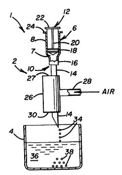
Fig. 1 is a diagrammatic elevational view of a
microcapsule generating system in accordance with the
principles of the present invention.
Fig. 2 is a diagrammatic elevational view of a further
embodiment of the microcapsule generating system of Fig. 1.
Fig. 3 is an enlarged sectional view of the distal
ends of the air knife discharge tubes used in the
microcapsule generating systems of Figs. 1 and 2, showing
the inner tube or needle arranged such that it abuts
against the inner surface of the outer tube or sleeve.
Fig. 4 is a sectional view taken along line 4-4 in
Fig. 3.
Fig. 5 is a side elevational view of the inner tube of
Fig. 3 shown rotated 90 degrees.
Fig. 6 is an enlarged sectional view of the distal
ends of the air knife discharge tubes of Figs. 1 and 2
showing the inner tube or needle arranged such that it is
WO95/19840 2 1 8 0 7 4 2 PCT~S95/00535
radially spaced from the inner surface of the outer tube of
sleeve.
Fig. 7 is a sectional view taken along line 7-7 in
Fig. 6.
Fig. 8 is an enlarged sectional view of the distal
ends of the air knife discharge tubes of Figs. l and 2,
showing the outer tube beveled according to another
embodiment of the invention.
Fig. 9 is a further view of the beveled sleeve and
needle of Fig. 8 with the outer tube shown rotated 90
degrees.
Fig. l0 is a graph illustrating the size distribution
of single-coated microcapsules formed from three different
preparations of hepatocytes under the same procedures using
the system illustrated in Figs. l and 3-5 (90% of the
microcapsules have a diameter less than 75 ~m).
Fig. ll is a graph illustrating the size distribution
of single-coated microcapsules containing a proliferating
cell line that provides Factor VIII using the system
illustrated in Figs. l and 3-5.
Fig. 12 is a graph illustrating the size distribution
of single-coated microcapsules containing a proliferating
cell line that secretes calcitonin.
DESCRIPTION OF THE PREFERRED EMBODIMENT
Referring to the drawings in detail wherein like
numerals indicate like elements, a microencapsulation
system is shown in Fig. l according to the principles of
the present invention. Although the present invention can
be used to encapsulate other materials, it will be
described in conjunction with the encapsulation of droplets
of an alginate suspension containing individual cells or
tissue for purposes of simplification.
Referring to Fig. l, microencapsulation system l
generally comprises an air knife 2, which forms droplets 34
containing individual cells or tissue suspended in a
- WO95/19U0 2 1 8 0 7 4 2 PCT~S95/00535
gelable polymer solution, and a collection vessel or tank
4, which contains a gelling solution 36. Gelling solution
36 is positioned below the air knife for collecting the
droplets 34 and causing microcapsules 38 containing the
desired biological material to be formed.
Air knife 2 includes a syringe 6 for dispensing the
material to be encapsulated, such as an alginate solution
7, and a tubular air sleeve 26 which will be described in
more detail below. The syringe 6 includes a barrel 8,
which is shown as containing the alginate suspension 7, a
needle assembly 10, and a plunger 12 for forcing the
alginate suspension through the needle assembly. Needle
assembly 10 includes a capillary tube or needle 14 and hub
16 which fluidly couples needle 14 to barrel 8, so that
fluid, such as alginate solution 7 can be dispensed from
barrel 8 through needle 14.
Plunger 12 includes a piston 18 and a stem 20 which
are interconnected so that piston 18 can be readily moved
to displace alginate suspension 7 from barrel 8 into and
through needle 14. A grip 22 and a finger ledge 24 also
are provided as is conventionally known in the field, to
facilitate the manual displacement of plunger 12.
Preferably, a mechanical drive (not shown) can be coupled
to the plunger to displace the plunger at a constant rate
as would be apparent to one of ordinary skill.
Referring to Figs. 1 and 3-5, tubular air sleeve 26 is
positioned around part of the axial length of needle 14,
i.e., needle 14 extends through sleeve 26. Sleeve 26
includes an end wall 27 through which needle 14 extends.
End wall 27 and needle 14 form a closed end for sleeve 26
which includes an open distal end 30. A feed pipe 28 is
fluidly coupled to air sleeve 26 for introducing
pressurized gas, preferably sterilized air, into the flow
path or space 53 formed between the outer wall surface of
needle 14 and the inner wall surface of air sleeve 26. As
Wo95tl9840 2 1 8 0 7 4 2 PCT~S95/OOS35
will be described in more detail below, the pressurized air
in air sleeve 26 -controls, in part, the size of the
droplets dispensed by needle 14. Air knife 2 is suspended
above collection tank 4 with any suitable fixture as would
be apparent to one of skill.
Fig. 2 illustrates a second embodiment of the
microencapsulation system. System 1' differs from system
1 in that system 1' includes air knife 2' which does not
include a plunger mechanism. In air knife 2', needle 14 is
coupled to a container 42, which is configured to hold the
fluid from which the droplets are to be made, such as
alginate solution 7. Container 42 is coupled to a source
of pressurized gas (preferably sterilized air) via a feed
pipe 44. The pressure of the gas introduced into container
42 is controlled by conventional means to regulate the
discharge rate of alginate suspension 7, for example,
through needle 14. The discharge rate preferably is
regulated to be constant.
Referring to Figs. 3, 4 and 5, the configuration and
position of tubular member or needle 14 relative to sleeve
26 will be discussed. In the preferred embodiment, needle
14 is positioned in sleeve 26, which has an elongated and
hollow or tubular shape, and extends beyond outlet opening
30 of sleeve 26. Sleeve 26 can be made of stainless steel
or any other suitable sterilizable material. Sleeve 26
terminates at its distal end into a generally blunt edge
31. That is, edge 31 is neither beveled nor sharp.
Needle 14 is an elongated tubular member that is
hollow throughout its entire axial length. For the purpose
of illustration, the size of needle 14 can range between 16
and 30 gauge, and preferably is 20 gauge. Distal end 32 of
needle 14 is beveled at an angle ~ (the angle formed
between side wall 51 of needle 14 and beveled surface 50).
Angle ~ can range from about 15 degrees to 45 degrees to
provide the desired results, and preferably is about 22.
- W095/19840 2 1 8 0 7 4 2 PCT~S9S/OOS35
As shown in Fig. 3, beveled surface 50 is positioned a
short distance below blunt edge 31 of sleeve 26.
Preferably the uppermost portion of beveled surface is
about 1 mm below blunt edge 31. It should be understood,
- 5 however, that the present invention contemplates
positioning beveled surface 50 at various locations
relative to blunt edge 31 of sleeve 26. In addition,
although beveled surface 50 is shown facing the air flow
path, variations to this position are also contemplated
within the scope of the present invention.
Referring to Fig. 5, beveled elliptical surface 50 is
bounded by an upper edge 60, and side edges 61 and 62 that
meet at and terminate into a pointed tip 52. While beveled
surface 50 is shown as being flat, it should be understood
that beveled surface 50 could alternatively be arcuately
shaped to provide an additional contact surface for the
droplets to be formed thereon and, thus, help to control
the droplet size. The beveled shape of edge 50 represents
an important aspect of the present invention, in that the
beveled edge allows air knife 2 (or 2') to generate smaller
size droplets 34.
As illustrated in Figs. 3 and 4, needle 14 preferably
is eccentrically (i.e., non-coaxially) positioned inside
sleeve 26. In other words, center 54 of needle outlet
opening 58 is offset from center line 56 of sleeve 26.
This eccentricity also is important to generate small
diameter droplets 34. In the preferred embodiment, side
wall 51 of needle 14 about the inner wall surface of sleeve
26, so as to prevent air from flowing therebetween.
The eccentric placement of needle 14 further
contributes to the regulation of the size of the droplets
formed and to the significant reduction of their size.
Additionally, the beveled shape of needle 14 and the
placement of needle 14 in contact with the inner wall
surface of sleeve 26 cause the formation of blank
WO95/19840 2 1 8 0 7 4 2 PCT~S95100535
microcapsules of much smaller size than the microcapsules
containing cells or tissue. Consequently, the latter
microcapsules are readily identifiable and distinguishable
from the blank microcapsules, and thus segregable
therefrom.
Referring now to Figs. 6 and 7, there is illustrated
an alternate embodiment of air knife 2. This embodiment is
similar to the embodiment illustrated in Figs. 3, 4 and 5,
with the single variation that needle 14 is not placed in
direct contact with the inner surface of sleeve 26. A
spacing 65 is allowed to be formed between needle 14 and
sleeve 26, through which pressurized air is allowed to
flow.
The size of spacing 65, i.e., the distance between
needle 14 and sleeve 26, varies with the desired
application of microcapsule generating system 1.
Generally, the farther needle 14 is from center axis 56 of
sleeve 26 (i.e., the closer needle 14 is to the inner wall
of sleeve 26), the fluid extruded through needle 14 tends
to accumulate on the back side of beveled surface 50. In
contrast to a configuration where the needle 14 is
coaxially aligned with, or close to center axis 56 of
sleeve 26, control to reduce the size of the capsules can
be exercised due to increased air flow rate over tip 52 as
the needle 14 is moved radially outward.
Turning now to Figs. 8 and 9, there is illustrated
another embodiment of air knife 2. The embodiment is
similar to that shown in Figs. 3, 4 and 5, except that air
knife 2 includes a sleeve 70 that is similar in design and
construction to sleeve 26, with the exception that sleeve
70 includes a beveled edge 72, rather than blunt edge 31.
The beveled edges 50 and 72 of needle 14 and sleeve
70, respectively, can be rotated relative to each other to
attain the optimal desired droplet size and shape. Fig. 8
shows the two beveled edges 50 and 72 having their slopes
WO9S/19840 2 1 8 0 7 4 2 PCT~S95/00535
in the same general direction, while Fig. g illustrates the
slopes as being oppositely positioned.
The general operation of the device will be described
in conjunction with the system illustrated in Figs. 1 and
3-5 using a particulates suspension in an alginate solution
for purposes of example and, thus, is not intended to limit
the scope of the invention. Syringe 6 is provided with an
alginate suspension and plunger 12 displaced at a constant
rate (e.g., 0.1 - 2 ml/min depending on the size of needle
14 sleeve and the material to be encapsulated~. As
suspension 7 is discharged from outlet opening 58 in
beveled surface 50 of needle 14, pressurized gas (e.g.,
air) as designated by arrow 29 concurrently is flowed
downwardly through a chamber 53 formed between the inner
surface of sleeve 26 and the outer surface of needle 14.
Some of the gas exiting outlet opening 30 flows toward
beveled surface 50 to agitate accumulation 64 of suspension
7 on surface 50. The air currents push the alginate
suspension which accumulates at beveled surface 50 of
needle 14, toward tip 52, thus forming droplets 34 having
a desired shape and size. That is, the gas introduced
through feed pipe 28 enters sleeve 26 and exits sleeve 26
via outlet opening 30 whereafter air currents agitate the
alginate suspension accumulated on distal end 32 of needle
or capillary tube 14 to form very small droplets 34. The
droplets are captured in a gelling solution 36, such as
calcium chloride solution. In the case where CaCl2 is used,
the calcium interacts-with the carboxylic acid groups of
the alginate to form calcium alginate gel. In general,
droplets 34 have a substantially spherical shape. The air
flow pressure and plunger speed are regulated so as to
generate the desired droplet size and shape as discussed
above.
Using the air knife of the present invention,
biological material containing microcapsules having
WO95/19840 2 1 8 0 7 4 2 PCT~SsS/00535
diameters from about 20-300 ~m are formed. The blanks,
microcapsules without biological material, are generally
about ten to fiftyfold smaller. Thus, the microcapsules
containing biological material are clearly distinguishable
from the blank microcapsules based on size and are readily
separated as described below.
The very small blank microcapsules are separated from
the remaining microcapsules containing the coated
biological material, by allowing the latter microcapsules,
having a larger size, to settle out, and then by washing
away the smaller blank microcapsules. The foregoing
process of allowing the larger microcapsules to settle out
in tank 4, and the washing away of the smaller blank
microcapsules is repeated as many times as needed until the
desired concentration of encapsulated islets or other
tissue is attained.
Then, the collected encapsulated biological material
may be over coated by repeating the process described
above. Such over coating will ensure that tissue or cells
are completely encapsulated.
The present invention will hereinafter be described
more specifically by the following Examples which are
provided for illustrative purposes and are not intended to
limit the invention. In these examples, a system
constructed according to microcapsule generation system l
was used. The cell suspension in alginate solution was
placed in the barrel of the syringe which includes a 20-
gauge needle having a pointed tip beveled at a 22 angle.
The uppermost edge 60 of beveled surface 50 was positioned
about l mm below edge 30 of sleeve 26.
EXAMPLE l
Encapsulation of Pancreatic Islets
A suspension of pancreatic islets in alginate is
placed in the syringe barrel. The syringe plunger 12 is
displaced to provide a flow rate of 0.3 ml/min to dispense
-- WO9S/19~0 2 1 8 0 7 4 2 PCT~S95/00535
the cell suspension in alginate solution from the needle
while air is delivered to outer sleeve 26, which has a 2 mm
inner diameter approximately, to provide an entry pressure
in sleeve 26 of about 30 psi. Droplets of the suspension
fall into collecting vessel 4 containing 120 mM CaCl2 and 10
mM HEPES. The vessel is positioned so that the CaCl2 is
about 160-165 mm from tip 52 of the needle. The
microcapsules containing pancreatic islets recovered with
this procedure had a diameter of about 50 to 300 ~m. On
the other hand, the blank microcapsules obtained with this
procedure had a diameter ranging between 1 ~m and 20 ~m.
Consequently, the blank microcapsules containing islets are
readily identifiable and distinguishable from the blank
microcapsules for subsequent separation.
EXAMPLE 2
EncaPsulation of HePatocytes
An cell suspension in alginate solution comprising rat
hepatocytes is prepared. The syringe plunger 12 is
displaced to provide a flow rate of 0.3 ml/min. to dispense
the suspension from the needle while air is delivered to
outer sleeve 26, which has a 2 mm inner diameter
approximately, to maintain an internal sheath entry
pressure of about 30-33 psi. Droplets of the suspension
fall into collecting vessel 4 containing 120 mM CaCl2 and 10
mM HEPES. The vessel is positioned so that the CaCl2
solution is about 160 mm from tip 52 of the needle. Over
90% of the microcapsules recovered with this procedure had
a diameter of less than 75 ~m. The size distribution is
illustrated in Fig. 10 which shows three different
preparations of hepatocytes which were run separately
according to the procedures described in this example. The
consistency of the data from each preparation indicates
that these results are reproducible as required for
commercial manufacturing.
WO95/19840 2 1 80742 PCT~S95/oo53s
EXAMPLE 3
EncaPsulation of Proliferating
Cells that Secrete Factor VIII
An alginate suspension comprising Factor VIII
secreting cells is prepared. The syringe plunger 12 is
displaced to provide a flow rate of 0.3 ml/min to dispense
the alginate suspension from the needle while air is
delivered to outer sleeve 26, which has a 2 mm inner
diameter approximately, to provide a pressure of about 33
psi entering sleeve 26. Droplets of the alginate
suspension fall into collecting vessel 4 containing 120 mM
CaCl2 and 10 mM HEPES. The vessel is positioned so that the
CaCl2 solution is about 154 mm from tip 52 of the needle.
go~ of the microcapsules recovered with this procedure had
a diameter between 25 and 75 ~m. This size distribution is
illustrated in Fig. 11 wherein (1) all, (2) pellet, and (3)
supernatant correspond to (1) microcapsules containing
cells together with blanks, (2) primarily microcapsules
containing cells and (3) primarily blanks, respectively.
EXAMPLE 4
EncaPsulation of Proliferatinq
Cells that Secrete Calcitonin
An suspension of cells secreting calcitonin is
prepared in alginate solution. The syringe plunger 12 is
displaced at a travel speed of 0.3 ml/min to dispense the
suspension from the needle while air is delivered to outer
sleeve 26, which has a 2 mm inner diameter approximately,
to maintain an internal pressure of about 33 psi entering
sleeve 26. Droplets of the suspension fall into collecting
vessel 4 containing 120 mM CaCl2 and 10 mM HEPES. The
vessel is positioned so that the CaCl2 solution is about 154
mm from tip 52 of the needle. 90~ of the microcapsules
recovered with this procedure had a diameter between 25 and
75 ~m. This size distribution is illustrated in Fig. 12 in
which all, pelleted and supernatant have the same meanings
as described in conjunction with Example 3 and Fig. 11.
-- WO95119840 2 1 8 0 7 4 2 PCT~S95/00535
It will be apparent from the description above to
persons skilled in the art that a method of using the above
mentioned structures is also disclosed. Such a method of
forming droplets comprising the steps of providing first
and second tubes 26 and 14, each having an outlet opening
30 and 50, respectively, positioning said outlet openings
30 and 50 with respect to one another such that the second
tube outlet opening 50 is located in the flow path taken by
gas discharged from the first tube outlet opening 30, a
center of the second tube outlet opening being offset from
a center axis 56 of the first tube outlet opening causing
pressurized gas to flow from the first tube outlet opening
30, and causing a fluid 7 to flow from and be suspended
from 64 the end of the second tube outlet opening 50 so
that the gas flowing from said first tube outlet opening 30
impinges fluid suspended from 64 the second tube outlet
opening to form droplets 34 therefrom.
It will be further understood by persons skilled in
the art that microcapsules 38 are formed by a particular
process having the steps of discharging a gas stream 29
having a central axis 56 of flow in a first direction from
a gas supply opening 30, wherein said first direction is
toward a container 4 of gelling medium 36 which when it
comes in contact with droplets 34 of a fluid 7 to be
encapsulated hardens the outer surface of the droplets 34
of the fluid 7 to be microencapsulated, placing the
discharge opening 50 of a source 8 or 42 of fluid 7 to be
microencapsulated within the gas stream flowing from said
gas supply opening such that a center of said discharge
3C opening 50 is disposed eccentric from said central axis 56
of flow; discharging a fluid 7 to be encapsulated from the
discharge opening 50 of the source of fluid 8 or 42 such
that fluid droplets 34 are formed which fall into the
container 4 of gelling medium 36 to form microcapsules 38.
While the system has been described as a whole,
W095/lg840 2 1 8~742 ~CT~S95/00535
persons skilled in the art will understand that the air
knife configurations 2, 2' described are not limited to
forming droplets 34 for the encapsulation of biological
media, but are exceptionally well suited for this
application thereby overcoming many drawbacks of the prior
art as previously described.
The above is a detailed description of particular
embodiments of the invention. It is recognized that
departures from the disclosed embodiment may be made within
the scope of the invention and that obvious modifications
will occur to a person skilled in the art. The full scope
of the invention is set out in the claims that follow and
their equivalents. Accordingly, the claims and
specifications should not be construed to unduly narrow the
lS full scope of protection to which the invention is
entitled.