Note: Descriptions are shown in the official language in which they were submitted.
~18f~849
W0 95/19804 PCTlU595/00906
1
MULTICHANNEL APPARATUS FOR EPIDURAL SPIN T CORD STIMULATION
BACKGROUND OF TFxE INVENTION _ -_
1. Field of the Invention
This invention relates to apparatus and method for -
electrically stimulating a spinal cord. More specifically,
~ this invention relates to an apparatus and method for
changing-the intensity and location of resulting spinal
cord stimulation by changing the pulse parameters of at
least two separate voltage or current controlled sources
applied to in line electrodes transverse to the spinal cord
axis.
2. Description of the Prior Art
In epidural spinal cord stimulation (ESCS) two major
practical problems reduce the efficacy of this therapy.
One is the difficulty of directing the stimulation induced
paresthesia to the desired skin areas and the other is the
problem of motor responses to the stimulation, which
reduces the amplitude range of the stimulation. It is
generally agreed that in ESCS, for chronic pain,
2~ paresthesia should cover the whole pain region. With
present stimulation methods and equipment only highly
skilled and experienced surgeons are able to position the
lead insuch a way that the desired overlap is reached and
desired results are obtained over time. It is difficult to
focus the stimulation on the desired region during surgery
and, with single channel approaches, impossible to refocus
it afterwards, even though some small readjustments can be
made by selecting a different contact combination, pulse
rate, pulse width or voltage.
Especially the possibility of refocusing paresthesia
after surgery would be highly desirable because, even if
during surgery paresthesia covers the pain area perfectly,
the required paresthesia pattern often changes later. This
may be caused by such things as lead migration or
~ 35 histological changes, such as the growth of connective
tissue around the electrode. The problem of lead placement
has-been addressed by U.S. Patent No. 5,121,754 by the use
of a lead with a deformable distal shape.
2180849
WO 95119804 PCT/US9i100906
2
Using mathematical modeling we have discovered that
the superposition of potential fields due to simultaneous
stimulation by multiple pulse generators and connected
electrodes will result in a significant change in the size ~..
and shape ofthe stimulated spinal cord area. This means
that post-operative changes in stimulation fields cah be ~
obtained by selective parametric changes in the pulse
generator outputs.-- Such changes in the stimulated spinal
cord area will not only improve pain suppression but
unwanted motor responses will be tainimized or eliminated as
well. These changes in stimulated area are impossible to
obtain using a single channel stimulation.
U.S. Patent No. 3,379,462 provides multiple electrodes
but does not address the problem of post operative field
changes and does not provide superimposed fields-due to
multiple channel stimulation.
U.S. Patent No. 3,646;940 provides electrical means
for locally stimulating masses of electrically excitable
tissue using several pulse generators which are
electrically connected to multiple electrodes at distant
sites. The problem addressed includes bladder evacuation
where an electrical pulse will contract the bladder but
simultaneously contract the sphincter thus inhibiting
evacuation. This problem is overcome by the use of a
second time shifted electrical pulse to inhibit the sphinc-
ter response. This approach using separate bipolar
electrodes to stimulate a nerve at multiple sites can not
address the problem of the field superposition necessary to
shift a stimulation field with respect to the spinal cord.
This is because the stimulation sites according to this
teaching are so far apart that the potential fields do not
overlap, and thus will not give another field by linear -
superposltion even if pulses are applied simultaneously to
the two bipolar electrodes. Moreover, theprecise and
stable positioning of bipolar electrodes relative to each
other necessary to establish desired and known field
superposition is not obtainable by surgical implantation of
separate electrode pairs. 'Therefore, this patent does not
CA 02180849 2001-O1-04
66742-557
3
address the use of varying superimposed fields to vary the
population of recruited nerve fibers.
The problems of directing stimulation induced
paresthesia to desired skin areas, of unwanted motor responses
to stimulation, of correcting for lead migration or incorrect
positioning during surgery, and of making significant
postoperative field changes have not been solved by existing
apparatus and methods.
SUMMARY OF THE INVENTION
The apparatus of this invention provides a number of
superimposed current generated electrical fields for epidural
spinal cord stimulation. The apparatus uses a multi-channel
neurological pulse generator which provides independently
controlled voltage or current pulses. A lead connected to the
pulse generator has electrodes at the distal end corresponding
to the number of channels. The lead is implanted a few mm
apart from the spinal cord with the electrode array transverse
and facing the spinal cord. The pulses given by the simulator
channels are selectably simultaneous or alternate in time, are
selectably equal or different in amplitude, or both. These
capabilities permit shifting the electrical field after
implantation to optimize the paresthesia effects or to
eliminate unwanted motor responses. This use of multiple,
superimposed potential fields, generated by transverse
combinations of electrodes, results in different and variable
stimulated spinal cord areas as compared to a single field, and
thus provides a better controllable paresthesia effect. The
various means provided for shifting and changing the stimulated
spinal area postoperatively, whether used individually or
collectively, permit tailoring the stimulated area to a
particular individual's spinal cord site.
CA 02180849 2001-O1-04
66742-557
3a
According to a broad aspect, the invention provides a
system for causing excitation in nerve fibers of a spinal
column, including the dorsal column of the spinal column, or
other neural tissue of a spinal cord comprising: a. an
electrode array comprising a first, a second and a third
electrode, the second and third electrodes located on either
side of the first electrode, each electrode adapted to be
placed in the epidural or intrathecal space of the spinal
column; b. a source of electrical pulses connected to and
sending pulses to the electrodes so that cathode/anode pairs
are formed between the first and second electrodes and the
first and third electrodes, respectively; whereby, and electric
field of variable strength and location is generated in the
neural tissue.
BRIEF DESCRIPTION OF THE DRAWINGS
FIG. 1 is a schematic view of a patient with an
implanted neurological stimulation system employing the present
invention.
WO 95119804 PCT/U595/00906
4
FIG. 2 is a cross sectional view of the spinal cord
showing implantation of an insulated lead of the present
invention.
FIG. 3 is a-simplified geometric model of the cross
section of the midcervical portion of the spinal cord used
for computer modeling.
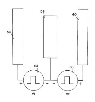
FIG. 4A is a schematic drawing of three in-line
electrodes and their connections to two pulse generators.
FIG. gB is a schematic drawing of a stimulating
cathodal electrode and a distant anodal electrode and their
connections to one pulse generator used in monopolar
stimulation.
Fig. 5 shows simultaneous pulses from two pulse
generators. FIG. 6 shows alternating pulses from two
- pulse generators.
FIG. 7 shows the resulting electrical potential field
when a single pulse train of FIG. 5, generated by the
circuit of FIG. 4B, is applied to the model, with the field
being represented by isopotential lines.
FIG. 8 shows the resulting potential field when two
simultaneous pulse trains of equal amplitude, generated by
the circuit of FIG. 4A, are applied to the model.
FIG. 9 shows the recruited area related to the
potential field shown in FIG. 7 using the single pulse
train circuit of FIG. 4B.
FIG. 10 shows the recruited area related to the
potential field of FIG. 8 with two simultaneous pulse
trains of equal amplitude when using the circuit of FIG.
4A.
FIG. 11-shows the resulting potential field when the
amplitude of the pulse train, generated by V2 of FIG. 4A,
is set equal to zero, with electrodes 58 and 6D having the
same negative voltage and both acting as cathodes.
FIG. 12 shows the recruited area related to the
potential field of FIG. 11, with the pulse train generated
between electrodes 58 and 6D having a zero amplitude such
that the electrodes have thesamenegative voltage.
FIG. 13 shows the resulting potential field when two
simultaneous pulse trains of equal amplitude are applied to
66742-557
CA 02180849 2000-03-24
the model with the center electrode offset 1.0 mm. from the
spinal cord midline.
FIG. 14 shows the recruited area related to the
potential field of FIG. 13 having two simultaneous pulse
5 trains with the same amplitude and with the center
electrode offset 1.0 mm from the spinal cord midline.
FIG. 15 shows the resulting potential field when two
simultaneous pulse trains are applied to the model with the
pulse amplitude between electrodes 50' and 58, V1, lower
than the pulse amplitude between electrodes 58 and 60, V2,
and the center electrode being offset 1.0 mm from the
spinal cord midline..
FIG. 16 shows the recruited area related to the
potential field of FIG. 15 having two simultaneous pulse
trains with different amplitudes, and with the center
electrode offset 1.0 mm from midline of the spinal cord.
FIG. 17 shows the recruited area when two simultaneous
pulse trains of equal amplitude are applied to the model,
with the center electrode centered at the spinal cord
midline.
FIG. 18 shows the recruited area when the alternating
pulse trains of equal amplitude from FIG. 6 are applied to
the model.
FIG. 19 shows a schematic of the pulse generator
driving a first embodiment of the lead.
FIG. 20 shows a schematic of the pulse generator
driving a second embodiment of the lead.
DETAILED DESCRIPTION OF THE PREFERRED EI~ODIMENTS
Fig. 1 is a schematic view of a patient IO having an
implant of a neurological stimulation system employing the
present invention to stimulate spinal cord 12 of the
patient. The preferred system employs implantable pulse
generator 14 to produce a number of i.~.depende.~-. stimulation
pulses wcich are sent to spinal cord .2 by insulated lead
15 and cou~le~ to the spinal cord by electrodes located at
point 18.
Implantable pulse generator 14 preferably is an ITRSL
IiR impla.~.tab_e pulse generator available from Medtronic,
*Trade-mark
21808~~~
W0 95119804 PCT/US95100906
6
Inc. with provisions for multiple pulse-outputs which are
selectably either simultaneous or with one shifted in time
with respect to the other, and which are selectably of
independently varying ata~ilitudes. This preferred system -
employs programmer 20 which is coupled via conductor 22 to
radio frequency antenna 24. This permits attending medical
personnel to select the various pulse output options after
implant using radio frequency communications. While the
preferred system employs fully implanted elements, systems
employing partially implanted generators and radio-
frequency coupling may also practice the present invention.
Fig. 2 is a cross sectional view of spine 12 showing
implantation of the distal end of insulated lead 16 at
point 18 within epidural space 26. Also shown is the
subdural space 28 filled with cerebrospinal fluid (cfs),
vertebral body 30, vertebral arch 31, and dura mater 32.
The following models were developed to compute the
effects of multiple superimposed field stimulation-of the
spinal cord particularly related to the problems of
paresthesia coverage and unwanted motor responses. The
results obtained show that using multiple field stimulation
it is possible to change the paresthesia pattern from
symmetrical to asymmetrical or vice versa'to correct for
changes in paresthesia pattern due to postoperative lead
displacement, and also--to reduce the activation of dorsal
root fibers in favor of dorsal column fibers to reduce the
occurrence of motor responses. After the explanation of
the models, the invention incorporating the information
provided by the models will be described.
Two complementary models provide the theoretical basis
for the instant invention. One model is a three
dimensional volume conductor model of the spinal cord and
its surroundings which incorporates the major macro
anatomical structures with the corresponding electrical
conductivities and the stimulating electrodes. -The second
model represents the electrical properties of the largest
myelinated dorsal root and dorsal column nerve fibers.
These models are extensively described by J. J. Struijk in
his Doctor of Philosophy thesis at the University of
CA 02180849 2000-03-24
" 66742-557
Twente, the Netherlands "Immediate Effects of Spinal Cord
Stimulation".
In order to assess the direct effects of stimulation
on the nerve fibers a two step procedure was followed.
First, the potential field in the volume conductor model
was calculated. Second, this field was applied to the
nerve fiber model to determine which fibers are excited by
the stimulation. The results of these calculations, shown
in later figures as isopotential lines and nerve fiber
recruitment areas in the -dorsal columns oW the spinal cord,
provide the effects of changing various stimulation
parameters.
Three dimensional volume conductor models of the spine
12 were developed using a simplified model of a transverse
section of the midcervical spinal cord as shown in Fig. 3.
A similar model of the midthoracic region was also studied.
Fig. 3 shows the spinal cord-composed of gray matter 40,
white mater 42, cerebrospinal fluid (csf) 44, epidural
space 46, vertebral bone 48, surrounding tissues
represented by layer 50, and a thin layer of dura mater 54.
This figure also shows electrode contact insulation 52 and
electrical contacts 56, 58 and 60 for two channel
stimulation. The electrodes 56, 58 and 60 are positioned
in the dorsal epidural space 46 next to the dura mater 54.
The elect_ical conductivities for these various
elements are given in the following table A. The thickness
of the dorsal csf Layer was measured from magnetic
resonance imaging (MRI) scans obtained from twenty six
subjects. In the midcervical and the midthoracic models
the average t::icknesses of the dorsal csf layers, 2.4 mm
and 5.6 mm respectively, were used.
she t'.~.=ee dimensional volume conductor model was
made up of disc=ete elements using a rectangular grid with
variable grid spacings. The length of the model was 60 mm.
The number o. grid points was 57 times 57 times 57 which is
equal to 185,193. A finite difference method was used to
2~ ~a~49
WO 95119804 PCT/US95/00906
8
apply the governing Laplace equation to discrete elements.
The resulting set of linear equations was solved using a
Red-Black Gauss-Seidel iteration, with variable
overrelaxation.
S A fiber model for the dorsal column fibers was based
upon D.R. McNeal's "Analysis of a model for excitation of
myelinated nerve", IEEE Transactions Biom. Eng., Vol. 23,
pp.329-337, 1976-. In the-model used here collaterals
entering the dorsal and ventral horns (grey matter) of the
spinal cord model are attached to every second node of
Ranvier of a 21-noded fiber. The diameters of these
collaterals were one third of the diameter of the
corresponding dorsal column fiber which was 10 micrometer.
For the dorsal root fibers a cable model with a curved
trajectory was used, the proximal end being connected to a
dorsal column fiber model. The dorsal root fiber model had
a diameter of 10 micrometer. In order to assess the direct
effects of stimulation on the nerve fibers, the potential
field in the volume conductor model is calculated and then
the potential field is applied to the nerve fiber models to
determine which fibers are excited by the.stimulation.
TABLE A
CONDUCTIVITIES OF fiHE VOLUME CONDUCTOR
COMPARTMENTS [S/sq. m.}
grey matter 0.23
white matter- (longitudinal) 0.60
white matter (transverse) 0.083
cerebrospinal fluid 1.70
epidural space 0.040
dura matter D.030
vertebral bone DØ40 .
surrounding layer 0.004
electrode insulation O.DOI
These models were used to evaluate the, differences
between a stimulation field developed by pulses from a
single pulse generator and a stimulation field developed by
2I8Q84~
WO 95119804 PCTlU595100906
9
two separate pulse sources. The circuit of Fig. 4A was
used for the two souYCes stimulation model having
electrodes 56, 58 and 60 and V1 voltage source 64 and V2
voltage source 66. Electrode 58 has a median position -
while electrodes 56 and 60 have lateral positions with
respect to the spinal cord.
The circuit of Fig. 4B was used for a single source
monopolar stimulation model having voltage source 65
applied between electrode 59 and the outside of layer 50 of
the spinal cord model of Fig. 3. The outside of layer 50
is used for-the reference connection because the positive
anode of V3 from voltage source 65 is assumed to connect to
the case of the implantable pulse generator, which is
distant from the spinal cord. The electrode areas used
in the models were approximately 12 square millimeters in
size because this size has been approved by the United
States Federal Drug Administration. The contact separation
is larger than the thickness of the dorsal csf layer to
reduce the shunting effect of this well conducting layer.
The contact separation is on the order of~the distance
between the dorsal root entry zone and the spinal cord
midline. In Fig. 4A, anode contacts 56 and 60 are longer
than cathode contact 58. This provides a shielding effect
by the outer (anodal) electrodes even if the lead is some-
what rotated in the coronal plane, which is the case if the _
lead has not been implanted perfectly rostrocaudally. The
shielding effect will diminish slightly if the outer anodal
electrodes 56 and 60 are shorter than the cathodal elec-
trode 58.
V1, V2 and V3 pulses, generated by voltage sources 64,
66 and 65 respectively of Figs. 4A and 4B, have a pulse
width of 210 microseconds. There are two modes of opera-
- tion for the two voltage sources 64 and 66. Mode one,
shown in Fig. 5, has simultaneous outputs of V1 and V2.
Mode two, shown in Fig. 6, has the outputs V1 and V2 offset
in time. There is also an independent amplitude control of
voltage sources 64 and 66 to provide different V1 and V2
amplitudes.
21~U849
W 0 95119804 PCTIUS95100906
Fig. 7 shows the resulting potential field represented
by isopotential lines 68 when th~,pulse is applied to the __
model by a single-cathode 59 and a distant anode 50 as
shown in Fig. 4B. Fig. 8 shows the resulting isopotential
5 lines 68 when two pulses with equal amplitudes are
simultaneously applied to the model, according to the '
scheme of Fig. 4A_ - Fig. 9 shows the resulting recruited
area 70 of dorsal column fibers with a diameter of 10
micrometer when a single cathode 59 is used with the same
10 model as that used in Fig. 7.- Fig. 10 shows the recruited
area 70 for two simultaneous pulses of equal amplitude
using the same model as in Fig. e. These figures show that
for stimulation with a transversely positioned tripole the
negative potentials and the recruited area of dorsal column
fibers are more restricted to the medial part of the dorsal
columns than in monopolar stimulation.
The shape of the recruited area of dorsal column
fibers does not differ significantly when mono-, bi-, tri-
or quadripolar stimulation with a conventional
longitudinal SCS electrode array is given, as was shown by
Holsheimer et al. using the same type of model (Stereotact
Funct Neurosurg 1991, Vol. 56, pp. 220-233). Calculations
also showed that dorsal root fibers need higher voltages
for their activation when stimulating with a transversely
positioned tripole, which will reduce the probability of
motor responses significantly.
The use of simultaneous pulses from two unbalanced
sources results in a controllable asymmetrical stimulation
which is impossible to attain with single source
stimulation. The. resulting isopotential lines 68 obtained
when V2 of Fig. 4A is set equal to zero, with electrodes 58
and 60 having the same potential, and applied to the model
is shown in Fig. 11. This shows how to obtain asymmetrical '
stimulation by merely using unbalanced sources with
multiple electrodes in a transverse plane, even though the '
electrode positions are perfectly symmetrical. Fig. -12
shows that a large shift in the recruited area 70 of dorsal
column fibers is also obtained using these unbalanced
2180849
WO 95119804 P(.°f1U595100906
11
sources. The example shown here is the.most extreme one
with V2 equal to zero volts.
If the lead is not at the spinal midline due to lead
migration, by lateral positioning during surgery, or to an
asymmetrical position of the spinal cord in the spinal
canal, it is still possible to obtain an almost symmetrical
stimulation. Fig. 13 shows the resulting isopotential
lines 6s and Fig. 14 shows recruited area 70 for an
electrode offset of 1.0 mm from~midline with V1 and V2
pulses simultaneous and of equal amplitude. The recruited
area is asymmetrical even though the voltage sources are
equal.
Figs. 15 and 16 show the results with an electrode
offset of 1.0 mm from midline and simultaneous inputs Vl
and V2 of Fig. 4A set equal to 2.26 volts and 4.52 volts
respectively to obtain an asymmetrical field. These
figures show that the shape of potential field and
recruited area are modified by this unbalanced input,
resulting in an almost symmetrical recruited area 70 in the
dorsal columns in Fig. 16.
Fig. 17 shows the recruited area 70 for equal
amplitude simultaneous pulses applied to the model by a
symmetrically positioned transverse electrode array, and
Fig. 18 shows the recruited areas 70 for equal amplitude
offset pulses of Fig. 6 applied to the model, which is the
union of two asymmetrical recruited areas.
The results of this modeling indicate that areas of
recruited spinal nerve fibers can be modified, when using
more than one source for stimulation of the spinal cord
versus single source stimulation, in that a variety of
parameters can now be changed to vary the stimulated area
and intensity. These parameter changes can obviously be
extended. For example, the effects of only two sources
were investigated here, but these same parameters can also
be changed if three, four or-more independent sources were
employed with analogous results.
The information developed using these models has been
incorporated into this invention in two embodimentsa Fig.
19 shows pulse generator 14 with positive going pulse
WO 95119804 218 0 ~3 4 9 PCTIUS95/00906
12
outputs 72, 74, 76, and 78 with respect to ground reference
80. The outputs at 72, 74, 76, and 78 are each selectable
in time as were V1 or--V2 of Fig.,6,-and each output can be
changed in amplitude independent of the other outputs or -
can be electrically disconnected. Line 16 has electrodes
38 connected to these outputs with wire 80A connecting
output 72 to electrode 38A, wire 80B connecting output 74
to electrode 38B, wire SOC connecting output 76 to
electrode 38D, wire 80D connecting output 78 to electrode
1D 38E, and wire 80E connecting ground reference 80 to
electrode 38C. Electrodes 38 have different sizes with
electrodes 38A, 38B, 38D and 38E, which are connected to
the voltage outputs of pulse generator 14, wider than
interspersed electrode 38C which is connected to ground
reference 80. This provides the Improved shielding effect
described previously.
With these connections and with the time and amplitude
variables of pulse generator 14 a stimulation field will be
set up between each electrode connected to a pulse
2D generator output and the interleaved electrode connected to
the pulse generator ground reference. The two modes of
stimulation, shown in Figs. 5 and 6, used in the modeling
- study are obtained by connecting pulse generator 14 to
electrodes 38 as described above. If a smaller number of
electrodes are used the unused outputs of pulse generator
14 are electrically disconnected.
Fig. 20 shows a second embodiment with pulse generator
14having additional outputs with the same characteristics
regarding the outputs, ground reference and capabilities as
before, and with lead 17 having electrodes 39. In this
second embodiment lead 17 has electrodes 39 connected to
the outputs of pulse generator 14 differently, with wire
80A connecting output 72 to electrode 39A, wire 80B
connecting output 74 to electrode 39C, wire 80C connecting
output 76 to electrode 39D, wire 80D connecting output 78 -
to electrode 39F, wire 80G connecting output 82 to-
electrode 39G, and wire 80H connecting output 84 to
electrode 39I. Wire 80E connecting electrode 39B to ground
reference 80, wire 80F connecting electrode 39E to ground
WO 95119804 PCTlUS9Sl00906
i
13
reference 80, and wire 80I connecting electrode 39H to
ground reference 80, establish the ground connections.
Electrode 39B is centered between the driven electrodes 39A
and 39C. Similarly, the ground referenced electrode 39E is
centered between the driven electrodes 39D and 39F, and
electrode 39H is centered between electrodes 39G and 39I.
With this second embodiment, the stimulation can be
applied at different spinal levels by using one out of
three combinations 39A, B, and C; 39D, E, and F; or 39G, H,
and I. Again, the unused outputs of pulse generator 14 are
electrically disconnected.
This system provides the capability to change the
depth and location of the stimulation by changing the
amplitude or timing of one field with respect to another.
The modeling of the fields described earlier shows that
results are changed markedly by the use of multiple pulse
generators connected to different electrodes positioned in
a transverse plane with respect to the spinal cord.
While this invention has been described with reference to
illustrative embodiments, this description is not intended
to be constructed in a limiting sense. Various modifica-
tions of the illustrative embodiments, as well as other
embodiments of the invention, will be apparent to persons
skilled in the art upon reference to this description. It
is therefore contemplated that the appended claims will
cover any such modifications or embodiments as fall within
the true scope of the invention.