Note: Descriptions are shown in the official language in which they were submitted.
~ W095119141 2 ! 8 1 1 7q PCT/US9~ClOû~52
BONE-CUTTING GUIDE
BAcK~ uNJ OF ~1~ INVENTION
The present invention relates to the field of
arthroscopic surgery, particularly anterior cruciate
ligament reconstruction.
one common approach to cruciate ligament reconstruction
is the use of the patellar tendon to form a bone-tendon-bone
graft. This involves cutting out a bone block from the top
of the patella. Deviations from proper technique for
removal of the patellar bone block have resulted in reports
10 of patellar tendinitis, patellar fractures and even, in some
instances, ruptures of the patellar ligament after the
grafting pLoceduLe. Whether or not these complaints are
properly related to the use of the patellar bone block in
cruciate 1 i~ 1_ reconstruction, these concerns are reason
15 enough to identify an alternate method for performing the
surgery .
Cruciate ligament reconstruction involves the drilling
of a tunnel through the tibia and into the femur. The
tunnels are used for in6ertion of a graft to replace the
20 damaged cruciate ligament. The accuracy in drilling these
tunnels is critical to providing a satisfactory repair. A
commonly u~ed method of directing a drill to form these
tunnels is by using a guide wire drilled through the bone
first. The location of the guide wire can be tested before
25 drilling out the tunnel. A cannulated drill is then
inserted over the guide wire to drill the full size tunnel.
When it has been desirable to retain the core of the bone
tunnel being drilled, a coring reamer is used. ~Iowever,
when a coring reamer is used over a guide wire, the guide
30 wire produces a stress riser in the bone core. Noreover,
the use of a guide wire is generally inadeguate to
accurately guide a coring reamer throughout the drilling of
an entire bone tunnel.
woss/1s1~1 2 ~ ~ ~ 1 7~ r~ sn~ls2
-- 2 --
SUMMAR~ QF rrTTF INV~NTI~N ~ = =
The present invention i8 directed, among other things,
to a bone-cutting guide and related method for drilling bone
to form, in one embodiment, a tibial tunnel and, if desired,
5 a femoral tunnel. The construction of the guide may be
replicated within the scope of the invention for use in
other areas of the body for accurate use of a coring reamer.
The tibial guide includes a base that has a cylindrical
tunnel f or guiding a cutting in:, LL ?r-t such as a coring
lO reamer therethrough. A positioning arm extends from the
base and has a distal end anchoring against the tibia within
a knee j oint. At least one anchoring pin is insertable
through the base alongside the cylindrical tunnel and into
the tibia for rigidly stabilizing the guide. A coring
15 reamer can be accurately guided by insertion through the
cylindrical tunnel.
In accordance with an ' _ '; r t of the invention, the
positioning arm can be adjustably mounted to the base within
a range of angles. The preferred range is from about 50 to
20 about 80~ between a seating portion of the positioning arm
and an axis through the cylindrical tunnel. In addition, a
distal end of the positioning arm is provided with one or
more spikes for stabilizing the end of the arm within the
knee j oint . The ~nho~; r L provides that the positioning
25 arm is curved between a first spike and a second spike so
that a cutting instrument such as a reamer operating through
the cylindrical tunnel is directed through an opening
created by the curve in the positioning arm.
The present invention in various embodiments is
30 directed to a bone-tendon-bone composite graft for use in
cruciate ligament reconstruction along with a tibial drill
guide for forming the tibial tunnel, a trefoil rasp for
forming ch~nn~l ~ in the bone tunnels and a bone block drill
guide for forming the bone plugs of the graft. The method
35 of the present invention is directed to 1; ~, L
reconstruction surgery. In a preferred embodiment, a bone
t~ ~1 s formed in e~h of two bo~-s of the joi-t. In knee
~ WO 951191~1 2 1 8 1 1 7 9 PCT/US9~100~52
-- 3 --
surgery, these are the femur and the tibia. Preferably, the
bone tunnel i5 formed by drilling a core out through the
bone such that the core might be used to form the bone plug
in the composite graft. The bone plugs are machined to form
5 two longitudinal substal~tially parallel grooves opposite one
another. At least one ligament replacement, ~uch as a
semitPnfl;n~ s~lC tendon, and/or gracilis, is extended between
both of two bone plugs along the parallel grooves in each
plug. The ligament rep~ A~- - L is attached to the two bone
10 plugs. Each bone plug is inserted into one of the bone
tunnels and secured therein by an interference screw. The
use of the bone-tendon-bone composite graft of the invention
results in a reconstructed cruciate ligament, also, in
accordance with an ~ ~;r L the present invention.
The invention further includes in another P~o~;r-rt a
bone block drill guide for forming the bone plugs required
in the composite graft of the present invention. The bone
block drill guide includes a main hole :Eor ~ Ating the
bone plug. First and second parallel holes that intersect
20 opposite sides of the main hole are used for directing a
drill ~it to cut a groove longi~ ;n~l ly along the bone
block .
A trefoil rasp in accordance with an ~ of the
present invention is used to file rh~nn~ in the bone
25 tunnels. Two ~hRnnPl ~, oppositely located from one another,
ac ~ -~lAte the l; ~ - L replacement attached between the
two bone grafts. The third channel is generally located
parallel to and equidistant from the other two ~hAnnPl ~:.
This third channel is us~d to guide an interference screw
30 along the bone tunnel adjacent to the bone portion of the
graft .
The composite graft in accordance with an ' ';- L of
the present invention is advantageously formed without
cutting into the patella. The trefoil rasp provides for
35 interference screw fixation without permitting the screw to
cut into the ligament replacement.
wo 95/191~1 2 1 8 1 1 7 9 PCT/US9S/00 152
-- 4 --
Other features of the present invention will become
apparent during the following description taken in
conjunction with the drawings.
BRIEF DESCRIPTION OF THE DRAWINGS
FIG. 1 is a peL:,~e-:Llve view in partial cross section
of a reconstructed l;, ~ formed with the present
invention .
FIG. 2 is an ;~ -tLiC view of a tibial drill guide of
the pre6ent invention.
FIG. 3a i5 an isometric view of a trefoil rasp.
FIG. 3b is an end view of the rasp of Fig. 3a.
FIG. 4a is an; ~ -tLiC view of a bone plug made by the
invention .
FIG. 4b is a side view of the bone plug of Fig. 4a.
FIG. 5 is a side view of a bone-tendon-bone composite
graft made by the present invention.
FIG. 6 is a cross-sectional view of FIG. l taken along
lines 6-6.
FIG. 7a is a side view of a bone block drill guide.
FIG. 7b is a plan view of the bone block drill guide of
FIG. 7a.
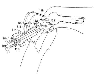
FIG. 8 is an isometric view of a tibial guide of the
present invention in use against a knee.
FIG. 9 is an isometric view of the distal end of the
25 positioning arm of the tibial guide of FIG. 8.
FIG. lO is an isometric view of a coring reamer of the
present invention.
nT~TATT Fn DESCRIPTION OF THE ~ ~ L1 EMBODIM~NTS
Referring now to the drawings, a lt:cu-._LLucted ligament
3 o lO for a knee j oint is shown in Fig . l in accordance with an
embodiment of the present invention. The cruciate ligament
reconstruction surgical operation can be conducted as an
open surgery, or preferably, through arthroscopic surgery.
The arthroscopic surgical method presently preferred for
35 carrying out the present invention shall now be described.
Arthroscopic diagnostic procedures are first conducted
without tournitauet control in order to allow sufficient time
-
2181 1 79
wo 95/191J1 . _I~u~ ?
-- 5 --
f or the ACL reconstruction procedure . Conventional
anteromedial and distal lateral portals are drilled to give
access to the knee joint for these procedures. The
procedures may include meniscotomy, mPn;C,~1 repair, removal
5 of loose bodies, debridement of anterior cruciate ligament
tear, etc. Notchplasty may be ~ c~cl under tourniquet
control. The boundary of the notchplasty should be
sufficiently wide (about 2 cm. ) and sufficiently posterior
to include the posterior lateral femoral cortex in order to
lO ensure accurate placemsnt and subseguent; c LLY .
In order to proceed with anterior cruciate ligament
L~cullaL,-ction, a vertical incision is made medial to the
tibial tubercle approximately 2 . 5 cm. in length. The skin
incision may be 1ln~9Prm;nPd in such a fashion as to provide
15 sufficient mobility for retraction, while harvesting the
tibial and femoral bon,e cores. A carefully placed
anteL, ~~;~tl tibial incision may begin approximately l cm.
medial to the tibial t1~lbercle and 2 cm. distal to the joint
line. Conventional surgical pLoceduL-~s are used to excise a
20 semitDn~l;nn~llC tendon, and, if desired, the ~cc -nying
gracilis . While the u~e of the semitPn~; no~cus tendon and
gracilis is one .~ nt of the invention, alternative
ligament repl~c- ~ materials may be substituted for use in
the composite graft of the invention.
The two major bones that meet at the knee joint are the
tibia 12 and the femur 14. A bone tunnel 16 is drilled
through each of these ~wo bones. The tunnels 16 may be
drilled with a regular drill that crushes and removes the
bone within the tunnel. However, it is preferable to use a
30 coring reamer to drill the bone tunnels. The reamer drills
out a core of bone through each of the bone tunnels. The
bone core can then be used to form a bone plug in the
composite graft that will be replaced when lecui,~LLucting
the ligament. In using the coring reamer to drill out a
35 core that may be reused in the composite graft, it is
important that a guide pin not be inserted into the core for
directing the reamer. The hole formed by the guide pin
WO 95/19l~l 2 f ~ 1 1 7 9 P~T/Uss~l00~52 ~
-6-
through the center of the core would form a stress riser in;
the bone plug making the bone plug su~ject to fracture. A
tibial guide 30 in accordance with an embodiment of the
present invention properly orients and guides a coring
5 reamer for making the bone tunnels without a guide wire.
Referring now to FIG. 2, the tibial guide 30 is shown.
A pipe 32 is oriented at approximately 55' to horizontal.
The pipe 32 provides a cylindrical tunnel that serves to
guide a coring reamer 33 or other drill inserted
10 therethrough. With the patient's leg held fixed at
approximately 110 to 120, the guide can be used for
drilling both the tibial tunnel and then the femoral tunnel.
Therefore, a portal for the drill is not required behind the
femur and a closed tunnel can be drilled. Both tunnels are
15 drilled through the tibia from the anter~ 1 ti~ial
incision.
A positioning arm 34 is attached to the pipe 32. The
positioning arm 34 has a fork 36 at its far end. The fork
36 has two rounded prongs. The fork 36 is attached to an
20 arcuate portion 38 of the arm 34. The arcuate portion 38
allows for maneuverability of the arm 3~1 within the knee
area upon insertion through the antt~ ;Al portal.
Molnwhile, the arthrosoope is inserted into the knee joint
through the distal lateral portal. The fork 36 needs to be
25 placed against the leading edge of the posterior cruciate
ligament. The positioning arm 34 is shaped and oriented
with respect to the pipe 32 so that the hole drilled by a
reamer or drill through the pipe 32 is directed through the
tibia to a point approximately 7 m; 11 ;--ters from the
30 leading edge of the posterior cruciate ligament. The center
of the tibial tunnel is further defined by the tangent to
the center of the inner circumference of the anterior one-
third of the lateral meniscus.
An adjustable rod 40 is attached to the pipe 32 at one
35 end . A calf strap 41 secures the guide to the patient ' s
leg. The guide has an ankle strap 42 proximate the opposite
end of the rod 40. The rod 40 can be adjusted in length to
W09S1191J,1 2 t 81 t 79 r~l~u. c~
-- 7 --
~c~ '~te different leg sizes. The calf strap 41 and
ankle strap 42 provide anchors for achieving and maintaining
proper orientation of the pipe 32. The straps are affixed
with the f ork 3 6 oriented properly around the PC~ attachment
5 on the tibia.
Another anchor to securely orient the pipe 32 i6
provided by a K-wire 44. The K-wire 44 i8 6hot through the
skin of the patient'6 leg and into the tibia. The K-wire 44
may be positioned on the guide 30 closely adjacent the pipe
10 32 so that the hole formed in the tibia by the K-wire is
adj acent and parallel to the hole to be drilled through the
pipe 32. The anchoring provided by the cup 36, the ankle
strap 42 and the K-wire 44 stably and correctly position the
pipe 32 for guiding a coring reamer or a drill. The tunnels
15 may thus be cored without a guide pin in the core. The
tibial tunnel i6 reamed f irst and the core removed . The
knee is flexed or extended a variable amount in order to
properly position the f emoral tunnel . A longer coring
reamer i5 then directed through the tibial tunnel for
20 drilling in and through the femur. The bone core from the
femur is removed. Stan,dard deburring and debridement
procedures are followed.
A presently preferred ~r,ho~lir l_ of the tibial guide is
shown in FIG. 8. Rigià, stabilization of the guide against
25 the knee is accomplished without the need for an ankle
strap. A base 100 has a longitudinal open pass2ge for
receiving a guide tube 102. The guide tube 102 has a
cylindrical tunnel 104 longit~ ;n~l ly therethrough. Guide
tubes may be made with different diameter cylindrical
30 tunnels to ~ te reamers or drills of ccLLc!~u-lding
diameter. The cylindrical tunnel 104 must be tight enough
around the reamer to accurately guide it but wide enough to
permit the reamer to rotate therein. On either side of the
cylindrical tunnel, anchoring pins 106 or outriggers may be
35 inserted. The two anchoring pins 106 provide two points of
stability for the base against the tibia. The anchoring
pins 106 have pointed ends 108 that can pierce skin.
WO 95/191~ 8 ~ ~ 7 9 PCT/Us9sMO~s2 ~
Therefore, it is not neces6ary to insert the pins through an
incision, and they can be used to poke their own holes. The
anchoring pins 106 securely engage the anterior margin of
the tibia. The anchoring pin6 are notched along their
5 length on one side. Inside the base loo, the notches llo
engage internal ridges to act like a ratohet. A6 the
anchoring pins 106 are extended out from the base the
notches click again6t the ridges. The anchoring pins 106
are prevented from retracting by the notches and ridges. To
10 remove an anchoring pin from a patient, the handle on the
proximal end of the anchoring pin is turned 180 to
disengage the notches from the ridges. Then the anchcring
pin can move freely longi~ inAl ly in either direction. The
handle of the anchoring pin may advantageously be arranged
15 as a flag which pcints horizontally outward ~rom the base
when the notches are engaged and points inward toward the
other anchoring pin when the notches are disengaged. The
adjustability of the anchcring pin6 a~ ~ ~Ates the
variation in leg 6ize encountered from patient to patient.
Once the anchoring pins 106 have been adjusted, the
guide tube 102 can also be reciprccally adjusted. The top
outer surface 112 of the guide tube preferably has a flat
portion for en~a~, t with a set screw. A knob 11~ on top~
of the base can be turned to tighten or release the screw
25 frcm the guide tube. With the screw released, the guide
tube 102 is pushed up against the tibia to thereby provide
three points of engagement near the entrance of the tunnel
to be drilled.
A curved track 116 is securely attached vertically to
30the base 100. A positioning arm 118 is adjustably mountable
on the curved track. A screw handle 120 on the positioning
arm is used to tighten the arm 118 against the vertical
track 116. The positioning arm 118 can be slid along the ` A
track 116 to assume a range of positions. The range alters
35 the angle made between an axis of the cylindrical tunnel of
the guide tube and a seating portion 122 at the distal end
of the positioning arm within a range of between about 45-
-
WO 95~19111 2 ~ 8 ~ ~ 7 ~ F~ g~ 2
g
50 and 80. By providing a sufficiently large minimum angle
of between 45 and 50, the tibial guide ensures that the
tibial tunnel is not dr illed at too shallow an angle .
The seating portion 122 of the positioning arm at the
5 distal end of the arm is provided with two anchoring spikes
124 and 126. The spikes project from the end of the
positioning arm for insertion into the top of the tibia.
The spikes precisely define the exit end of the tunnel to be
drilled through the tibia, and thus permit the surgeon to
10 know on inspection and control the tunnel ' s location.
Therefore, it is desirable to place the spikes so that the
anatomic center of the anterior cruciate ligament is located
midway therebetween. The spikes are sharp so that they may
dig into the tibia. A first spike 12~ extends from near the
15 end of the positioning arm. The seating portion 122 of the
po6itioning arm is curved between the first spike 124 and a
second spike 126. The curve defines and identifies an open
region to permit clearance for a coring reamer used in
operation to drill a hole through the bone. Thus, the
20 positioning arm does not inter~ere with the drilling
proce~s. Moreover, the seating portion 122 partially
encircles the tunnel to be drilled and the spikes are
inserted on opposite sides of the tunnel to be drilled. The
two spikes on the positioning arm and the two anchoring pins
25 through the base provide four points of stabilization which
make for a completely rigid attachment between the tibial
guide and the tibia. The attachment is advantageously rigid
in three dimensions.
With the tibial guide rigidly attached to the tibia, it
30 is a relatively simple matter to insert the coring reamer
through the cylindrical tunnel and drill an accurate tunnel.
Advantageously, the bone core is not exposed to the damage
ordinarily ?1~_ ~nying the use of a guide wire. However,
without the hole through the bone caused by a guide wire,
35 the bone core plugs up the coring reamer with almost fluid- -
tight engagement. Pulling the bone core out from a
conventional coring reamer would be difficult because they
WO 95/19111 ~ 1 7 9 PCT/US95100.152
- 10 -
are generally solid cylinders. Air or other fluid cannot
get in behind the bone core so that pulling on the bore core
tends to create a vacuum behind the bone core. The suction
of the vacuum pulls the bone core into the coring reamer
5 making it difficult to remove. In accordance with the
present invention, a coring reamer 130 is used that is
slotted to permit air or fluid in behind the bone core as
shown in FIG. lO. The slots or openings 132 are a greater
distance from the cutting edge 134 of the reamer than the
lO length of a bone core to be drilled out. A rod can be
inserted through a slot or opening behind the bone core to
easily push the bone core out from the reamer. A bone core
obt2ined using the tibial guide and coring reamer of the
invention may be used and replaced in the body as a part of
15 the graft used for the lcOcoll -L~ uCtiOn.
If cores have been drilled out from the bone tunnels
they may be used for the bone plugs 25 otherwise, donor
bone, namely allograft bone, can be used to make the bone
plugs. Referring now to FIGS. 4a and 4~, whatever bone plug
20 25 is used, two longitudinal substantially parallel grooves
50 are drilled on opposite sides of each bone plug. The
grooves provide a recess in which the semit~ ; nnsllC tendon
20 and gracilis 21 can be seated. A notch 52 may also be
drilled, if desired, across one end of the bone plug so that
25 the tendon can be wrapped alongside and around the end of
the bone plug, without protruding excessively from the plug.
The notch 52 is not required because the bone tunnel is open
at each end providing no restriction on the tendon
projecting above the end of the graft. It is also
30 advantageous to provide suture holes 27 through the bone
plug for attaching the tendon to the plug. The suture holes
27 are drilled into the grooves radially through the bone
plug and from one of the substantially parallel grooves 50
to the other. In a presently preferred ~mho~; L, three
35 such suture holes are drilled through the bone plug.
In order to easily and efficiently form a bone core
into the desired bone plug for a composite graft, a bone
.
wo 9~/191~1 2 1 8 1 1 7 ~ PCT/US9C100~2
block drill guide 60 of the invention as shown in Figs. 7a
and 7b may be used. Thl~ drill guide 60 features a central
substantially cylindrical column 62. The central column 62
includes a pair of opposing curved walls 64 having a center
5 of curvature substantially coincident with the center axis
through the column 62. The curved walls 6~ are shaped so as
to hold a bone core parallel with the axis of the column and
substantially centered within the column. A second pair of
opposing curved walls are arranged at 180 to each other
l~ with respect to the central column formed by the curved
walls 64. This second pair of walls are the drill guide
walls 66. The drill guide walls 66 form two parallel
columns on opposite sid2s of the central column. The drill
guide walls 66 have a shorter radius of ~:ULV-LULe th2n the
15 first pair of opposing curved walls 64. In accordance with
a presently preferred ~mho~ L, the inner diameter of the
drill guide walls 66 is 6 mm whereas the inner diameter of
the first pair of opposing walls 64 is ll mm. The central
column 62 is mounted over a base 68. A bone core standing
20 in the central column 62 rests on the base 68. The base 68
is provided with holes therethrough in alignment with the
open circular cylinder formed within the drill guide walls
66. The base 68 may also include legs 70 for supporting the
drill guide over a table. For drilling suture holes through
25 the bone block, holes 72 are arranged horizontally through
the drill guide walls 66. Three holes 72 are preferably
aligned in a line.
The substantially parallel grooves 50 are drilled by
inserting the bone core or allograft into the center chamber
30 of the column 62 formed by the opposing curved walls 64. A
drill is directed in the column 62 along each of the drill
guide walls 66 in succession. Thus, parallel grooves 50 are
formed on opposite sides of the bone core. The drill may be
equipped with a stop tc prevent the drill from being
35 directed too far down through the column where it may
contact the table beneath. A drill bit inserted through the
holes 72 can be easily directed through the center of a
WOgS/191~1 2 ~ P~ 'C~1S~ ~
-- 12 --
groove drilled along the bone core. The suture holes
drilled through guide holes 72 preferably extend from one
groove to the opposite groove in the bone block.
The semi~Pn~; nr~511c tendon 20 and/or gracilis 21 is
5 extended between both of the bone plugs 2~. The tendons are
seated inside the two substantially parallel grooves 50 and
about an end of each bone plug. The tendons are preferably
sutured to themselves to form a double loop as shown in FIG.
5. Sutures are also used through the suture holes to attach
l0 the tendon to each of the bone plugs. The tendon strands
may be straight or twisted between the bone plugs. Twisting~
will shorten the length of the graft. A ligament
replacement of an emho~ L of the invention may include
both the semi~n~;n~sllC tendon and the gracilis. As such
15 four strands will connect the two bone plugs. Other
Ls of the invention may use one or the other of the
semitan~;nrlsllc tendon and gracilis. still further
Ls of the invention may substitute or combine man
made or arti ~ fibers or human tissue for the tendons
20 for use as the ligament rep~ ~c. t.
In affixing the composite graft 80 within a bone
tunnel, contact between a screw 82 and the tendon should be
avoided 80 as not to cut or tear the tendon. To better
insure that the screw is out of contact with the tendon, an
25 interference screw should be driven along the bone portion
of the graft between the graft and the bone tunnel wall. A
trefoil rasp 9o of the present invention is re~ 1e~ for
use prior to fixation of the graft. As shown in Figs. 3a
and 3b, the trefoil rasp 90 has three longitudinal lobes for
3 0 use in cutting three ch 1nnPl ~ into each of the bone tunnels .
Reciprocating v~ L of the trefoil rasp 90 in and out of
the bone tunnels 16 serves to file away ~he tunnel walls to
form the desired ch~nnPl 8. Two of the longitudinal lobes 92
are 180 apart on the rasp, These longitudinal lobes 92 are
35 used to form ~h~nnPl5 for A~ ting the semitPn~9;n~s11c
tendons 20 and gracilis 21 seated in the parallel grooves of;
the bone graft. When the gracilis 21 is attached along and
.. _ .... _ , . . . .. _ _ _ _ . , .
1~ WO 95119111 2 ~ 9 PCTIUS95/00~C2
-- 13 --
on top of the semitendinosus tendon 20, the chAnnP1~ are
required to provide roolm for the graft to fit within the
tunnel .
The third longitudinal lobe 94 is located parallel to
5 and equidistant from the two opposed lobes. Looking at the
end of the ra6p as in Fig. 3b, the third lobe 94 is
preferably 90 to each of the other two lobes. In the
presently preferred ~_a; L, the third lobe 94 projects 2
mm. from the rasp shaft whereas the other two lobes 92 each
10 project 3 mm. from the shaft. The third lobe 94
advantageously files aw~ly a channel along which an
interference screw is d~-iven as shown in FIG. 6. The
channel helps to maintain the screw 6traight adjacent the
bone portion of the gra~ t. Advantageously, the tibial guide
15 30 and the trefoil rasp 90 can complement one another in
forming the channel for the screw. The hole formed by the
X-wire 44 of the tibial guide may be used as the screw
channel. To achieve thi s result, the trefoil rasp should be
aligned in the tibial tunnel with its third lobe 94
2 0 overlapping into the K-~ire hole .
After the rhAnnPl ~ have been filed in the bone tunnels,
the 6utures 84 hanging from one end of the composite graft
are attached to a needl~, a passer or other conventional
graft plA~ - L tool. The passer is inserted through tibial
25 and femoral bone tunnels and out through the skin on the
posterior side of the lcrlee. The passer is removed leaving
the suture hanging from the posterior end of the graft and a
suture at the other end of the graft hanging out through the
tibial incision. The sutures may be pulled on to properly
30 tension and locate the graft within the bone tunnels.
Alternatively, the graft: may be positioned within the bone
tunnels using a pushing device instead of a suture pulling
the graft into position.
Fixation of the graft is preferably acc~ l; ChPd with a
35 headless cannulated interference screw. The cannulated
interference screw can ~e carried by a guide wire extending
from the tip of an angled driver. The guide wire is
Wo 95/191~ 9 PcT/uss~loo~s2
-- 14 --
preferably a springy wire made of a material such as
Nitinol'm. The wire extends about 2 centimeters past the end
of a screw carried by the driver. For securing the
interference screw in the femoral tunnel, the angled or
5 flexible driver and screw are preferably inserted through
the anteL, -~1; A 1 portal . An angled driver and use thereof
i5 described in co-pending United States Patent Application
Serial No. 07/956,733 filed October 2, 1992. A flexible
slide may be used to provide a track to rollow from the
0 ant~ iAl portal to the channel in the femoral tunnel for
the interference screw. The insertion of a flexible slide
simplifies the guidance of the interference screw into the
channel of the femoral tunnel. Once the screw is properly
positioned in the tunnel, the driver can initiate screwing
15 and the slide can be removed. The oppositely located
~h~nn~ in the femoral tunnel hold the semit~n~l;nr~sue
tendon in position away from the interference screw as it is
screwed between the bone portion of the gra~t and the
channel of the bone tunnel . Upon f ixation of the
20 interference screw in the femoral tunnel, the angled driver
is removed.
The proper tension is then applied to the graft by
pulling on the suture hanging out from the tibial incision.
A driver and a headless cannulated interference screw are
25 then inserted through the tibial incision for driving the
screw along the channel formed in the tibial tunnel. The
sutures are cut and the incisions are closed. The
reconstructed knee upon fixation of the graft appears as in
Fig. 1.
While this operation has been ~ c~l~s~cl in terms of
using autogenou5 bone cores, alternative sources of bone
plugs may be substituted. Allografts, in which donor bone
is freeze-dried or fresh frozen for preservation, are one
alternative. The freeze drying process kills cells in the
35 bone and may reduce the risk of transmission of infection.
Another alternative bone plug is the use of synthetic graft
material. With any of these alternatives, the bone plugs
W095119~41 2 ~ 9 I~ sr ~52
-- 15 --
may be shaped to appear as described above for the
autogenous graft. With the allograft and the synthetic
graft, the coring reamer is no longer required and an
ordinary drill may be used instead for drilling the bone
5 tunnels.
The surgical technique of the present invention
advantageously makes use of the fact that the semitPn~l;nocus
and gracilis has less morbidity associated with harvesting
than does the patellar tendon. It is further advantageous
lO to use a coring reamer and a bone block drill guide of the
invention to remove the bone core6 from the bone tunnels in
the tibia and femur and shape them to A~ Ate the
6emitPn~9; n-~s11c tendon. ~he trefoil rasp provides the still
further advantage of maintaining alignment of the graft and
15 interference screws in the bone tunnel so that the screw is
directed adjacent only the bone portion of the graft.
Of course, it should be understood that various changes
and modifications to thl~ preferred ~ ts de6cribed
above will be apparent to those skilled in the art. For
20 example, the bone block~3 in the composite graft may be
autogenous, allogenic or synthetic. The tendon or other
ligament replacement used on the graft may be one or more
strands sutured to both of the bone blocks along the
grooves. Noreover, all:ernative equipment may be used for
25 drilling the grooves and the bone plugs. ~hese and other
changes can be made without departing from the spirit and
the 13COpe of the invention and without ~l;m;n;ch;nq its
attendant advantages. l:t is therefore intended that such
changes and modifications be covered by the following
3 0 claims .