Note: Descriptions are shown in the official language in which they were submitted.
3 389
BIOPSY NEEDLE
The present invention relates to biopsy
needle equipment for acquiring samples of
body specimens from a living patient.
- Description of the Prior Art -- ~
Biopsy needle equipment is available
which locks a stylet to a needle and which
also provides some means of connecting a plug
or aspirating equipment to the proximal end
of the needle lumen. The problem with this
equipment- has been that secure attachment of
the stylet to the needle is not provided, and
that a connector for a plug or aspirating
equipment normally extends proximally from a
handle used to force the needle and stylet
through tissue. The first presents a
considerable problem since a great deal of
force must be exerted on the stylet and
needle when bone is penetrated. The second
presents a problem in exerting this force
- 2181389
since the connector is opposite the hand
which makes generating a considerable amount
of force uncomfortable, if not impossible.
The instant invention overcomes the
first problem by dividing the handle used for
exerting force into proximal and distal-parts
which can be attached securely together but
separated readily. The handle parts are
attached respectively to the needle and the
stylet which results in these parts also
being attached securely together. The second
problem is solved by having a Luer connector,
mounted within the handle and connected to
the needle lumen, exposed only when the
handle parts are separated. These handle
parts provide an ergonomically shaped handle
only when required and are separated after
penetration.
- 2181389
Summary of the Invention
The present invention is a device for
acquiring a sample of tissue or fluid from a
patient, generally referred to herein as a
biopsy needle assembly. The invention may
also be packaged as a kit which includes all
necessary components for performing sampling
of tissue. The invention also includes a
method for utilizing the device of the
present invention in acquiring the biopsy,
fluid or tissue sample. The biopsy needle
assembly is specifically designed to include
features which prevent the loss of any
portion of the tissue or fluid sample during
extraction of the biopsy needle from the
incision track after a sample has been
captured within the lumen of the needle or
remove a sample by aspiration. Thus, the
biopsy needle assembly includes a concealed
male portion of a Luer connector to sealingly
receive a mating plug or aspiration
apparatus.
S~1~1,3~9
Entering a biopsy needle through bone
into bone marrow requires that a great deal
of force be exerted on the needle by the
physician. This is greatly facilitated here
by an ergonomically shaped handle which
closely fits the hand and permits generating
a great deal of force against the needle with
the force being optimally distributed.
The biopsy assembly includes a handle, a
stylet and a needle with a lumen extending
from the distal to the proximal end being
sized to accept the stylet. The handle is
formed of two parts, a proximal part and a
distal part. The opposing surfaces of the
handle parts are generally planar. The
proximal handle part is attached to the
proximal end of the stylet, and the distal
handle part is attached near the proximal end
of the needle. When the stylet is inserted
into the needle lumen, the planar handle
surfaces face each other.
~18 1 383
An extension, shaped like a cylinder
with a proximal planar surface, is formed
around the proximal end of the needle and
attached extending proximally from the planar
surface of the distal handle part. The
extension has two outwardly extending opposed
~ flanges of different widths, offset from but
lying in the plane of the extension proxi-mal
end. A male Luer connection, attached to the
proximal end of the extension and around the
needle, extends proximally from the
extension.
A cylindrical shaped recess into the
planar distal surface of the proximal handle
part is sized to accept the proximally
projecting extension and Luer connector
extension. This recess has wings of opposed
outward unequal width extensions sized to
mate with and receive the opposing flanges,
when the extension is placed within the
recess with the handle parts in a single
orientation with respect to each other. The
13~9
unequal width flanges and mating unequal
width wings provide only this single
orientation, where the extension and flanges
can be inserted into the recess and its
wings. This single orientation occurs when
the handle parts are at right angles to each
- other. - -
Two slots, each beginning at a wing,
extend outwardly around the recess in the
same direction approximately one-quarter of
the distance around the recess circumference.
The slots are located such that each is
aligned with the edge of the adjacent flange,
when the extension and flanges are placed
within the recess and wings with the handles
at the required right angle relationship.
Each slot is made wide enough to receive the
edges of the adjacent flange, and deep enough
to permit rotation of the flange through the
slot. Since each slot extends in only one
direction around the recess circumference
from its respective wing, the two handle
2181389
parts can only be rotated in one direction
relative to each other. Since the slots
extend around the recess about one-quarter of
the recess circumference, this permits
rotating the two handle parts from the
insertion right angle orientation into
alignment. The terminus of each slot also
extends through the side of the handle to-
permit observation of alignment.
Restricting the final angular
relationship of the two parts to one aligned
angle of the handle parts is important,
because the distal end of the stylet and
needle may be inclined. If they are
inclined, then the orientation of the stylet
with respect to the needle must be fixed so
the inclination angles will match each other.
Stops are provided to ensure that the
handle parts are locked in alignment. The
stops are provided by two studs and two
arcuate grooves. The studs project distally
Z~ ~3~9
from the planar surface of the proximal
handle part near each end. These studs
engage arcuate grooves in the proximal planar
surface of the distal handle part, which are
formed to be in the path of the studs when
the two parts are rotated with respect to one
other. These arcuate grooves extend only far
enough to permit rotation of the two hand~le
parts into alignment.
After the two handle parts are locked
together in alignment they form a complete
ergonomically shaped handle, which permits
the surgeon to exert greater force. In use,
the handle parts are gripped in one hand.
Being gripped together assures that the two
handle parts stay in alignment and locked
together in use.
The handle parts are attached before
forcing the needle and stylet through the
bone, since this requires a great deal of
force. After forcing the needle and stylet
- ~1813~
through the bone the male Luer connector
extension is then exposed by removing the
stylet from the needle after merely rotating
the two handle parts in the proper direction
to right angles to each other. This permits
attaching a plug, which mates with the Luer
connector, and is provided for sealing the
needle lumen before removing the sample. ~
This improves this procedure by blocking air
flow. Aspiration apparatus can also be
attached to the male Luer connector instead
of the plug in order to aspirate the sample.
A Luer attachment connector, of
necessity, has a relatively sharp end which
is covered here by an ergonomically shaped
handle. This ergonomic handle shape greatly
increases the force that can be generated by
the physician because the resulting close
hand match distributes the applied force more
evenly. Locating an attachment means for a
plug or aspirating apparatus within the
handle provides an attachment capability for
~1~138~
a plug or aspirating apparatus without
changing this optimum ergonomic handle shape.
In addition, this method of attachment
provides a secure attachment of the stylet to
the needle with known orientations between
the two to permit using inclined distal ends
~ on these parts. However, since the handle-
parts are readily separable, this attachment
capability is provided with a minimum of
operator inconvenience.
218138~
Brief Description of the Drawings
In the drawings, in which like reference
numerals indicate corresponding parts or
elements of preferred embodiments of the
present invention throughout the several
views:
_
Figure 1 is a front elevational view-of
the invention;
Figure 2 is a perspective view;
Figure 3 is an exploded perspective
view;
Figure 4 is a top plan view of biopsy
needle and distal handle part with the distal
handle part removed;
Figure 5 is a bottom plan view of the
proximal handle part with the distal handle
part removed and the needle in place inside
the distal handle part;
- 21813~
Figure 6 is a fragment of the handle
parts in cross-section showing only the
extension from the distal handle part and the
mating and adjacent portions of the proximal
handle part;
~ Figure 7 is a view of the proximal--
handle part and needle with a cut-out to show
the interior construction;
Figure 8 is a front elevational view of
the invention with proximal handle part and
needle removed;
Figure 9 is a side elevational view of a
plug;
Figure 10 is a side elevational view of
an alternative embodiment of the invention;
and
Figure 11 is a side view of the probe.
- 2181~89
Detailed Description of the Invention
Detailed embodiments of the present
invention are disclosed herein. It is to be
understood, however, that the disclosed
embodiments are merely exemplary of the
present invention which may be embodied-in-
various forms. Therefore, specific details
disclosed herein are not to be interpreted as
limiting, but rather as a basis for the
claims and as a representative basis for
teaching one of skill in the art to variously
practice the invention.
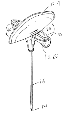
Referring now to Figure 1 a front
elevational view of a biopsy needle assembly
10 is shown. Assembly 10 uses a two part
handle 12 consisting of a proximal handle
part 12A and a distal handle part 12B which
together form a complete handle. Proximal
handle part 12A is formed around the distal
end of stylet 14, and distal handle part 12B
is formed around biopsy needle 16 near the
~1~13~9
distal end. As depicted here, stylet 14 is
shown extending beyond needle 16 through a
lumen 18, not shown, in the needle which
extends from the distal to the proximal ends
of the needle. The extension of stylet 14
beyond needle 16 provides a distal cutting
~ surface 20. - ~
In Figure 3, an exploded perspective
view of assembly 10, shows proximal handle
part 12A rotated 90 degrees from alignment
with distal handle part 12B, and with stylet
14 partially withdrawn from needle 16. In
Figures 3, 4, and 8 a cylindrical shaped
extension 22 formed around the distal end of
needle 16 extending proximally from surface
24 of distal handle portion 12B is shown. A
wide flange 26 and a narrow flange 28 extend
outwardly from extension 22 offset from and
generally parallel with the proximal end of
extension 22. A male Luer connector 27 is
attached proximately to the proximal end of
extension 22 and around needle 16. Four
14
~1 81389
voids 25, into distal handle part 12B from
surface 24, are provided for weight
reduction. Except for a stop to be described
later and the above described features
surface 24 is planar.
In Figure 5 proximal handle part 12A is
shown. Distal surface 30 of proximal handle
part 12A is also shown with a cylindrically
shaped recess 32 extending proximally inward.
Wing 34 and wing 36 are outward extensions of
recess 32 with wing 34 being wider than wing
36. Recess 32 is sized to accept extension
22 and wings 34 and 36 are sized to accept
flanges 26 and 28 respectively when stylet 14
in inserted through needle 16 with the
orientation shown in Figure 2. Only in this
orientation is wide flange 26 opposite wide
wing 34 and narrow flange 28 opposite narrow
wing 36 to permit inserting stylet 14
completely into needle 16.
~1813~9
In Figure 7, one of the two opposed
slots 38 which extend around approximately
one-quarter of the circumference of recess 32
and outwardly therefrom is shown. Slots 38
are aligned with flanges 26 and 28 when
extension 28 is inserted fully into recess 42
and are wide enough to accept the flange
edges. These opposed slots 38 begin at t-he
sides of wings 34 and 36 respectively and at
opposite sides of recess 42. The terminus
end of slots 38 extend outward through
proximal handle part 12A to provide a visual
indication of alignment as shown in Figure 1.
Slots 38 permit rotating distal handle part
12A with respect to proximal handle part 12B
one-quarter of a turn into alignment but only
in the slot direction. Figure 6 shows flange
28 rotating into one of the slots 28. While
flanges 26 and 28 are offset the same amount
here, this is not a requirement, since slots
38 can be offset different amounts to match
unequal offsets of flanges 26 and 28. This
engagement of flanges 26 and 28 with slots 38
16
~1813~
locks distal handle part 12B securely to
proximal handle part 12A, which also locks
attached stylet 14 securely within needle 16.
Figures 2, 3 and 4 show arcuate grooves
40 in surface 24 of distal handle part 12B.
- Figures 5 and 7 shows studs 42 which extend
distally from proximal handle part 12A. --When
stylet 14 is inserted through lumen 18
completely into needle 16, as shown in Figure
2, and proximal handle part 12A rotated into
alignment with distal handle part 12B, as
shown in Figure 1, studs 42 will engage and
track grooves 40 to their terminus when the
two handle parts are aligned. This provides
a stop to ensure that the handle parts 12A
and 12B are aligned at the end of this
rotation. If the angle between handle parts
12A and 12B were reversed 180 degrees, then
studs 42 would be opposite the closed ends of
grooves 40 which would prevent insertion.
This reversal is prevented by the different
~18~3~9
widths of wings 34 and 36 and of flanges 26
and 28 described earlier.
Figure 9 shows plug 44 with a female
Luer connection 46 and Figure 10 shows plug
48 with a stopple end 50. Female Luer
~ connection 46 of plug 44 is sized and - -
arranged to mate with male Luer connector- 27.
Stopple 50 of plug 48 is sized and shaped to
wedge within and stopple the proximal opening
15 . of male Luer connector 27. If desired, when
plug 52 is used, male Luer connector can be
replaced by a simple cylinder.
Figure 11 shows probe 52 which has a
circular cross-section sized to slidingly fit
within lumen 18 of needle 16. Probe 52 has a
length greater than needle 16 and distal
handle part 12B together to permit removing
specimens from within the needle .
In use, this biopsy needle apparatus 10
can aspirate marrow material as well as take
'~181a~9
biopsy specimens. The arrangement is such
that it is particularly adapted to obtaining
specimens from the patient's iliac crest.
In the procedure of obtaining specimens
from a patient's iliac crest, typically a
- skin incision is made using aseptic - -
techniques and an incision made with a
scalpel blade in the appropriate area.
Biopsy needle assembly 10 is assembled as
shown in Figure 1, with proximal handle part
12A locked to distal handle part 12B as
described earlier, to provide a complete
ergonomically shaped handle for the physician
and a distal cutting surface 20 for
penetrating bone.
An incision is then made and handle 12
of biopsy needle apparatus 10, held in the
hand between the thumb and fingers and braced
against the juncture of the thumb and
forefinger, is introduced through the
incision and brought in contact with the
19
~1813~9
, .
posterior iliac spine. The needle 16 is then
rotated in a alternating clockwise
counterclockwise direction by handle 12 and
simultaneously entered into the iliac spine
by exerting force against the handle while
pointing the needle in the direction of the
- anterior superior iliac spine. ~-
Lower resistance to needle 16 is felt
once the needle enters the marrow cavity
whereupon proximal handle part 12 and
attached stylet 14 is removed by rotating the
two handle parts 12A and 12B in the proper
direction relative to each other until the
two parts are 90 degrees to each other to the
attitude shown in Figure 2. This removes
flanges 26 and 28 from slots 38 and unlocks
the two handle parts 12A and 12B from each
other. Proximal handle part 12A and attached
stylet 14 is then removed from apparatus 10
to expose male Luer connector 27 as shown in
Figure 3.
21813~9
For aspiration procedures, aspirator
apparatus having a female Luer connector, is
then attached to male Luer connector 27 and
this procedure accomplished.
For biopsy procedures, needle 16 is
~ slowly advanced millimeter by millimeter by
firm pressure on distal handle part 12B,-
using alternating clockwise and
counterclockwise rotation, to advance the
needle into the marrow two to three
centimeters or until adequate marrow sample
is obtained. The force required for this and
subsequent operations is not as great as that
for the previous part of these procedures,
and therefore the proximal handle part 12A is
not required since an ergonomically shaped
handle is no longer required.
Alternatively, to remove needle 16 with
the biopsy specimen lodged in lumen 16 distal
handle part 12B is used to manipulate the
needle. In this procedure needle 16 is first
g
pulled back two to three millimeters and its
tip then redirected with gentle pressure to
push it into the marrow cavity the same
distance that it was pulled back; second, the
needle is rotated several times in
alternating clockwise and counterclockwise
rotations to secure the specimen in lumen 18
of the needle. At this time either plug 44
is used to close the proximal end of lumen 18
at male Luer connector 27, or plug 48 is
inserted into the Luer connector as a
stopple. This permits removing needle 16
with the specimen remaining secure because
air is not permitted to enter lumen 18.
Needle 10 is then removed from the patient's
ilium very slowly and in a rotary fashion to
avoid losing the specimen using distal handle
part 12B. After removal from the ilium, the
specimen is removed from lumen 18 by
introducing probe 52 into lumen 18 through
2S the distal end of needle 16, and pushing the
specimen out of the proximal end. Use of a
different method than this may crush the
~1813~
S specimen and make it undesirable for
interpretation.
While this invention has been described
with respect to specific embodiments, these
description are not intended to be construed
~ in a limiting sense. Various modifications
of the illustrative embodiments, as well-as
other embodiments of the invention, will be
apparent to persons skilled in the art upon
reference to this description. It is
therefore contemplated that the appended
claims will cover any such modifications or
embodiments as fail within the true scope of
the invention.
23