Note: Descriptions are shown in the official language in which they were submitted.
~ wossnossl r~ s~ 3~
21~158~
DIALYSIS COMBINATION AND MICRODIALYSIS PROBE AND INSERTION
MEANS INTENDENT FOR SAID COMsINATION
The invention relates to a dialysis probe combination
5 according to the y~ e of Claim 1, a microdialysis probe
according to the preamble of Claim 3 and an insertion device
~rr~ n~ to Claim 5.
By microdialysis is meallt an investigatory procedure in which
10 a probe i8 inserted in vivo in tissue, such that one side of
a semi-p~ - -hl e membrane will be in contact with tissue and
body fluid, while the other side is flushed with a dialysis
l _ quid which takes UE) substances through the - ' a~
wherewith these ~uI,~Lan~s can then be analyzed in the li~uid
15 that flows past. A microdialysis probe is known from SE-C-434
214. This method has m~de significant advances over recent
years, and the number of research publications that are
c~ mected with microdialysis have increased a thousand-fold
in ten years.
The use in ol~n~c~l and polyrl~n;r~l activities for diagnos-
tic purposes on human beings, however, has been retarded,
pr--~arily because dialysis probes are naturally quite
fr~gile, which makes th~am ~llff~rl~lt to insert. At least one
25 part of the probe must have a surface which is comprised of
a thin, semi-pl~ -hl e ' alle, which is easily broken.
US-A 4,354,491 teaches single-slot canula tubes which after
inserting a cdtll~ into a vein can be withdrawn from the
30 body, whereafter the outer part of the catheter can be
withdrawn out of the slot. This patent publication also
describes a canulâ tube which in~ two mutually opposing,
longitv~lin~lly extending slots of which one terminates
immediately prior to th~ tip, and with which the two parts
35 of the canula tube are held toyt:Ll.eL at the other end by
means of a separable fitting. However, neither solution can
be used with a microdia3ysis ~,~LI.el,eL, due to the fact that
_ _ _ _ _ _ _ _ _ _ _ _ _ _ _ _ _ , . _, , . , _ . . , . , .. , _ _ _
tJ) ~ t~
tn O tn O tn O tn
ID n ~ t ' ~ :~ D
~D ~ ~ n ~ , n ~
1 ~D . ~ J 1l C (D n 11 ~ J n ~ O ~ O
~ _ ~ .
WO 95/20991 l ~ 5~: 5
1 ~
3 2181~
set forth in respective characterizing clauses of the
following Claims 3 and 5.
When applying the inve1ntion, a ~epaLaLe canula tube having
5 an inwardly lying probe is inserted to the position in which
the probe shall lie, possibly after having pierced the actual
skin as a yLt:yaLCl~Oly measure. During insertion the probe
lies within the canula tube, wherewith at least the distal
part of the probe and parts of its proximal part are suf f ici-
10 ently large to be unable to leave the canula tube through itslongit~ ;n~l ly extendin,g slot. When the canula tube is to be
withdrawn, the probe is withdrawn through the canula tube
slot at the proximal end of the canula tube, at which
position the probe is narrowed or flattened so as to be able
15 to pass through the slot. This n~LL. _1 part is held firmly
adjacent the surface of the skin, whereafter the canula tube
can be easily withdrawll past the narrowed part. The canula
tube and the probe can 1~e gripped in each hand, particularly
when the canula tube and the probe are each provided with a
20 suitable handgrip, thereby ~nAhl~n~ withdrawal to be effected
by one single person. ~ fairly handy person can even carry
this out entirely alo1ne, at least when the position is
ArC~cl:~; hle to the patient. The invention therewith; , .,v~:s
the pr~cc;h;l;ty of using this method of examination rl;n;rAl-
25 ly and polyrlin~rAlly~ h~hich is highly yL, ~c~ng with respectto the investigation of many systemic states, such as
diabetes, for instance. It is also possible to deliver
different substances to the body.
30 The invention will no~r be described in more detail with
reference to a non-limiting ,~ fying ~ thereof
ahd also with Lt:fe,~1-~;~ to the Al , ylng drawings. Fig.
lA illustrates a microdialysis probe. Fig. lB is a sectional
view of a microdialysis probe, taken on the line B-B in Fig.
35 lA. Fig. 2 is a side Vilr~w of an insertion device. Fig. 3 is
a section view of the canula tube of the insertion device
shown in Fig. 2, taken on the lines III-III. Fig. 4 shows the
W0 95/20991 r~ 0
Q
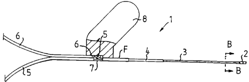
insertion device of Fig. 2 from above. Fig. 5 is a schematic
illustration of an insertion device having a microdialysis
probe inserted therein.
5 The microdialysis probe 1 shown in Fig . lA i n~ lPF~ between
an end part 2 and an inL, ~ te part 4 a mantle part 3
which is semi-p~ -hl ~ at least at a part of its surface.
As shown in Fig. lB, a thin inner tube 5 ' is located within
the mantle part 3 such as to form in cross-section an annular
10 space between the inner tube S ' and the mantle part 3.
Although not shown, the inner tube has an opening located
close to the end part 2. The inner tube and the annular space
are each connected to a respective line 5, 6 which can be
connected ~ e~; Llvely to a source of dialysis liquid and a
15 sampler. Since these _ L~, form no part of the inven-
tion, their details will not be further described.
Figs. 2-4 show an insertion device for the microdialysis
probe shown in Fig. 1. This insertion device includes a
20 canulQ tube 9 having a pointed tip 11. The canula tube 9 is
fastened to a handle means 12, 13, which in the illustrated
case is made of plastic. The particular feature of the canula
tube 9 is that it is provided with a slot 10 best seen from
the cross-sectional view of Fig. 3, which extends approxima-
25 tely along the full length of the canula tube. In order toprovide a holding facility, the slot must occupy in an angle
of less than 180, and preferably occupies an angle of about
65 .
30 The inner .11~ Ler of the canula tube 9 and the rl1 i r~n~
of the slot 10 are adapted so that the major part of the
insertible portion of the microdialysis probe 1 will fit
11n~1y in the canula tube, while the slot 10 is too narrow
to permit the probe to be removed radially. Thus, at least
35 the end part 2 of the probe and the illL ~'1ate part 4
should be larger than the slot 10, with one exception which
enables the function of the invention: The plastic hose 4 is
W0 95/20991 r~
.
5 ~8~5
namely flattened from one side at F, such that its essential-
ly elliptical form at this point has a minor axis which is
slightly smaller than the width of the slot. This enables the
probe to be curved at F after having inserted the canula tube
5 into tissue with the ~icrodialysis probe inside the canula
tube, such that the outer probe end will be located outside
the ~r-gln~ry outward extension of the bore of the canula
tube 9 at the positi~n F. When the canula tube is then
withdrawn from the tissue, it can be drawn past the stationa-
10 ry flattening F, so as to leave the microdialysis probe inposition after the can~lla tube has been fully withdrawn.
The afoledesuL ~ bed operation is facilitated by the various
handle parts shown in the Figures: The microdialysis probe
15 has a flat wing 8 which is suitably held firmly so as to
prevent the microdialysis probe 1 from being withdrawn at the
same time as the canula tube. The canula tube handle part
consists in two upstanding parts 12 which form a slot in the
extension of the canula tube slot 10, said wing 8 fitting
20 into the slot as will b,Pst be seen from Fig. 5. The upstand-
ing parts carry finger-grip wings 13. This enabl~s the canula
tube to be withdrawn with the aid of two finger~ of one hand
hooked ~around the finger-grip wings 13, while the wing 8
fixedly mounted on the microdialysis probe 1 is held firmly
25 with the oth~r hand, in order to prevent simultaneous with-
drawal of the probe. After the canula tube has been withdrawn
and discarded, the wing 8 can be suitably folded down against
the skin and secured t~lereto with adhesive tape, therewith
providing certain resi stance to withdrawing and bending
30 forces in the connection lines 5 and 6.
A canula tube of this kind, provided with a slot 10, can be
~Luduut:~ conveniently by drawing a rolled band of stAinl~s
steel around a mandril through an ~ u~Llately configured
35 draw plate of a wire drawing kind.
The illustrated canula tube can, in many cases, be fully
WO 95/20991 r~
21~ 0 ~
sufficient for inserting a canula through the skin. In other
cases, when the skin is very tough, it is suitable to first
pierce the skin with a lancet-like point, so that the hole
through the skin will not clamp elastically around the canula
5 tube and make insertion of the probe-fitted canula difflcult.
~he invention greatly facilitates the insertion of a micro-
dialysis probe, because only two hands are required and not
three, and because patients pf~P~ n~ normal subtle motoria
lO will often be able to insert the probe themselves.