Note: Descriptions are shown in the official language in which they were submitted.
218201P
_ PHOTOCHEMICAL ABLATION OF GASTRO-INTESTINAL TISSUE
This invention relates generally to the medical field and, more
particularly, to the use of photodynamic therapy in organ augmentation, for
example,
enterocystoplasty or colocystoplasty or the use of other gastro-intestinal
segments for
organ augmentation. The present invention involves a technique in which a
photosensitive composition selectively accumulates in the mucosal tissues of a
human
or animal subject.
It is to be understood that the present invention is useful for
augmentation of various organs. For ease of illustration, the specification
herein
describes in detail augmentation of a bladder organ using gastro-intestinal
tissue. It
is contemplated that the augmentation of other organs using various tissues as
augment is within the scope of the present invention.
The technique involved in augmentation of an organ involves exposing
a portion of gastro-intestinal tissue (including stomach, large bowel (colon)
and small
intestine) and transplanting or inserting the exposed gastro-intestinal tissue
into the
organ. Various recipient organs such as the bladder, stomach and other
portions of
the gastro-intestinal tract, such as esophagus and the like, are contemplated
as being
within the scope of the present invention. In particular, bladder augmentation
has been
principally used in the treatment of patients with tuberculosis of the
bladder, interstitial
cystitis and bladder cancer. Currently, bladder augmentation is gaining wider
acceptance as a therapeutic option for patients with small, non-compliant
bladders and
for treating the variety of congenital, inflammatory and neoplastic problems
in the
urinary bladder which are refractory to medical management. Medical
indications for
bladder augmentation include fibrosed and scarred bladders from previous
surgery,
radiation therapy, or trauma; small non-compliant bladders associated with
extrophy
and epispadias; and, neurogenic bladders associated with myelodysplasia.
Despite significant advances in the use of organ augmentation, there
is a need for an improved augmentation procedure since numerous complications
are
associated with incorporating intestinal mucosa into the recipient organ.
These
complications include metabolic and electrolyte disturbances, such as
hyperchloremic
metabolic acidosis and hypokalemia; chronic bacterial colonization, which
results in
infection and/or sepsis; formation of stones or lithiasis; or malignant
transformation at
-1-
21 8201f~
the vesicoenteric anastomosis. Still other complications arise from the fact
that the
intestinal mucosa continues to produce mucus after being transplanted into the
recipient organ. The continued mucus production causes problems in patients.
In
bladder augmentation, for example, the continued mucus production requires
frequent
catherization to prevent blockages in the genitourinary tract.
One attempt to overcome these complications involves mechanical
stripping of the bowel mucosa layer while leaving the underlying submucosa and
muscular layers intact. This mechanical stripping leads to a decrease in mucus
production. However, in the animal models tested, there is often marked
retraction and
fribrosis of the intestinal segment with little or no gain in organ capacity.
The retraction
and lack of increased organ capacity defeats the purpose of organ
augmentation.
Further, this type of mechanical stripping of the mucosa is technically
tedious and has
limited potential application to humans.
Therefore, it is important that a technique for organ augmentation
includes a way to decrease or prevent mucus production by the transplanted
intestinal
tissue while maintaining the elastic and muscular integrity of the
transplanted tissue.
The present invention addresses this problem.
In photodynamic therapy, photosensitive compositions are used to
selectively diagnose, alter or destroy pathologic tissue. For example,
photosensitive
compounds are used which differentially localize in a target tumorous tissue
where the
compositions absorb electromagnetic radiation when irradiated. The
photosensitive
compositions are useful due to their ability to differentially localize in the
target tissue
as compared to the amount absorbed by the surrounding non-cancerous or normal
tissues and produce toxic reactions when activated.
For example, photosensitive compositions have been proposed as
useful compounds for topical applications for diagnosis and treatment of skin
diseases.
In addition, photosensitive compositions have been proposed for use in the
sterilization
of biological samples containing infectious agents such as bacteria and
viruses. The
bactericidal effects are induced by irradiation of tissues containing
photosensitive
compositions effective against gram-negative and gram-positive microorganisms
(Martinetto et al., Drugs Exp. Clin. Res. XII (4) 335-342, 1986).
Photosensitive
compositions have also been used to decontaminate blood and blood components.
In addition, photosensitive compositions have been used in the treatment of
blood
-2-
~,~ p~ ~:, o la
vessel occlusions such as atherosclerotic plaques, thrombi and the like.
Photodynamic
therapy in combination with hyperthermia has also been proposed as useful in
treating
many of these disorders or diseases. Photosensitive compositions have also
been
proposed as useful in the diagnosis of disease. These photosensitive
compositions
have fluorescent properties and since the photosensitive compositions
sequester in
diseased tissues, fluorescent visualization and/or measurement can be used to
diagnose and localize the disease or to direct therapy to the affected
tissues.
Until the present invention, there has been no suggestion of using
photodynamic therapy in the in vivo treatment of benign protocol for subjects
who
could benefit from intestinal mucosa transplanted into an organ.
The present invention provides the use of a technique for treating
various organ disorders and in particular, for organ disorders amenable to
organ
augmentation. The technique of the present invention is less invasive than
currently
used methods of organ augmentation, requires less hospitalization, and avoids
the
possible complications which accompany other organ augmentation techniques.
Additionally, the present invention provides the use of photochemical
ablation of gastro-intestinal tissue for organ augmentation.
According to one aspect of the invention, there is herein defined an
organ augmentation technique wherein gastro-intestinal tissue is transplanted
into a
human or animal subject's organ, which comprises sensitizing the gastro-
intestinal
tissue with an effective amount of a photosensitive composition which
accumulates in
the gastro-intestinal tissue and exposing the gastro-intestinal tissue to a
source of
electromagnetic radiation energy for a predetermined period of time and at a
predetermined wavelength and intensity, whereby the photosensitive composition
accumulated in the irradiation-exposed gastro-intestinal tissue absorbs the
electromagnetic radiation or undergoes a photochemical reaction.
According to another aspect of the invention, there is additionally
herein defined a technique for treating a subject who will have, is having or
has had
an organ augmentation with gastro-intestinal tissue comprising the steps of:
(a) providing an apparatus for diagnosing or treating tissue comprising:
a catheter for insertion into the subject's organ, said catheter having
a proximal end and a distal end;
-3-
~~ ~ g~~ o~) a
said catheter comprising at least one axially extending opening
therethrough for receiving an electromagnetic radiation delivery means;
said delivery means having a transparent or translucent distal end and
an opaque and/or reflective proximal end; and
said delivery means being operatively connected to a source of energy
for delivery of electromagnetic radiation energy to the distal end of the
delivery means;
(b) determining the position of the delivery means relative to the
gastro-intestinal tissue and surrounding tissue of the subject to ensure that
the distal
end of the delivery means is adjacent to the gastro-intestinal tissue;
(c) administering to the subject an effective amount of a photosensitive
composition at a point in time prior to or during irradiation of the gastro-
intestinal
tissue; and
(d) irradiating the gastro-intestinal tissue by delivering electromagnetic
radiation energy through the distal end of the delivery means to the gastro-
intestinal
tissue; the energy being delivered for a predetermined time and at a
predetermined
wavelength and intensity sufficient to effectively treat the gastro-intestinal
tissue.
According to the present invention, a photosensitive composition is
administered to the subject which preferentially accumulates in gastro-
intestinal tissue,
which will be or has been transplanted into a recipient organ. Electromagnetic
radiation is applied to the tissue. The absorption of the electromagnetic
radiation by
the photosensitive composition damages or destroys the mucosal tissue cells
without
damaging the underlying submucosal or muscle layers of the transplanted
tissue. The
transplanted tissues maintain the desired elastic and strength properties.
After
damage or destruction of the gastro-intestinal mucosal tissue cells,
transitional
epithelial cells migrate in from the surrounding tissue and repopulate to
cover the
transplanted tissue segment. The migration of the epithelial cells of the
organ obviates
many of the problems normally associated with the retained transplanted tissue
within
the organ, since there is a substantial decrease both in mucus production and
in the
absorption of fluid and chemicals by the recipient organ.
In one embodiment, the present invention is particularly useful in
bladder augmentation (i.e. enterocystoplasty) procedures in which a subject's
bladder
is opened and a segment of the subject's gastro-intestinal tissue is attached
to the
bladder in order to increase the bladder capacity and/or repair damage to the
bladder.
-4-
op
This operation is used in subjects with small contracted bladders or in
subjects with
bladder trauma or neoplasms. The lining of the transplanted tissue continues
to
secrete mucus and absorb fluid and chemicals from the urine. The technique
used in
the present invention causes the mucosal lining of the transplanted gastro-
intestinal
tissue to diminish or cease mucus production without injuring or destroying
the
submucosal or muscular layers of the transplanted gastro-intestinal tissue,
thereby
maintaining the transplanted tissue's elastic and strength properties.
Figure 1 is a simplified sectional view of a region of a subject showing
a urethra and bladder, schematically illustrating one embodiment of the
present
invention.
According to the present invention, a photosensitive composition is
administered to the subject. The photosensitive composition is preferentially
retained
or absorbed by gastro-intestinal (bowel) tissue and is sequestered in bowel
tissue at
a much higher level than in a recipient organ tissue. The photosensitive
composition
is taken up by various cells in the transplanted segment of bowel. The
transplant
segment is exposed to electromagnetic radiation either before, during or after
being
transplanted into the recipient organ. The electromagnetic radiation is
absorbed by the
photosensitive composition and causes a series of chemical reactions which
lead to
damage or destruction of the mucosal layer of tissue of the transplanted bowel
segment, while sparing the submucosal and muscular layers of the transplanted
tissue.
The structures of the recipient organ (i.e. the walls, blood vessels and
muscle layers)
are not damaged because the photosensitive composition does not accumulate in
these structures in sufficient amounts to cause damage. The epithelial cells
of the
recipient organ grow in and cover the bowel submucosa and muscle such that
there
is little or no production of mucus by the transplanted tissue.
The photosensitive composition can be administered pre-operatively,
intraoperatively or post-operatively to the subject. It is also contemplated
that the
transplanted tissue can be exposed to the electromagnetic radiation pre-
operatively,
intraoperatively or post-operatively after a pre-determined recovery period
has passed.
It is to be understood that the exact procedure of photosensitive composition
administration and electromagnetic radiation treatment is dependent upon the
parameters of each subject's situation and the localization properties of the
photosensitive composition. In various preferred embodiments, the organ
-5-
~~ ~g~.~ ova
augmentation will be followed by a suitable recovery period. Thereafter, at
least one
preferred photosensitive composition is administered to the subject. After a
suitable
period of time has elapsed in order to preferentially allow the transplanted
mucosal
tissue to absorb and/or retain the photosensitive composition, electromagnetic
radiation
is administered to ablate the mucosal layer of the transplanted tissue. The
photodynamic therapy damages or destroys the mucosa of the transplanted
tissue,
thereby allowing recovery of the submucosal layer of transplanted tissue with
neothelium originating from the surrounding recipient organ tissue.
It is to be understood that in certain circumstances, exposing the
intestinal segment to electromagnetic radiation intraoperatively may be
difficult and it
may be preferable to treat the subject after a post-operative period of
convalescence.
It is also contemplated that subjects who have previously undergone organ
augmentations and who are still experiencing continued mucus production and/or
absorption of chemicals, can be treated with the technique of the present
invention.
In such situations, the organ augmentation subjects may receive the
photosensitive
composition and thereafter, receive the electromagnetic radiation treatment
using a
cystoscope to expose the intestinal augment to the electromagnetic radiation.
The
preferential retention of the photosensitive composition within the intestinal
mucosa
minimizes any possible damage to the underlying submucosa or intestinal muscle
of
the transplanted tissue and to the surrounding recipient organ tissue. It is
contemplated that this electromagnetic radiation therapy treatment can be
performed
on an outpatient under local or small amounts of IV sedation.
It is contemplated that various photosensitive compositions are useful
in the present invention. There are various classes of useful photosensitive
compositions, including, for example, porphyrins, chlorins (such as
benzochlorins,
benzochlorin metal complexes, bacteriochlorins and the like), purpurins,
verdins,
phthalocyanines and iminium salts of these compositions and other
compositions.
Various photosensitive compositions include tin ethyl eitopurpurin dichloride
(SnET2),
photorin, benzoporphyrin derivative, monaspartyl chlorin e6, and Zn-
phthalcyanine. In
addition, it is possible to use a photosensitive precursor, such as 5-
aminolevulinic acid
(ALA), which is a precursor to the production of the photosensitizer
protoporphyin-IX
in vivo. It is to be understood that the present invention envisions the use
of these and
-6-
~. ~ I '~ a-~ a ~ o
other classes of photosensitive compositions, and the present invention is not
limited
to particular photosensitive compositions.
Examples of various known photosensitive compositions include those
compounds disclosed in Loh et al., J. Photochem. Photobiol., 20:47-54(1993),
Selman
et al., Photochem. Photobiol., 57:681-685 (1993), Morgan et al., J. Org. Chem.
51:1347-1350 (1986), Skalkos, et al., Med. Chem. Res. 2:276-281 (1992), U.S.
Patent
Application Serial No. 07/901,597 and Morgan, et al., U.S. Patents Nos.
4,877,872,
4,988,808, 5,051,415 and 5,216,012. These compositions are physiologically
acceptable for subcutaneous, intravenous, intravesical, or oral administration
as
solutions, emulsions or suspensions.
In addition to the required photosensitive composition, additional
components may be chemically attached to or physically combined with the
photosensitive composition for administration to the subject. These additional
components may include labeling compositions, cytotoxins, immunoglobins,
monoclonal antibodies and/or receptor ligands, which may enhance the
photosensitive
composition's selectivity for the desired tissue.
The photosensitive compositions and any additional components are
formulated into a final pharmaceutical formulation for administration to the
subject
using techniques generally known in the art. The pharmaceutical formulation
can be
administered singly or as components of mixtures as solutions, emulsions or
suspensions. It is to be understood that the final pharmaceutical formulations
can be
prepared in conventional forms either as liquid suspensions or solutions,
solid forms
suitable for dissolution or suspension in liquid prior to injection or as
emulsions. The
formulation may include suitable excipients such as saline, dextrose,
glycerol, water
and the like. The final pharmaceutical formulation may also contain additional
components such as pH buffering agents, wetting or emulsifying agents and the
like.
The photosensitive compositions can be administered by any suitable
route or method. These methods include, for example, subcutaneous,
intravascular,
introperitoneal or intramuscular injection, oral or topical administration or
a suppository
administration. It is further contemplated that the photosensitive composition
can be
an extended release formulation, such that it is delivered over a period of
time and
there is a sustained release of the photosensitive composition. The extended
release
-7-
_" can be administered by a vascular stent or implantable device, or can be
orally
administered by a tablet or capsule, for example.
Various modes of administration are well known in the art and the
administration can be implemented in a manner which is most suitable for
delivery of
the photosensitive composition. This administration can include a slower
sustained
release system or, if properly formulated, an oral administration. The
quantity of the
formulation being administered is dependent upon the choice of the active
photosensitive composition, the condition to be treated, the mode of
administration and
the individual subject. As such, smaller or larger doses may be needed
depending
upon the specificity of the formulation. It is contemplated that in
formulations having
additional components such as highly specific monoclonal antibody preparations
or
specific receptor ligands, the dosages may be less than formulations which are
less
specific to the target tissue. It is contemplated that dosages within the
range of about
0.05-10 mg/kg are suitable. It is also understood that these ranges are merely
suggestive and many variables must be taken into consideration in the
treatment of
individual subjects and variations from these recommended values are expected.
Other ingredients which can be included in the formulation include
antimicrobial agents and/or preservatives as necessary. Many variations of the
above,
along with other suitable vehicles will suggest themselves to those skilled in
the art in
light of the description herein.
The photosensitive composition is administered in an effective amount
such that a sufficient amount of the photosensitive composition accumulates in
the
desired target or transplanted tissue. In certain embodiments of the present
invention,
a predetermined period of time is allowed to pass in order to optimize the
accumulation
and retention of the photosensitive composition in the target tissue. It is
contemplated
that various protocols of treatment using the method of the present invention
may
involve irradiating the photosensitive compositions after a suitable period of
time has
elapsed. It is contemplated that these time periods can range from a
relatively short
time of approximately one hour or less to a longer time of three to four days
after
administration of the photosensitive composition to the patient. However, it
should be
understood that the optimum time lapse (if any) between drug administration
and
irradiation depends on the type and amount of photosensitive composition
administered
and the subject's history.
_g_
O
__ After the photosensitive composition accumulates in the transplant
tissue, the tissue is irradiated with electromagnetic radiation of a
predetermined
wavelength and intensity at which the composition absorbs energy. This
absorption
of energy by the photosensitive composition causes a reaction which damages or
destroys the desired cells or tissue in which the composition has accumulated.
It is to be understood that most photosensitive compositions both
fluoresce and sensitize. Both fluorescence and sensitization are de-excitation
pathways which are competitive with each other and are generally activated by
any
wavelength of electromagnetic radiation absorbed by the photosensitive
composition.
For example, it is not the case that excites only fluorescence while another
wavelength
causes the sensitization reaction.
It is contemplated that various types of electromagnetic radiation are
useful with the present invention. Such electromagnetic radiation envisions
the use
of all of the electromagnetic spectrum which is made up of photons. Useful
electromagnetic radiation includes, for example, light in the ultraviolet,
visible and
infrared ranges and ultrasound. Such luminescence is dependent upon the
photosensitive composition being used and the method of treatment. Both the
photosensitive composition and the electromagnetic radiation can be
administered by
any suitable method. These methods include both the in vivo and ex vivo
administration of the radiation and/or photosensitive composition. Further,
the
administration of both the drug and radiation therapy can be in a single
application or
if desired, multiple applications. Further, the sustained release
administration of the
photosensitive composition can be utilized to take advantage of the properties
of the
photosensitive composition and the electromagnetic radiation therapy treatment
being
administered.
It is to be understood that the particular wavelength and intensity of the
electromagnetic radiation energy delivered to the tissue is dependent, in
part, upon the
type of photosensitive composition being used. In certain embodiments,
photosensitive
compositions which have absorbance peaks at shorter wavelengths and show
greater
absorbencies may be used. In various embodiments, the shorter wavelength peaks
are advantageous because the light of the shorter wavelength is less capable
of
penetration into underlying tissue, while greater absorbencies in the
photosensitive
_g_
~-1 i~~.lblb
composition are desirable because less light energy is required to cause a
given
degree of reaction.
The wavelength of irradiating energy is chosen to match an absorbance
peak of the photosensitive composition. The suitable wavelengths for the
photosensitive compositions are readily determined by the composition's
absorption
spectrum. For example, in the deeper tissue penetration red wavelength range
of the
visible electromagnetic radiation spectrum, the photosensitive composition tin
ethyl
etiopurpurin dichloride is illuminated with light that includes the wavelength
corresponding to the absorption peak at about 665+/-5 nm. 5-Aminolevulinic
acid is
a precursor to protoporphyin-IX in vivo, which is the active photosensitive
composition
and which absorbs energy at about 630+/-5 nm. The irradiation dosages are
readily
determined and dependent upon the method of delivery of the photosensitive
composition and the type and amount of photosensitive composition being
administered and retained by the target tissue. Thus, the intensities of light
illumination will typically be in the range of more than about 5 to less than
about 500
joules/cm2 of light.
Irradiation of the target tissue containing the photosensitive composition
in accordance with the instant invention can be achieved by delivering
electromagnetic
radiation energy from a conventional light source, a laser, or by sending an
electromagnetic signal from any other appropriate transmitting device. The
particular
method of irradiation of the tissue depends upon the location in the subject
of the
affected tissue.
It has been found that one particularly useful transmitting device
comprises a laser which delivers highly accurate intensities and wavelengths
of light
through at least one optical fiber. For example, in one embodiment, the light
energy
is delivered through an optical fiber which can optionally have a light
diffusing means
operatively attached thereto. The light energy is delivered by placing the
delivery
means in a catheter. The delivery means is properly located and positioned
adjacent
the target tissue. A portion of the catheter is sufficiently transparent or
translucent to
allow the light energy to adequately irradiate the adjacent target tissue. The
remaining
portion of the catheter may be coated with an opaque or reflective shield type
material,
such that light does not penetrate the adjacent muscle or recipient organ
tissues, which
is comprised of a diffusing material which allows the light to radiate from
the optic fiber.
-10-
~~ j $a~ ~I~
For example, useful light sources are described in U.S. Patent Nos. 5,169,395
and
5,196,005.
It is also contemplated that the electromagnetic radiation can be
delivered using a transmitting device with a spatially localized illuminator,
such as a
microlens fiber which provides a circular illumination field with good uniform
density
and sharp demarcation boundaries. For example, useful devices include that
defined
in U.S. Patent No. 5,231,684.
The delivery means can be in the form of a light guide, such as a
single optical fiber or a fiber optic bundle, which in preferred embodiments,
comprises
at least one optical fiber having an appropriate provision for lighting
thereof. The
delivery means and catheter each have a sufficiently small cross section so
that the
delivery means and catheter may be fabricated within the appropriate
dimensions to
comfortably fit within the patient's body or desired orifice. The catheter may
be made
of a rigid type material or may be made of sufficiently flexible material for
positioning
the light delivery means and catheter throughout a tortuous path.
Also, it is contemplated that various other apparatuses may be
employed within the scope of the present invention in order to ease the use of
the
method of the present invention by cleansing, heating and/or cooling the
tissue being
treated with the photodynamic therapy of the present invention. For example,
when
about 40-45°C heat is delivered to the tissue by, for example,
microwave (not shown)
or laser (not shown), the effects of the photodynamic therapy are enhanced.
It is also contemplated that the electromagnetic radiation can be
provided with various means for guiding the delivery means through a lumen,
and
means for measuring light intensity, temperature and drug fluorescence or
illumination.
It is also contemplated that the delivery means can possess an irrigation
apparatus to
provide a source of irrigation to the area as desired and to keep the area
being
irradiated relatively clear.
Accurate positioning of the delivery means assures that there is limited
penetration of electromagnetic radiation into the tissue and that only the
desired tissue
is irradiated. Such accurate positioning can be aided by using an ultrasound
probe.
It is also contemplated that other methods of accurately positioning the
delivery means
can be used. For example, the catheter and/or delivery means can have
graduated
-11-
~.,~~~L~QI~
marks thereon so that the actual position of the delivery means can be
accurately
located.
After proper localization of the delivery means is achieved, the
transmitting device is operatively engaged and energy irradiates the adjacent
target
tissue. The preferred length of time of irradiation and wavelength of
electromagnetic
radiation are determined by the type and amount of photosensitive composition
being
used and other factors as described above. The irradiation of the
photosensitive
composition causes the photosensitive composition to absorb the
electromagnetic
radiation generally or induces a photochemical reaction of the photosensitive
composition, thereby inducing damage or destruction of the desired tissue. The
photosensitive composition may cause a hemorrahagic necrosis of the affected
tissue.
Further, with the passage of time there is subsequent diminishment or
cessation of the
cellular and/or tissue functions and subsequent atrophy of the affected
tissue. For
example, it is surprisingly found that in bladder augmentation procedures, the
bladder
tissue and the submucosa of the transplanted bowel tissue are spared any
damage
while the mucosal layer of the transplanted bowel tissue remains in the
destroyed and
atrophied state. The submucosal and muscular layers of the transplanted bowel
tissue
are repopulated with transitional epithelium that migrates in from the
adjacent bladder
tissue.
Various transmitting devices can be utilized in delivering the
electromagnetic radiation to the desired target tissue. For ease of
illustration of the
present invention, the following description relates to photochemical ablation
of gastro-
intestinal mucosal tissue for a bladder augmentation. However, as described in
detail
above, various other organs are contemplated as being treated, along with
other types
of transplant tissue, as well as other types of electromagnetic radiation in
addition to
the ones described below.
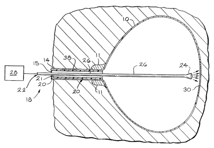
Referring to Figure 1, there is illustrated in simplified form a sectional
view of a subject showing a bladder 10 (including the bladder sphincter muscle
11 ),
and a urethra 14 in a distended condition and defining an opening or lumen 15.
A photodynamic therapy system or apparatus 18 generally comprises
a catheter 20, a delivery means 22 and a source of electromagnetic radiation
energy
28. The catheter 20 defines an opening 21 extending axially therethrough for
receiving
the delivery means 22. The delivery means 22 can comprise at least one, or
-12-
~~ J ~ ~~ b f~
alternately multiple, long, small diameter optic fibers. The delivery means 22
coaxially
extends through the catheter 20. In an alternative embodiment (not shown), the
delivery means 22 may be a part of the catheter 20. It is to be understood
that the
catheter 20 and the delivery means 22 may generally have a rounded or tapered
configuration to minimize any damage to the urethral lining and to ease
insertion of the
catheter 20 and delivery means 22 into the opening 15 of the urethra 14 and
into the
bladder 10. At least a portion of the delivery means 22 is disposed within the
bladder
10. The delivery means 22 has a directional distal end 24 (which is generally
transparent or translucent) and a proximal end 26 which extends from the
distal end
24 out of the subject's body to the electromagnetic radiation source 28 such
as a laser,
LED device, or lamp. The proximal end 26 of the delivery means 22 is
preferably of
an opaque and/or reflective material such that no light is delivered to any
surrounding
tissue.
The axial length of the distal end 24 is sufficient to generally illuminate
an affected area 30, comprising the transplanted bowel tissue. In preferred
embodiments, the length of the distal end 24 can vary depending upon the
extent
amount of light energy to be administered. It is understood that the
preferable length
of the distal end 24 will differ among subjects in order to accurately deliver
the required
light to the desired tissues. In certain embodiments, it is preferred that the
distal end
24 be of a directional material, such that electromagnetic radiation, rather
than
radiating outwardly from the axis of the distal end into the tissues, is
focused within
well-defined boundaries. In the embodiment shown in Figure 1, the affected
area 30
receives the diffused light (schematically indicated with arrows). In other
embodiments, it may be preferred that the distal end 24 be of a diffusing
material such
that the light radiates outwardly from the axis of the distal end 24 into the
tissues.
It is contemplated that monitors (not shown) can be placed in the
bladder 10 for measuring light intensity and temperature. This positioning of
the
catheter 20 and delivery means 22 can be aided using an ultrasound probe (not
shown) and/or by direct visualization using an endoscope (not shown).
After the delivery means 22 is localized in the bladder 10, the energy
source 28 is activated and energy is delivered to the affected tissue 30. The
intensity,
wavelength and duration of the energy are dependent upon many variables
including
the type and amount of photosensitive composition used. During this
irradiation, it is
-13-
~~ l ~ ~.~ ~l ~
possible to continuously monitor the position of the distal end 24 of the
delivery means
22 such that there is little damage to the surrounding tissues. After
irradiation, the
catheter 20 and delivery means 22 are removed from the bladder 10 and urethra
14.
The following examples are intended to illustrate the present invention
but not to limit its scope.
Example
33 Fischer 344 female exbreeder rats weighing between 200-250
grams were used. Eight (8) rats died in the immediate postoperative period
(first 24
hours), secondary to either bleeding complications or anesthesia related
causes. Of
the remaining 25 rats, there were three control groups of five rats each and
one
treatment group consisting of ten rats. The treatment group was given five
micrograms
per kg of the photosensitizer hematoporphyrin derivative (HpD), intravenously
24 hours
prior to bladder augmentation. At the time of surgery, approximately 1.5 cm of
terminal
ileum was isolated and used for ileocystoplasty. Primary anastomosis of the
small
bowel was performed using 7-0 Vicryl suture in an interrupted fashion. The
isolated
segment of ileum was opened along its antimesenteric border and aluminum foil
was
placed around this segment to protect the underlying tissues and mesenteric
blood
supply from the effects of the light. HpD has a peak absorption at about 630
nm. A
diffuse non-coherent red light source was used to irradiate the bowel mucosa
for 20
minutes for a total delivered fluorescence of 240J/cm2. An infrared filter was
used to
limit the light spectrum to 590-750 nm. After this, the treated intestinal
segment was
used to perform the augmentation using the Goodwin cup-patch technique. 7-0
Vicryl
suture in running fashion was used to complete the anastomosis.
Each of the three control groups underwent bladder augmentation as
well. One group had no further treatment, another group was given HpD only,
and a
third group was treated with light only. Each rat received 20,000 units/kg
penicillin
intramuscular prior to surgery. Post-operatively, the rats received 80,000
units PCN
per 100mL in their drinking water for one week. After this, the oral
antibiotics were
discontinued. Rompun (12 mg/kg) and ketamine (80 mg/kg) anesthesia were used.
Each rat underwent a pre-operative cycstometrogram (CMG) to
determine the bladder capacity prior to augmentation. A five french pediatric
feeding
tube was placed transurethrally into the bladder and the urine was removed.
Next,
saline was infused into the rat's bladder at a rate of 0.25 mUminute. Bladder
pressure
-14-
0
was measured concomitantly. Bladder capacity was defined as the volume of
saline
required to achieve a pressure of 30 mm Hg (40 cm water) or the volume at
which
saline leaked around the feeding tube. The value selected was the one that
occurred
fi rst.
Following a recovery period of six weeks, each rat was placed in a
metabolic cage for 24 hours to collect urine to measure the amount of mucus
production. The urine collected was cooled to four degrees centigrade
overnight, then
centrifuged for three minutes. The urine was aspirated off and the remaining
mucus
was air dried. The amount of mucus production was quantified as the dry weight
obtained.
Urine cultures were also obtained in each rat to check for evidence of
factorial colonization. Rompun and ketamine anesthesia were used as described
above. A small vertical suprapubic incision was made under sterile conditions
to
expose the bladder. Urine for culture was obtained by direct bladder
aspiration in
order to avoid contaminated specimens. The incision was closed with 4-0 silk.
Bacterial colonization was defined as greater than 100,000 organisms per mL.
After a period of at least 48 hours, a repeat CMG was performed to
determine post-operative bladder capacity prior to euthanasia. This was done
in the
same fashion as described above. Next, a median sternotomy was performed and
blood was obtained via direct cardiac aspiration for measurement of
electrolytes (Na,
K, CI, C02. Prior to euthanasia, 10% neutral buffered formalin was instilled
into the
bladder through a five french feeding tube and the bladder neck was tied off
using 2-0
silk. The bladder and augment were removed and placed in formalin to fix the
tissues
overnight. The next day, the bladders were opened to determine the presence of
stone formation. Representative sections were obtained and submitted for
histological
preparation.
Histological examination of the bladders in all three control groups
revealed an obvious transition zone between the native bladder and the
augment.
There was no evidence of transitional epithelium ingrowth from the bladder
onto the
surface of the augment. Alcian Blue and PAS stains confirmed the presence of
mucin
production within the small bowel mucosa. However, each of the treated
bladders
demonstrated ingrowth of transitional epithelium to completely cover the
intestinal
augment. The underlying intestinal muscular and serosal layers were left
intact. In all
-15-
~.., ~ ~ ~, a ~ o
but one case, there was no evidence of mucus producing epithelium using the
special
stains. In this instance, there were only two small foci of remaining mucin
producing
small bowel mucosa. There was also no evidence of fibrosis or collagen
deposition
in either the control or treated groups.
Since there was no difference histologically between any of the control
groups, the data obtained from all groups were combined for comparison with
the
treatment group. In order to confirm the histological findings, urine
collections were
performed to measure the amount of mucus excreted in a 24 hour time period.
The
treated rats produced significantly less mucus than the control rats. The mean
amount
of mucus excreted by the controls (n=15) was 18.9 micrograms over 24 hours.
The
treated rats (n=10) produced only 5.7 micrograms of mucus over 24 hours. The
treated rats also had a lower incidence of factorial colonization when
compared to the
controls. Two controls had significant bacteria counts in their urine while
none of the
treated animals did. No difference was seen in the electrolyte values between
both
groups. Two treated rats developed stones whereas none of the control rats
did. This
is presumably secondary to the fact that when the intestinal mucosa sloughs
off after
photodynamic therapy, the suture used to make the anastomosis is exposed to
the
urine. This can be obviated by using a suture like chromic with a shorter half
life and
by allowing a two to four week recovery period after bladder augmentation.
After this,
a light source can be passed transurethrally to perform the treatment. Lastly,
the
bladder capacity in all groups increased after bladder augmentation. The mean
pre-
and post-operative bladder capacities in the control rats were 0.89 mL and
1.97 mL
pre-and post-operatively. The larger post-operative bladder capacity in the
control rats
can be explained by the fact that large amounts of mucus were found in these
bladders at the time of fixation. This mucus created a functional bladder
outlet
obstruction with poor emptying of the bladder and therefore a larger capacity.
It will be appreciated by a person of ordinary skill in the art that while
the present invention has been disclosed and described herein with respect to
certain
preferred embodiments and alternatives thereof, various changes in form and
detail
may be made therein without departing from the scope and spirit thereof.
-16-