Note: Descriptions are shown in the official language in which they were submitted.
2182511
P-314 - 1 -
MICROBIOLOGICAL CULTURE BOTTLE AND, AND METHOD
OF MARING AND USING SAME
BACKGROUND OF THE INVENTION
1. Technical Field
The present invention generally relates the
detection of aerobic microorganisms. More particularly,
the present invention relates to a culture bottle for
use in systems for detecting aerobic microorganisms.
2. Background Art
Culturing bodily fluids such as blood, sputum,
and urine is commonly employed in the medical field in
order to ascertain the presence or absence of micro-
organisms.
Typically, a sample of bodily fluid to be
tested is obtained from a patient. The sample is then
analyzed in order to determine the presence or absence
of microorganisms. Several methods of determining the
presence or absence of microorganisms are commonly
employed. The most common technique employed involves
preparing a culture by inoculating a growth medium with
a sample of the bodily fluid and incubating the culture.
After sufficient incubation, a visual inspection by a
technician is performed in order to observe and assess
for the presence or absence of bacterial growth.
It is the standard practice in microbiology to
detect the presence and assess numbers of microorganisms
in samples. Medical test samples include body fluids
such as blood, spinal fluid and urine. Industrial
samples include pharmaceuticals, foods and any other
sample that must be tested for presence or levels of
organisms. All such samples are cultured by inserting
them into a vessel containing sterile growth medium.
CA 02182511 1998-08-12
P-314 - 2 -
The growth medium contains the appropriate nutrient to
support the growth of the target organisms.
Microbial presence is detected through changes
in the liquid medium or in the atmosphere over the
specimen after a period of time. For example, United
States Patent No. 4,812,656 to Ahnell et al. uses media
with carbon 13 labelled substrates. After subjecting
the sample to conditions conducive to microbial growth,
the ratio of carbon 13 to carbon 12 in the gaseous
atmosphere is determined. United States Patent No.
5,232,839 to Eden et al., assigned to the assignee of the
present invention discloses a method for timely detecting
microbiological growth in a sealed container by
monitoring consumption of the oxygen in the headspace or
production of C02 or any other gas as an indication of
microbial metabolism. United States Patent No.
5,217,876 describes a C02 sensor present at the bottom
of a vial, which detects presence of microorganisms by
detecting changes in the pH of the specimen or the
production of C02. United States Patent No. 5,047,331
to Swaine et al. discloses a blood culturing bottle
including a sterile container and nutrient growth media
increase in pressure in the head space is monitored.
Other known methods for measuring microbial
contamination in samples include measuring minute
changes in temperature, pH, turbidity; color,
bioluminescence and impedance. All these methods
determine microbial contamination by determining
microbial end products or metabolites.
For diagnostic purposes it is advantageous to
determine as quickly as possible whether or not any
microorganisms are present in a clinical sample.
Diagnosis and the commencement of efficacious drug
2182511
P-314 - 3 -
therapy are greatly enhanced by prompt evaluation of a
clinical sample for the presence or absence of
microorganisms. Therefore, optimizing a microorganism's
growth speeds up the diagnostic process. In order to
achieve optimal growth rates of aerobic microorganisms,
the concentration of dissolved oxygen in the culture can
be increased. In other words, preventing the culture
medium from becoming anaerobic enhances aerobic
microbial growth.
Oxygen has a low solubility in water and poor
diffusion across the air-water interface limits
attainable oxygen concentration in the culture medium.
Shaking, agitating, or bubbling air through a porous
sparger may be used to increase the dissolved oxygen
content in the culture. Shaking, agitating, or bubbling
air through the culture increases the amount of oxygen
in the growth medium and, thereby, increases oxygenation
of the aerobic bacteria enhancing their metabolism and
growth while preventing the culture medium from becoming
anaerobic. In order to achieve better oxygen
concentrations in the growth media agitation of the
bottles during growth is taught. (United States Patent
No. 5,047,331 and United States Patent No. 5,217,876).
However, shaking or agitating a culture requires more
complex and expensive apparatuses adds a potential for
culture bottle or tube breakage or contamination, and
can cause splashing of the culture. Additionally, the
shaking apparatus is typically expensive and is prone to
mechanical difficulty or failure.
It would, therefore, be advantageous to
provide means for increasing oxygenation of the bacteria
by increasing the amount of oxygen available to the
organism in the medium thereby, increasing the
oxygenation of the aerobic bacteria and enhancing their
CA 02182511 1998-08-12
- 4 -
metabolism and growth rate without the need for shaking,
agitating, or bubbling air through the media.
The present invention provides a container
adapted for use in the detection of aerobic microorganisms
including a non-toxic insert which can hold microorganisms
in suspension and increase microbial exposure to oxygenated
media and enhance microbial metabolism. A method for
making the container is further provided. Finally, the
present invention provides a process for detecting aerobic
microbiolical growth utilizing the novel container of the
present invention.
SUMMARY OF THE INVENTION
In a broad aspect, the present invention relates
to a container (10) adapted for use in the detection of
aerobic microorganisms in a sample, said container (10)
comprising: an inner chamber (12) defining a headspace
above a non-toxic porous insert means (22) said insert
means disposed within said inner chamber (12) for
( increasing surface area allowing microorganisms suspended
within said surface and on the surface thereof to increase
microbial exposure to oxygenated media (24) and enhance
microbial metabolism.
In another broad aspect, the present invention
relates to a method for making a container (10) adapted for
use in the detection of aerobic microorganisms in a sample
comprising the steps of: providing a sealable container
with a closed chamber; inserting an unexpanded non-toxic
porous insert (22) into a container (lo) below a headspace
of said chamber; and adding to the porous insert (22)
within the container (10) microbial growth media (24) said
porous insert providing a quantity of oxygen to said media
increasing the microorganisms to oxygen exposure; and
sterilizing said media.
CA 02182511 1998-08-12
- 4 (a) -
In yet another broad aspect, the present
invention relates to a method of detecting aerobic
microbiological growth in a sealed sample container (10)
having a headspace (16) and which contains a sample which
contains an unknown microorganism, said method comprising
the steps of: providing a sealed sample container (10)
having a headspace (16) and non-toxic porous insert (22)
saturated with microbiological growth media (24);
inoculating the insert (22) within the sealed sample
container (10) with said sample containing said unknown
microorganisms; monitoring metabolism within the container
(10) as an indicator of the presence of microorganisms to
detect microorganisms in the sample.
~
CA 02182511 1998-08-12
P-314 - 5 -
FIGURES IN THE DRAWINGS
Other advantages of the present invention will
be readily appreciated as the same becomes better
understood by reference to the following detailed
description when considered in connection with the
accompanying drawings wherein:
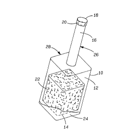
FIG. l is a perspective view of the
microbiological culture bottle of the present i nvention;
FIG. 2a is a graphic illustration of pressure
change in a sample for containing M. tuberculosis
in a
20% oxygen environment without the sponge inse rt;
FIG. 2b is a graphic illustration of pressure
change in a sample for containing M. tuberculosis
in a
200 oxygen environment with the sponge insert;
FIG. 3a is a graphic illustration of pressure
change in a sample containing M. tuberculosis in a 40%
oxygen environment without the sponge insert; and
FIG. 3b is a graphic illustration of pressure
change in a sample containing M. tuberculosis in a 40%
oxygen environment with the sponge insert.
FIG. 4a is a graphic illustration of pressure
change in a sample containing C. neoformans in a 20%
oxygen environment without the sponge insert.
FIG. 4b is a graphic illustration of pressure
change in a sample containing C. neoformans in a 20%
oxygen environment with the sponge insert.
FIG. 5a is a graphic illustration of pressure
change in a sample containing C. neoformans in a 40%
oxygen environment without the sponge insert; and
2182511
P-314 - 6 -
FIG. 5b is a graphic illustration of pressure
change in a sample containing C. neoformans in a 40%
oxygen environment with the sponge insert.
DETAILED DESCRIPTION OF THE INVENTION
The present invention generally shown at 10 in
FIG. 1 provides a container 10 for use in the detection
of aerobic microorganisms such as Mycobacterium
tuberculosis, Mycobacterium avium, and fungi, or other
microorganisms capable of growth within an oxygenated
environment. The container or vial 10 comprises a
bottle having an inner chamber 12 having a bottom
surface 14, a head space 16, a cap 18 with a resilient
rubber stopper 20, and a non-toxic insert 22 hydrated
with microbial growth promoting media 24 disposed within
the inner chamber 12 for better dispersion of the
microorganisms and to increase microbial exposure to
oxygenated media 24 and enhance microbial metabolism.
Additionally, the container has a neck portion 26 and a
shoulder portion 28.
The container 10 may be constructed of any
suitable material such as glass or plastic. Suitable
plastics include polystyrenes, polypropylenes, and
polycarbonates. Of course, any suitable material must
be non-toxic to the microorganisms and be capable of
being sterilized by suitable means such as by an
autoclave or irradiation. Preferably, the container 10
will be constructed of a transparent material~to aid not
only in the visual detection of microorganisms but will
also allow for a technician or user to visually confirm,
prior to introduction of a sample, such as bodily fluid,
that the container 10 is free contamination.
The non-toxic insert 22 is disposed within the
inner chamber 12 of the container 10. In the preferred
embodiment, the insert is made from highly porous
2182511
P-314 - ~ -
material which greatly increases surface area for
microbial exposure to the oxygenated growth media 24.
Increasing microbial exposure to oxygenated growth media
is a critical feature of the non-toxic insert 22. By
increasing exposure to oxygenated media in this manner,
shaking of the container is not required. In other
words, the insert 22 provides sufficient oxygenation of
the growth media 24 to promote and sustain microbial
proliferation without the need for other methods of
supplemental oxygenation.
In the preferred embodiment, the non-toxic
insert 22 is made of sponge. Sponge is an ideal
material for the insert means 22 because its high
porosity provides for greater oxygenation of the growth
media. The large surface area provided by the porosity
of the sponge allows for enhanced oxygen exchange
between the air and the growth media 24. Other
materials for the insert include cotton; fiber glass;
glass beads, plastic (resinous material) and sponge
beads and Porex'~ porous plastics (made of polyethylene,
polypropylene, polyvinylidene fluoride, ethylene-vinyl
acetate, stryeneacrylonitrite, etc.). It must be noted
that whatever material is selected to serve as the
insert 22, the material must be non-toxic to
microorganisms, that is, the material must be
essentially inert and not affect microbial growth.
When hydrated with a sufficient growth media
24, the non-toxic insert 22 occupies between about 25
80% of the volume of the inner chamber 12. By occupying
a volume in this range of volumes within the inner
chamber 12, growth conditions within the container 10
are optimized. In other words, the relationship between
growth media 24, surface area, and oxygen are optimal
when the hydrated insert 22 occupies a volume of the
2182511
P-314 -
container 10 within the above-stated range and,
therefore, increasing microorganism metabolism. Since
a number of aerobic microorganisms grow better suspended
in the liquid air interface where 02 is most available,
the insert 22 greatly enhances oxygenation of the
microbial growth media and, hence, oxygenation of the
aerobic microorganisms. Another means of increasing
the availability of oxygen is by increasing the oxygen
concentration in the headspace.
In essence, the insert 22 establishes an
environment with conditions similar to those found in
lungs. Establishing an "artificial lung" environment
enables growth in vitro of microorganisms, such as M.
tuberculosis and M. avium, which were previously
difficult to culture in vitro. This effect is also
observed with other oxygen requiring microorganisms such
as fungi. This micro-environment exposes the
microorganisms to highly oxygenated growth media 24 to
promote and support microbial growth.
The microbial growth medium 24 comprises of
all the nutrients required for growth of the target
organism. For example, microbiological growth media
such as Middlebrook 7H9 is used for growing
Mycobacterium sp. It is understood by those skilled in
the art that the microbiological growth media 24 is
chosen based on the particular microorganism being
selected for. In other words, the particular~microbial
growth medium 24 is selected based on biochemical or
nutritional requirements of the microorganism one
desires to culture.
In addition to the liquid culture medium, the
microbial growth medium 24 can include other additives
selective or differential additives such as antibiotics.
These additional additives can be used in order to
2182511
P-314 - 9 -
select for the presence of or differentiate particular
microorganisms based on specific and unique
microorganism characteristics i.e., antibiotic
resistance/susceptibility or growth requirements.
The present invention also includes a method
for making the container 10 adapted for use in the
detection of aerobic microorganisms. The method
comprises the steps of inserting an unexpanded non-toxic
insert 22 into the container 10. The unexpanded non-
toxic insert 22 is- preferably a dehydrated and/or
compressed sponge material. Additionally, the non-toxic
insert 22 can be an unfoamed or unexpanded material such
as polyurethane which is inserted into the container 10.
once inside the container 10, the unexpanded non-toxic
insert 22 is expanded by means known in the foaming art.
Glass or plastic (resin) beads as well as sponge beads
can also be added to containers. All the insert
materials serve the same purpose of increasing the
oxygen media interface thereby allowing more available
oxygen to the microorganisms.
When foam is used for the insert, expanding
the unexpanded non-toxic insert 22 within the container
10 includes the step of rehydrating the sponge material
with microbial growth media 24 such as Middlebrook 7H9
media or other suitable growth media. Thus, upon
expansion, the insert 22 is hydrated throughout with
media thereby providing a homogenous growth_promoting
environment throughout the material.
Foamable material can be casted within a
bottle followed by the addition of media. It is
critical that the material used for the insert 22 be
non-toxic to microorganisms as previously described
above.
2182511
P-314 - 10 -
The present invention also includes a method
for detecting aerobic microbiological growth in a sealed
sampled container 10 having a headspace 16 and non-toxic
insert 22 saturated with microbiological growth media
24. The method includes the steps of providing a sealed
sample container 10 having a headspace 16 and non-toxic
insert 22 saturated with microbiological growth media
24. The insert 22 disposed within the sealed sample
container 10 is inoculated with a sample, such as bodily
fluid, to be analyzed for the presence or absence of
microorganisms. The sealed sample container 10
containing the inoculated insert 22 is monitored for
evidence of microbial metabolism.
The sealed sample container 10 containing the
insert 22 saturated with microbiological growth media
can be provided in a sterile, ready to use form.
Additionally, the sealed sample container 10 containing
the insert 22 may be obtained in a form in which a
sterile, sealed container 10 having a dehydrated insert
22 is provided and the user aseptically adds their own
specific or preferred microbiological growth media 24 to
the sealed container 10 via the rubber stopper 20.
Inoculation of the insert 22 within the
container 10 is generally accomplished by injecting a
sample, such as bodily fluid using a sterile syringe and
needle. The needle is pierced through the resilient
rubber stopper 20 and the contents of the syringe is
injected onto the porous insert 22.
The inoculated container 10 is then monitored
for indicia of microbial metabolism such as pressure
change in the headspace of the container 10 as a
function of rate of changes of headspace pressure, or
visual indicia such as changes in turbidity (clarity) of
the microbiological growth media 24. This list of
2182511
P-314 - 11 -
possible indicia of microbial metabolism is merely for
illustrative purposes and is not intended to be provided
as a complete list. Other suitable methods of detecting
microbial metabolism known to those skilled in the art
may be substituted.
It should be noted that the present invention
is not limited to detection of microorganisms in bodily
fluid. Various types of samples, such as food stuffs or
other industrially tested samples, can be inoculated in
the container 10 by means well known in the art.
The following examples illustrate the
preparation of, use of and utility of the present
invention.
Examples
Example 1.
Materials and Methods
Containers containing sponge material hydrated
with an amount of Middlebrook 7H9 broth media sufficient
to completely wet the sponge (approximately 30 ml) were
sterilized by autoclave. The sponge material occupied
approximately 80% of the volume of container. Samples
containing 2.0 x 10² cfu/ml (colony forming
units/milliliter) Mycobacterium tuberculosis H37RV were
inoculated into the containers. The inoculated
containers were fitted with a ESP connecter (Difco
Laboratories, Inc.) and connected to an ESP machine
(headspace pressure sensing device, Difco Laboratories,
Inc.) and were statically incubated at 35°C. The
initial amount of oxygen in the headspace was 20~. An
experimental control was run in tandem with the
experimental container and varied on in that it did not
contain the sponge material.
2182511
P-314 - 12 -
Results
Referring to FIGS. 2a and 2b, after two
hundred and ten (210) hours of monitoring the change in
headspace pressure, the experimental container including
the sponge material insert (see FIG. 2b) exhibited a
much better and faster signal indicating the presence of
a microorganism than did the control container (see FIG.
2a). The experimental container displayed a more
defined signal to noise ratio than did the control
container, that is, the point at which detection was
possible was much more distinct for the experimental
container than for the control container. This
indicates that even in the absence of shaking, exposure
of the microorganisms to oxygenated media is enhanced by
using the non-toxic insert.
Example 2.
Materials and Methods
Containers containing sponge material hydrated
with an amount of Middlebrook 7H9 broth medium
sufficient to completely wet the sponge (approximately
ml) were sterilized by autoclave. The sponge
material occupied approximately 80% of the volume of
container. Samples containing 2.0 x 10² cfu/ml (colony
forming units/milliliter) Mycobacterium tuberculosis
25 H37RV were inoculated into the containers. The
inoculated containers were fitted with a ESP connecter
(Difco Laboratories, Inc.) and connected to an ESP
machine (headspace pressure sensing device, Difco
Laboratories, Inc.) and were statically incubated at
30 35°C. The initial amount of oxygen in the headspace was
40~. An experimental control was run in tandem with the
experimental container and varied on in that it did not
contain the sponge material.
2182511
P-314 - 13 -
Results
Referring to FIGS. 3a and 3b, after two
hundred and thirty (230) hours of monitoring the change
in headspace pressure, the experimental container
including the sponge material insert (see FIG. 3b)
exhibited a much better and faster signal indicating the
presence of a microorganism than did the control
container (see FIG. 3a). The experimental container
displayed a more defined signal to noise ratio than did
the control container, that is, the point at which
detection was possible was much more distinct for the
experimental container. These results also indicate
that growth in a higher concentration of oxygen yields
faster and more distinctive results i.e., a more
definite signal to noise ratio indicating the detection
of the presence of microorganisms and, is also
indicative of enhanced microbial metabolism.
Example 3.
Materials and Methods
Containers containing sponge material hydrated
with an amount of ESP medium sufficient to completely
wet the sponge (approximately 30m1) were sterilized by
a autoclave. The sponge material occupied approximately
80% of the volume of the container. Samples containing
0.6 cfu/ml (colony forming units/milliliters)
Cryptococcus neoformans ATCC 14116 were fitted
inoculated into the containers. The inoculated
containers were with a ESP connector (Difco
Laboratories, Inc.) and connected to an ESP machine
(headspace pressure sensing device, Difco Laboratories,
Inc.) and were statically incubated at 35°C. The
initial amount of oxygen in the bottle in the headspace
was 20%. An experimental control was run in tandem with
the experimental container and varied on in that did not
2182511
P-314 - 14 -
contain the sponge material.
Results
Referring to FIGS. 4a and 4b, after fifty-four
(54) hours of monitoring the change in headspace
pressure, the experimental container including the
sponge material insert (see FIG. 4b) exhibited a much
better and faster signal indicating the presence of a
microorganism than did the control container (FIG. 4a).
The experimental container displayed a more defined
signal to noise ratio than did the control container,
that is, the point at which detection was possible was
much more distinct for the experimental container than
for the control container. This indicates that even in
the absence of shaking, exposure of the microorganisms
to oxygenated media is enhanced by using the non-toxic
insert.
Example 4
Materials and Methods
Containers containing sponge material hydrated
with an amount of ESP aerobic medium sufficient to
completely wet the sponge (approximately 30 ml) were
sterilized by a autoclave. The sponge material occupied
approximately 80% of the volume of the container.
Samples containing 0.6 cfu/ml (colony forming
units/milliliter) Cryptococcus neoformans ATCC 14116
were inoculated into the containers. The inoculated
containers were fitted with a ESP connector (Difco
Laboratories, Inc.) and connected to an ESP machine
(headspace pressure sensing device, Difco Laboratories,
Inc.) and were incubated without agitation at 35° C.
The initial amount of oxygen in the headspace was 40~.
An experimental control was run in tandem with the
experimental container and varied on in that it did not
contain the sponge material.
2182511
P-314 - 15 -
Results
Referring to FIGS. 5a and 5b, after fifty-two
(52) hours of monitoring the change in headspace
pressure, the experimental container including the
sponge material insert (see FIG. 5b) exhibited a much
better and faster signal indicating the presence of a
microorganism than did the control container (see FIG.
5a). The experimental container displayed a more
defined signal to noise ratio than did the control
container, that is, the point at which detection was
possible was much more distinct for the experimental
container. These results also indicate that growth in
a higher concentrations of oxygen yields faster and more
distinctive results, i.e., a more definite signal to
noise ratio indicating the detection of the presence of
microorganisms and, is also indicative of enhanced
I microbial metabolism.
The invention has been described in an
illustrative manner, and it is to be understood that the
terminology which has been used is intended to be in the
nature of words of description rather than of
limitation.
Obviously, many modifications and variations
of the present invention are possible in light of the
above teachings. It is, therefore, to be understood
that within the scope of the appended claims wherein
reference numerals are merely for convenience and are
not to be in any way limiting, the invention may be
practiced otherwise than as specifically described.