Note: Descriptions are shown in the official language in which they were submitted.
CA 02183542 2000-08-01
WO 95/22611 PCTIUS95/02251
- 1 -
DESCRIPTION
Methods-an d Comr)o itions for Stimulating Bone Cells
,10
1. Field of the InverLtion
The present invention relates generally to the field
of bone cells and tissues. More particularly, certain
embodiments concern the transfer of genetic material into
bone and other embodiments concern type II collagen. In
certain examples, the invention concerns the use of type
II collagen and nucleic acids to stimulate bone growth,
repair and regeneration. Methods, compositions, kits and
devices are provided for transferring an osteotropic gene
into bone progenitor cells, which is shown to stimulate
progenitor cells and to promote increased bone formation
in vivo .
2. Description of the Related Art
Defects in the process of bone repair and
regeneration are linked to the development of several
human diseases and disorders, e.g., osteoporosis and
osteogenesis imperfecta. Failure of the bone repair
mechanism is, of course, also associated with significant
complications in clinical orthopaedic practice, for
example, fibrous non-union following bone fracture,
WO 95/22611 2183 5 42 PCTIUS95102251
c~v'f 2 -
implant interface failures and large allograft failures.
The lives of many individuals would be improved by the
development of new therapies designed to stimulate and
strengthen the fracture repair process.
Naturally, any new technique to stimulate bone
repair would be a valuable tool in treating bone
fractures. A significant portion of fractured bones are
still treated by casting, allowing natural mechanisms to
effect wound repair. Although there have been advances
in fracture treatment in recent years, including improved
devices, the development of new processes to stimulate,
or complement, the wound repair mechanisms would -
represent significant progress in this area.
A very significant patient population that would
benefit from new therapies designed to promote fracture
repair, or even prevent or lessen fractures, are those
patients suffering from osteoporosis. The term
osteoporosis refers to a heterogeneous group of disorders - -
characterized by decreased bone mass and fractures.
Clinically, osteoporosis is segregated into type I and
type II. Type I osteoporosis occurs predominantly in
middle aged women and is associated with estrogen loss at
the menopause, while osteoporosis type II is associated
with advancing age.
An estimated 20-25 million people are at increased
risk for fracture because of site-specific bone loss.
The cost of treating osteoporosis in the United States is
currently estimated to be in the order of $10 billion per
year. Demographic trends, i.e., the gradually increasing
age of the US population, suggest that these costs may
increase 2-3 fold by the year 2020 if a safe and
WO 95/22611 2183542 PCT/US95/02251
3 -
effective treatment is not found. -
= The major focus of current therapies for
osteoporosis is fracture prevention, not fracture repair.
= 5 This is an important consideration, as it is known that
significant morbidity and mortality are associated with
prolonged bed rest in the elderly, especially those who
have suffered hip fracture. New methods are clearly
needed for stimulating fracture repair, thus restoring
mobility in these patients before the complications
arise.
Osteogenesis imperfecta (01) refers to a group of
inherited connective tissue diseases characterized by
bone and soft connective tissue fragility (Byers and
Steiner, 1992; Prockop, 1990). Males and females are
affected equally, and the overall incidence is currently
estimated to be 1 in 5,000-14,000 live births. Hearing
loss, dentinogenesis imperfecta, respiratory
insufficiency, severe scoliosis and emphysema are just
some of the conditions that are associated with one or
more types of 01. While accurate estimates of the health
care costs are not available, the morbidity and mortality
associated with 01 certainly result from the extreme
propensity to fracture (01 types I-IV) and the
deformation of abnormal bone following fracture repair
(01 types II-IV) (Bonadio and Goldstein, 1993). The most
relevant issue with 01 treatment is to develop new
methods by which to improve fracture repair and thus to
improve the quality of life of these patients.
The techniques of bone reconstruction, such as is
used to reconstruct defects occurring as a result of
trauma, cancer surgery or errors in development, would
also be improved by new methods to promote bone repair.
Reconstructive methods currently employed, such as using
autologous bone grafts, or bone grafts with attached soft
WO 95/22611 2183542 PCT/US95/02251
- 4 -
tissue and blood vessels, are associated with significant
drawbacks of both cost and difficulty. For example,
harvesting a. useful amount of autologous bone is not
easily achieved, and even autologous grafts often become
infected or suffer from resorption.
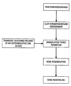
The process of bone repair and regeneration
resembles the process of wound healing in other tissues.
A typical sequence of events includes; hemorrhage; clot
formation; dissolution of the clot with concurrent
removal of damaged tissues; ingrowth of granulation
tissue; formation of cartilage; capillary ingrowth and
cartilage turnover; rapid bone formation (callus tissue);
and, finally, remodeling of the callus into cortical and
trabecular bone. Therefore, bone repair is a complex
process that involves many cell types and regulatory
molecules. The diverse cell populations involved in
fracture repair include stem cells, macrophages,
fibroblasts, vascular cells, osteoblasts, chondroblasts,
and osteoclasts.
Regulatory factors involved in bone repair are known
to include systemic hormones, cytokines, growth factors,
and other molecules that regulate growth and
differentiation. Various osteoinductive agents have been
purified and shown to be polypeptide growth-factor-like
molecules. These stimulatory factors are referred to as
bone morphogenetic or morphogenic proteins (BMPs), and
have also been termed osteogenic bone inductive proteins
or osteogenic proteins (OPs). Several BMP (or OP) genes
have now been cloned, and the common designations are
BMP-1 through BMP-8. New BMPs are in the process of
discovery. Although the BMP terminology is widely used,
it may prove to be the case that there is an OP
WO 95122611 2 18 3 5 4 PCT/US95102251
- 5 -
counterpart term for every individual BMP (Alper, 1994).
BMPs 2-8 are generally thought to be osteogenic,
although BMP-1 is a more generalized morphogen (Shimell
at al., 1991). BMP-3 is also called osteogenin (Luyten
at al., 1989) and BMP-7 is also called OP-1 (Ozkaynak et
al., 1990). BMPs are related to, or part of, the
transforming growth factor-a (TGF-Q) superfamily, and
both TGF-$1 and TGF-/32 also regulate osteoblast function
(Seitz at al.,, 1992). Several BMP (or OP) nucleotide
sequences and polypeptides have been described in U.S.
Patents, e.g., 4,795,804; 4,877,864; 4,968,590;
5,108,753; including, specifically, BMP-1 disclosed in
U.S. Patent 5,108,922; BMP-2A (currently referred to as
BMP-2) in U.S. Patents 5,166,058 and 5,013,649; BMP-2B
(currently referred to as BMP-4) disclosed in U.S. Patent
5,013,649; BMP-3 in 5,116,738; BMP-5 in 5,106,748; BMP-6
in 5,187,076; BMP-7 in 5,108,753 and 5,141,905; and OP-1,
COP-5 and COP-7 in 5,011,691.
Other growth factors or hormones that have been
reported to have the capacity to stimulate new bone
formation include acidic fibroblast growth factor
(Jingushi at al., 1990); estrogen (Boden at al., 1989);
macrophage colony stimulating factor (Horowitz at al.,
1989); and calcium regulatory agents such as parathyroid
hormone (PTH) (Raisz and Kream, 1983).
Several groups have investigated the possibility of
using bone stimulating proteins and polypeptides,
particularly recombinant BMPs, to influence bone repair
in vivo. For example, recombinant BMP-2 has been
employed to repair surgically created defects in the
mandible of adult dogs (Toriumi at al., 1991), and high
doses of this molecule have been shown to functionally
repair segmental defects in rat femurs (Yasko at al.,
1992). Chen and colleagues showed that a single
WO 95122611 21{38 3 5 4 2 PCT/US95102251
6
application of 25-100 mg of recombinant TGF-a1 adjacent
to cartilage induced endochondral bone formation in the
rabbit ear full-thickness skin wounds (Chen et al.,
1991). It has also been reported that an application of
TGF-p1 in a 3%k methylcellulose gel was able to repair
surgically induced large skull defects that otherwise
heal by fibrous connective tissue and never form bone
(Beck et al., 1991).
However, there are many drawbacks associated with
these type of treatment protocols, not least the
expensive and time-consuming purification of the
recombinant proteins from their host cells. Also,
polypeptides, once administered to an animal are more
unstable than is generally desired for a therapeutic
agent, and they are susceptible to proteolytic attack.
Furthermore, the administration of recombinant proteins
can initiate various inhibitive or otherwise harmful
immune responses. It is clear, therefore, that a new
method capable of promoting bone repair and regeneration
in vivo would represent a significant scientific and
medical advance with immediate benefits to a large number
of patients. A method readily adaptable for use with a
variety of matrices and bone-stimulatory genes would be
particularly advantageous.
SW RY OF THE INVENTION
The present invention overcomes one or more of these
and other drawbacks inherent in the prior art by
providing novel methods, compositions and devices for use
in transferring nucleic acids into bone cells and
tissues, and for promoting bone repair and regeneration.
Certain embodiments of the invention rest, generally,
with the inventors' surprising finding that nucleic acids
can be effectively transferred to bone progenitor cells
WO 95/22611 1 Q `~ C~ 2 PCT/US95102251
ic1U+o
in vivo and that, in certain embodiments, the transfer of
an osteotropic gene stimulates bone repair in an animal.
= The invention, in general terms, thus concerns
methods, compositions and devices fortransferring a
nucleic acid segment into bone progenitor cells or -
tissues. The methods of the invention generally comprise
contacting bone progenitor cells with a composition
comprising a nucleic acid segment in-a manner effective
to transfer the nucleic acid segment into the cells. The
cells may be cultured cells or recombinant cells
maintained in vitro, when all that is required is to add
the nucleic acid composition to the cells, e.g., by
adding it to the culture media.
- -
Alternatively, the progenitor cells may be located
within a bone progenitor tissue site of an animal, when
the nucleic acid composition would be applied to the site
in order to effect, or promote, nucleic acid transfer
into bone progenitor cells in vivo. In transferring
nucleic acids into bone cells within an animal, a
preferred method involves first adding the genetic
material to a bone-compatible matrix and then using the
resultant matrix to contact an appropriate tissue site
within the animal. The "resultant" matrix may, in
certain embodiments, be referred to as a matrix
impregnated with genetic material, or it may take the
form of a matrix-nucleic acid mixture, or even conjugate.
An extremely wide variety of genetic material can be
transferred to bone progenitor cells or tissues using the
compositions and methods of the invention. For example,
the nucleic acid segment may be DNA (double or single-
stranded) or RNA (e.g., mRNA, tRNA, rRNA); it may also be
= 35 a "coding segment", i.e., one that encodes a protein or
polypeptide, or it may be an antisense nucleic acid
molecule, such as antisense RNA that may function to
WO 95122611 2 1 8 3 5 4 2 PCT/US95/02251
- 8 -
disrupt gene expression. The nucleic acid segments may
thus be genomic sequences, including exons or introns
alone or exons and introns, or.coding cDNA regions, or in
fact any construct that one.'3esires to transfer to a bone
progenitor cell or tissue. Suitable nucleic acid
segments may also be in virtually any form, such as naked
DNA or RNA, including linear nucleic acid molecules and
plasmids; functional inserts within the genomes of
various recombinant viruses, including viruses with DNA
genomes and retroviruses; and any form of nucleic acid
segment, plasmid or virus associated with a liposome or a
gold particle, the latter of which may be employed in
connection with the gene gun technology.
The invention may be employed to promote expression
of a desired gene in bone cells or tissues and to impart
a particular desired phenotype to the cells. This
expression could be increased expression of a gene that
is normally expressed (i.e., "over-expression"), or it
could be used to express a gene that is not normally
associated with bone progenitor cells in their natural
environment. Alternatively, the invention may be used to
suppress the expression of a gene that is naturally
expressed in such cells and tissues, and again, to change
or alter the phenotype. Gene suppression may be a way of
expressing a gene that encodes a protein that exerts a
down-regulatory function, or it may utilize antisense
technology.
1. Bone Progenitor Cells and Tissues
In certain embodiments, this invention provides
advantageous methods for using genes to stimulate bone
progenitor cells. As used herein, the term "bone
progenitor cells" refers to any or all of those cells
that have the capacity to ultimately form, or contribute
to the formation of, new bone tissue. This includes
WO 95/22611 218 3 5 4 2 PCT/US95102251
- 9 -
various cells in different stages of differentiation,
such as, for example, stem cells, macrophages,
fibroblasts, vascular cells, osteoblasts, chondroblasts,
osteoclasts, and the like. Bone progenitor cells also
include cells that have been isolated and manipulated in
vitro, e.g., subjected to stimulation with agents such as
cytokines or growth factors or even genetically
engineered cells. The particular type or types of bone
progenitor cells that are stimulated using the methods
and compositions of the invention are not important, so
long as the cells are stimulated in such a way that they
are activated and, in the context of in vivo embodiments,
ultimately give rise to new bone tissue.
The term "bone progenitor cell" is also used to
particularly refer to those cells that are located
within, are in contact with, or migrate towards (i.e.,
"home to"), bone progenitor tissue and which cells
directly or indirectly stimulate the formation of mature
bone. As such, the progenitor cells may be cells that
ultimately differentiate into mature bone cells
themselves, i.e., cells that "directly" form new bone
tissue. Cells that, upon stimulation, attract further
progenitor cells or promote nearby cells to differentiate
into bone-forming cells (e.g., into osteoblasts,
osteocytes and/or osteoclasts) are also considered to be
progenitor cells in the context of this disclosure - as
their stimulation "indirectly" leads to bone repair or
regeneration. Cells affecting bone formation indirectly
may do so by the elaboration of various growth factors or
cytokines, or by their physical interaction with other
cell types. Although of scientific interest, the direct
or indirect mechanisms by which progenitor cells
CVO 95/22611 2 1 8 3 5 4 2 PCT/US95/02251
-
stimulate bone or wound repair is not a consideration in
practicing this invention.
Bone progenitor cells and bone progenitor tissues
5 may be cells and tissues that, in their natural
environment, arrive at an area of active bone growth,
repair or regeneration (also referredto as a wound
repair site). In terms of bone progenitor cells, these
may also be cells that are attracted or recruited to such
10 an area. These may be cells that are present within an
artificially-created osteotomy site in an animal model,
such as those disclosed herein. Bone progenitor cells
may also be isolated from animal or human tissues and
maintained in an in vitro environment. Suitable areas of
the body from which to obtain bone progenitor cells are
areas such as the bone tissue and fluid surrounding a
fracture or other skeletal defect (whether or not this is
an artificially created site), or indeed, from the bone
marrow. Isolated cells may be stimulated using the
methods and compositions disclosed herein and, if
desired, be returned to an appropriate site in an animal
where bone repair is to be stimulated. In such cases,
the nucleic-acid containing cells would themselves be a
form of therapeutic agent. Such ex vivo protocols are
well known to those of skill-in the art.
In important embodiments of the invention, the bone
progenitor cells and tissues will be those cells and
tissues that arrive at the area of bone fracture or
damage that one desires to treat. Accordingly, in
treatment embodiments, there is no difficulty associated
with the identification of suitable target progenitor
cells to which the present therapeutic compositions
should be applied. All that is required in such cases is
to obtain an appropriate stimulatory composition, as
disclosed herein, and contact the site of the bone
fracture or defect with the composition. The nature of
WO 95/22611 CA 02183542 2000-08-01
PCTIUs95/02251
- 11 -
this biological environment is such that the appropriate
cells will become activated in the absence of any further
targeting or-cellular identification by the practitioner.
Certain methods of the invention involve, generally,
contacting bone progenitor cells with a composition
comprising one or more osteotropic genes (with or without
additional genes, proteins or other biomolecules) so as
to promote expression of said gene in said cells. As
outlined above, the cells may be contacted in vitro or in
vivo. This is achieved, in the most direct manner, by
simply obtaining a functional osteotropic gene construct
and applying the construct to the cells. The present
inventors surprisingly found that there are no particular
molecular biological modifications that need to be
performed in order to promote effective expression of the
gene in progenitor cells. Contacting the cells with DNA,
e.g., a linear DNA molecule, or DNA in the form of a
plasmid or other recombinant vector, that contains the
gene of interest under the control of a promoter, along
with the appropriate termination signals, is sufficient
to result in uptake and expression of the DNA, with no
further steps necessary.
In preferred embodiments, the process of contacting
the progenitor cells with the osteotropic gene
composition is conducted in vivo. Again, a direct
consequence of this process is that the cells take up and
express the gene and that they, without additional steps,
function to stimulate bone tissue growth, repair or
regeneration.
An assay of an osteoinductive gene may be conducted
using the bone induction bioassay of Sampath and Reddi
(1981). This is a rat bone formation assay thiLt is routinely used to evaluate
the osteogenic activity of bone inductive factors.
WO 95/22611 2183542
- 12 - PCT/US95/02251
However, for analyzing the effects ofosteotropic genes
on bone growth, one is generally directed to use the
novel osteotomy model disclosed herein.
2. Osteotropic Genes
As used herein, the terms"osteotropic and
osteogenic gene"- are used to refer to a gene or DNA
coding region that encodes a protein, polypeptide or
peptide that is capable of promoting, or assisting in the
promotion of, bone formation, or one that increases the
rate of primary bone growth or healing (or even a gene
that increases the rate of skeletal connective tissue
growth or healing). The terms promoting, inducing and
stimulating are used interchangeably throughout this text
to refer to direct or indirect processes that ultimately
result in the formation of new bone tissue or in an
increased rate of bone repair. Thus, an osteotropic gene
is a gene that, when expressed, causes the phenotype of a
cell to change so that the cell either differentiates,
stimulates other cells to differentiate, attracts bone-
forming cells, or otherwise functions in a manner that
ultimately gives rise to new bone tissue.
In using the new osteotomy model of the invention,
an osteotropic gene is characterized as a gene that is
capable of stimulating proper bone growth in the
osteotomy gap to any degree higher than that observed in
control studies, e.g., parallel studies employing an
irrelevant marker gene such as Q-galactosidase. This
stimulation of "proper bone growth" includes both the
type of tissue growth and the rate of bone formation. In
using the model with a 5 mm osteotomy gap, an osteotropic
gene is generally characterized as a gene that is capable
of promoting or inducing new bone formation, rather than
abnormal bone fracture repair, i.e., fibrous non-union.
In using the 2 mm osteotomy gap, one may characterize
WO 95/22611 CA 02183542 2000-08-01 PCT/US95/02251
- 13 -
osteotropic-genes as crenes that increase the rate of
primary bone healing as compared to controls, and more
preferably, genes capable of stimulating repair of the
osteotomy defect in a time period of less than nine
weeks.
In general terms, an osteotropic gene may also be
characterized as a gene capable of stimulating the growth
or regeneration of skeletal connective tissues such as,
3.0 e.g., tendon, cartilage, and ligament. Thus, in certain
embodiments, the methcds and compositions of the
invention may be employed to stimulate the growth or
repair of both bone tissue itself and also of skeletal
connective tissues.
A variety of osteotropic genes are now known, all of
which are suitable for use in connection with the present
invention. Osteotropic genes and the proteins that they
encode include, for example, systemic hormones, such as
parathyroid hormone (PTH) and estrogen; many different
growth factors and cytokines; chemotactic or adhesive
peptides or polypeptides; molecules such as activin (U.S.
Patent 5,208,219;
specific bone morphoge:netic proteins (BMPs); and even
growth factor receptor genes.
Examples of suitable osteotropic growth factors
include those of the transforming growth factor (TGF)
gene family, including TGFs 1-3, and particularly TGF-#1,
TGF-Q2 and TGF-fl3, (U.S. Patents 4,886,747 and
4,742,003), with TGF-a (U.S. Patent 5,168,051) also
being of possible use; and also fibroblast growth factors
(FGF), previously referred to as acidic and basic FGF and
now referred to as FGF1-9; granulocyte/macrophage colony
stimulating factor (GMCSF); epidermal growth factor
(EGF); platelet derived growth factor (PDGF); insulin-
CVO 95122611 2183542
- 14 PCT1L'S95/02251 I
-
like growth factors (IGF), including TGF-I and IGF-II;
and leukemia inhibitory factor (LIF), also known as HILDA
and DIA. Any of the above or other related genes, or DNA
segments encoding the active, portions of such proteins,
may be used in the novel methods and compositions of the
invention.
Certain preferred osteotropic genes and DNA segments
are those of the TGF superfamily, such as TGF-$l, TGF-$2,
TGF-(33 and members of the EMP family of genes. For
example, several BMP genes have been cloned that are
ideal candidates for use in the nucleic acid transfer or
delivery protocols of the invention. Suitable BMP genes
are those designated BMP-2 through BMP-12. BMP-1 is not
considered to be particularly useful at this stage.
There is considerable variation in the terminology
currently employed in the. literature in referring to
these genes and polypeptides. It will be understood by
those of skill in the art that all BMP genes that encode
an active osteogenic protein are considered for use in
this invention, regardless of the differing terminology
that may be employed. For example, BMP-3 is also called
osteogenin and BMP-7 is also called OP-i (osteogenic
protein-1). It is likely that the family of factors
termed OP(s) is as large as that termed BMP(s), and that
these terms, in fact, describe the same set of molecules
(Alper, 1994).
The DNA sequences for several BMP (or OP) genes have
been described both in scientific articles and in U.S.
Patents such as 4,877,864; 4,968,590; 5,108,753.
Specifically, BMP-1 sequences are disclosed in U.S.
Patent 5,108,922; BMP-2A (currently referred to as BMP-2)
in U.S. Patents 5,166,058 and 5,013,649; BMP-2B - '
(currently referred to as BMP-4) disclosed in U.S. Patent
5,013,649; BMP-3 in 5,116,738; BMP-5 in 5,106,748; BMP-6
CA 02183542 2000-08-01
WO 95/22611 PCT1LIS95/02251
- 15 -
in 5,187,076; and BMP-7 in 5,108,753 and 5,141,905,
The article by Wozney
et: al., (1988) is
considered to be particularly useful for describing BMP
molecular clones and their activities. DNA sequences
encoding the osteogenic proteins designated OP-1, COP-5
and COP-7 are also disclosed in U.S. Patent 5,011,691.
All of the above issued U.S. Patents are
intended to be -
used in order to supplement the present teachings
regarding the preparation of BMP and OP genes and DNA
segments that express osteotropic polypeptides. As
disclosed in the above patents, and known to those of
skill in the art, the original source of a recombinant
gene or DNA segment to be used in a therapeutic regimen
need not be of the same species as the animal to be
treated. In this regard, it is contemplated that any
recombinant PTI:i, TGF or BMP gene may be employed to
promote bone repair or regeneration in a human subject or
an animal, e.g., a horse. Particularly preferred genes
are those from human, murine and bovine sources, in that
such genes and DNA segments are readily available, with
the human or murine forms of the gene being most
preferred for use in ;human treatment regimens.
Recombinant proteins and polypeptides encoded by isolated
DNA segments and genes are often referred to with the
prefix "r" for recombinant and "rh" for recombinant
human. As such, DNA segments encoding rBMPs, such as
rhBMP-2 or rhBMP-4, are contemplated to be particularly
useful in connection with this invention.
The definition o.E a "BMP gene", as used herein, is a
gene that hybridizes, under relatively stringent
hybridization conditions (see, e.g., Maniatis et al.,
1982), to DNA sequences presently known to include BMP
gene sequences.
CA 02183542 2000-08-01
WO 95/22611 PCTIUS95102251
- 16 -
To prepare an osl:eotropic gene segment or cDNA one
may follow the teachings disclosed herein and also the
teachings of any of patents or scientific documents
specifically referenced herein. Various nucleotide
sequences encoding active BMPs are disclosed in U.S.
Patents 5,166,058, 5,013,649, 5,116,738, 5,106,748,
5,187,076, 5,108,753 and 5,011,691.
By way of example only, U.S. Patent
5,166,058, teaches that hBMP-2 is encoded by a nucleotide
sequence from nucleotide #356 to nucleotide #1543 of the
sequence shown in Table II of the patent. One may thus
obtain a hBMP-2 DNA segment using molecular biological
techniques, such as polymerase chain reaction (PCRT") or
screening a cDNA or ge:nomic library, using primers or
:L5 probes with sequences based on the above nucleotide
sequence. The practice of such techniques is a routine
matter for those of skill in the art, as taught in
various scientific articles, such as Sambrook et al.,
(1989), incorporated herein by reference. Certain
documents further particularly describe suitable
mammalian expression vectors, e.g., U.S. Patent
5,168,050,
Osteotropic genes and DNA segments that are
particularly preferred for use in certain aspects of the
present compositions and methods are the TGF, PTH and BMP
genes. TGF genes are described in U.S. Patents
5,168,051; 4,886,747 and 4,742,003
TGFa may not be as widely
applicable as TWO, but is proposed for use particularly
in applications involving skeletal soft tissues. The PTH
gene, or a DNA segment encoding the active fragment
thereof, such as a DNA segment encoding a polypeptide
that includes the amino acids 1-34 (hPTH1-34; Hendy et
al., 1981; is another
CA 02183542 2000-08-01
WO 95/22611 PCT/US95/02251
- 17 -
preferred gene; as are the BMP genes termed BMP-4 and
BMP-2, such as the gene or cDNA encoding the murine BMP-4
disclosed herein.
It is also contemplated that one may clone further
genes or cDNAs that encode an osteotropic protein or
polypeptide. The techniques for cloning DNA molecules,
i.e., obtaining a specific coding sequence from a DNA
library that is distinct from other portions of DNA, are
well known in the art. This can be achieved by, for
example, screening an appropriate DNA library, as
disclosed in Example XV herein, which relates to the
cloning of a wound healing gene. The screening procedure
may be based on the hybridization of oligonucleotide
:15 probes, designed from a consideration of portions of the
amino acid sequence of known DNA sequences encoding
related osteoge:nic proteins. The operation of such
screening protocols are well known to those of skill in
the art and are described in detail in the scientific
:20 literature, for example, in Sambrook et al., (1989).
Osteotropic genes: with sequences that vary from
those described in the literature are also encompassed by
:25 the invention, so long as the altered or modified gene
still encodes a protein that functions to stimulate bone
progenitor cells in any direct or indirect manner. These
sequences include those caused by point mutations, those
due to the degeneracies of the genetic code or naturally
:30 occurring allelic variants, and further modifications
that have been introduced by genetic engineering, i.e.,
by the hand of man.
Techniques for introducing changes in nucleotide
:35 sequences that are designed to alter the functional
properties of the encoded proteins or polypeptides are
well known in the art, e.g., U.S. Patent 4,518,584,
CA 02183542 2000-08-01
WO 95/22611 PCTIUS95/02251
- 18 -
which techniques are
also described in further detail herein. Such
modifications include the deletion, insertion or
substitution of base:;, and thus, changes in the amino
acid sequence. Changes may be made to increase the
osteogenic activity of a protein, to increase its
biological stability or half-life, to change its
glycosylation pattern, and the like. All such
modifications to the nucleotide sequences are encompassed
by this invention.
It will, of course, be understood that one or more
than one osteotropic gene may be used in the methods and
compositions of the invention. The nucleic acid delivery
methods may thus entail the administration of one, two,
three, or more, osteotropic genes. The maximum number of
genes that may be applied is limited only by practical
considerations, such as the effort involved in
simultaneously preparing a large number of gene
constructs or even the possibility of eliciting a
significant adverse cytotoxic effect. The particular
combination of genes may be two or more distinct BMP
genes; or it may be such that a growth factor gene is
combined with a hormone gene, e.g., a BMP gene and a PTH
gene; a hormone or growth factor gene may even be
combined with a gene encoding a cell surface receptor
capable of interacting with the polypeptide product of
the first gene.
In using multiple genes, they may be combined on a
single genetic construct under control of one or more
promoters, or they may be prepared as separate constructs
of the same of different types. Thus, an almost endless
combination of different genes and genetic constructs may
be employed. Certain gene combinations may be designed
to, or their use may otherwise result in, achieving
synergistic effects on cell stimulation and bone growth,
CA 02183542 2000-08-01
WO 95/22611 PCT/US95/02251
- 19 -
any and all such combinations are intended to fall within
the scope of the present invention. Indeed, many
synergistic effects have been described in the scientific
literature, to that one of ordinary skill in the art
would readily be able to identify likely synergistic gene
combinations, or even gene-protein combinations.
It will also be understood that, if desired, the
nucleic segment or gene could be administered in
3.0 combination with further agents, such as, e.g., proteins
or polypeptides or various pharmaceutically active
agents. So long as genetic material forms part of the
composition, there is virtually no limit to other
components which may also be included, given that the
1.5 additional agents do not cause a significant adverse
effect upon contact with the target cells or tissues.
The nucleic acids may thus be delivered along with
various other agents, for example, in certain embodiments
one may wish to administer an angiogenic factor, and/or
20 an inhibitor of bone resorption, as disclosed in U.S.
Patents 5,270,300 and 5,118,667, respectively,
3. Gene Constructs and DNA Segments
As used herein, the terms "gene" and "DNA segment"
are both used to refer to a DNA molecule that has been
isolated free of total genomic DNA of a particular
species. Therefore, a gene or DNA segment encoding an
osteotropic gene refers to a DNA segment that contains-
sequences encoding an osteotropic protein, but is
isolated away from, or purified free from, total genomic
DNA of the species from which the DNA is obtained.
Included within the term "DNA segment", are DNA segments
and smaller fragments of such segments, and also
recombinant vectors, including, for example, plasmids,
cosmids, phage, retroviruses, adenoviruses, and the like.
CA 02183542 2000-08-01
WO 95/22611 PCT/L'S95/02251
- 20 -
The term "gene" is used for simplicity to refer to a
functional protein o:: peptide encoding unit. As will be
understood by those in the art, this functional term
includes both genomic, sequences and cDNA sequences.
"Isolated substantially away from other coding sequences"
means that the gene of interest, in this case, an
osteotropic gene, forms the significant part of the
coding region of the DNA segment, and that the DNA
segment does not contain large portions of naturally-
occurring coding DNA, such as large chromosomal fragments
or other functional genes or cDNA coding regions. Of
course, this refers to the DNA segment as originally
isolated, and does not exclude genes or coding regions,
such as sequences encoding leader peptides or targeting
sequences, later added to the segment by the hand of man.
This invention provides novel ways in which to
utilize various known osteotropic DNA segments and
recombinant vectors. As described above, many such
vectors are readily available, one particular detailed
example of a suitable vector for expression in mammalian
cells is that described in U.S. Patent 5,168,050.
However, there is no ---
requirement that a highly purified vector be used, so
long as the coding segment employed encodes a osteotropic
protein and does not. include any coding or regulatory
sequences that would have a significant adverse effect on
bone progenitor cells. Therefore, it will also be
understood that useful nucleic acid sequences may include
additional residues, such as additional non-coding
sequences flanking either of the 5' or 3' portions of the
coding region or may include various internal sequences,
i.e., introns, which are known to occur within genes.
After identifying an appropriate osteotropic gene or
DNA molecule, it may be inserted into any one of the many
vectors currently known in the art, so that it will
WO 95/22611 - 21 - 2183542 PCT/US9',102251
direct the expression and production of the osteotropic
protein when incorporated into a bone progenitor cell.
In a recombinant expression vector, the coding portion of
the DNA segment is positioned under the control of a
promoter. The promoter may be in the form of the
promoter which is naturally associated with an
osteotropic gene, as may be obtained by isolating the 5'
non-coding sequences located upstream of the coding
segment or exon, for example, using recombinant cloning
and/or PCR' technology, in connection with the
compositions disclosed herein.
In other embodiments, it is contemplated that
certain advantages will be gained by positioning the
coding DNA segment under the control of a recombinant, or
heterologous, promoter. As used herein, a recombinant or
heterologous promoter is intended to refer to a promoter
that is not normally associated with an osteotropic gene
in its natural environment. Such promoters may include
those normally associated with other osteotropic genes,
and/or promoters isolated from any other bacterial,
viral, eukaryotic, or mammalian cell. Naturally, it will
be important to employ a promoter that effectively
directs the expression of the DNA segment in bone
progenitor cells.
The use of recombinant promoters to achieve protein
expression is generally known to those of skill in the
art of molecular biology, for example, see Sambrook et
al., (1989). The promoters employed may be constitutive,
or inducible, and can be used under the appropriate
conditions to direct high level or regulated expression
of the introduced DNA segment. The currently preferred
promoters are those such as CMV, RSV LTR, the SV40
promoter alone, and the SV40 promoter in combination with
various enhancer elements.
WO 95122611 q 1Op ed~ 5 4 - 22 PCTIUS95/02251
fr -
Osteotropic genes and DNA segments may also be in
the form of a DNA insert which is located within the
genome of a recombinant virus, such as, for example a
recombinant adenovirus, adeno-associated virus (AAV) or
retrovirus. In such embodiments, to place the gene in
contact with a bone progenitor. cell, one would prepare
the recombinant viral particles; the genome of which
includes the osteotropic ggne insert, and simply contact
the progenitor cells or tissues with the virus, whereby
the virus infects the cells and transfers the genetic
material.
In certain preferred embodiments, one would
impregnate a matrix or implant material with virus by
soaking the material in recombinant virus stock solution,
e.g., for 1-2 hours, and then contact the bone progenitor
cells or tissues with the resultant, impregnated matrix.
Cells then penetrate, or grow into, the matrix, thereby
contacting the virus and allowing viral infection which
leads to the cells taking up the desired gene or cDNA and
expressing the encoded protein.
In other preferred embodiments, one would form a
matrix-nucleic acid admixture, whether using naked DNA, a
plasmid or a viral vector, and contact the bone
progenitor cells or tissues with the resultant admixed
matrix. The matrix may then deliver the nucleic acid
into the cells following disassociation at the cell
surface, or in the immediate cellular environment.
Equally, the matrix admixture itself, especially a
particle- or fiber-DNA admixture, may be taken up by
cells to provide subsequent intracellular release of the
genetic material. The matrix may then be extruded from
the cell, catabolized by the cell, or even stored within
the cell. The molecular mechanism by which a bone-
compatible matrix achieves transfer of DNA to a cell is
immaterial to the practice of the present invention.
NO 95122611 21 SJg 5 4 2 PCTIUS95/02251
- 23 -
4. Bone-Compatible Matrices
In certain preferred embodiments, the methods of the
invention involved preparing a composition in which the
osteotropic gene, genes, DNA segments, or cells already
incorporating such genes or segments, are associated
with, impregnated within, or even conjugated to, a bone-
compatible matrix, to form a "matrix-gene composition"
and the matrix-gene composition is then placed in contact
with the bone progenitor cells or tissue. The matrix may
become impregnated with a gene DNA segment simply by
soaking the matrix in a solution containing the DNA, such
as a plasmid solution, for a brief period of time of
anywhere from about 5 minutes or so, up to and including
about two weeks.
Matrix-gene compositions are all those in which
genetic material is adsorbed, absorbed, impregnated,
conjugated to, or otherwise generally maintained in
contact with the matrix. "Maintained in contact with the
matrix" means that an effective amount of the nucleic
acid composition should remain functionally associated
with the matrix until its transfer to the bone progenitor
cell or its release in the bone tissue site.
The type of matrix that may be used in the
compositions, devices and methods of the invention is
virtually limitless, so long as it is a "bone-compatible
matrix". This means that the matrix has all the features
commonly associated with being "biocompatible", in that
it is in a form that does not produce a significant
adverse, allergic or other untoward reaction when
administered to an animal, and that it is also suitable
for placing in contact with bone tissue. A "significant"
adverse effect is one that exceeds the normally accepted
side-effects associated with any given therapy.
CA 02183542 2000-08-01
WO 95122611 PCT/US95/02251
24 -
"Bone-compatible", as used herein, means that the
matrix (and gene) does not produce a significant adverse
or untoward reaction when placed in contact with bone.
In certain embodiments, when electing to use a particular
bone compatible matrix, one may, optionally, take various
other factors into consideration, for example, the
capacity of the matrix to provide a structure for the
developing bone, its capacity to be resorbed into the
body after the bone has been repaired, and such like.
However, these proper-:ies are not required to practice
the invention and are merely exemplary of the factors
that may be considered.
In other embodiments, one may also consider the
likelihood that the matrix will be transported into the
cell, e.g., by active or passive membrane transport.
Where such transport and subsequent nucleic acid release
is contemplated, other properties of the matrix and gene
may be assessed in optimizing the matrix-gene
formulation. For example, adenovirus vectors may provide
for advantageous DNA :release in such embodiments.
Matrices that are readily metabolized in the cytoplasm
would also likely be preferred in such embodiments.
Matrices that are later released from the cell, and
preferably, also removed from the surrounding tissue
area, would be another preferred form of matrix for use
in such embodiments.
The choice of matrix material will differ according
to the particular circumstances and the site of the bone
that is to be treated. Matrices such as those described
in U.S. Patent 5,270,300
may be employed. Physical and chemical
characteristics, such as, e.g., biocompatibility,
biodegradability, strength, rigidity, interface
properties, and even cosmetic appearance, may be
considered in choosing a matrix, as is well known to
CA 02183542 2000-08-01
WO 95/22611 PCTIUS95/02251
- 25 -
those of skill in the art. Appropriate matrices will
deliver the gene composition and, in certain
circumstances, may be incorporated into a cell, or may
provide a surface for new bone growth, i.e., they may act
as an in situ scaffolding through which progenitor cells
may migrate.
A particularly important aspect of the present
invention is its use in connection with orthopaedic
:L0 implants and interfaces and artificial joints, including
implants themselves and functional parts of an implant,
such as, e.g., surgical] screws, pins, and the like. In
preferred embodiments, it is contemplated that the metal
surface or surfaces of an implant or a portion thereof,
:L5 such as a titanium surface, will be coated with a
material that has an affinity for nucleic acids, most
preferably, with hydroxyl apatite, and then the coated-
metal will be further coated with the gene or nucleic
acid that one wishes to transfer. The available chemical
20 groups of the absorptive material, such as hydroxyl
apatite, may be readily manipulated to control its
affinity for nucleic acids, as is known to those of skill
in the art.
25 In certain embodiments, non-biodegradable matrices
may be employed, such as sintered hydroxylapatite,
aluminates, other bioceramic materials and metal
materials, particularly titanium. A suitable ceramic
delivery system is that described in U.S. Patent
?30 4,596,574. Polymeric matrices may also be employed, including acrylic
ester
polymers, lactic acid polymers, and polylactic
polyglycolic acid (PLGA) block copolymers, have been
disclosed (U.S. Patent 4,526,909, U.S. Patent 4,563,489,
35 Simons et al., 1992, and Langer and Folkman, 1976,
respectively,
CA 02183542 2000-08-01
WO 95/22611 PCf/US95/02251
- 26 -
In certain embodiments, it is contemplated that a
biodegradable matrix will likely be most useful. A
biodegradable matrix is generally defined as one that is
capable of being rescrbed into the body. Potential
biodegradable matrices for use in connection with the
compositions, devices and methods of this invention
include, for example, biodegradable and chemically
defined calcium sulfate, tricalciumphosphate,
hydroxylapatite, PLGA. block copolymers, polyanhydrides,
matrices of purified proteins, and semi-purified
extracellular matrix compositions.
One preferred group of matrices are collagenous
matrices, including those obtained from tendon or dermal
collagen, e.g., type I collagen, which is generally
prepared from dermis; those obtained from cartilage, such
as type II collagen; and various other types of collagen.
Collagens may be obtained from a variety of commercial
sources, e.g., Sigma that supplies type II collagen
obtained from bovine trachea; and Collagen Corporation.
Collagen matrices may also be prepared as described in
U.S. Patents 4,394,370 and 4,975,527,
The various collagenous materials may also be in the
form of mineralized collagen. One preferred mineralized
collagenous material is that termed UltraFiber,
obtainable from Norian Corp. (Mountain View, CA). U.S.
Patent 5,231,169,
describes the preparation of mineralized collagen through
the formation of' calcium phosphate mineral under mild
agitation in situ in the presence of dispersed collagen
fibrils. Such a formulation may be employed in the
WO 95/22611 27 - 2183542 PCT/US95102251
context of delivering a nucleic acid segment to a bone
tissue site.
Certain other preferred collagenous materials are
those based upon type II collagen. Type II collagen
preparations have been discovered to have the surprising
and advantageous property of, absent any osteotropic
gene, being capable of stimulating bone progenitor cells.
Prior to the present invention, it was thought that
type II collagen only had a structural role in the
cartilage extracellular matrix and the present finding
that type II collagen is actually an
osteoconductive/osteoinductive material is unexpected.
The present invention thus contemplates the use of -a
variety of type II collagen preparations as gene transfer
matrices or bone cell stimulants, either with or without
DNA segments, including native type II collagen, as
prepared from cartilage, and recombinant type II
collagen.
--
PLGA block copolymers may also be employed as gene
transfer matrices. Such polymers have been shown to
readily incorporate DNA, are commercially available, non-
toxic, and hydrolyze at defined rates, (i.e. they
facilitate the sustained release of pharmaceutical
agents). PLGA block copolymers have two particular
advantageous properties in that, first, they exhibit
reversible thermal gelation, and second, may be combined
with other agents that allow for radiographic
visualization.- - - -- -
5. Nucleic Acid Transfer Embodiments
Once a suitable matrix-gene composition has been
prepared or obtained, all that is required to deliver the
osteotropic gene to bone progenitor cells within an
animal is to place the matrix-gene composition in contact
WO 9512261 1 2 1 8 3 5 4 2i. - 28 - PCTIU595/02251
with the site in the body in which one wishes to promote
bone growth. This may be achieved by physically -
positioning the matrix-gene composition in contact with
the body site, or by injecting a syringeable form of the
S matrix-gene composition intothe appropriate area. -
The matrix-gene composition may be applied to a
simple bone fracture site that one wishes to repair; an
area of weak bone, such as in a 'patient with
osteoporosis, or a bone cavity site that one wishes to
fill with new bone tissue. Bone cavities may arise as a
result of an inherited disorder, birth defect, or may
result following dental or periodontal surgery or after
the removal of an osteosarcoma.
The use of PLGA and like compounds as matrices
allows the matrix-DNA composition to be syringeable,
which is achieved by, generally, admixing the matrix-gene
composition with a pluronic agent. The resultant matrix-
gene-pluronic may be stored within a thermal-jacket
syringe, maintained at a temperature of about 4 C,
immediately prior to administration to the body. In this
temperature and environment, the composition will be a
liquid. Following insertion into the body, the
composition will equilibrate towards body temperature,
and in so-doing will form a gelatinous matrix.
The above phenomenon is termed "reversible thermal
gelation", and this allows for a controlled rate of
gelation to be achieved. The manner of using pluronic
agents in this context will be known to those of skill in
the art in light of the present disclosure. Matrix-gene-
pluronic compositions may also be admixed, or generally
associated with, an imaging agent so that the present
gene transfer technology may be used in imaging
modalities. In these cases, the attending physician or
veterinarian will be able to monitor the delivery and
WO 95/22611 ?1835; 4 PCT/US95/02251
~
29 -
positioning of the matrix-gene composition. Many safe
and effective imaging agents, such as the radiographic
compound calcium phosphate, areavailable that may be
used in conjunction with fluoroscopy, or even with
tomography, to image the body or tissue site while the
composition is being delivered.
Where an image of the tissue site is to be provided,
one will desire to use a detectable imaging agent, such
as a radiographic agent, or even a paramagnetic or
radioactive agent. Many radiographic diagnostic agents
are known in the art to be useful for imaging purposes,
including e.g., calcium phosphate.
In the case of paramagnetic ions, examples include
chromium (III), manganese (II), iron (III), iron (II),
cobalt (II), nickel (II), copper (II), neodymium (III),
samarium (III), ytterbium (III), gadolinium (III),
vanadium (II), terbium (III), dysprosium (III), holmium
(III) and erbium (III), with gadolinium being generally
preferred. Ions useful in other contexts, such as X-ray
imaging, include but are not limited to, lanthanum (III),
gold (III), lead (II), and especially bismuth (III).
Although not generally preferred, radioactive
isotopes are not excluded and may be used for imaging
purposes if desired. Suitable ions include iodine131
iodine123 technicium99m indium111 rhenium'88, rhenium'86,
gallium67, copper67, yttrium90, iodine" and astatine211
The amount of gene construct that is applied to the
matrix and the amount of matrix-gene material that is
applied to the bone tissue will be determined by the
attending physician or veterinarian considering various
biological and medical factors. For example, one would
wish to consider the particular osteotropic gene and
matrix, the amount of bone weight desired to be formed,
WO 95122611 (rlOel 2] 1 4 `~ 5 - 30 - PCT/U595/02251
rt
the site of bone. damage, thecondition of the damaged
bone, the patient's or animal's age, sex, and diet, the
severity of any infection, the time of administration and
any further clinical factors that may affect bone growth,
such as serum levels of various factors and hormones.
The suitable dosage regimen willtherefore be readily
determinable by one of skill in the art in light of the
present disclosure, bearing in mind the individual
circumstances.
-
In treating humans and animals, progress may be
monitored by periodic assessment of bone growth and/or
repair, e.g., using X-rays. The therapeutic methods and
compositions of the invention are contemplated for use in
both medical and veterinary applications, due to the lack
of species specificity in bone inductive factors. In
particular, it is contemplated that domestic, farm and
zoological animals, as well as thoroughbred horses, would
be treatable using the nucleic acid transferprotocols
disclosed herein.
The present methods and compositions may also have
prophylactic uses in closed and open fracture reduction
and also in the improved fixation of artificial joints.
The invention is applicable to stimulating bone repair in
congenital, trauma-induced, or oncologic resection-
induced craniofacial defects, and also is useful in the
treatment of periodontal disease and other tooth repair
processes and even in cosmetic plastic surgery. The
matrix-gene compositions and devices of this invention
may also be used in wound healing and related tissue
repair, including, but not limited to healing of burns,
incisions and ulcers.
The present invention also encompasses DNA-based
compositions for use in cellular transfer to treat bone
defects and disorders. The compositions of the invention
WO 95/22611 9 PCT/US95/02251
Q
31 -
generally comprise( an osteotropic gene in association
with a bone-compatible matrix, such as type II collagen,
wherein the composition is capable of stimulating bone
growth, repair or regeneration upon administration to, or
implantation within, a-bone progenitor tissue site of an
animal. The osteotropic gene or genes may be any of
those described above, with TGF-a (for soft skeletal
tissues), TGF-(31, TGF-02, TGF-03, PTH, BMP-2 and BMP-4
genes being generally preferred. Likewise, irrespective
of the choice of gene, the bone-compatible matrix may be
any of those described above, with biodegradable matrices
such as collagen and, more particularly, type II
collagen, being preferred.
In still further embodiments, the present invention
concerns osteotropic devices, which devices may be
generally considered as molded or designed matrix-gene
compositions. The devices of the invention naturally
comprise a bone-compatible matrix in which an osteotropic
gene is associated with the matrix. The combination of
genes and matrix components is such that the device is
capable of stimulating bone growth or healing when
implanted in an animal. The devices may be of virtually
any size or shape, so that their dimensions are adapted
to fit a bone fracture or bone cavity site in the animal
that is to be treated, allowing the fracture join and/or
bone regrowth to be more uniform. Other particularly
contemplated devices are those that are designed to act
as an artificial joint. Titanium devices and
hydroxylapatite-coated titanium devices will be preferred
in certain embodiments. Parts of devices in combination
with an osteotropic nucleic acid segment, such as a DNA-
coated screw for an artificial joint, and the like, also
fall withinthe scope of the invention.
Therapeutic kits comprising, in suitable container
means, a bone compatible matrix, such as type II collagen
WO 95/22611 21 O354 2 - 32 - PCT/US95/02251
!J !u
or a PLGA block polymer, and an osteotropic gene form
another aspect of the invention. Such kits will
generally contain a pharmaceutically acceptable
formulation of the matrix and a pharmaceutically
acceptable formulation of an osteotropic gene, such as
PTH, EMP, TGF-$, FGF, GMCSF, EGF, PDGF, IGF or a LIF
gene. Currently preferred genes. include PTH, TGF-/31,
TGF-/32, TGF-/33, and BMP-4 genes.
The kits may comprise a single container means that
contains both the biocompatible matrix and the
osteotropic gene. The container means may, if desired,
contain a pharmaceutically acceptable sterile syringeable
matrix, having associated with it, the osteotropic gene
composition and, optionally, a detectable label or
imaging agent. The syringeable matrix-DNA formulation
may be in the form of a gelatinous composition, e.g., a
type II collagen-DNA composition, or may even be in a
more fluid form that nonetheless forms a gel-like
composition upon administration to the body. In these
cases, the container means may itself be a syringe,
pipette, or other such like apparatus, from which the
matrix-DNA material may be applied to a bone tissue site
or wound area. However, the single container means may
contain a dry, or lyophilized, mixture of a matrix and
osteotropic gene composition, which may or may not
require pre-wetting before use.
Alternatively, the kits of the invention may
comprise distinct container means for each component. In
such cases, one container would contain the osteotropic
gene, either as a sterile DNA solution or in a
lyophilized form, and the other container would include
the matrix, which may or may not itself be pre-wetted
with a sterile solution, or be in a gelatinous, liquid or:
othersyringeable form.
PCTIUS95/02251
WO 95122611 ?183542
- 33 -
The kits may also comprise a second or third
container means for containing a sterile,
pharmaceutically acceptable buffer, diluent or solvent.
Such a solution may be required to formulate either the
DNA component, the matrix component, both components
separately, or a pre-mixed combination of the components,
into a more suitable form for application to the body,
e.g., a more gelatinous form. It should be noted,
however, that all components of a kit could be supplied
in a dry form (lyophilized), which would allow for
"wetting" upon contact with body fluids. Thus, the
presence of any type of pharmaceutically acceptable
buffer or solvent is not a requirement for the kits of
the invention. The kits may also comprise a second or
third container means for containing a pharmaceutically
acceptable detectable imaging agent or composition.
The container means will generally be a container
such as a vial, test tube, flask, bottle, syringe or
other container means, into which the components of the
kit may placed. The matrix and gene components may also
be aliquoted into smaller containers, should this be
desired. The kits of the present invention may also
include a means for containing the individual containers
in close confinement for commercial sale, such as, e.g.,
injection or blow-molded plastic containers into which
the desired vials or syringes are retained.
Irrespective of the number of containers, the kits
of the invention may also comprise, or be packaged with,
an instrument for assisting with the placement of the
ultimate matrix-gene composition within the body of an
animal. Such an instrument may be a syringe, pipette,
forceps, or any such medically approved delivery vehicle.
WO 95122611 2183542
- 34 - PCT/US95/02251
6. Type II Collagen as an Osteoconductive/inductive
Material
The present invention also provides methods for
stimulating bone progenitor c_ls,, as may be applied, in
certain circumstances, to promote new bone formation, or
to stimulate wound-healing.` `As such, the bone progenitor
cells that are the targets of the invention may also be
termed "wound healing bone progenitor cells". Although
the function of wound healing itself may not always be
required to practice all aspects of the invention, and
although a mechanistic understanding is not necessary to
practice the invention, it is generally thought that the
wound healing process does operate during execution of
the invention.
To stimulate a bone progenitor cell in accordance
with these aspects of the invention, generally one would
contact a bone progenitor cell with a composition
comprising a biologically effective amount of type II
collagen. Although preparations of crushed bone and
mineralized collagen have been shown to be
osteoconductive, this property has not previously been
ascribed to type II collagen. The present inventors have
found that type II collagen alone is surprisingly
effective at promoting new bone formation, it being able
to bridge a 5 mm osteotomy gap in only eight weeks in all
animals tested (FIG. 5A, FIG. 5B, FIG. 6A, FIG. GB, FIG.
6C, FIG. 6D, FIG. 7A, FIG. 7B, FIG. 8A, FIG. 8B, and FIG.
8C).
The forms of type II collagen that may be employed
in this invention are virtually limitless. For example,
type II collagen may be purified from hyaline cartilage
of bovine trachea, or as isolated from diarthrodial
joints or growth plates. Purified type II collagen is
commercially available and may be purchased from, e.g.,
Sigma Chemical Company, St. Louis, MO. Any form of
WO 95/22611 21835 42', PCT(US95/02251
- 35 -
recombinant type II collagen may also be employed, as may
I
be obtained from a type II collagen-expressing
recombinant host cell, including bacterial, yeast,
mammalian, and insect cells. One particular example of a
recombinant type II collagen expression system is a yeast
cell that includes an expression vector that encodes type
II collagen, as disclosed herein in Example VI.
The type II collagen used in the invention may, if
desired, be supplemented with additional minerals, such
as calcium, e.g., in the form of calcium phosphate. Both
native and recombinant type II collagen may be
supplemented by admixing, adsorbing, or otherwise
associating with, additional minerals in this manner.
1s Such type II collagen preparations are clearly
distinguishable from the types of "mineralized collagen"
previously described, e.g., in U.S. Patent 5,231,169 that
describes the preparation of mineralized total collagen
fibrils.
An object of this aspect of the invention is to
provide a source of osteoconductive matrix material, that
may be reproducibly prepared in a straightforward and
cost-effective manner, and that may be employed, with or
without an osteotropic gene segment, to stimulate bone
progenitor cells. Recombinant type II collagen was
surprisingly found to satisfy these criteria. Although
clearly not required for effective results, the
combination of native or recombinant type II collagen
with mineral supplements, such as calcium, is encompassed
by this invention.
A biologically effective amount of type II collagen
is an amount of type II collagen that functions to
stimulate a bone progenitor cell, as described herein.
By way of example, one measure of a biologically
effective amount is an amount effective to stimulate bone
WO 95122611 2183542 PCTIUS95/02251
36 -
progenitor cells to the extent that new bone formation is
evident. In this regard, the inventors have shown that
mg of lyophilized collagen functions effectively to
close a 5 mm osteotomy gap in three weeks. This
5 information may be used by those of skill in the art to
optimize the amount of type II collagen needed for any
given situation.
Depending on the individual case, the artisan would,
10 in light of this disclosure, readily be able to calculate
an appropriate amount, or dose, of type II collagen for
stimulating bone cells and promoting bone growth. In
terms of small animals or human subjects, suitable
effective amounts of collagen include between about 1 mg
and about 500 mg, and preferably, between about 1 mg and
about 100 mg, of lyophilized type II collagen per bone
tissue site. Of course, it is likely that there will be
variations due to, e.g., individual responses, particular
tissue conditions, and the speed with which bone
formation is required. While 10 mg were demonstrated to
be useful in the illustrative example, the inventors
contemplate that 1, 5, 10, 15, 20, 30, 40, 50, 75, 100,
125, 150, 200, 300 mg, and the like, may be usefully
employed for human patients and small animals. Of
course, any values within these contemplated ranges may
be useful in any particular case.
Naturally, one of the main variables to be accounted
for is the amount of new bone that needs to be generated
in a particular area or bone cavity. This can be largely
a function of the size of the animal to be treated, e.g.,
a cat or a horse. Therefore, there is currently no upper-
limit on the amount of type II collagen, or indeed on the
amount of any matrix-gene composition, that can be
employed in the methods of the invention, given careful
supervision by the practitioner.
WO 95/22611 2{ 8 3 5 4 2 PCT/US95102251
- 37 - i V e~
In contacting or applying type II collagen, with or
without a DNA segment, to bone progenitor cells located
within a bone progenitor tissue.site of an animal, bone
tissue growth will be stimulated. Thus, bone cavity
sites and bone fractures may be filled and repaired.
The use of type II collagen in combination with a
nucleic acid segment that encodes a polypeptide or
protein that stimulates bone progenitor cells when
expressed in said cells is preferred, as described above.
Nucleic acid segments that comprise an isolated PTH gene,
BMP gene, growth factor gene, growth factor receptor
gene, cytokine gene or a chemotactic factor gene are
preferred, with PTH, TGF-$ and BMP genes being most
preferred. The genes function subsequent to their
transfer into, and expression in, bone progenitor cells
of the treated animal, thus promoting bone growth.
Although type II collagen alone is effective, its
combined use with an osteotropic gene segment may prove
to give synergistic and particularly advantageous
effects. Type II collagen, whether native or
recombinant, may thus also be formulated into a
therapeutic kit with an osteotropic gene segment, in
accordance with those kits described herein above. This
includes the use of single or multiple container means,
and combination with any medically approved delivery
vehicle, including, but not limited to, syringes,
pipettes, forceps, additional diluents, and the like.
BRIEF DESCRIPTION OF THE DRAWINGS
The drawings form part of the present specification
and are included to further demonstrate certain aspects
of the present invention. The invention may be better
understoodby reference to one or more of these drawings
WO 95/22611 PCT/US95/02251
2183542 0
- 38 -
in combination with the detailed description of specific
embodiments presented herein.-
FIG. 1. A model of DNA therapy for bone repair.
FIG. 2A. A schematic model of the cellular and
molecular basis of the direct DNA transfer mechanism into
osteogenic cells in vivo. Shown i,s the method of
creating osteotomy and placing gene-activated matrix in
situ.
FIG. 2B. A schematic model of the cellular and
molecular basis of the direct DNA transfer mechanism into
osteogenic cells in vivo. Shown is the method of - --
fracturing repair cells where blood vessels grow into the
gene-activated matrix (FIG. 2A).
FIG. 2C. A schematic model of the cellular and
molecular basis of the direct DNA transfer mechanism into
osteogenic cells in vivo. Shown are fractured cells
taking up DNA as an episomal element, i.e. direct gene
transfer in vivo.
FIG. 2D. A schematic model ofthe cellular and
molecular basis of the direct DNA transfer mechanism into
osteogenic cells in vivo. Shown are fractured repair
synthesizing and secreting recombinant proteins encoded
by the episomal DNA.
FIG. 2E. A schematic model of the cellular and
molecular basis of the direct DNA transfer mechanism into
osteogenic cells in vivo. Shown is the resulting new
bone formation.
FIG. 3A. Achilles' tendon gene transfer is shown as
a time course overview at 3 weeks post-surgery.
WO 95/22611 - 39 - 2183542 PCT/US95/02251
~
FIG. 3B. Achilles' tendon gene transfer is shown as
a time course overview at 9 weeks post-surgery.
FIG. 3C. Achilles' tendon gene transfer is shown as
a time course overview at 12 weeks post-surgery.
FIG. 3D. Achilles' tendon gene transfer is shown as
a time course immunohistochemistry study. Shown is the
microscopy of tendon tissue that received SIS implant
impregnated with expression plasmid DNA. Note the
positive cytoplasmic staining of fibroblastic cells 9
weeks post-surgery.
FIG. 3E. Achilles' tendon gene transfer is shown as
a time course immunohistochemistry study. Shown is the
microscopy of tendon tissue that received SIS implant
alone, without DNA. Note the relative absence of
cytoplasmic staining.
FIG. 4. Monitoring of cruciate ligament gene
transfer using a substrate utilization assay. Three
weeks following the implantation of SIS soaked in a
solution of the pSV40$-gal expression plasmid, tendon
tissue was harvested, briefly fixed in 0.5%
glutaraldehyde, and then incubated with X-gal according
to published methods. Tissues were then embedded in
paraffin and sections were cut and stained with H and E.
Note the positive (arrows) staining in the cytoplasm of
granulation tissues fibroblasts.
FIG. 5A. Direct DNA transfer into regenerating
bone: Q-gal activity. The figure compares /3-
galactosidase activity in homogenates of osteotomy gap
tissue from two Sprague-Dawley rats. In animal #1, the
UltraFiber' implant material was soaked in a solution of
pSV40/3-gal DNA, Promega) encoding bacterial
/3-galactosidase. In animal #2, the implant material was
WO 95122611 2~! Q 354 - 40 - PCTIUS95/02251
1V
soaked in a pure solution of-pGL2-Promoter Vector DNA
(Promega) encoding insect luciferase. Enzyme activity
was determined using substrate assay kits
galactosidase and LuciferaseAssay Systems, Promega).
Note that significant ,6-galactosidase activity was found
only in the homogenate prepared from animal #1.
FIG. 5B. Direct DNA transfer into regenerating
bone: luciferase activity. The figure compares -
luciferase activity in aliquots of the homogenates
described in FIG. 5A. Luciferase activity was determined
using the commercial reagents and protocols (Promega)
described in FIG. 5A. Note that significant luciferase
activity is found only in the homogenate prepared from
animal #2.
FIG. 6A. Osteotomy gene transfer monitored by PTH
studies. In this study an expression plasmid coding for
a functional 34 amino acid peptide fragment of human
parathyroid hormone (PTH1-34) was transferred and
expressed in vivo using the GAM technology. The progress
of new bone formation in the gap was monitored
radiographically for three weeks and the animals were
sacrificed. Shown is a radiograph of the osteotomy gap
of the control animal that received an antisense hPTH1-34
GAM construct. There was no evidence of radiodense
tissue in the gap.
FIG. 6B. Osteotomy gene transfer (FIG. 6A)
monitored by PTH studies. Shown is a histological
section of osteotomy repair tissue from the same control
animal. The section is characterized by the presence of
granulation tissue fibroblasts and capillaries.
FIG. 6C. Osteotomy gene transfer (FIG. 6A)
monitored by PTH studies. Shown is a radiograph of the
osteotomy gap that receivedthe sense PTH1-34 GAM
WO 95/22611 21 p 3C g 2 PCT/US95102251
41 -
construct. Note the presence of radiodense tissue in the
gap (arrow).
FIG. 6D. Osteotomy gene transfer (FIG. 6A)
monitored by PTH studies. Shown is a histological
section of osteotomy repair tissue from the same control
animal. The section is characterized by the presence of
trabecular bone plates that extend into the gap from the
surgical margin.
FIG. 7A. Osteotomy gene transfer BMP-4 studies.
Shown is immunohistochemical evidence of BMP-4 transgene
expression by granulation tissue fibroblasts near the
center of an osteotomy gap three weeks after surgery.
Note the positive (arrows) staining of spindled cells.
The BMP-4 transgene included an epitope tag (HA epitope,
Pharmacia) that facilitated the identification of
transgenic BMP-4 molecules. Tissue staining was
performed using commercially available polyclonal anti-HA
antibodies and standard procedures. Immunostaining was
localized only to gap tissues. Control sections included
serial sections stained with pre-immune rabbit serum and
tissue sections from 13 control osteotomy gaps. In both
instances all controls were negative for peroxidase
staining of granulation tissue fibroblasts.
FIG. 78. Osteotomy gene transfer BMP-4 studies.
Shown is the histology of newly formed bone as early as
three weeks following gene transfer (FIG. 7A).
-
FIG. 8A. Radiographic evidence of bridging new bone
formation (arrows) as a consequence of BMP-4 gene
transfer and expression at six weeks' post surgery. 9
and 16 weeks post-op, are presented in FIG. 8B and FIG.
8C, respectively, to demonstrate the orderly growth of
new bone in situ over time. This animal, which has been
maintained for 23 weeks, has been ambulating normally
CVO 95122611 2! 0 35 2 PCTIUS95/02251
(+1O 3 42
without an external fixator for the past 7 weeks.
Similar results have been obtained in a second long term
animal (of two)-that is now 17 weeks post-op.
FIG. SB. Radiographic evidence of bridging new bone
formation (arrows) as a consequence of BMP-4 gene
transfer and expression at nine weeks' post surgery (see
FIG. BA).
FIG. 8C. Radiographic evidence of bridging new bone
formation (arrows) as a consequence of BMP-4 gene
transfer and expression at sixteen weeks' post surgery
(see FIG. 8A).
FIG. 9A. The animal shown here is representative
of the control group that received an osteotomy plus a
collagen sponge without DNA of any type. The animal was
maintained for 9 weeks following surgery and then
sacrificed. Progress of new bone formation in the gap
was monitored radiographically and histologically. Shown
is a radiograph of the osteotomy gap at 9 weeks. Note
the absence of radiodense tissue in the gap.
FIG. 9B. Shown is a histological section of
osteotomy gap tissue from the control animal used in FIG
9A. The section is characterized by the presence of
granulation tissue fibroblasts and capillaries.
FIG. 10. PLJ-HPTH1-34 expression construct. A cDNA
fragment coding for a prepro-hPTH1-34 peptide was
generated by PCRm (Hendy et al., 1981) and then ligated
into a BamHI cloning site in the PLJ retroviral
expression vector (Wilson et al., 1992). Several
independent clones with the insert in the coding
orientation were isolated and characterized.
FIG. il. Southern analysis of retroviral
WO 95/22611 PCTJUS95/02251
~ 2183542
43
integration in the YZ-15 clone. 10 mg of YZ-15 genomic
DNA were digested with KpnI (for which there is a unique
site in the vector LTR) and analyzed by Southern
blotting. A cDNA fragment that coded for prepro-hPTHl-35
was used as a probe. The positive control for the
Southern hybridization conditions was a Kpnl digest of
genomic DNA from Rat-1 cells infected and selected with
the recombinant, replication-defective retrovirus PLJ-
hPTHl-84 (Wilson et al., 1992). KpnI digests of DNA were
also prepared from two negative controls: native Rat-1
cells and Rat-1 cells infected and selected with BAG
("BAG cells", (Wilson et al., 1992), a replication-
defective recombinant retrovirus that encodes /3-
galactosidase, which is an irrelevant marker gene in
these studies. Lane assignments were as follows: 1,
PLJ-hPTHi-84 cells; 2 BAG cells; 3, YZ-15; 4, native Rat-
1 cells. DNA sizes (kb) are shown at the left of the
figure. As expected, a fragment of the predicted size
(e.g., 4.3 kb) is seen only in lane 1 (the positive
control) and in lane 3 (YZ-15 DNA).
FIG. 12. Northern blot analysis of a transduced
Rat-1 clone. Poly-A(')RNA was prepared from the YZ-15
clone and analyzed by Northern blotting as described
(Chen et al., 1993). FIG. 12 contains two panels on a
single sheet. Poly-A(') RNA prepared from PLJ-hPTH1-84
cells, BAG cells, and native Rat-1 cells were used as
positive and negative controls. Four probes were applied
to a single blot following sequential stripping: hPTH1-
34, 3-gal, Neo, and /3-actin. Lane assignments were as
follows: 1, PLJ-hPTH1-84 cells; 2, BAG cells; 3, YZ-15
cells; 4, native Rat-1 cells. As expected, the hPTHl-34
transcript is seen only in lane 1 (positive control) and
in lane 3-4; a Neo transcript is seen in lanes 1-3; a
/3-gal transcript is seen only in lane 2; and 3-actin
transcripts are seen in lanes 1-4.
- 44 - PCT/US95/02251
WO 95/22671 2183542,
FIG. 13. Northern analysis of poly-A(') RNA
demonstrating expression of the PTH/PTHrP receptor in
osteotomy repair tissue.
FIG. 14. Overlapping murine cDNA clones
representing the LTBP-like (I,TBP-3) sequence. A partial
representation of restriction sites is shown. N, NcoI;
P, PvuII; R, RsaII; B, BamHI; H, HindIII. The numbering
system at the bottom assumes that the "A" of the
initiator Met codon is nt #1.
FIG. 15A. A schematic showing the structure of the
murine fibrillin-1 gene product. Structural domains are
shown below the diagram. Symbols designating various
structural elements are defined in the legend to FIG.
15B.
FIG. 15B. A schematic showing the structure of the
murine LTBP-like (LTBP-3) molecule. Domains #1-5 are
denoted below the diagram. Symbols designate the
following structural elements: EGF-CB repeats: open
rectangles; TGF-bp repeats: open ovals; Fib motif: open
circle; TGF-bp-like repeat: patterned oval; cysteine-rich
sequences: patterned rectangles; proline/glycine-rich
region: thick curved line, domain #2; proline-rich
region, thick curved line, domain #3. Note that symbols
designating the signal peptide have been deleted for
simplicity. Additionally, the schematic assumes that
EGF-like and EGF-CB repeats may extend for several amino
acids beyond the C. position.
FIG. 15C. A schematic showing the structure of
human LTBP-1. Domains #1-5 are denoted below the
diagram. The symbols designating the structural elements
are defined in the legend to FIG. 15B.
FIG. 16. Overview of expression of the new LTBP-
WO 95122611 2183542
PCT/US95/02251
45 -
like (LTBP-3) gene during murine development as
determined by tissue in situ hybridization. FIG. 16
consists of autoradiograms made by direct exposure of
tissue sections to film after hybridization with
radiolabeled probes. Day 8.5-9.0 sections contained
embryos surrounded by intact membranes, uterine tissues,
and the placental disk, cut in random planes. Day 13.5
and 16.5 sections contain isolated whole embryos
sectioned in the sagittal plane near or about the mid-
line. Identical conditions were maintained throughout
autoradiography and photography, thereby allowing a
comparison of the overall strength ofhybridization in
all tissue sections. The transcript is expressed in
connective tissue, mesenchyme, liver, heart and CNS.
FIG. 17A. Selected microscopic views of mouse
LTBP-3 gene expression in day 8.5-9.0 p.c. mouse
developing tissues. All photographs in FIG. 17A- FIG.
17D were taken from the same slides used to prepare whole
mount sections (after dipping slides in radiographic
emulsion). Shown is the neural tube, brightfield image.
1 cm = 20 mm.
FIG. 17B. Selected microscopic views of mouse
LTSP-3 gene expression in day 8.5-9.0 p.c. mouse
developing tissues. Shown is the neural tube, darkfield
image. Note expression by neuroepithelial cells and by
surrounding mesenchyme. 1 cm = 20 mm.
FIG. 17C. Selected microscopic views of mouse
LTBP-3 gene expression in day 8.5-9.0 p.c. mouse
developing tissues. Shown is the heart, brightfield
image. The figure demonstrates expression by myocardial
and endocardial (arrowheads) cells. 1 cm = 20 mm.
FIG. 17D. Selected microscopic views of mouse
LTSP-3 gene expression in day 8.5-9.0 p.c. mouse
WO 95/22611 PC1'1US95/02251
218354Z
- 46 -
developing tissues. Shown is the heart, darkfield image.
The figure demonstrates expression by myocardial and
endocardial (arrowheads) cells. Darkfield
photomicrographs were taken after exposure of tissues to
photographic emulsion for 2 weeks. In this image and
the one shown in FIG. 17B, red.blood cell and other
plasma membranes give a faint`white signal that
contributes to the-background of the experiment. 1 cm =
20 mm.
FIG. 18A. Microscopy of mouse LTBP-3 gene
expression in day 13.5 and day 16 p.c. mouse developing
tissues. All photographs in FIG. 18A - FIG. 18P were
taken from the same slides used to prepare whole mount
sections (after dipping slides in radiographic emulsion).
Shown is the cartilage model of developing long bone from
lower extremity, brightfield image. Expression by
chondrocytes and by perichondrial cells is seen in FIG.
18B. 1 cm = 20 mm.
FIG. 18B. Microscopy of mouse LTBP-3 gene
expression in day 13.5 and day 16 p.c. mouse developing
tissues. Shown is the cartilage model of developing long
bone from lower extremity, darkfield image. Note
expression by chondrocytes and by perichondrial cells.
In all darkfield views of FIG. 18, red blood cell and
other plasma membranes give a faint white signal that
contributes to the background of the experiment. Note
the absence of spurious hybridization signal in areas of
the slide that lack cellular elements. 1 cm = 20 mm.
FIG. 18C. Microscopy of mouse LTBP-3 gene
expression in day 13.5 and day 16 p.c. mouse developing
tissues. Shown is the lung, brightfield image. 1 cm =
20 mm.
FIG. 18D. Microscopy of mouse LTBP-3 gene
WO 95/22611 2183549 PCTIUS95102251
=
47 -
expression in day 13.5 and day 16 p.c. mouse developing
tissues. Shown is the heart, brightfield image. 1 cm =
20 mm.
FIG. 18E. Microscopy of mouse LTBP-3 gene
expression in day 13.5 and day 16 p.c. mouse developing
tissues. Shown is the lung, darkfield image. Note
expression by epithelial cells of developing airway and
by the surrounding parenchymal cells. 1 cm = 20 mm.
FIG. 18F. Microscopy of mouse LTBP-3 gene
expression in day 13.5 and day 16 p.c. mouse developing
tissues. Shown is the heart, darkfield image. Note
continuing expression by myocardial cells. 1 cm = 20 mm.
- -
FIG. 18G. Microscopy of mouse LTBP-3 gene
expression in day 13.5 and day 16 p.c. mouse developing
tissues. Shown is the pancreas, brightfield image. 1 cm
= 20 mm.
FIG. 18H. Microscopy of mouse LTBP-3 gene
expression in day 13.5 and day 16 p.c. mouse developing
tissues. Shown is the intestine, brightfield image. 1
cm = 20 mm.
FIG. 181. Microscopy of mouse LTBP-3 gene
expression in day 13.5 and day 16 p.c. mouse developing
tissues. Shown is the pancreas, darkfield image. Note
expression by acinar epithelial cells. 1 cm = 20 mm.
FIG. 18J. Microscopy of mouse LTBP-3 gene
expression in day 13.5 and day 16 p.c. mouse developing
tissues. Shown is intestine, darkfield image. Note the
expression in epithelial and subepithelial cells. 1 cm =
20 mm.
FIG. 18K. Microscopy of mouse LTBP-3 gene
WO 95/22611 48 - PCT/US95/02251
2183542
-
expression in day 13.5 and day 16 p.c. mouse developing
tissues. Shown is kidney, brightfield image. 1 cm = 20
mm.
FIG. 18L. Microscopy of mouse LTEP-3 gene
expression in day 13.5 and day 16 p.c. mouse developing
tissues. Shown is skin, brightfield image. 1 cm = 20
mm.
FIG. 18M. Microscopy of mouse LTBP-3 gene
expression in day 13.5 and day 16 p.c. mouse developing
tissues. Shown is kidney, darkfield image. Note
expression by blastemal cells beneath the kidney
capsule, epithelial cells of developing nephrons and
tubules, and the interstitial mesenchyme. 1 cm = 20 mm.
FIG. 18N. Microscopy of mouse LTBP-3 gene
expression in day 13.5 and day 16 p.c. mouse developing
tissues. Shown is the skin, darkfield image. Note the
expression by epidermal, adnexal and dermal cells of
developing skin. 1 cm = 20 mm.
FIG. 180. Microscopy of mouse LTBP-3 gene
expression in day 13.5 and day 16 p.c. mouse developing
tissues. Shown is the retina, brightfield image. 1 cm =
20 mm.
FIG. 18P. Microscopy of mouse LTBP-3 gene
expression in day 13.5 and day 16 p.c. mouse developing
tissues. Shown is the retina, darkfield image. Note
expression by retinal epithelial cells and by adjacent
connective tissue cells. 1 cm = 20 mm.
FIG. 19. Time-dependent expression of the.LTBP-3
gene by MC3T3-E1 cells. mRNA preparation and Northern
blotting were preformed as described in Example XIV.
Equal aliquots of total RNA as determined by W
CA 02183542 2000-08-01
WO 95/22611 PCT/US95/02251
- 49 -
spectroscopy were loaded in each lane of the Northern
gel. As demonstrated by UV spectroscopy were loaded in
each lane of the Northern gel. As demonstrated by
methylene blue staining (Sambrook et al., 1989), equal
amounts of RNA were transferred to the nylon membrane.
The results demonstrate a clear, strong peak in LTBP-3
gene expression by 14 days in culture. Weaker signals
denoting LTBP-3 gene expression also can be observed
after 5 days and 28 days in culture.
:L0
FIG. 20. Antisera. #274 specifically binds LTBP-3
epitopes. Transfection of 293T cells with a full length
mouse LTBP-3 expression plasmid followed by
radiolabeling, preparation of medium sample,
:L5 immunoprecipitation, and 4-18's gradient SDS-PAGE were
performed as described in Example XIV. The figure
presents a SDS-PAGE autoradiogram of medium samples
following a 2 day exposure to film. Lane assignments are
as follows: Lane 1, radiolabeled 293T medium (prior to
1:0 transfection) immunoprecipitated with preimmune serum;
Land 2, radiolabeled 293T medium (prior to transfection)
immunoprecipitated with antibody #274; Lane 3,
radiolabeled 393T medium (following transfection and
preincubation with 10 Ag of LTBP-3 synthetic peptide
25 cocktail) immunoprecipitated with antibody #274; and Lane
4, radiolabeled 293T medium (following transfection)
immunoprecipitated with antibody #274. As indicated by
the bar, the full length LTBP-3 molecule migrated at
180-190 kDa.
3,0
FIG. 21. Co-immunoprecipitation of LTBP-3 and
TGR-01 produced by MC3T3-E1 cells. Aliquots (-106
incorporated CPM) of radiolabeled media produced by
MC3T3-E1 cells after 7 days in culture were
35 immunoprecipitated as described in Example XIV. Bars
indicate the position of cold molecular weight standards
used to estimate molecular weight (RainbowTM mix,
Ni'O 95/22611 2183542 PCTIUS95/02251
- 50 -
Amersham). Immunoprecipitates were separated using
4%-18l gradient SDS-PAGE and reducing conditions. The
figure shows a negative control lane I consisting of
MC3T3-E1 medium immunoprecipitated with anti-LTBP-3
antibody #274. Western blotting was performed using the
lower portion of the gradient gel and a commercially
available antibody to TGF-$1 (Santa Cruz Biotechnology,
Inc.). Antibody staining was detected using commercially
available reagents and protocols (ECL Western Blotting
Reagent, Amersham). MC3T3-E1 medium was
immunoprecipitated with anti-LTBP-2 antibody #274.
FIG. 22A. Radiographic analysis of the type II
collagen osteotomy gap three weeks after surgery.
FIG. 22B. Radiographic analysis of the type I
collagen osteotomy gap three weeks after surgery.
FIG. 22C. Histologic analysis of the type II
collagen osteotomy shown in FIG. 22A.
FIG. 23A. Adenovirus-mediated gene transfer into
bone repair/regeneration cells in vivo. Positive
(arrows) (3-gal cytoplasmic staining is observed in the
fracture repair cells.
FIG. 23B. Adenovirus-mediated gene transfer into
bone repair/regeneration cells in vivo. Serial section
negative control strained with the vehicle of the (3-gal
antibody plus a cocktail of non-specific rabbit IgG
antibodies.
FIG. 23C. Adenovirus-mediated gene transfer into
bone repair/regeneration cells in vivo. Osteotomy site
was filled with a fibrous collagen implant material
soaked in a solution of the replication-defective
recombinant adenovirus AdRSV$-gal (-1013 plaque forming
WO 95/22611 2183542 PCT/US95/02251
51 -
units/ml). Note the. positive.(arrow) $-gal nuclear
staining of chondrocytes within the osteotomy site, as
demonstrated by immunohistochemi:stry using a specific
anti-,6-gal antibody.
FIG. 24. The murine BMP-4 amino acid sequence, SEQ
ID NO:1. The HA epitope is shown in bold at the extreme
carboxy terminus of the sequence.
FIG. 25. DNA sequence of the murine LTBP-3 gene
(SEQ ID NO:2).
FIG. 26. Amino acid sequence of the murine LTBP-3
gene product (SEQ ID NO:3).
FIG. 27. DNA sequence of the murine LTBP-2 gene
(SEQ ID NO:17).
FIG. 28. Amino acid sequence of the murine LTBP-2
gene product (SEQ ID NO:18).
DESCRIPTION OF THE PREFERRED EMBODIMENT
1. Applications of Bone Repair Technology to Human
Treatment
The following is a brief discussion of four human
conditions to exemplify the variety of diseases and
disorders that would benefit from the development of new
technology to improve bone repair and healing processes.
In addition to the following, several other conditions,
such as, for example, vitamin D deficiency; wound healing
in general; soft skeletal tissue repair; and cartilage
and tendon repair and regeneration, may also benefit from
technology concerning the stimulation of bone progenitor
cells.
WO 95/22611 2 ~iOOJh 5 4 2 PCT1US95102251
s.1 - 52 - -
The first example is the otherwise healthy
individual who suffers a fracture. Often, clinicalbone
fracture is treated by casting to alleviate pain and
allow natural repair mechanisms to repair the wound.
While there has been progress in the treatment of
fracture in recent times, even without considering the
various complications that may arise in treating
fractured bones, any new procedures to increase bone
healing in normal circumstances would represent a great
advance.
A second example which may benefit from new
treatment methods is osteogenesis imperfecta (0I). 01
encompasses various inherited connective tissue diseases
that involve bone and soft connective tissue fragility in
humans (Byers and Steiner, 1992; Prockop, 1990). About
one child per 5,000-20,000 born is affected with 01 and
the disease is associated with significant morbidity
throughout life. A certain number of deaths also occur,
resulting in part from the high propensity for bone
fracture and the deformation of abnormal bone after
fracture repair (01 types II-IV; Bonadio and Goldstein,
1993). The relevant issue here is quality of life;
clearly, the lives of affected individuals would be
improved by the development of new therapies designed to
stimulate and strengthen the fracture repair process.
01 type I is a mild disorder characterized by bone
fracture without deformity, blue sclerae, normal or near
normal stature, and autosomal dominant inheritance
(Bonadio and Goldstein, 1993). Osteopenia is associated
with an increased rate of lone bone fracture upon
ambulation (the fracture frequency decreases dramatically
at puberty and during young adult life, but increases
once again in--late middle age). Hearing loss, which
often begins in the second or third decade, is a feature
of this disease in about half the families and can
CVO 95/22611 - 53 - 21 p 3 5 A 2 PCT/US95/02251
progress despite the general decline in fracture
frequency. Dentinogenesis imperfecta is observed in a
subset of individuals.
In contrast, 01 types II-VI represent a spectrum of
more severe disorders associated with a shortened life-
span. 01 type II, the perinatal lethal form, is
characterized by short stature, a soft calvarium, blue
sclerae, fragile skin, a small chest, floppy appearing
lower extremities (due to external rotation and abduction
of the femurs), fragile tendons and ligaments, bone
fracture with severe deformity, and death in the
perinatal period due to respiratory insufficiency.
Radiographic signs-of bone weakness include compression
of the femurs, bowing of the tibiae, broad and beaded
ribs, and calvarial thinning.
01 type III is characterized by short stature, a
triangular facies, severe scoliosis, and bone fracture
with moderate deformity. Scoliosis can lead to emphysema
and a shortened life-span due to respiratory
insufficiency. 01 type IV is characterized by normal
sclerae, bone fracture with mild to moderate deformity,
tooth defects, and a natural history that essentially is
intermediate between 01 type II and 01 type I.
More than 200 01 mutations have been characterized
since 1989 (reviewed in Byers and Steiner, 1992; Prockop,
1990). The vast majority occur in the COL1A1 and COL1A2
genes of type I collagen. Most cases of 01 type I appear
to result from heterozygous mutations in the COL1A1 gene
that decrease collagen production but do not alter
primary structure, i.e., heterozygous null mutations
affecting COL1A1 expression. Most cases of 01 types II-
IV result from heterozygous mutations in the COL1A1 and
COL1A2 genes that alter the structure of collagen.
WO 93122611 2183542 - 54 - PCT/US95102251
A third important example is osteoporosis. The term
osteoporosis refers to a heterogeneous group of disorders
characterized by decreased bone mass and fractures. An
estimated 20-25 million people are at increased risk for -
fracture because of site-specific bone loss. Risk
factors for osteoporosis include increasing age, gender
(more females), low bone mass, early menopause, race
(Caucasians), low calcium intake, reduced physical
activity, genetic factors, environmental factors
(including cigarette smoking and abuse of alcohol or
caffeine), and deficiencies in neuromuscular control that
create a propensity to fall.
More than a million fractures in the USA each year
can be attributed to osteoporosis, and in 1986 alone the
treatment of osteoporosis cost an estimated 7-10 billion
health care dollars. Demographic trends (i.e., the
gradually increasing age of the US population) suggest
that these costs may increase 2-3 fold by the year 2020
if a safe and effective treatment is not found. Clearly,
osteoporosis is a significant health care problem.
Clinically, osteoporosis is segregated into type I
and type II. Type I osteoporosis occurs predominantly in
middle aged women and is associated with estrogen loss at
the menopause, while osteoporosis type II is associated
with advancing age. Much of the morbidity and mortality
associated with osteoporosis results from immobilization
of elderly patients following fracture.
Current therapies for osteoporosis patients focus on
fracture prevention, not fracture repair. This remains
an important consideration because of the literature,
which clearly states that significant morbidity and
mortality are associated with prolonged bed rest in the
elderly, particularly those who have suffered hip
fractures. Complications of bed rest include blood clots
WO 9522611 2183542 PCT[US95102251
- 55 -
and pneumonia. These complications are recognized and
measures are usually taken to avoid them, but these
measures hardly represent the best approach to therapy.
Thus, the osteoporotic patient population would benefit
from new therapies designed to strengthen bone and speed
up the fracture repair process, thereby getting these
people on their feet before the complications arise.
A fourth example is related to bone reconstruction
and, specifically, the ability to reconstruct defects in
bone tissue that result from traumatic injury; cancer or
cancer surgery; birth defect; a developmental error or
heritable disorder; or aging. There is a significant
orthopaedic need for more stable total joint implants,
and cranial and facial bone are particular targets for
this type of reconstructive need. The availability of
new implant materials, e.g., titanium, has permitted the
repair of relatively large defects. Titanium implants
provide excellent temporary stability across bony
defects. However, experience has shown that a lack of
viable bone bridging the defect can result in exposure of
the appliance, infection, structural instability and,
ultimately, failure to repair the defect.
Autologous bone grafts are another possible
reconstructive modality, but they have several
demonstrated disadvantages in that they must be harvested
from a donor site such as iliac crest or rib, they
usually provide insufficient bone to completely fill the
defect, and the bone that does form is sometimes prone to
infection and resorption. Partially purified xenogeneic
preparations are not practical for clinical use because
microgram quantities are purified from kilograms of
bovine bone, making large scale commercial production
both costly and impractical. Allografts and
demineralized bone-preparations are therefore often
employed.
WO 95122611 2 1 8 3 5 4 2 - 56 - PCTIUS95/02251
Microsurgical transfers of free bone grafts with
attached soft tissue and blood vessels can close bony
defects with an immediate source of blood supply to the
graft. However, these techniques are time consuming,
have been shown to produce a great deal of morbidity, and
can only be used by specially trained individuals.
Furthermore, the bone implant is often limited in
quantity and is not readily contoured. In the mandible,
for example, the majority of patients cannot wear dental
appliances using presently accepted techniques (even
after continuity is established), and thus gain little
improvement in the ability to masticate. Toriumi et al.,
have written that, "reconstructive surgeons should have
at their disposal a bone substitute that would be
is reliable, biocompatible, easy to use, and long lasting
and that would restore mandibular continuity with little
associated morbidity."
In connection with bone reconstruction, specific
problem areas for improvement are those concerned with
treating large defects, such as created by trauma, birth
defects, or particularly, following tumor resection. The
success of orthopaedic implants, interfaces and
artificial joints could conceivably be improved if the
surface of the implant, or a functional part of an
implant, were to be coated with a bone stimulatory agent.
The surface of implants could be coated with one or more
appropriate materials in order to promote a more
effective interaction with the biological site
surrounding the implant and, ideally, to promote tissue
repair.
2. Bone Repair
Bone tissue is known to have the capacity for repair
and regeneration and there is a certain understanding of
the cellular and molecular basis of these processes. The
WO 95/22611 2 1 8 3 5 4 2 PCT/US95/02251
- 57 -
initiation of new-bone formation involves the commitment,
clonal expansion, and differentiation of progenitor
cells. Once initiated,' bone formation is promoted by a
variety of polypeptide growth factors, Newly formed bone
is then maintained by a series of local and systemic
growth and differentiation factors.
The concept of specific bone growth-promoting agents
is derived from the work of Huggins and Urist. Huggins
et al., 1936, demonstrated that autologous
transplantation of canine incisor tooth to skeletal
muscle resulted in local new bone formation (Huggins et
al., 1936). Urist and colleagues reported that
demineralized lyophilized bone segments induced bone
formation (Urist, 1965; Urist et al., 1983), a process
that involved macrophage chemotaxis; the recruitment of
progenitor cells; the formation of granulation tissue,
cartilage, and bone; bone remodeling; and marrow
differentiation. The initiation of cartilage and bone
formation in an extraskeletal site, a process referred to
as osteoinduction, has permitted the unequivocal
identification of initiators of bone morphogenesis
(Urist, 1965; Urist et al., 1983; Sampath et al., 1984;
Wang et al., 1990; Cunningham et al., 1992).
Significant progress has now been made in
characterizing the biological agents elaborated by active
bone tissue during growth and natural bone healing.
Demineralized bone matrix is highly insoluble; Sampath
and Reddi (1981) showed that only 31 of the proteins can
be extracted using strong combinations of denaturants and
detergents. They also showed that the unfractionated
demineralized bone extract will initiate bone
morphogenesis, a critical observation that led to the
-purification of "osteoinductive" molecules. Families of
proteinaceous osteoinductive factors have now been
purified and characterized. They have been variously
CA 02183542 2000-08-01
WO 95/22611 PCTIUS95/02251
58 -
referred to in the literature as bone morphogenetic or
morphogenic proteins (BMPs), osteogenic bone inductive
proteins'or osteogeni:: proteins .(OPs).
3. Bone Repair and Bone Morphogenetic Proteins (BMPs)
Following their initial purification, several bone
morphogenetic protein genes have now been cloned using
molecular techniques (Wozney et al., 1988; Rosen et al.,
1989; summarized in Alper, 1994). This work has
established BMPs as members of the transforming growth
factor-Q (TGF-16) superfamily based on DNA sequence
homologies. Other TGF molecules have also been shown to
participate in new bone formation, and TGF-0 is regarded
as a complex multifunctional regulator of osteoblast
function (Centrella et al., 1988; Carrington et al.,
1988; Seitz et al., 1992). Indeed, the family of
transforming growth factors (TGF-B1, TGF-#2, and TGF-P3)
has been proposed as potentially useful in the treatment
of bone disease (U.S. Patent 5,125,978,
The cloning of distinct BMP genes has led to the
designation of individual BMP genes and proteins as BMP-1
through BMP-8. BMPs 2-8 are generally thought to be
osteogenic (BMP-1 may be a more generalized morphogen;
Shimell et al., 1991). BMP-3 is also called osteogenin
(Luyten et al., 1989) and BMP-7 is also called OP-1
(Ozkaynak et al., 1990). TGFs and BMPs each act on cells
via complex, tissue-specific interactions with families
of cell surface receptors (Roberts and Sporn, 1989;
Paralkar et al., 1991).
Several BMP (or OP) nucleotide sequences and
vectors, cultured host cells and polypeptides have been
described in the patent literature. For example, U.S.
Patents, 4,877,864, 4,968,590 and 5,108,753 all concern
CA 02183542 2000-08-01
WO 95122611 PCT/US95/02251
- 59 -
osteogenic factors. More specifically, BMP-1 is
disclosed in U.S. Patent 5,108,922; BMP-2 species,
including MBP-2A and EBMP-2B, are disclosed in U.S.
Patents 5,166,058, 5,013,649, and 5,013,649; BMP-3 in
5,116,738; BMP-5 in 5,106,748; BMP-6 in 5,187,076; and
BMP-7 in 5,108,753 and 5,141,905.
Various BMP clones and their activities
are particularly described by Wozney et al., (1988;
incorporated herein by reference). DNA sequences
encoding the osteogenc proteins designated OP-1, COP-5
and COP-7 are also disclosed in U.S. Patent 5,011,691.
Although the BMP term:.nology is widely used, it may prove
to be the case that there is an OP counterpart term for
every individual BMP (Alper, 1994).
4. Bone Repair and Growth Factors and Cytokines
Transforming growth factors (TGFs) have a central
role in regulating tissue healing by affecting cell
proliferation, gene expression, and matrix protein
synthesis (Roberts and Sporn, 1989). While not
necessarily a direct effect, Bolander and colleagues have
provided evidence tha,: TGF-01 and TGF-02 can initiate
both chondrogenesis and osteogenesis (Joyce et al., 1990;
Izumi et al., :L992; Jingushi et al., 1992). In these
studies new cartilage and bone formation appeared to be
dose dependent (i.e., dependent on the local growth
factor concentration). The data also suggested that TGF-
Pi and TGF-02 stimulated cell differentiation by a
similar mechanism, even though they differed in terms of
the ultimate amount of new cartilage and bone that was
formed.
Other growth factors/hormones besides TGF and BMP
may influence new bone formation following fracture.
Bolander and colleagues injected recombinant acidic
fibroblast growth factor into a rat fracture site
WO 95122611 2183542 - 60 - PCT/US95/02251 .
(Jingushi et al., 1990). The major effect of multiple
high doses (1.0 mg/50 ml) was a significant increase in
cartilage tissue in the fracture gap, while lower doses
had no effect. These investigators also used the reverse
transcriptase-polymerase chain-,xeaction (PCRm) technique
to demonstrate expression of estrogen receptor
transcripts in callus tissue (Boden et al., 1989). These
results suggested a role for estrogen in normal fracture
repair. -
Horowitz and colleagues have shown that activated
osteoblasts will synthesize the cytokine, macrophage
colony stimulating factor (Horowitz et al., 1989). The
osteotropic agents used in this study included
lipopolysaccharide, PTH1-84, PTH1-34, vitamin D and
all-trans retinoic acid. This observation has led to the
suggestion that osteoblast activation following fracture
may lead to the production of cytokines that regulate
both hematopoiesis and new bone formation. Various other
proteins and polypeptides that have been found to be
expressed at high levels in osteogenic cells, such as,
e.g., the polypeptide designated Vgr-1 (Lyons et al.,
1989), also have potential for use in connection with the
present invention.
5. Bone Repair and Calcium Regulating Hormones
Calcium regulating hormones such as parathyroid
hormone (PTH) participate in new bone formation and bone
remodeling (Raisz and Kream, 1983). PTH is an 84 amino
acid calcium-regulating hormone whose principle function
is to raise the Ca" concentration in plasma and
extracellular fluid. Studies with the native hormone and
with synthetic peptides have demonstrated that the amino-
terminus of the molecule (aa 1-34) contains the
structural requirements for biological activity (Tregear
et al., 1973; Hermann-Erlee et al., 1976; Riond, 1993).
WO 95121611 2183542 PCTIUS95102251
- 61 -
PTH functions by binding to a specific cell surface
receptor that belongs to the G protein-coupled receptor
superfamily (Silve et al., 1982;'Rizzoli et al., 1983;
Juppner et al., 1991).
Using a retroviral approach, a human full-length PTH
gene construct has been introduced into cultured rat
fibroblasts to create recombinant PTH-secreting cells.
These cells were then transplanted into syngeneic rat
recipients that were observed to develop hypercalcemia
mediated by the increased serum concentrations of PTH
(Wilson et al., 1992). The object of these studies was
to create an animal model of primary hyperparathyroidism.
PTH has a dual effect on new bone formation, a
somewhat confusing aspect of hormone function despite
intensive investigation. PTH has been shown to be a
potent direct inhibitor of type I collagen production by
osteoblasts (Kream et al., 1993). Intact PTH was also
shown to stimulate bone resorption in organ culture over
years ago, and the hormone is known to increase the
number and activity of osteoclasts. Recent studies by
Gay and colleagues have demonstrated binding of
['25I]PTH(1-84) to osteoclasts in tissue sections and that
25 osteoclasts bind intact PTH in a manner that is both
saturable and time- and temperature dependent (Agarwala
and Gay, 1992). While these properties are consistent
with the presence of PTH/PTHrP receptors on the
osteoclast cell surface, this hypothesis is still
30 considered controversial. A more accepted view, perhaps,
is that osteoclast activation occurs via an osteoblast
signaling mechanism.
On the other hand, osteosclerosis may occur in human
patients with primary hyperparathyroidism (Seyle, 1932).
It is well known that individuals with
hyperparathyroidism do not inexorably lose bone mass, but
WO 95/22611
2183542 PCT/U595/02251
62 -
eventually achieve a new bone remodeling steady state
after an initial period of net bone loss. Chronic, low
dose administration of the amino-terminal fragment of PTH
(aa 1-34) also can induce new bone formation-according to
a time- and dose-dependent schedule (Seyle, 1932; Parsons
and Reit, 1974).
Human PTH1-34 has recently been shown to: stimulate
DNA synthesis in chick osteoblasts and chondrocytes in
culture (van der Plas, 1985; Schluter at al., 1989;
Somjen at al., 1990); increase bone cell number in vivo
(Malluche at al., 1986); enhance the in vitro growth of
chick embryonic cartilage and bone (Kawashima, 1980;
Burch and Lebovitz, 1983; Lewinson and Silbermann, 1966;
Endo at al., 1980; Klein-Nulend at al., 1990); enhance
surface bone formation (both cortical and trabecular
bone) in normal and osteogenic animals and in humans with
osteoporosis (Reeve at al., 1976; Reeve at al., 1980; Tam
at al., 1982; Hefti at al., 1982; Podbesek at al., 1983;
Stevenson and Parsons, 1983; Slovik at al., 1986;
Gunness-Hey and Hock, 1984; Tada at al., 1988; Spencer at
al., 1989; Hock and Fonseca, 1990; Liu and Kalu, 1990;
Hock and Gera, 1992; Mitlak at al., 1992; Ejersted et
al., 1993); and delay and reverse the catabolic effects
of estrogen deprivation on bone mass (Hock at al., 1988;
Hori at al., 1988; Gunness-Hey and Hock, 1989; Liu at
al., 1991). Evidence of. synergistic interactions between
hPTH-1-34 and other anabolic molecules has been
presented, including insulin-like growth factor, BMP-2,
growth hormone, vitamin D, and TGF-f3 (Slovik at al.,
1986; Spencer et al., 1989; Mitlak at al., 1992; Canalis
at al., 1989; Linkhart and Mohan, 1989; Seitz at al.,
1992; Vukicevic at al., 1989).
Anecdotal observation has shown that serum PTH
levels may be elevated following bone fracture (Meller at
al., 1984; Johnston at al., 1985; Compston at al., 1989;
W O 95/22611 l PCTNS95/02251
= 1
- 63 -
Hardy et al., 1993), but the significance of this
observation is not understood. There are apparently no
reports in the literature concerning attempts to localize
either PTH or the PTH/PTHrP receptor in situ in human
fracture sites or in experimental models. Furthermore,
no attempt has been made to augment bone repair by the
exogenous addition of PTH peptides. Although hPTH1-34 is
known to function as an anabolic agent for bone, prior to
the present invention, much remained to be learned about
the role (if any) of PTH during bone regeneration and
repair.
6. Protein Administration and Bone Repair
Several studies have been conducted in which
preparations of protein growth factors, including BMPs,
have been administered to animals in an effort to
stimulate bone growth. The results of four such
exemplary studies are described blow.
Toriumi et al., studied the effect of recombinant
BMP-2 on the repair of surgically created defects in the
mandible of adult dogs (Toriumi et al., 1991). Twenty-
six adult hounds were segregated into three groups
following the creation of a 3 cm full thickness
mandibular defect: 12 animals received test implants
composed of inactive dog bone matrix carrier and human
BMP-2, 10 animals received control implants composed of
carrier without BMP-2, and BMP-4 animals received no
implant. The dogs were euthanized at 2.5-6 months, and
the reconstructed segments were analyzed by radiography,
histology, histomorphometry, and biomechanical testing.
Animals that received test implants were euthanized after
2.5 months because of the presence of well mineralized
bone bridging the defect. The new bone allowed these
animals to chew a solid diet, and the average bending
strength of reconstructed mandibles was 27% of normal
WO 95/22611 2 1 8 4 2 - 64 - PCTIUS95/02251
('normal' in this case represents the unoperated,
contralateral hemimandible). In contrast, the implants
in the other two groups werenon-functional even after
6 months and showed minimal bone formation.
Yasko et al., published a related study in which the
effect of BMP-2 on the repair of segmental defects in the
rat femur was examined (Yasko et al., 1992). The study
design included a group that received a dose of 1.4 mg of
BMP-2, another group that received 11.0 mg of BMP-2, and
a control group that received carrier matrix alone.
Endochondral bone formation was observed in both groups
of animals that received BMP-2. As demonstrated by
radiography, histology, and whole bone (torsion) tests of
mechanical integrity, the larger dose resulted in
functional repair of the 5-mm defect beginning 4.5 weeks
after surgery. The lower dose resulted in radiographic
and histological evidence of new bone formation, but
functional union was not observed even after 9 weeks post
surgery. There was also no evidence of bone formation in
control animals at this time.
Chen et al., showed that a single application of 25-
100 mg of recombinant TGF-01 adjacent to cartilage
induced endochondral bone formation in the rabbit ear
full thickness skin wounds (Chen et al., 1991). Bone
formation began 21 days following the creation of the
wound and reached a peak at day 42, as demonstrated by
morphological methods. Active bone remodeling was
observed beyond this point.
In a related study, Beck et al., demonstrated that a
single application of TGF-$1 in a 3'k methylcellulose gel
was able to repair surgically induced large skull defects
that otherwise heal by fibrous connective tissue and
never form bone (Beck et al., 1991). Bony closure was
achieved within 28 days of the application of 200 mg of
WO 95122611 21835 ~# !. PCT/US95/02251
65 -
TGF-ail and the rate of healing-was shown to be dose
dependent.
Studies such as those described above have thus
established that exogenous growth factors can be used to
stimulate new bone formation/repair/regeneration in vivo.
Certain U.S. Patents also concern methods for treating
bone defects or inducing bone formation. For example,
U.S. Patent 4,877,864 relates to the administration of a
therapeutic composition of bone inductive protein to
treat cartilage and/or bone defects; U.S. Patent
5,108,753 concerns the use of a device containing a pure
osteogenic protein to induce endochondral bone formation
and for use in periodontal, dental or craniofacial
reconstructive procedures.
However, nowhere in this extensive literature does
there appear to be any suggestion that osteogenic genes
themselves may be applied to an animal in order to
promote bone repair or regeneration. Indeed, even
throughout the patent literature that concerns genes
encoding various bone stimulatory factors and their in
vitro expression in host cells to produce recombinant
proteins, there seems to be no mention of the possibility
of using nucleic acid transfer in an effort to express an
osteogenic gene in bone progenitor cells in vivo or to
promote new bone formation in an animal or human subject.
7. Biocompatible Matrices for use in Bone Repair
There is a considerable amount of work that has been
directed to the development of biocompatible matrices for
use in medical implants, including those specifically for
bone implantation work. In context of the present
invention, a matrix may be employed in association with
the gene or DNA coding region encoding the osteotropic
polypeptide in order to easily deliver the gene to the
CA 02183542 2000-08-01
WO 95/22611 PCT/US95/02251
66 -
site of bone damage. Such matrices may be formed from a
variety of materials ;presently in use for implanted
medical applications.
in certain cases, the matrix may also act as a
"biofiller" to provide a structure for the developing
bone and cartilage. However, the formation of such a
scaffolding structure is not a primary requirement,
rather, the main requirements of the matrix are to be
biocompatible and to be capable of delivering a nucleic
acid segment to a bone cell or bone tissue site.
Matrices that may be used in certain embodiments
include non-biodegradable and chemically defined
matrices, such as sintered hydroxyapatite, bioglass,
aluminates, and other ceramics. The bioceramics may be
altered in composition, such as in calcium-aluminate-
phosphate; and they may be processed to modify particular
physical and chemical characteristics, such as pore size,
particle size, particle shape, and biodegradability.
Certain polymeric matrices may also be employed if
desired, these include acrylic ester polymers and lactic
acid polymers, as disclosed in U.S. Patents 4,526,909,'
and 4,563,489, respectively,
Particular examples of useful polymers are
those of orthoesters, anhydrides, propylene-cofumarates,
or a polymer of one or more a-hydroxy carboxylic acid
monomers, e.g., a-hyd.roxy acetic acid (glycolic acid)
and/or a-hydroxy propionic acid (lactic acid).
Some of the preferred matrices for use in present
purposes are those that are capable of being resorbed
into the body. PoterLt;ial biodegradable matrices for use
in bone gene transfer include, for example, PLGA block
copolymers, biodegradable and chemically defined calcium
sulfate, tricalciumphosphate, hydroxyapatite, and
polyanhydrides. Furthermore, biomatrices comprised of
CA 02183542 2000-08-01
R'095/22611 PCT/US95/02251
- 67 -
pure proteins and/or extracellular matrix components may
be employed.
The inventors have shown the use of bone or dermal
collagenous materials as matrices, as may be prepared
from various commerciELlly-available lyophilized collagen
preparations, such as those from bovine or rat skin, as
well as PLGA block copolymers. Collagen matrices may
also be formulated as described in U.S. Patent 4,394,370,
:10 which concerns the use of collagenous matrices as delivery vehicles for
osteogenic protein. U1traFiber, as may be obtained from
Norian Corp. (Mountain View, CA), is a preferred matrix.
Preferred matrices are those formulated with type II
collagen, and most preferably, recombinant type II
collagen and mineralized type II collagen.
Further suitable matrices may also be prepared from
combinations of materals, such as PLGA block copolymers,
which allow for sustained release; hydroxyapatite; or
collagen and tricalciumphosphate. Although sufficient
sequestration and subsequent delivery of an osteotropic
gene is in no way a limitation of the present invention,
should it be desired, a porous matrix and gene
combination may also be administered to the bone tissue
site in combination with an autologous blood clot. The
basis for this is that blood clots have previously been
employed to increase sequestration of osteogenic proteins
for use in bone! treatment (U.S. Patent 5,171,579),
and their use in -- -
connection with the present invention is by no means
excluded (they may even attract growth factors for
cytokines).
CA 02183542 2000-08-01
WO 95/22611 PCTIUS95/02251
- 68 -
8. Collagen
Although not previously proposed for use with a
nucleic acid molecule, the use of collagen as a
pharmaceutical delivery vehicle has been described. The
biocompatibility of collagen matrices is well known in
the art. U.S. Patents 5,206,028, 5,128,136, 5,081,106,
4,585,797, 4,390,519, and 5,197,977
describe the biocompatibility of
collagen-containing matrices in the treatment of skin
lesions, use as a wound dressing, and as a means of
controlling bleeding. In light of these documents,
therefore, there is no question concerning the
suitability of applying a collagen preparation to a
tissue site of an animal.
U.S. Patent 5,197,977 describes the preparation of a
collagen-impregnated vascular graft including drug
materials complexed with the collagen to be released
slowly from the graft following implant. U.S. Patent
4,538,603 is directed to an occlusive dressing useful for
treating skin lesions and a granular material capable of
interacting with wound exudate. U.S. Patent 5,162,430
describes a pharmaceutically acceptable, non-immunogenic
composition comprising a telopeptide collagen chemically
conjugated to a synthetic hydrophilic polymer.
Further documents that one of skill in the art may
find useful include U. S. Patents 4,837,285, 4,703,108,
4,409,332, and 4,347,234.
These references describe the uses of collagen as a non-immunogenic,
biodegradable, and
bioresorbable :binding agent.
The inventors contemplate that collagen from many
sources will be useful in the present invention.
Particularly useful are the amino acid sequences of type
WO 95122611 2183542 PCT[US95102251
- 69 -
II collagen. Examples of type II collagen are well known
in the art. For example, the amino acid sequences of
human (Lee et al., 1989), rat (Michaelson et al., 1994),
and murine (Ortman et al., 1994) have been determined
(SEQ ID NO:10, SEQ ID NO:12, and SEQ ID NO:14,
respectively).
Although not previously known to be capable of
stimulating bone progenitor cells itself, type II
collagen is herein surprisingly shown to possess this
property, which thus gives rise to new possibilities for
clinical uses.
9. Nucleic Acid Delivery
The transfer of nucleic acids to mammalian cells has
been proposed a method for treating certain diseases or
disorders. Nucleic acid transfer or delivery is often
referred to as "gene therapy". Initial efforts toward
postnatal (somatic) gene therapy relied on indirect means
of introducing genes into tissues, e.g., target cells
were removed from the body, infected with viral vectors
carrying recombinant genes, and implanted into the body.
These type of techniques are generally referred to as ex
vivo treatment protocols. Direct in vivo gene transfer
has recently been achieved with formulations of DNA
trapped in liposomes (Ledley et al., 1987); or in
proteoliposomes that contain viral envelope receptor
proteins (Nicolau et al., 1983); calcium phosphate-
coprecipitated DNA (Benvenisty and Reshef, 1986); and DNA
coupled to a polylysine-glycoprotein carrier complex
(Wu and Wu, 1988). The use of recombinant replication-
defective viral vectors to infect target cells in vivo
has also been described (e.g., Seeger et al., 1984).
In recent years, Wolff et al., demonstrated that
direct injection of purified preparations of DNA and RNA
WO 95/22611 2183542 PCT/US95/02251
70 -
into murine skeletal muscle resulted in significant
reporter gene expression (Wolff et al., 1990). This was
an unexpected. finding, and the mechanism of gene transfer
couldnot be defined. The authors speculated that muscle
cells may be particularly suited to'take up and express
polynucleotides in vivo or that damage associated with
DNA injection may allow transfection to occur.
Wolff et al_, suggested several potential -
applications of the direct injection method, including
(a) the treatment of heritable disorders of muscle, (b)
the modification of non-muscle disorders through muscle
tissue expression of therapeutic transgenes, (c) vaccine
development, and (d) a reversible type of gene transfer,
in which DNA is administered much like a conventional
pharmaceutical treatment. In an elegant study Liu and
coworkers recently showed that the direct injection
method can be successfully applied to the problem of
influenza vaccine development (Ulmer et al., 1993).
The use of gene transfer to synoviocytes as a means
of treating arthritis has also been discussed (Bandara et
al., 1992; Roessler et al., 1993). The protocols
considered have included both the ex vivo treatment of
isolated synoviocytes and their re-introduction into the
animal and also direct gene transfer in which suitable
vectors are injected into the joint. The transfer of
marker genes into synoviocytes has already been
demonstrated using both retroviral and adenoviral -
technology (Bandara et al., 1992; Roessler et al., 1993).
Despite the exclusive emphasis on protein treatment
by those working in the field of new bone growth, the
present inventors saw that there was great potential for
using nucleic acids themselves to promote bone
regeneration/repair in vivo. This provides for a more
sophisticated type of pharmaceutical delivery. In
WO 95/22611 fw sj 1OQ fraj 5 4 ip PCTIU59i/02251
Le
- 71 -
addition to the ease and cost of preparing DNA, it was
also reasoned that using DNA transfer rather than peptide
transfer'.would provide many further advantages. For
example, DNA transfer allows for the expression or over-
expression of integral membrane receptors on the surface
of bone regeneration/repair cells, whereas this cannot be
done using peptide transfer because the latter (a priori)
is an extracellular manipulation. Importantly, DNA
transfer also allows for the expression of polypeptides
modified in a site-directed fashion with the very minimum
of additional work (i.e., straightforward molecular
biological manipulation without protein purification) as
well as sustained release of therapies delivered by an
injectable route.
The advantages of using DNA are also manifold
regarding the development of pharmaceutical products and
effective means of delivery. Here, important advantages
include the ability to prepare injectable formulations,
especially those compositions that exhibit reversible
thermal gelation, and the opportunity to combine such
injectables with imaging technology during delivery.
"Sustained release" is also an important advantage of
using DNA, in that the exogenously added DNA continues to
direct the production of a protein product following
incorporation into a cell. The use of certain matrix-DNA
compositions also allows for a more typical "sustained
release" phenomenon in that the operative release of DNA
from the matrix admixture can also be manipulated.
The inventors contemplated that both naked DNA and
viral-mediate DNA could be employed in an effort to
= transfer genes to bone progenitor cells. In beginning to
study this, the most appropriate animal model had to be
employed, that is, one in which the possibilities of
using nucleic acids to promote bone repair could be
adequately tested in controlled studies.
WO 951226 i1 2183542 72 - PCT/US95102251
-
10. Osteotomy Model
Prior to the present invention, three model systems
were available for study in this area; including Mov13
mice, an animal model of 01. Unfortunately, each of the
models suffers from significant drawbacks. With the
Mov13 mice, first, these mice typically die in young
adulthood because of retrovirus-induced leukemia
(Schnieke et al., 1983); second, gene transfer studies in
Mov13 mice conducted between=postnatal weeks 8-16 (i.e.,
prior to the development of leukemia) may be complicated
by a natural adaptation in which a significant amount of
new bone is deposited on the periosteal surface (Bonadio
et al., 1993); and third, an osteotropic gene transferred
into an osteotomy site may synergize with the active
retrovirus and make it even more virulent.
Another system is the in vivo bone fracture model
created by Einhorn and colleagues (Bonnarens and Einhorn,
1984). However, this model is a closed system that would
not easily permit initial studies of gene transfer in
vivo. The organ culture model developed by Bolander and
colleagues (Joyce et al., 1990) was also available, but
again, this model is not suitable for studying gene
transfer in vivo. Due to the unsuitability of the above
models for studying the effects of gene transfer on bone
repair and regeneration-, the inventors employed a rat
osteotomy system, as described below.
The important features of the rat osteotomy model
are as follows: under general anesthesia, four 1.2 mm
diameter pins are screwed into the femoral diaphysisof
normal adult Sprague-Dawley rats. A surgical template
ensures parallel placement of the pins. An external
fixator is then secured on the pins, and a 2 mm, or 5 mm,
segmental defect is created in the central diaphysis with
a Hall micro 100 oscillating saw. A biodegradable
PCT/US95/02251
WO 95/22611 2183542
- 73 -
implant material, soaked in a solution of plasmid DNA,
other genetic construct or recombinant virus preparation,
is then placed in the intramedullary canal and the defect
is closed (FIG. 5A, FIG. 5B, FIG GA, FIG. 613, FIG. 6C,
FIG. 6D, FIG. 7A, FIG. 73, FIG. SA, FIG. 8B, FIG. 8C).
New bone formation can be detected as early as three
weeks later in the 2 mm gap, although up to 9 weeks is
generally allowed for new bone formation to occur. The
fixator provided the necessary stability, and there were
no limitations on animal ambulation. The surgical
protocol has been successfully performed on 21/21 animals
to date. None of these animals have died. Assays of new
bone formation are performed after sacrifice, except
plain film radiography, which is performed weekly from
the time of surgery to sacrifice.
Previous studies in Sprague-Dawley rats have shown
that the 5 mm osteotomy gap will heal as a fibrous non-
union, whereas a gap of less than 3 mm, (such as the 2 mm
gap routinely employed in the studies described herein)
will heal by primary bone formation. Studies using the 5
mm gap thus allow a determination of whether transgene
expression can stimulate new bone formation when fibrous
tissue healing normally is expected. On the other hand,
studies with the 2 mm gap allow a determination of
whether transgene expression can speed up natural primary
bone healing. Controls were also performed in which
animals received no DNA (FIG. 9A and FIG. 9B).
11. Gene Transfer Promotes Bone Repair In Vivo
The present inventors surprisingly found that gene
transfer into bone progenitor cells in vivo (i.e., cells
in the regenerating tissue in the osteotomy gap) could be
readily achieved. Currently, the preferred methods for
achieving gene transfer generally involve using a fibrous
WO 95/22611 2183542 PCT/US95/02251
- 74 -
collagen implant material soaked in a solution of DNA
shortly before being placed in the site in which one
desires to promote bone growth. As the studies presented
herein show, the implant material facilitates the uptake
of exogenous plasmid constructs by'dells (in the
osteotomy gap) which clearly participate in bone
regeneration/repair. The transgenes, following cellular
uptake, direct the expression of recombinant
polypeptides, as evidenced by the in vivo expression of
functional marker gene products.
Further studies are presented herein demonstrating
that the transfer of an osteotropic gene results in
cellular expression of a recombinant osteotropic
molecule, which expression is directly associated with
stimulation of new bone formation. After considering a
relatively large number of candidate genes, a gene
transfer vector coding for a fragment of human
parathyroid hormone (hPTHi-34) was chosen for the
inventors' initial studies. Several factors were
considered in making this choice: (a), recombinant
hPTH1-34 peptides can be discriminated from any
endogenous rat hormone present in osteotomy tissues;
(b), hPTH1-34 peptides will stimulate new bone formation
in Sprague-Dawley rats, indicating that the human peptide
can efficiently bind the PTH/PTHrP receptor on the rat
osteoblast cell surface; and (c), there is only one
PTH/PTHrP receptor, the gene for this receptor has been
cloned, and cDNA probes to the receptor are available.
Thus, in terms of understanding the mechanism of
action of the transgene on new bone formation in vivo,
the inventors reasoned it most straightforward to
correlate the expression of recombinant hPTHl-34 peptide
and its receptor with new bone formation in the rat
osteotomy model. Of course, following these initial
studies, it is contemplated that any one of a wide
WO 95122611 2183542 PCT1US95102251
- 75 -
variety of genes may be employed in connection with the
bone gene transfer embodiments of the present invention.
Previous studies have indicated that hPTHl-34 is a
more powerful anabolic agent when given intermittently as
opposed to continuously. Despite the fact that an
anabolic effect would still be expected with continuous
dosing, as documented by the studies of Parsons and co-
workers (Tam et al., 1982; Spencer et al., 1989), there
was a concern that the PLJ-hPTHl-34 transgene may not
function very effectively as transfected cells would be --
expected to express recombinant hPTH1-34 molecules in a
constitutive manner. The finding that transfection and
expression of the LPH-hPTH1-34 transgene did effectively
stimulate bone formation in the rat osteotomy model was
therefore an important result.
As the osteotomy site in this model is highly
vascularized, one possible complication of the studies
with the PLJ-hPTH1-34 transgene is the secretion of
recombinant human PTH from the osteotomy site with
consequent hypercalcemia and (potentially) animal death.
Weekly serum calcium levels should therefore be
determined when using this transgene. The fact that no
evidence of disturbed serum calcium levels has been found
in this work is therefore a further encouraging finding.
These studies complement others by the inventors in
which direct gene transfer was employed to introduce
genes into Achilles' tendon and cruciate ligament, as
described in Example XI.
Various immediate applications for using nucleic
acid delivery in connection with bone disorders became
apparent to the inventors following these surprising
findings. The direct transfer of an osteotropic gene to
promote fracture repair in clinical orthopaedic practice
'O 95/22611 2 1 8 3 5 4 2 PCT/US95/02251
76 -
is just one use. Other important aspects of this
technology include the use of gene transfer to treat
patients with "weak bones", such as in diseases like
osteoporosis; to improve poor healing which may arise for
unknown reasons, e.g., fibrous non-union; to promote
implant integration and the function of-artificial
joints; to stimulate healing of other skeletal tissues
such as Achilles' tendon; and as an adjuvant to repair
large defects. In all such embodiments, DNA is being
used as a direct pharmaceutical agent.
12. Biological Functional Equivalents
As mentioned above, modification and changes may be
made in the structure of an osteotropic gene and still
obtain a functional molecule that encodes a protein or
polypeptide with desirable characteristics. The
following is a discussion based upon changing the amino
acids of a protein to create an equivalent, or even an
improved, second-generation molecule. The amino acid
changes may be achieved by changing the codons of the DNA
sequence, according to the following codon table:
WO 95/22611 ?t83542 FCTIUS95102251
77
Table 1
Amino Acids Codons
Alanine Ala A GCA GCC GCG GCU
Cysteine Cys C UGC UGU
Aspartic acid Asp D GAC GAU
Glutamic acid Glu E GAA GAG
Phenylalanine Phe F UUC UUU
Glycine Gly G GGA GGC GGG GGU
Histidine His H CAC CAU
Isoleucine Ile I AUA AUC AUU
Lysine Lys K AAA AAG
Leucine Leu L UUA UUG CUA CUC CUG CUU
Methionine Met M AUG
Asparagine Asn N AAC AAU
Praline Pro P CCA CCC CCG CCU
Glutamine Gln Q CAA CAG
Arginine Arg R AGA AGG CGA CGC CGG CGU
Serine Ser S AGC AGU UCA UCC UCG UCU
Threonine Thr T ACA ACC ACG ACU
Valine Val V GUA GUC GUG GUU
Tryptophan Trp W UGG
Tyrosine Tyr Y UAC DAD
For example, certain amino acids may be substituted
for other amino acids in a protein structure without
appreciable loss of interactive binding capacity with
structures such as, for example, antigen-binding regions
of antibodies or binding sites on substrate molecules.
Since it is the interactive capacity and nature of a
protein that defines that protein's biological functional
activity, certain amino acid sequence substitutions can
be made in a-protein sequence, and, of course, its
underlying DNA coding sequence, and nevertheless obtain a
protein with like properties. It is thus contemplated by
the inventors that various changes may be made in the DNA
sequences of osteotropic genes without appreciable loss
of their biological utility or activity.
CA 02183542 2000-08-01
WO 95/22611 PCTIUS95/02251
- 78 -
In making such changes, the hydropathic index of
amino acids may be considered. The importance of the
hydropathic amino acid index in conferring interactive
biologic function on a protein is generally understood in
the art (Kyte and Doolittle, 1982, incorporate herein by
reference). It is accepted that the relative hydropathic
character of the amino acid contributes to the secondary
structure of the resultant protein, which in turn defines
the interaction of the protein with other molecules, for
example, enzymes, substrates, receptors, DNA, antibodies,
antigens, and the like.
Each amino acid has been assigned a hydropathic
index on the basis of their hydrophobicity and charge
characteristics (Kyte and Doolittle, 1982), these are:
Isoleucine (+4:.5); valine (+4.2); leucine (+3.8);
phenylalanine (+2.8),~ cysteine/cystine (+2.5); methionine
(+1.9) ; alanine (+1.0); glycine (-0.4); threonine (-0.7);
serine (-0.8); tryptophan (-0.9); tyrosine (-1.3);
proline (-1.6); hist:.dine (-3.2); glutamate (-3.5);
glutamine (-3.5); aspartate (-3.5); asparagine (-3.5);
lysine (-3.9); and ar-ginine (-4.5).
It is known in the art that certain amino acids may
be substituted by other amino acids having a similar
hydropathic index or score and still result in a protein
with similar biological activity, i.e., still obtain a
biological functionally equivalent protein. In making
such changes, the substitution of amino acids whose
hydropathic indices are within 2 is preferred, those
which are within 1 are particularly preferred, and those
within 0.5 are even more particularly preferred.
It is also understood in the art that the
substitution of like amino acids can be made effectively
on the basis of hydraphilicity. U.S. Patent 4,554,101
states that the - -
WO 95/22611 218 3542 PCT/US95102251
- 79 -
greatest local average hydrophilicity of a protein, as
governed by the hydrophilicity of its adjacent amino
acids, correlates with a biological property of the
protein.
As detailedin U.S. Patent 4,554,101, the following
hydrophilicity values have been assigned to amino acid
residues: arginine (+3.0); lysine (+3.0); aspartate
(+3.0 1); glutamate (+3.0 1); serine (+0.3);
asparagine (+0.2); glutamine (+0.2); glycine (0);
threonine (-0.4); proline (-0.5 t 1); alanine (-0.5);
histidine *-0.5); cysteine (-1.0); methionine (-1.3);
valine (-1.5); leucine (-1.8); isoleucine (-1.8);
tyrosine (-2.3); phenylalanine (-2.5); tryptophan (-3.4).
It is understood that an amino acid can be
substituted for another having a similar hydrophilicity
value and still obtain a biologically equivalent, and in
particular, an immunologically equivalent protein. In
such changes, the substitution of amino acids whose
hydrophilicity values are within 2 is preferred, those
which are within 1 are particularly preferred, and those
within 0.5 are even more particularly preferred.
As outlined above, amino acid substitutions are
generally therefore based on the relative similarity of
the amino acid side-chain substituents, for example,
their hydrophobicity, hydrophilicity, charge, size, and
the like. Exemplary substitutions which take various of
the foregoing characteristics into consideration are well
known to those of skill in the art and include: arginine
and lysine; glutamate and aspartate; serine and
threonine; glutamine and asparagine; and valine, leucine
and isoleucine.
WO 95/22611 PCT/US95/02251
2183542 S
_
_80
13. Site-Specific Mutagenesis
Site-specific mutagenesis is a technique useful in
the preparation of individual-peptides, or biologically
functional equivalent proteins or peptides, through
specific mutagenesis of the underlying DNA. The
technique further provides a ready ability to prepare and
test sequence variants, for example, incorporating one or
more of the foregoing considerations, by introducing one
or more nucleotide sequence changes into the DNA. Site-
specific mutagenesis allows the production of mutants
through the use of specific oligonucleotide sequences
which encode the DNA sequence of the desired mutation, as
well as a sufficient number of adjacent nucleotides, to
provide a primer sequence of sufficient size and sequence
complexity to form a stable duplex on both sides of the
deletion junction being traversed. Typically, a primer
of about 17 to 25 nucleotides in length is preferred,
with about 5 to 10 residues on both sides of the junction
of the sequence being altered.
In general, the technique of site-specific
mutagenesis is well known in the art, as exemplified by
various publications. As will be appreciated, the
technique typically employs a phage vector which exists
in both a single stranded and double stranded form.
Typical vectors useful in site-directed mutagenesis
include vectors such as the M13 phage. These phage are
readily commercially available and their use is generally
well known to those skilled in the art. Double stranded
plasmids are also routinely employed in site directed
mutagenesis which eliminates the step of transferring the
gene of interest from a plasmid to a phage.
In general, site-directed mutagenesis in accordance
herewith is performed by first obtaining a single-
stranded vector or melting apart of two strands of a
WO 95/22611 2 1 8 3 5 4 2 PCTIUS95/02251
= U
- 81 -
double stranded vector-which includes within its sequence
a DNA sequence which encodes the desired osteotropic
protein. An oligonucleotide primer bearing the desired
mutated sequence is prepared, generally synthetically.
This primer is then annealed with the single-stranded
vector, and subjected to DNA polymerizing enzymes such as
E. coli polymerase I Klenow fragment, in order to
complete the synthesis of the mutation-bearing strand.
Thus, a heteroduplex is formed wherein one strand encodes
the original non-mutated sequence and the second strand
bears the desired mutation. This heteroduplex vector is
then used to transform appropriate cells, such as E. coli
cells, and clones are selected which include recombinant
vectors bearing the mutated sequence arrangement.
The preparation of sequence variants of the selected
osteotropic gene using site-directed mutagenesis is
provided as a means of producing potentially useful
species and is not meant to be limiting as there are
other ways in which sequence variants of osteotropic
genes may be obtained. For example, recombinant vectors
encoding the desired osteotropic gene may be treated with
mutagenic agents, such as hydroxylamine, to obtain
sequence variants.
WO 95/22611 CA 02183542 2000-08-01 PCT/US95/02251
- 82 -
14. Monoclonal Antibody Generation
Means for preparing and characterizing antibodies
are well known in the art (See, e.g., Antibodies: A
Laboratory Manual, Cold Spring Harbor Laboratory, 1988:
The methods for generating monoclonal antibodies
(MAbs) generally begin along the same lines as those for
preparing polyclonal antibodies. Briefly, a polyclonal
antibody is prepared by immunizing an animal with an
immunogenic composition in accordance with the present
invention and collecting antisera from that immunized
animal. A wide range of animal species can be used for
the production of antisera. Typically the animal used
for production of anti-antisera is a rabbit, a mouse, a
rat, a hamster, a guinea pig or a goat. Because of the
relatively large blood volume of rabbits, a rabbit is a
p_eferred choice for production of polyclonal antibodies.
As is well known in the art, a given composition may
vary in its immunoge:nicity. It is often necessary
therefore to boost the host immune system, as may be
achieved by coupling a peptide or polypeptide immunogen
to a carrier. Exemplary and preferred carriers are
keyhole limpet hemocyanin (KLH) and bovine serum albumin
(BSA). Other albumins such as ovalbumin, mouse serum
albumin or rabbit serum albumin can also be used as
carriers. Means for conjugating a polypeptide to a
carrier protean are well known in the art and include
glutaraldehyde, m-maleimidobencoyl-N-hydroxysuccinimide
ester, carbodiimyde and bis-biazotized benzidine.
As is also well known in the art, the immunogenicity
of a particular immunogen composition can be enhanced by r
the use of non-specific stimulators of the immune
response, known as adjuvants. Exemplary and preferred
CA 02183542 2000-08-01
WO 95/22611 PCT/US95/02251
- 83 -
adjuvants include complete Freund's adjuvant (a non-
specific stimulator of the immune response containing
killed Mycobacterium tuberculosis), incomplete Freund's
adjuvants and aluminum hydroxide adjuvant.
The amount of immunogen composition used in the
production of polyclunal antibodies varies upon the
nature of the immunocren as well as the animal used for
immunization. A variety of routes can be used to
administer the immunogen (subcutaneous, intramuscular,
intradermal, intravenous and intraperitoneal). The
production of polyclonal antibodies may be monitored by
sampling blood of the immunized animal at various points
following immunization. A second, booster injection, may
also be given. The process of boosting and titering is
repeated until a suitable titer is achieved. When a
desired level of immunogenicity is obtained, the
immunized animal can be bled and the serum isolated and
stored, and/or the animal can be used to generate MAbs.
MAbs may be readily prepared through use of well-
known techniques, such as those exemplified in U.S.
Patent 4,196,265.
Typically, this technique involves immunizing a suitable
animal with a selected immunogen composition, e.g., a
purified or partially purified LTBP-3 protein,
polypeptide or peptide. The immunizing composition is
administered in a manner effective to stimulate antibody
producing cells. Rodents such as mice and rats are
preferred animals, however, the use of rabbit, sheep frog
cells is also possible. The use of rats may provide
certain advantages (Goding, 1986, pp. 60-61), but mice
are preferred, with the BALB/c mouse being most preferred
CVO 95/22611 PCTIU595/02251
2183542
- 84 -
as this is most routinely used and generally gives a
higher percentage of stable fusions.
Following immunization, somatic cells with the
potential for producing antibodies, specifically B
lymphocytes (B cells), are selected.for use in the MAb
generating protocol. These cells may be obtained from
biopsied spleens, tonsils or lymph nodes, or from a
peripheral blood sample. Spleen cells and peripheral
blood cells are preferred, the former because they are a
rich source of antibody-producing cells that are in the
dividing plasmablast stage, and the latter because
peripheral blood is easily accessible. Often, a panel of
animals will have been immunized and the spleen of animal
with the highest antibody titer will be removed and the
spleen lymphocytes obtained by homogenizing the spleen
with a syringe. Typically, a spleen from an immunized
mouse contains approximately 5 X 10' to 2 X 10⁸
lymphocytes.
The antibody-producing B lymphocytes from the
immunized-animal are then fused with cells of an immortal
myeloma cell, generally one of the same species as the
animal that was immunized. Myeloma cell lines suited for
use in hybridoma-producing fusion procedures preferably
are non-antibody-producing, have high fusion efficiency,
and enzyme deficiencies that render then incapable of
growing in certain selective media which support the
growth of only the desired fused cells (hybridomas).
Any one of a number of myeloma cells may be used, as
are known to those of skill in the art (Goding, pp.
65-66, 1986; Campbell, pp. 75-83, 1984). For example,
where the immunized animal is a mouse, one may use
P3-X63/Ag8, X63-Ag8.653, NS1/1.Ag 4 1, Sp210-Agl4, FO,
NSO/U, MPC-11, MPC11-X45-GTG 1.7 and S194/5XXO Bul; for
rats, one may use R210.RCY3, Y3-Ag 1.2.3, IR983F and
WO 95/22611 e PCTIUS95102251
L~
- 85 -
43210; and U-266, GM1500-GRG2, LICK-LON-HMy2 and UC729-6
are all useful in connection with human cell fusions.
One preferred murine myeloma cell is the NS-1
myeloma cell line (also termed P3-NS-1-Ag4-1), which is
readily available from the NIGMS Human Genetic Mutant
Cell Repository by requesting cell line repository number
GM3573. Another mouse myeloma cell line that may be used
is the 8-azaguanine-resistant mouse murine myeloma SP2/0
non-producer cell line.
Methods for generating hybrids of antibody-producing
spleen or lymph node cells and myeloma cells usually
comprise mixing somatic cells with myeloma cells in a 2:1
proportion, though the proportion may vary from about
20:1 to about 1:1, respectively, in the presence of an
agent or agents (chemical or electrical) that promote the
fusion of cell membranes. Fusion methods using Sendai
virus have been described by Kohler and Milstein (1975;
1976), and those using polyethylene glycol (PEG), such as
37% (v/v) PEG, by Gefter et al. (1977). The use of
electrically induced fusion methods is also appropriate
(Goding pp. 71-74, 1986).
Fusion procedures usually produce viable hybrids at
low frequencies, about 1 x 10⁻⁶ to 1 x 10⁻⁸. However, this
does not pose a problem, as the viable, fused hybrids are
differentiated from the parental, unfused cells
(particularly the unfused myeloma cells that would
normally continue to divide indefinitely) by culturing in
a selective medium. The selective medium is generally
one that contains an agent that blocks the de novo
synthesis of nucleotides in the tissue culture media.
Exemplary and preferred agents are aminopterin,
methotrexate, and azaserine. Aminopterin and
methotrexate block de novo synthesis of both purines and
pyrimidines, whereas azaserine blocks only purine
PCT/US95/02251
NVO 9512261 183542
86 -
synthesis. Where aminopterin or methotrexate is used,
the media is supplemented with hypoxanthine and thymidine
as a source of nucleotides (HAT.medium). Where azaserine
is used, the media is supplemented with hypoxanthine.
The preferred selection medium is HAT. Only cells
capable of operating nucleotide salvage pathways are able
to survive in HAT medium. The myeloma cells are
defective in key enzymes of the salvage pathway, e.g.,
hypoxanthine phosphoribosyl transferase (HPRT), and they
cannot survive. The B cells can operate this pathway,
but they have a limited life span in culture and
generally die within about two weeks. Therefore, the
only cells that can survive in the selective media are
those hybrids formed from myeloma and B cells.
This culturing provides a population of hybridomas
from which specific hybridomas are selected. Typically,
selection of hybridomas is performed by culturing the
cells by single-clone dilution in microtiter plates,
followed by testing the individual clonal supernatants
(after about two to three weeks) for the desired
reactivity. The assay should be sensitive, simple and
rapid, such as radioimmunoassays, enzyme immunoassays,
cytotoxicity assays, plaque assays, dot immunobinding
assays, and the like.
The selected hybridomas would then be serially
diluted and cloned into individual antibody-producing
cell lines, which clones can then be propagated
indefinitely to provide MAbs. The cell lines may be
exploited for MAb production in two basic ways. A sample
of the hybridoma can be injected (often into the
peritoneal cavity) into a histocompatible animal of the
type that was used to provide the somatic and myeloma
cells for the original fusion. The injected animal
develops tumors secreting the specific monoclonal
WO 95/22611 2d Q `35~#4 2 PCT/US95102251
1 O J
- 87 -
antibody produced by the fused cell hybrid. The body
fluids of the animal, such as serum or ascites fluid, can
then be tapped to provide MAbs in high concentration.
The individual cell lines could also be cultured in
vitro, where the MAbs are naturally secreted into the
culture medium from which they can be readily obtained in
high concentrations. MAbs produced by either means may
be further purified, if desired, using filtration,
centrifugation and various chromatographic methods such
as HPLC or affinity chromatography.
15. LTBP-3
Other aspects of the present invention concern
isolated DNA segments and recombinant vectors encoding
LTBP-3, and the creation and use of recombinant host
cells through the application of DNA technology, that
express LTBP-3 gene products. As such, the invention
concerns DNA segment comprising an isolated gene that
encodes a protein or peptide that includes an amino acid
sequence essentially as set forth by a contiguous
sequence from SEQ ID NO:3. These DNA segments are
represented by those that include a nucleic acid sequence
essentially as set forth by a contiguous sequence from
SEQ ID NO:2 (FIG. 25). Compositions that include a
purified protein that has an amino acid sequence
essentially as set forth by the amino acid sequence of
SEQ ID NO:3 (FIG. 26) are also encompassed by the
invention.
The TGF-$s represent a family of structurally
related molecules with diverse effects on mammalian cell
shape, growth, and differentiation (Roberts and Sporn,
1990). Initially synthesized as a precursor consisting
of an amino-terminal propeptide followed by mature TGF-$,
two chains of nascent pro-TGF-$ associate in most tissues
to form a Mr -106,000 inactive disulfide-bonded dimer.
WO 95122611 2183542
PCT/US95102251
88 -
Homodimers are most common, but heterodimers have also
been described (Cheifetz et al., 1987; Ogawa et al.,
1992). During biosynthesis the mature TGF-/3 dimer is
cleaved from the propeptide dither. TGF-(3 latency results
in part from the non-covalent association ofpropeptide
and mature TGF-/3 dimers (Pincher et al., 1984, 1986;
Wakefield et al., 1987; Millan et al., 1992; Miyazono and
Heldin, 1989). Consequently, the propeptide dimer is
often referred to as the latency associated protein
(LAP), and LAP plus the disulfide-bonded TGF-/3 dimer are
also known as the small latent complex. In the
extracellular space small latent complexes must be
dissociated to activate mature TGF-/3. The mechanism of
activation of the latent complex is thought to be one of
the most important steps governing TGF-0 effects (Lyons
et al., 1986; Antonelli-Orlidge et al., 1989; Twardzik et
al., 1990; Sato et al., 1993).
In certain lines of cultured cells small latent
growth factor complexes may contain additional high
molecular weight proteins. The best characterized of
these high molecular weight proteins is the latent TGF-R
binding protein, or LTBP (Miyazono et al., 1988; Kanzaki
et al., 1990; Tsuji et al., 1990; Olofsson et al., 1992;
Taketazu et al., 1994). LTBP produced by different cell
types is heterogeneous in size, perhaps because of
alternative splicing or because of tissue-specific-
proteolytic processing (Miyazono et al., 1988; Wakefield
et al., 1988; Kanzaki et al., 1990; Tsuji et al., 1990).
Latent TGF-g complexes that contain LTBP are known as
large latent complexes. LTBP has no known covalent
linkage to mature TGF-Q, but rather it is linked-by a
disulfide bond to LAP. -
WO 95/22611 2{ 8 3542 PCT/US95/02251
lUerJ`Y
89 - -
Regarding the novel protein LTBP-3, the present
invention concerns DNA segments,'that can be isolated
from virtually any mammalian source, that are free from
total genomic DNA and that encode proteins having LTBP-3-
like activity. DNA segments encoding LTBP-3-like species
may prove to encode proteins, polypeptides, subunits,
functional domains, and the like.
As used herein, the term "DNA segment" refers to a
DNA molecule that has been isolated free of total genomic
DNA of a particular species. Therefore, a DNA segment
encoding LTBP-3 refers to a DNA segment that contains
LTBP-3 coding sequences yet is isolated away from, or
purified free from, total genomic DNA of the species from
which the DNA segment is obtained. Included within the
term "DNA segment", are DNA segments and smaller
fragments of such segments, and also recombinant vectors,
including, for example, plasmids, cosmids, phagemids,
phage, viruses, and the like.
Similarly, a DNA segment comprising an isolated or
purified LTBP-3 gene refers to a DNA segment including
LTBP-3 coding sequences and, in certain aspects,
regulatory sequences, isolated substantially away from
other naturally occurring genes or protein encoding
sequences. In this respect, the term "gene" is used for
simplicity to refer to a functional protein, polypeptide
or peptide encoding unit. As will be understood by those
in the art, this functional term includes.both genomic
sequences, cDNA sequences and smaller engineered gene
segments that express, or may be adapted to express,
proteins, polypeptides or peptides.
"Isolated substantially away from other coding
sequences" means that the gene of interest, in this case,
a gene encoding LTBP-3, forms the significant part of the
NVO 95/22611 PCTIUS95/02251
2183542
90 -
coding region of the DNA segment, and that the DNA
segment does not contain large portions of naturally-
occurring coding DNA, such as large chromosomal fragments
or other functional genes or cDNA coding regions. -Of
course, this refers to the DNA segment as originally
isolated, and does not exclude genes or coding regions
later added to the segment by the hand of man.
In particular embodiments, the invention concerns
isolated DNA segments and recombinant vectors
incorporating DNA sequences-that encode an LTBP-3 species
that includes within its amino acid sequence an amino
acid sequence essentially as set forth in SEQ ID NO:3.
In other particular embodiments, the invention concerns
isolated DNA segments and recombinant vectors
incorporating DNA sequences that include within their
sequence a nucleotide sequence essentially as set forth
in SEQ ID NO:2.
The term "a sequence essentially as set forth in SEQ
ID NO:3" means that the sequence substantially
corresponds to a portion of SEQ ID NO:3 and has
relatively few amino acids that are not identical to, or
a biologically functional equivalent of, the amino acids
of SEQ ID NO:3. The term "biologically functional
equivalent" is well understood in the art and is further
defined in detail herein (for example, see section 7,
preferred embodiments). Accordingly, sequences that have
between about 70% and about 80%; or more preferably,
between about 81% and about 90%; or even more preferably,
between about 91% and about 99%; of amino acids that are
identical or functionally equivalent to the amino acids
of SEQ ID NO:3 will be sequences that are "essentially as
set forth in SEQ ID NO:3".
In certain other embodiments, the invention concerns
isolated DNA segments and recombinant vectors that
WO 95/22611 2183542 P.CT1US95/02251
r
- 91 -
include within their sequence a nucleic acid sequence
essentially as set forth in SEQ ID NO:2. The term
"essentially as set forth in SEQ ID NO:2" is used in the
same sense as described above and means that the nucleic
acidsequence substantially corresponds to a portion of
SEQ ID NO:2 and has relatively few codons that are not
identical, or functionally equivalent, to the codons of
SEQ ID NO:2. Again, DNA segments that encode proteins
exhibiting LTBP-3-like activity will be most preferred.
It will also be understood that amino acid and
nucleic acid sequences may include additional residues,
such as additional N- or C-terminal amino acids or 5' or
3' sequences, and yet still be essentially as set forth
in one of the sequences disclosed herein, so long as the
sequence meets the criteria set forth above, including
the maintenance of biological protein activity where
protein expression is concerned. The addition of
terminal sequences particularly applies to nucleic acid
sequences that may, for example, include various non-
coding sequences flanking either of the 5' or 3' portions
of the coding region or may include various internal
sequences, i.e., introns, which are known to occur within
genes.
Naturally, the present invention also encompasses
DNA segments that are complementary, or essentially
complementary, to the sequence set forth in SEQ ID NO:2.
Nucleic acid sequences that are "complementary" are those
that are capable of base-pairing according to the
standard Watson-Crick complementarity rules. As used
herein, the term "complementary sequences" means nucleic
acid sequences that are substantially complementary, as
may be assessed by the same nucleotide comparison set
forth above, or as defined as being capable of
hybridizing to the nucleic acid segment of SEQ ID NO:2,
WO 95/22611 PCTIUS95102251
2183542
92 -
under relatively stringent conditions such as those
described herein.
The nucleic acid segments of the present invention,
regardless of the length of the coding sequence itself,
may be combined with other DNA sequences, such as
promoters, polyadenylation signals, additional
restriction enzyme sites, multiple cloning sites, other
coding segments, and the like, such that their overall
length may vary considerably. It is therefore
contemplated that a nucleic acid fragment of almost any
length may be employed, with the total length preferably
being limited by the ease of preparation and use in the
intended recombinant DNA protocol. For example, nucleic
acid fragments may be prepared that include a short
contiguous stretch identical to or complementary to SEQ
ID NO:2, such as about 14 nucleotides, and that are up to
about 10,000 or about 5,000 base pairs in length, with
segments of about 3,000 being preferred in certain cases.
DNA segments with total lengths of about 1,000, about
500, about 200, about 100 and about 50 base pairs in
length (including all intermediate lengths) are also
contemplated to be useful.
It will be readily understood that "intermediate
lengths", in these contexts, means any length between the
quoted ranges, such as 14, 15, 16, 17, 18, 19, 20, etc.;
21, 22, 23, etc.; 30, 31, 32, etc.; 50, 51, 52, 53, etc.;
100, 101, 102, 103, etc.; 150, 151, 152, 153, etc.;-
including all integers through the 200-500; 500-1,000;
1,000-2,000; 2,000-3,000; 3,000-5,000; 5,000-10,000
ranges, up to and including sequences of about 12,001,
12,002, 13,001, 13,002 and the like.
It will alsobe understood that this invention is
not limited to the particular nucleic acid and amino acid
sequences of SEQ ID NO:2 and SEQ ID NO:3. Recombinant
WO 95/22611 2183542 PCT/US95102251
93 -
vectors and isolated DNA segments may therefore variously
include the LTBP-3 coding regions themselves, coding
regions bearing selected alterations or modifications in
the basic coding region, or they may encode larger
polypeptides that nevertheless include LTBP-3-coding
regions or-may encode biologically functional equivalent
proteins or peptides that have variant amino acids
sequences.
The DNA segments of the present invention encompass
biologically functional equivalent LTBP-3 proteins and
peptides. Such sequences may arise as a consequence of
codon redundancy and functional equivalency that are
known to occur naturally within nucleic acid sequences
and the proteins thus encoded. Alternatively,
functionally equivalent proteins or peptides may be
created via the application of recombinant DNA
technology, in which changes in the protein structure may
be engineered, based on considerations of the properties
of the amino acids being exchanged. Changes designed by
man may be introduced through the application of site-
directed mutagenesis techniques, e.g., to introduce
improvements to the antigenicity of the protein or to
test mutants in order to examine activity at the
molecular level.
If desired, one may also prepare fusion proteins and
peptides, e.g., where the LTBP-3 coding regions are
aligned within the same expression unit with other
proteins or peptides having desired functions, such as
for purification or immunodetection purposes (e.g.,
proteins that may be purified by affinity chromatography
and enzyme label coding regions, respectively).
Recombinant vectors form further aspects of the
present invention. Particularly useful vectors are
contemplated to be those vectors in which the coding
WO 95/22611 PCT/US95/02251
nMaz - 94 -
portion of the DNA segment, whether encoding a full
length protein or smaller peptide, is positioned under
the control of a promoter.- The promoter may be in the
form of the promoter that is naturally associated with a
LTBP-3 gene, as may be obtained by isolating the 5' non-
coding sequences located upstream of the coding segment
or exon, for example, using recombinantcloning and/or
PCR' technology, in connection with the compositions
disclosed herein.
In other embodiments, it is contemplated that
certain advantages will be gained by positioning the
coding DNA segment under the control of a recombinant, or
heterologous, promoter. As used herein, a recombinant or
heterologous promoter is intended to refer to a promoter
that is not normally associated with an LTBP-3 gene in
its natural environment. Such promoters may include
LTBP-3 promoters normally associated with other genes,
and/or promoters isolated from any bacterial, viral,
eukaryotic, or mammalian cell. -Naturally, it will be
important to employ a promoter that effectively directs
the expression of the DNA segment in the cell type,
organism, or even animal, chosen for expression. The use
of promoter and cell type combinations for protein
expression is generally known to those of skill in the
art of- molecular biology, for example, see Sambrook et
al., 1989. The promoters employed may be constitutive,
or inducible, and can be used under the appropriate
conditions todirect high level expression of the
introduced DNA segment, such as is advantageous in the
large-scale- production of recombinant proteins or
peptides. Appropriate promoter systems contemplated for -
use in high-level expression include, but are not limited
WO 95/22611 PCT/US95102251
2183542
- 95
to, the Pichia expression vector system (Pharmacia LKB
Biotechnology) (see Example XVI herein).
in connection with expression embodiments to prepare
recombinant LTBP-3 proteins and peptides, it is
contemplated that longer DNA segments will most often be
used, with DNA segments encoding the entire LTBP-3
protein or functional domains, subunits, etc. being most
preferred. However, it will be appreciated that the use
of shorter DNA segments to direct the expression of LTBP-
3 peptides or epitopic core regions, such as may be used
to generate anti-LTBP-3 antibodies, also falls within the
scope of the invention. DNA segments that encode peptide
antigens from about 15 to about 50 amino acids in length,
or more preferably, from about 15 to about 30 amino acids
in length are contemplated to be particularly useful.
The LTBP-3 gene and DNA segments may also be used in
connection with somatic expression in an animal or in the
creation of a transgenic animal. Again, in such
embodiments, the use of a recombinant vector that directs
the expression of the full length or active LTBP-3
protein is particularly contemplated.
In addition to their use in directing the expression
of the LTBP-3 protein, the nucleic acid sequences
disclosed herein also have a variety of other uses. For
example, they also have utility as probes or primers in
nucleic acid hybridization embodiments. As such, it is
contemplated that nucleic acid segments that comprise a
sequence region that consists of at least a 14 nucleotide
long contiguous sequence that has the same sequence as,
or is complementary to, a 14 nucleotide long contiguous
sequence of SEQ ID NO:2 will find particular utility.
Longer contiguous identical or complementary sequences,
e.g., those of about 20, 30, 40, 50, 100, 200, 500, 1000
CVO 95/22611 218 3 5(~` 2 PCT/US95102251
96 -
(including all intermediate lengths) and even up to full
length sequences will also be of use in certain
embodiments.
The ability of such nucleic acid probes to
specifically hybridize to LTBP-3-encoding sequences will
enable them to be of use in detecting t.ie presence of
complementary sequences in a given sample. However,
other uses are envisioned, including the use of the
sequence information for the preparation of mutant
species primers, or primers for use in preparing other
genetic constructions.
Nucleic acid molecules having sequence regions
consisting of contiguous nucleotide stretches of 10-14,
15-20, 30, 50, or even of 100-200 nucleotides or so,
identicalor complementary to SEQ ID NO:2, are
particularly contemplated as hybridization probes for use
in, e.g., Southern and Northern blotting. This would
allow LTBP-3 structural or regulatory genes to be
analyzed, both in diverse cell types and also in various
mammalian cells. The total size of fragment, as well as
the size of the complementary stretch(es), will
ultimately depend on the intended use or application of
the particular nucleic acid segment. Smaller fragments
will generally find use in hybridization embodiments,
wherein the length of the contiguous complementary region
may be varied, such as between about 10-14 and about 100
nucleotides, but larger contiguous complementarity
stretches may be used, according to the length
complementary sequences one wishes to detect.
The use of a hybridization probe of about 10-14
nucleotides in length allows the formation of a duplex
molecule that is both stable and selective. Molecules
having contiguous complementary sequences over stretches
greaterthan 10 bases in length are generally preferred,
CA 02183542 2000-08-01
WO 95/22611 PCT/tS95/02251
- 97 -
though, in order to increase stability and selectivity of
the hybrid, and thereby improve the quality and degree of
specific hybrid molecules obtained. One will generally
prefer to design. nucleic acid molecules having gene-
, complementary stretches of 15 to 20 contiguous
nucleotides, or even longer where desired.
Hybridization probes may be selected from any
portion of any of the sequences disclosed herein. All
that is required is to review the sequence set forth in
SEQ ID NO:2 and to select any continuous portion of the
sequence, from about 1C-14 nucleotides in length up to
and including the full length sequence, that one wishes
to utilize as a probe or primer. The choice of probe and
primer sequences may be governed by various factors, such
as, by way of example only, one may wish to employ
primers from towards the termini of the total sequence.
The process of selecting and preparing a nucleic
acid segment that includes a contiguous sequence from
within SEQ ID NO:2 may alternatively be described as
preparing a nucleic acid fragment. Of course, fragments
may also be obtained by other techniques such as, e.g.,
by mechanical shearing or by restriction enzyme
digestion. Small nucleic acid segments or fragments may
be readily prepared by, for example, directly
synthesizing the fragment by chemical means, as is
commonly practiced using an automated oligonucleotide
synthesizer. Also, fragments may be obtained by
application of nucleic acid reproduction technology, such
as the PCR' technology of U.S. Patent 4,603,102,
by introducing
selected sequences into recombinant vectors for
recombinant production, and by other recombinant DNA
techniques generally known to those of skill in the art
of molecular biology.
WO 95/22611 2183542 - 98 - PCTIUS95/02251
Accordingly, the nucleotide sequences of the
invention may be used for their ability to selectively
form duplex molecules with complementary stretches of
LTBP-3 gene or cDNA fragments., Depending on the
application envisioned, one will desire to employ varying
conditions of hybridization to achieve varying degrees of
selectivity of probe towards target sequence. For
applications requiring high selectivity, one will
typically desire to employ relatively stringent
conditions to form the hybrids, e.g., one will select
relatively low salt and/or high temperature conditions,
such as provided by about 0.02 M to about 0.15 M NaCl at
temperatures of 50 C to 70 C. Such selective conditions
tolerate little, if any, mismatch between the probe and
the template or target strand, and would be particularly
suitable for isolating LTBP-3 genes.
Of course, for some applications, for example, where
one desires to prepare mutants employing a mutant primer
strand hybridized to an underlying template or where one
seeks to isolate LTBP-3-encoding sequences from related
species, functional equivalents, or the like, less
stringent hybridization conditions will typically be
needed in order to allow formation of the heteroduplex.
In these circumstances, one may desire to employ
conditions such as about 0.15 M to about 0.9 M salt, at
temperatures ranging from 20 C to 55 C. Cross-
hybridizing species can thereby be readily identified as
positively hybridizing signals with respect to control -
hybridizations. In any case, it is generally appreciated
that conditions can be rendered more stringent by the
addition of increasing amounts of formamide, which serves
to destabilize the hybridduplex in the same manner as
increased temperature. Thus, hybridization conditions
can be readily manipulated, and thus will generally be a
method of choice depending-on the desired results.
WO 95/22611 218 3 5 4 2 PCT/US95/02251
99 -
In certain embodiments, it will be advantageous to
employ nucleic acid sequences of the present invention in
combination with an appropriate means, such as a label,
for determining hybridization. A wide variety of
appropriate indicator means are known in the art,
including fluorescent, radioactive, enzymatic or other
ligands, such as avidin/biotin, which are capable of
giving a detectable signal. Inpreferred embodiments,
one will likely desire to employ a fluorescent label or
an enzyme tag, such as urease, alkaline phosphatase or
peroxidase, instead of radioactive or other environmental
undesirable reagents. In the case of enzyme tags,
colorimetric indicator substrates are known that can be
employed to provide a means visible to the human eye or
spectrophotometrically, to identify specific
hybridization with complementary nucleic acid-containing
samples.
In general, it is envisioned that the hybridization
probes described herein will be useful both as reagents
in solution hybridization as well as in embodiments
employing a solid phase. In embodiments involving a
solid phase, the test DNA (or RNA) is adsorbed or
otherwise affixed to a selected matrix or surface. This
fixed, single-stranded nucleic acid is then subjected to
specific hybridization with selected probes under desired
conditions. The selected conditions will depend on the
particular circumstances based on the particular criteria
required (depending, for example, on the G+C content,
type of target nucleic acid, source of nucleic acid, size
of hybridization probe, etc.). Following washing of the
hybridized surface so as to remove nonspecifically bound
probe molecules, specific hybridization is detected, or
even quantitated, by means of the label.
The following examples are included to demonstrate
preferred embodiments of the invention. It should be
WO 95/22611 PCTIUS95/02251
2j83542
100 -
appreciated by those of skill in the art that the
techniques disclosed in the-examples which follow
represent techniques discovered by the inventor to
function well in the practice of the invention, and thus
can be considered to constitute preferred modes for its
practice. However, those of skill in the art should, in
light of the present disclosure, appreciate that many
changes can be made in the specific embodiments which are
disclosed and still obtain a like or-similar result
without departing from the spirit and scope of- the
invention.
EXAMPLE I
ANIMAL MODEL FOR ASSESSING NEW BONE FORMATION
As various animal models were not suitable for
studying the effects of nucleic acid transfer on bone
formation, the inventors employed the following model
system. The important features of the rat osteotomy
model-are as described in the following protocol (which
is generally completed in 25-35 minutes).
The osteotomy was performed on one femur per animal.
Right to left differences have not been apparent, but
such differences are monitored in these studies, since
the limb receiving the osteotomy is randomized.
After pre-operative preparation (i.e., shaving and
Betadine scrub), adult male Sprague Dawley rats (-500
- gm, retired male breeders) were anesthetized using a 3%
halothane 97% oxygen mixture (700 ml/min. flow rate). A
lateral approach to the femur was made on one limb.
Utilizing specially designed surgical guides, four 1.2-mm
diameter pins were screwed into the diaphysis after pre-
drilling with a high speed precision bit. A surgical
template ensured precise and parallel placement of-the
pins. The order of pin placement was always the same:
WO 95122611 2183542 PCTIUS95/02251
.
- 101 -
outer proximal first and then outer distal, inner
proximal and inner distal (with "outer" and "inner"
referring to the distance from the hip joint). Pin
placement in the center of the femur was ensured by
fluoroscopic imaging during pin placement. The external
fixator was secured on the pins and a t mm or 2 mm
segmental defect was created in the central diaphysis
through an incision using a Hall Micro 100 Oscillating
saw (#5053-60 Hall surgical blades) under constant
irrigation. Other than the size of the segmental defect,
there is no difference between the 5 mm and 2 mm
osteotomy protocols (FIG. 5A, FIG. 5B, FIG. 6A, FIG. 6B,
FIG. 6C, FIG. 6D, FIG. 7A, FIG. 7B, FIG. 8A, FIG. 8B,
FIG. 8C).
The contents of the osteotomy site were irrigated
with sterile saline and the fibrous collagen implant
material, previously soaked in a solution of plasmid DNA
or other DNA construct, if appropriate, was placed in
situ. The wound was then closed in layers. Since the
fixator provided the necessary stability no limitations
on animal ambulation existed, and other supports were not
required. The surgical protocol has been successfully
performed on 53 animals to date, including 35 controls
(Table 2 and FIG. 24). None of these animals have died
and no significant adverse effects have been observed,
other than complications that might be associated with
surgical fracture repair. Minor complications that were
experienced include 1 animal that developed a post-
operative osteomyelitis and l animal in which 2/4 pins
loosened as a consequence of post-operative bone
fracture.
CA 02183542 2000-08-01
WO 95/22611 PC=T/US95/02251
102 -
EXAMPLE II
IMPLANT MATERIAL FOR USE IN BONE GENE TRANSFER
Various implant materials may be used for
transferring genes into the site of bone repair and/or
regeneration in vivo. These materials are soaked in a
solution containing the DNA or gene that is to be
transferred to the bone regrowth site. Alternatively,
DNA may be incorporated into the matrix as a preferred
method of making.
One particular example of a suitable material is
fibrous collagen, which may be lyophilized following
extraction and partial purification from tissue and then
sterilized. A particularly preferred collagen is the
fibrous collagen implant material termed UltraFiber'", as
may be obtained from Norian Corp., (Mountain View, CA).
Detailed descriptions; of the composition and use of
UltraFiber"" are provided in Gunasekaran et al., (1993a,
b.
A more particularly preferred collagen is type II
collagen, with most particularly preferred collagen being
either recombinant type II collagen, or mineralized type
II collagen. Prior to placement in osteotomy sites,
implant materials are: soaked in solutions of DNA (or
virus) under sterile conditions. The soaking may be for
any appropriate and convenient period, e.g., from 6
minutes to over-night. The DNA (e.g., plasmid) solution
will be a sterile aqueous solution, such as sterile water
or an acceptable buffer, with the concentration generally
being about 0.5 - 1.0 mg/ml. Currently preferred
plasmids are those such as pGL2 (Promega), pSV409-gal,
pAd.CMV1acZ, and pLJ.
WO 95/22611 CA 02183542 2000-08-01 PCTIUS95/02251
103 -
EXAMPLE III
PARATHYROXD HORMONE GENE CONSTRUCTS
The active fragment of the human parathyroid hormone
gene (hPTH1-39:) was chosen as the first of the
osteotropic genes to be incorporated into an expression
vector for use in gene transfer to promote new bone
formation in the rat osteotomy model.
The inventors chose to construct the hPTH1-34
transgene in the pLJ expression vector (FIG. 10), since
this vector was appropriate for studies of transgene
function both in vitro and in vivo. A schematic of the
PLJ-hPTHi-34 transger.Le is shown in FIG. 10. The DNA and
amino acid sequences of the hPTH1-34 are well known,
e.g., see Hendy et a].., (1981).
To insert the transgene into the PLJ
expression vector P(M of a full-length PTH recombinant
clone was employed, followed by standard molecular
biological manipulation.
A retroviral stock was then generated following
CaPO,-mediated transfection of 0 crip cells with the PLJ-
hPTHi-34 construct, all according to standard protocols
(Sambrook et al., 1989). Independent transduced Rat-i
clones were obtained by standard infection and selection
procedures (Sambrook et al., 1989).
One clone (YZ-15) was analyzed by Southern analysis,
demonstrating that the PLJ-hPTH1-34 transgene had stably
integrated into the Rat-1 genome (FIG. 11). A Northern
analysis was next performed to show that the YZ-15 clone
expressed the :PLJ-hPTH1-34 transgene, as evidenced by the
presence of specific PLJ-hPTHl-34 transcripts (FIG. 12).
WO 95122611 2183542 - 104 - PCT/US95/02251
EXAMPLE TV
PARATHYROID HORMONE POLYPEPTIDE EXPRESSION AND ACTIVITY
A sensitive and specific radioimmunoassay was --
performed to demonstrate that the YZ-15 cells expressed
and secreted a recombinant hPTH1-34 molecule (Table 2).
The radioimmunoassay was performed on media from
transduced Rat-1 clones. To quantify secretion of the
recombinant hPTH-1-34 peptide produced by YZ-15 cells,
the culture medium from one 100 mm confluent dish was
collected over a 24 hour period and assayed with the NH2-
terminal hPTH RIA kit (Nichols Institute Diagnostics)
according to the manufacturer's protocol. PLJ-hPTH1-87
cells and BAG cells served as positive and negative
controls, respectively.
Protein concentrations in Table 2 are expressed as
the average of three assays plus the standard deviation
(in parenthesis). The concentration of the 1-34 and full
length (1-84) peptides was determined relative to a
standard curve generated with commercially available
reagents (Nichols Institute Diagnostics).
Table 2
CELL LINES - PTH(pg/ml)
YZ-15 - - - - 247 (t 38)
PLJ-hPTHl-84 2616 (t 372)
BAG 13 (f 3)
As shown in Table 2, PTH expression was detected in
both YZ-15 cells and PLJ-hPTHl-84 cells. BAG cells
produced no detectable PTH and served as a baseline for
the RIA. These results demonstrate that YZ-15 cells
expressed recombinant hPTHl-34 protein.
The recombinant hPTH1-34-molecule was added to rat
CVO 95/22611 x+ ~} {10p 3 5 4 rj PCTIUS95/02251
~+
- 105 -
osteosarcoma cells and a cAMP response assay conducted in
order to determine whether the secreted molecule had
biological activity. Unconcentrated media was collected
from YZ-15 cells, PLJ-hPTHl-84 cells, and BAG cells and
was used to treat ROS17/2.8 cells for 10 minutes, as
described (Majmudar et al., 1991). cAMP was then
extracted from treated cells and quantified by RIA (Table
3). The amount of CAMP shown is the average ofthree
assays. The standard deviation of the mean is shown in
parenthesis.
Table 3
CELL LINES cAMP (pmol)
YZ-15 20.3 (t 0.25)
PLJ-hPTH184 88.5 (t 4.50)
BAG 7.6 (t 0.30)
A cAMP response was induced by the recombinant PTH
secreted by the YZ-15 cells and by PLJ-hPTH1-84 cells.
BAG cells produced no PTH and served as the baseline for
the cAMP assay. These results provide direct in vitro
evidence that the PLJ-hPTH1-34 transgene directs the
expression and secretion of a functional osteotropic
agent.
EXAMPLE V
BONE MORPHOGENETIC PROTEIN (BMP) GENE CONSTRUCTS
The murine bone morphogenetic protein-4 (BMP-4) was
chosen as the next of the osteotropic genes to be
incorporated into an expression vector for use in
promoting bone repair and regeneration.
A full length murine BMP-4 cDNA was generated by
screening a murine 3T3 cell cDNA library (Stratagene).
The human sequence for BMP-4 is well known to those of
WO 95/22611 2183 5YA 2 PCT1US95102251
rC - 106 -
skill in the art and has been deposited in Genbank.
Degenerate oligonucleotide primers were prepared and
employed in a standard PCRN to obtain a murine cDNA
sequence. -
,
The ends of the cDNA clone were further modified
using the polymerase chain reaction so that the full
length cDNA (5'-.3' direction) encodes the natural murine
initiator Met codon, the full length murine coding
sequence, a 9 amino acid tag (known as the HA epitope),
and the natural murine stop codon. The amino acid
sequence encoded by the murine BMP-4 transgene is shown
in FIG. 24; this entire sequence, including the tag, is
represented by SEQ ID NO:1.
Placement of the HA epitope at the extreme carboxy
terminus should not interfere with the function of the
recombinant molecule sequence in vitro or in vivo. The
advantage of the epitope is for utilization in
immunohistochemical methods to specifically identify the
recombinant murine BMP-4 molecule in osteotomy tissues in
vivo, e.g., the epitope can be identified using a
commercially available monoclonal antibody (Boehringer-
Mannheim), as described herein.
Studies to demonstrate that the murine BMP-4
transgene codes for a functional osteotropic agent
include, for example, (a) transfection of COS cells and
immunoprecipitation of a protein band of the correct size
using a monoclonal anti-HA antibody (Boehringer-
Mannheim); and (b) a quantitative in vivo bone induction
bioassay (Sampath and Reddi, 1981) that involves
implanting proteins from the medium of transfected COS
cells beneath the skin of male rats and scoring for new
bone formation in the ectopic site.
CA 02183542 2000-08-01
WO 95/22611 PCT/US95/02251
- 107 -
EXAMPLE VI
DETECTION OF mRNA- BY TISSUE IN SITU HYBRIDIZATION
The following technique describes the detection of
mRNA in tissue obtained from the site of bone
regeneration. This may be useful for detecting
expression of the transgene mRNA itself, and also in
detecting expression of hormone or growth factor
receptors or other molecules. This method may be used in
place of, or in addition to, Northern analyses, such as
those described in FI3. 13.
DNA from a plasmid containing the gene for which
mRNA is to be detected is linearized, extracted, and
precipitated with ethanol. Sense and antisense
transcripts are generated from 1 mg template with T3 and
T7 polymerases, e.g., in the presence of [355] UTP at >6
mCi/ml (Amersham Corp., >1200 Ci/mmol) and 1.6 U/ml
RNasin (Promega), with the remaining in vitro
transcription reagents provided in a kit (SureSiteTM
Novagen Inc.). After transcription at 37 C for 1 hour,
DNA templates are removed by a 15 minute digestion at
37 C with 0.5 U/mi RNase-free DNase I, extracted, and
precipitated with ethanol. Riboprobes are hydrolyzed to
an average final lengi_h of 150 bp by incubating in 40 mM
NaHCO3 , 60 mM Na2CO3 1 110 mM DTT at 600C, according to
previously determined formula. Hydrolysis is terminated
by addition of sodium acetate, pH 6.0, and glacial
acetic acid to 0.09 M and 0.005's (v/v), respectively, and
the probes are then ethanol precipitated, dissolved in
0.1 M DTT, counted, and stored at -20 C until use.
RNase precautions are taken in all stages of slide
preparation. Bouins ffixed, paraffin embedded tissue
:35 sections are heated to 65 C for 10 minutes,
deparaffinized in 3 changes of xylene for 5 minutes, and
rehydrated in at descending ethanol series, ending in
WO 95122611 2 1 8 3 5 4 2 PCT/US95/02251
108 -
phosphate-buffered saling,':(PBS). Slides will be soaked
in 0.2 N HC1 for 5 min., rinsed in PBS, digested with
0.0002% proteinase K in PBS for 30 minutes at 37 C and
rinsed briefly with DEPC-treated water. After
equilibrating for 3 minutes in 0.1 M triethanolamine-HC1
(TEA-HC1), pH 8.0, sections are acetylated in 0.25% (v/v)
acetic anhydride in 0.1 M TEA-HC1 for 10 minutes at room
temperature, rinsed in PBS, and dehydrated in an
ascending ethanol series. Each section receives 100-200
ml prehybridization solution (0.5 mg/ml denatured RNase-
free tRNA (Boehringer-Mannheim), 10 mM DTT, 5 mg/ml
denatured, sulfurylated salmon sperm DNA, 50% formamide,
10% dextran sulfate, 300 mM NaCl, 1X RNase-free
Denhardt's solution (made with RNase-free bovine serum
albumin, Sigma), 10 mM Tris-HC1, pH 7.4, 1 mM EDTA) and
then incubate on a 500C slide warmer in a humidified
enclosure for 2 hours. The sulfurylated salmon-sperm DNA
blocking reagent is used in both prehybridization and
hybridization solutions to help reduce nonspecific
binding to tissue by 35SH groups on the probe. It is
prepared by labeling RNase-free salmon sperm DNA (Sigma)
with non-radioactive a-thio-dCTP and a-thio-dATP
(Amersham) in a standard random oligonucleotide-primed
DNA labeling reaction. Excess prehybridization solution
is removed with a brief rinse in 4X SSC before
application of probe.
Riboprobes, fresh tRNA and sulfurylated salmon sperm
DNA will be denatured for 10 minutes at 70 C, and chilled
on ice. Hybridization solution, identical to
prehybridization solution except with denatured probe
added to 5 x 10⁶ CPM/ml, is applied and slides incubated
at 50 C overnight in sealed humidified chambers on a
slide warmer. Sense and antisense probes are applied to
serial sections. Slides are rinsed 3 times in 4X SSC,
washed with 2X SSC, 1 mM DTT for 30 min. at 50 C,
digested with RNase A (20 mg/ml RNase A, 0.5 M NaCl, 10
PCT/US95102251
WO 95122611 2183542
S
- 109 -
mM Tris, pH 8.0, 1 mM EDTA, pH 8.0) for 30 min. at 37 C,
and rinsed briefly with 2X SSC, 1 mM DTT. Three
additional washes are performed, each at 50 C for 30
minutes: once in 2X SSC, 50% formamide, 1 mM DTT, and
twice in 1X SSC, 0.13% (w/v) sodium pyrophosphate, 1 mm
DTT.
Slides are dehydrated in an ascending ethanol series
(with supplementation of the dilute ethanols (50% and
70%) with SSC and DTT to 0.1X and 1 mM, respectively).
Slides are exposed to X-ray film for 20-60 hours to
visualize overall hybridization patterns, dipped in
autoradiographic emulsion (Kodak NTB-2, diluted to 50%
with 0.3 M ammonium acetate), slowly dried for 2 hours,
and exposed (4 C) for periods ranging from 8 days to 8
weeks. After developing emulsion, sections are counter
strained with hematoxylin and eosin, dehydrated, and
mounted with xylene-based medium. The hybridization
signal is visualized under darkfield microscopy.
The above in situ hybridization protocol may be
used, for example, in detecting the temporal and spatial
pattern of PTH/PTHrP receptor expression. A suitable rat
PTH/PTHrP receptor cDNA probe (R15B) is one that consists
of a 1810 bp region encoding the full length rat bone
PTH/PTHrP receptor (Abou-Samra et al., 1992). The cDNA
fragment is subcloned into pcDNA 1 (Invitrogen Corp., San
Diego, CA) and is cut out using XbaI and BamHI. This
probe has provided positive signals for northern blot
analysis of rat, murine, and human osteoblastic cell
lines, rat primary calvarial cells, and murine bone
tissue. The pcDNA I plasmid contains a T7 and SPG
promoter that facilitate the generation of cRNA probes
for in situ hybridization. The full length transcript
has been used to detect PTH/PTHrP receptor in sections of
bone (Lee et al., 1994). The PTHrP cDNA probe (Yasuda
et al., 1989) is a 400 bp subcloned fragment in
WO 9/22611 CA 02183542 2000-08-01 PCT/US95102251
- 110 -
pBluescript TM (Stratagene). This probe has been used for in
situ hybridization, generating an antisense cRNA probe
using BamHI cleavage and the T3 primer and a sense cRNA
probe using EcoRI cleavage and the T7 primer.
EXAMPLE VII
IN VIVO PROTEIN DETECTION FOLLOWING TRANSGENE EXPRESSION
1. jS-galactosidase Transgene
Bacterial R-galactosidase can be detected
immunohistochemically" Osteotomy tissue specimens are
fixed in Bouins fixative, demineralized, and then split
in half along the longitudinal plane. One-half of each
specimen is embedded in paraffin for subsequent
immunohistochemical identification of the bacterial ~B-
galactosidase protein.
For immunohistochemistry, cross-Sections (2-3 mm
thick) were transferred to poly-L-Lysine coated
microscope slides and fixed in acetone at 0 C for at
least 20 min. Sections were rehydrated in PBS.
Endogenous peroxidase activity was quenched by immersion
of tissue sections in 0.1% hydrogen peroxide (in 95%
methanol) at room temperature for 10 min, and quenched
sections were washed 3x in PBS. In some cases, sectioned
calvariae were demineralized by immersion in 4% EDTA, 5%
polyvinyl pyrrolidone, and 7% sucrose, pH 7.4, for 24 h
at 4 C. Demineralized sections were washed 3x before
application for antibodies. Primary antibodies were used
without dilution in the form of hybridoma supernatant.
Purified antibodies were applied to tissue sections at a
concentration of 5 mg/ml. Primary antibodies were
detected with biotinylated rabbit antimouse IgG and
peroxidase conjugated streptavidin (Zymed Histostain-TM
SPkit). After peroxidase staining, sections were
counterstained with hernatoxylin.
WO 95122611 2183542 PCTIUS95102251
-111 -
Bacterial /3-gal can also be detected by substrate
utilization assays. This is conducted using commercially
available kits (e.g., Promega) according to the
manufacturers' instructions.
2. Luciferase Transgene
Luciferase can be detected by substrate utilization
assays. This is conducted using commercially available
kits (e.g., Promega) according to the manufacturers'
instructions.
3. PTH Transgenes
Recombinant PTH, such as hPTHl-34 peptide, is
assayed in homogenates of osteotomy gap tissue, for
example, using two commercially available
radioimmunoassay kits according to the manufacturer's
protocols (Nichols Institute Diagnostics, San Juan
Capistrano, CA).
One kit is the intact PTH-Parathyroid Hormone 100T
Kit. This radioimmunoassay utilizes an antibody to the
carboxy terminus of the intact hormone, and thus is used
to measure endogenous levels of hormone in gap osteotomy
tissue. This assay may be used to establish a baseline
value PTH expression in the rat osteotomy model.
The second kit is a two-site immunoradiometric kit
for the measurement of rat PTH. This kit uses affinity
purified antibodies specific for the amino terminus of
the intact rat hormone (PTH1-34) and thus will measure
endogenous PTH production as well as the recombinant
protein. Previous studies have shown that these
antibodies cross-react with human PTH and thus are able
to recognize recombinant molecules in vivo.
WO 95/22611 2 18 35 t 2 - 112 - PCT/US95102251
tic
Values obtained with kit #1 (antibody to the carboxy
terminus) are subtracted froinlvalues obtained with kit #2
(antibody to the amino terminus) to obtain an accurate
and sensitive measurements-. The level o-recombinant
peptide is thus correlated with the degree of new bone
formation.
4. BMP Transgene
Preferably, EMP proteins, such as the murine BMP-4
transgene peptide product, are detected
immunohistochemically using a specific antibody that
recognizes the HA epitope (Majmudar et al., 1991), such
as the monoclonal antibody available from Boehringer-
Mannheim. Antibodies to EMP proteins themselves may also
be used. Such antibodies, along with various immunoassay
methods, are described in U.S. Patent 4,857,456,
incorporated herein by reference.
Osteotomy tissue specimens are fixed in Bouins
fixative, demineralized, and then split in half along the
longitudinal plane. One-half of each specimen is
embedded in paraffin for subsequent immunohistochemical
identification of the recombinant murine BMP-4 molecule.
EXAMPLE VIII
DIRECT GENE TRANSFER INTO REGENERATING BONE IN VIVO
- To assess the feasibility of direct gene transfer
into regenerating bone in vivo, marker gene transfer into
cells in the rat osteotomy model was employed. These
studies involved two marker genes: bacterial /i-
galactosidase and insect luciferase.
Aliquots of a fibrous collagen implant material were
soaked in solutions of pure marker gene DNA. The implant
- PCT/US95/02?51
V1'O 95122611 218 3 5 4
113 -
materials were then placed in the osteotomy site, and
their expression determined as described above.
It was found that both marker genes were
successfully transferred and expressed, without any
failures, as demonstrated by substrate utilization assays
(FIG. 5A, FIG. 5B, FIG. 6A, FIG. 6B, FIG. 6C and FIG.
6D). Since mammalian cells do not normally synthesize
either marker gene product, this provides direct evidence
that osteotomy repair cells were transfected in vivo and
then expressed the $-galactosidase and luciferase
transgenes as a functional enzymes.
EXAMPLE IX
ADENOVIRAL GENE TRANSFER INTO REGENERATING BONE IN VIVO
One of the alternative methods to achieve in vivo
gene transfer into regenerating bone is to utilize an
adenovirus-mediated transfer event. Successful
adenoviral gene transfer of a marker gene construct into
bone repaircells in the rat osteotomy model has been
achieved (FIG. 23A, FIG. 23B, and FIG. 23C).
The inventors employed the adenoviral vector pAd.
CMV1acZ, which is an example of a replication-defective
adenoviral vector which can replicate in permissive cells
(Stratford-Perricaudet et al., 1992). In pAd.CMV1acZ,
the early enhancer/promoter of the cytomegalovirus (CMV)
is used to drive transcription of lacZ with an SV40
polyadenylationsequence cloned downstream from this
reporter (Davidson et al., 1993).
= The vector pAd.RSV4 is also utilized by the
inventors. This vector essentially has the same backbone
as pAdCMVlacZ, however the CMV promoter and the single
Bg1II cloning site have been replaced in a cassette-like
fashion with BglII fragment that consists of an RSV
R'O 95122611 q ^! 83 5 4 2 114 - PCT/US95/02251
(r 10 -
promoter, a multiple cloning site, and a poly(A') site.
The greater flexibility of this vector is contemplated to
be useful in subcloning osteotropic genes, such as the
hPTH1-34 cDNA fragment, for use in further studies.
To generate recombinant PTH adenovirus, a 100-mm
dish of 293 cells is transfected using calcium phosphate
with 20 mg of a plasmid construct, e.g., the plasmid
containing the hPTH1-34 insert linearized with NheI, plus
2 mg of wild type adenovirus DNA digested with Xbal and
ClaI. The adenovirus DNA is derived from adenovirus type
5, which contains only a single Xbal and ClaI sites and
has a partial deletion of the E3 region. Approximately 7
days post-transfection, cells and media are harvested and
a lysate prepared by repeated freeze-thaw cycles. This
lysate is diluted and used to infect 60 -mm dishes of
confluent 293 cells for 1 hour. The cells are then
overlaid with 0.8% agar/iX MEM/2% calf serum/12.5 mM
MgCl,. Ten days post-infection, individual plaques are to
be picked and used to infect 60-mm dishes of 293 cells to
expand the amount of virus. Positive plaques are
selected for further purification and the generation of
adenoviral stocks.
To purify recombinant adenovirus, 150-mm dishes of
75-90% confluent 293 cells are infected with 2-5
PFU/cell, a titer that avoids the potential cytotoxic
effects of adenovirus. Thirty hours post-infection, the
cells are rinsed, removed from the dishes, pelleted, and
resuspended in 10 mM Tris-HC1, pH 8.1. A viral lysate is
generated by three freeze-thaw cycles, cell debris is
removed by centrifugation for 10 min. at 2,000 rpm, and
the adenovirus is purified by density gradient
centrifugation. The adenovirus band is stored at -20 C
in sterile glycerol/BSA until needed.
The solution of virus particles was sterilized and
PCT/US95102251
WO 95122611 2183542
115 -
incubated with the implant material (from 6 min to
overnight), and the virus-impregnated material was
implanted into the osteotomy gap, where viral infection
of cells clearly occurred. The results obtained clearly
demonstrated the exquisite specificity of the anti-f-gal
antibody (Sambrook et al., 1989), and conclusively
demonstrated expression of the marker gene product in
chondrocyte-like cells of the osteotomy gap. The
nuclear-targeted signal has also been observed in pre-
osteoblasts.
EXAMPLE X
TRANSFER OF AN OSTEOTROPIC GENE
STIMULATES BONE REGENERATION/REPAIR IN VIVO
In order for a parathyroid hormone (PTH) transgene
to function as an osteotropic agent, it is likely that
there is a requirement for the PTH/PTHrP receptor to be
expressed in the bone repair tissue itself. Therefore,
the inventors investigated PTH/PTHrP receptor expression
in the rat osteotomy model.
A Northern analysis of poly-A(') RNA was conducted
which demonstrated that the PTH/PHTrP receptor was
expression in osteotomy repair tissue (FIG. 13).
The inventors next investigated whether gene
transfer could be employed to create transfected cells
that constitutively express recombinant hPTH1-34 in vivo,
and whether this transgene can stimulate bone formation.
The rate of new bone formation is analyzed as follows.
At necropsy the osteotomy site is carefully dissected for
histomorphometric analysis. The A-P and M-L dimensions
of the callus tissue are measured using calipers.
Specimens are then immersion fixed in Bouins fixative,
washed in ethanol, and demineralized in buffered formic
acid. Plastic embedding of decalcified materials is used
because of the superior dimensional stability of
CA 02183542 2000-08-01
W'095/22611 PCT/US95/02251
- 116 -
methacrylat-e during s&mple preparation and sectioning.
Tissue blocks are: dehydrated in increasing alcohol
concentrations and embedded. 5 mm thick sections are cut
in the coronal plane using a Reichert TM Polycut microtome.
Sections are prepared from midway through the width of
the marrow cavity to guard against a sampling bias.
Sections for light microscopy are stained using a
modified Goldner's trichrome stain, to differentiate
:L0 bone, osteoid, cartilage, and fibrous tissue. Sections
are cover-slipped using Eukitt's mounting medium
(Calibrated Instruments, Ardsley, NY). Histomorphometric
analyses are performed under brightfield using a Nikon
Optiphot TM Research micros;,ope. Standard point count
3.5 stereology techniques using a 10 mm x 10 mm eyepiece grid
reticular are used.
Total callus area is measured at 125X magnification
as an index of the overall intensity of the healing
20 reaction. Area fractions of bone, cartilage, and fibrous
tissue are measured at.250 X magnification to examine the
relative contribution of each tissue to callus formation.
Since the dimensions of the osteotomy gap reflect the
baseline (time 0), a measurement of bone area at
25 subsequent time intervals is used to indicate the rate of
bone infill. Statistical significance is assessed using
analysis of variance, with post-hoc comparisons between
groups conducted using Tukey's studentized range t test.
30 In the 5-mm rat osteotomy model described above, it
was found that l?TH transgene expression can stimulate
bone regeneration/repair in live animals (FIG. 6A, FIG.
6B, FIG. 6C, and FIG. 5D). This is a particularly
important finding as i,: is known that hPTH1-34 is a more
35 powerful anabolic ageni_ when given intermittently as
opposed to continuously, and it is the continuous-type
delivery that results from the gene transfer methods used
WO 95122611 PCT/US95/02251
2183542
- 117 -
here. -
Although the present inventors have already
demonstrated success of direct gene transfer into
regenerating bone in vivo, the use of ex vivo treatment
protocols is also contemplated. In such embodiments,
bone progenitor cells would be isolated from a particular
animal or human subject and maintained in an in vitro
environment. Suitable areas of the body from which to
obtain bone progenitor cells are areas such as the bone
tissue and fluid surrounding a fracture or other skeletal
defect (whether or not this is an artificially created
site) and from the bone marrow. Isolated cells would
then be contacted with the DNA (or recombinant viral)
composition, with, or preferably without, a matrix, when
the cells would take up the DNA (or be infected by the
recombinant virus). The stimulated cells would then be
returned to the site in the animal or patient where bone
repair is to be stimulated.
EXAMPLE XI
TRANSFER OF GENES TO ACHILLES' TENDON
AND TO CRUCIATE LIGAMENT IN VIVO
The studies on regenerating bone described above
complement others by the inventors in which gene transfer
was successfully employed to introduce genes into
Achilles' tendon (FIG 3A, FIG. 3B, FIG. 3C, FIG. 3D, and
FIG. 3E) and cruciate ligament (FIG. 4).
The Achilles' tendon consist of cells and
extracellular matrix organized in a characteristic tissue
architecture. Tissue wounding can disrupt this
architecture and stimulate a wound healing response. The
wounded tendon will regenerate, as opposed to scar, if
its connective tissue elements remain approximately
intact. Regeneration is advantageous because scar tissue
is not optimally designed to support normal mechanical
WO 95/22611 2 1 $ 3 5 42 118 - PCT/US95/02251
-
function. Segmental defects in tendon due to traumatic
injury may be treated with biological or synthetic
implants that encourage neo-tendon formation. This
strategy is limited, however,' by the availability of
effective (autologous) biological grafts, the long term
stability and compatibility of synthetic prostheses, and
the slow rate of incorporation often observed with both
types of implants.
The inventors hypothesized that the effectiveness of
biological grafts may be enhanced by the over-expression
of molecules that regulate the tissue regeneration
response. Toward this end, they developed a model system
in which segmental defects in Achilles' tendon are
created and a novel biomaterial, is used as a tendon
implant/molecular delivery agent. In the present
example, the ability to deliver and express marker gene
constructs into regenerating tendon tissue is
demonstrated.
Plasmid (pSVpgal, Promega) stock solutions were
prepared according to standard protocols (Sambrook
et al., 1989). SIS graft material was prepared from a
segment of jejunum of adult pigs (Badylak et al., 1989).
At harvest, mesenteric tissues were removed, the segment
was inverted, and the mucosa and superficial submucosa
were removed by a mechanical abrasion technique. After
returning the segment to its original orientation, the
serosa and muscle layers were rinsed, sterilized by
treatment with dilute peracetic acid, and stored at 4 C
until use.
Mongrel dogs (all studies) were anesthetized,
intubated, placed in right-lateral recumbency upon a
heating pad, and maintained with inhalant anesthesia. A
lateral incision from the musculotendinous junction to
the plantar fascia was used to expose the Achilles'
WO 95/22611 218 3 5 4 2 PCT/US95102251
- 119 -
tendon. A double thickness sheet of SIS was wrapped
around a central portion of the tendon, both ends were
sutured, a 1.5 cm segment of the tendon was removed
through a lateral opening in the graft material, and the
graft and surgical site were closed. The leg was
= immobilized for 6 weeks and then used freely for 6 weeks.
Graft tissues were harvested at time points indicated
below, fixed in Bouins solution, and embedded in
paraffin. Tissue sections (8 m) were cut and used for
immunohistochemistry.
In an initial study, SIS material alone (SIS-alone
graft) engrafted and promoted the regeneration of
Achilles' tendon following the creation of a segmental
defect in mongrel dogs as long as 6 months post surgery.
The remodeling process involved the rapid formation of
granulation tissue and eventual degradation of the graft.
Scar tissue did not form, and evidence of immune-mediated
rejection was not observed.
In a second study, SIS was soaked in a plasmid DNA
solution (SIS+plasmid graft) and subsequently implanted
as an Achilles' tendon graft (n=2 dogs) or a cruciate
ligament graft (n=2 dogs) in normal mongrel dogs. A
pSVj3gal plasmid that employs simian virus 40 regulatory
sequences to drive /3-galactosidase (/3-gal) activity was
detectable by immunohistochemistry using a specific
antibody in 4/4 animals. As a negative control, /3-gal
activity was not detected in the unoperated Achilles'
tendon and cruciate ligament of these animals. it
appeared, therefore, that SIS facilitated the uptake and
subsequent expression of plasmid DNA by wound healing
cells in both tendon and ligament.
A third study was designed to evaluate the time
course of 3-gal transgene expression. SIS + plasmid
grafts were implanted for 3, 6, 9, and 12 weeks (n=2 dogs
'O 95/22611 CA 02183542 2000-08-01 PCTIUS95/02251
- 120 -
pr time point) and t:ansgene expression was assayed by
immunohistochemistry and by in situ hybridization.
Cross-sections (8- m of Bouins fixed, paraffin embedded
tissue were cut and mounted on ProbeOnTM Plus slides
(Fisher). Immunohist:ochemistry was performed according
to the protocol provided with the Histostain-SP kit
(Zymed). In brief, slides were incubated with a well
characterized anti-p-galactosidase antibody (1:200
dilution, 5'-,3'), washed in PBS, incubated with a
biotinylated second antibody, washed, stained with the
enzyme conjugate plus a substrate-chromogen mixture, and
then counterstained with hematoxylin and eosin.
Bacterial P-gal activity was detected in tendons
that received the SIS+plasmid graft (8/8 animals).
Although not rigorously quantitative, transgene
expression appeared to peak at 9-12 weeks. Bacterial
#-gal gene expression was not detected in animals that
received SIS-a:Lone grafts (N=2, 3 weeks and 12 weeks).
Again, scar tissue did not form and evidence of immune-
mediated rejection was not observed.
This study demonstrated that the mucosal biomaterial
SIS can function as an autologous graft that promotes the
regeneration of tissues such as Achilles' tendon and
anterior cruciate ligament. SIS can also be used to
deliver a marker gene construct to regenerating tissue.
EXAMPLE XIII
:30 MECHANICAL PROI?ERTIES OF NEW SOME FORMATION
The mechanical properties of new bone formed during
gene transfer may be measured using, e.g., whole bone
torsion tests which create a stress state in which the
maximum tensile stresses will occur on planes that lie
obliquely to the bone's longitudinal axis. Such tests
may provide important inferences about the mechanical
CA 02183542 2000-08-01
WO 95/22611 PCT/US95/02251
121 -
anisotropy-of callus tissue and the degree of osseous
integration of new bone tissue. These tests are
particularly advantageous in the evaluation of fracture
specimens, e.g., the irregular shape of callus tissue
typically precludes the use of whole bone 4-point bending
tests because it is impossible to reproducibly align the
points from specimen to specimen.
Femurs are tested on an MTS Servohydraulic Testing
Machine while moist and at room temperature. A torque
sensor and rotary variable displacement transduces
provides data for torque-angular displacement curves.
Specially designed fixtures support each bone near the
metaphyseal-diaphyseal junctions, and apply a 2-point
load to the diaphysis. Tests are conducted at a constant
rate of displacement equal to 20 degrees/sec. A 250
inch-ounce load cell measures the total applied force.
All bones are tested while moist and room temperature.
Torque and angular displacement data are acquired using
an analog- to-digitalconverter and a Macintosh TM computer
and software. From this data, the following variables
are calculated: a) maximum torque, b) torsional
stiffness, the slope of the pre-yield portion of the
curve determined from a linear regression of the data, c)
energy to failure, the area under the torque-angular
displacement curve to the point of failure, and d) the
angular displacement ratio, the ratio of displacement at
failure to displacement at yield. Statistical
significance is determined Analysis of Variance followed
by multiple comparisons with appropriate corrections
(e.g., Bonferroni).
This invention also provides a means of using
osteotropic gene transfer in connection with
reconstructive surgery and various bone remodelling
procedures. The techniques described herein may thus be
employed in connection with the technology described by
WO 95/22611 CA 02183542 2000-08-01 PCT/US95/02251
- 122 -
Yasko et al., :1992; Chen et al., 1991; and Beck et al.,
1991:
EXAMPLE XIV
TYPE II COLLAGEN PROMOTES NEW BONE GROWTH
Certain matrix materials are capable of stimulating
at least some new growth in their own right, i.e., are
"osteoconductive materials". Potential examples of such
materials are well known in the field of orthopedic
research and include preparations of hydroxyapatite;
preparations of crushed bone and mineralized collagen;
PLGA block copolymers and polyanhydride. The ability of
these materials to stimulate new bone formation
distinguishes them from inert implant materials such as
methylcellulose, which have in the past been used to
deliver BMPs to sites of fracture repair.
This Example relates to a study using the rat
osteotomy model with implants made of collagen type I
(Sigma), collagen type II (Sigma), and UltraFiber"
(Norian Corp.). These materials have been placed in situ
without DNA of any type. Five animals received an
osteotomy with 10 mg of a type II collagen implant alone
(10 mg refers to the original quantity of lyophilized
collagen). Five of five control animals received an
osteotomy with 10 mg of a type I collagen implant alone.
Animals were housed for three weeks after surgery and
then sacrificed.
_10
The results of these studies were that SIS appeared
to retard new bone formation; type I collagen incited a
moderately intense inflammatory response; and UltraFiber'
acted as an osteoconductive agent. The type II collagen
implant studies yielded surprising results in that 10 mg
of this collagen was found to promote new bone formation
in the 5-mm osteotomy model (FIG. 22A, FIG. 22B, and FIG.
WO 95/22611 218 3 5 A 2 PCTIUS95/02251
~J u ~t L+
- 123 -
22C). New bone - bridging the osteotomy gap - was
identified three weeks after surgery in 5/5 animals that
received a type II collagen implant alone (i.e., minus
DNA of any type). In contrast, fibrous granulation
tissue, but no evidence of new bone formation, was
obtained in 5/5 animals receiving a type I collagen
implant alone.
Radiographic analysis demonstrated conclusively that
all animals receiving an osteotomy with a type II
collagen implant without exception showed radio-dense
material in the osteotomy gap (FIG. 22A). In sharp
contrast, radiographic analysis of all animals receiving
a type I collagen implant revealed no radio-dense
material forming in the osteotomy gap (FIG. 22B). The
arrow in FIG. 22A point to the new bone growth formed in
the osteotomy gap of type II collagen implanted-animals.
No such new bone growth was observed in the animals
receiving type I collagen implants (FIG. 22B).
FIG. 22C demonstrates the results of the osteotomy
with a type II collagen implant. The arrow points to the
area of new bone formed in the osteotomy gap. In
contrast, only fibrous granulation tissue was identified
in the type I collagen gap.
Previous studies have suggested that type II
collagen plays only a structural role in the
extracellular matrix. The results of the type II
collagen implant studies are interesting because they
demonstrate a novel and osteoconductive role for type II
collagen during endochondral bone repair. To further
optimize the osteoconductive potential of type II
collagen, a yeast expression vector that encodes for type
II collagen (full length al(II) collagen) will be
employed to produce recombinant al(II) collagen protein.
'O 95/22611 2183542 PCT/US95/02251 =
- 124 -
EXAMPLE XV
IDENTIFICATION OF FURTHER OSTEOTROPIC GENES:
ISOLATION OF A NOVEL LATENT TGF-fl
BINDING PROTEIN-LIKE (LTBP-3) GENE
The TGF-$s represent a family of structurally
related molecules with diverse effects on mammalian cell
shape, growth, and differentiation (Roberts and Sporn,
1990). Initially synthesized as a precursor consisting
of an amino-terminal propeptide followed by mature TGF-/3,
two chains of nascent pro-TGF-0 associate in most tissues
to form a Mr -106,000 inactive disulfide-bonded dimer.
Homodimers are most common, but heterodimers have also
been described (Cheifetz et al., 1987; Ogawa et al.,
1992). During biosynthesis the mature TGF-/3 dimer is
cleaved from the propeptide dimer. TGF-a latency results
in part from the non-covalent association of propeptide
and mature TGF-/3 dimers (Pircher et al., 1984 and 1986;
Wakefield et al., 1987; Millan et al., 1992; see also
Miyazono and Heldin, 1989). Consequently, the propeptide
dimer is often referred to as the latency associated
protein (LAP), and LAP plus the disulfide-bonded TGF-/3
dimer are also known as the small latent complex. In the
extracellular space small latent complexes must be
dissociated to activate mature TGF-/3. The mechanism of
activation of the latent complex is thought to be one of
the most important steps governing TGF-/3 effects (Lyons
et al., 1988; Antonelli-Orlidge et al., 1989; Twardzik et
al., 1990; Sato et al., 1993).
In certain lines of cultured cells small latent
growth factor complexes may contain additional high
molecular weight proteins. The best characterized of
these high molecular weight proteins is the latent TGF-a
binding protein, or LTBP (Miyazono et al., 1988; Kanzaki
et al., 1990; Tsuji et al., 1990; Olofsson et al., 1992;
Taketazu et al., 1994). LTBP produced by different cell
PCTIUS95/02251
WO 95/22611 2183542
=
- 125 -
types is heterogeneous in size, perhaps because of
alternative splicing or because of tissue-specific
proteolytic processing (Miyazono at al., 1988; Wakefield
at al., 1988; Kanzaki at al., 1990; Tsuji at al., 1990).
Latent TGF-f3 complexes that contain LTBP are known as
large latent complexes. LTBP has no known covalent
linkage to mature TGF-f3, but rather it is linked by a
disulfide bond to LAP.
Two LTBPs have been isolated to date. The deduced
human LTBP-1 amino acid sequence is comprised of a signal
peptide, 16 epidermal growth factor-like repeats with the
potential to bind calcium (EGF-CB repeats), 2 copies of a
unique motif containing 8 cysteine residues, an RGD cell
attachment motif, and an 8 amino acid motif identical to
the cell binding domain of the laminin B2 chain (Kanzaki
at al., 1990). There is evidence that LTBP-1 binds
calcium, which, in turn, induces a structural change that
protects LTBP from proteolytic attack (Colosetti at al.,
1993). LTBP-2 shows 41% sequence identity to LTBP-1, and
its structural domains show a similar overall
organization (Moren at al., 1994).
While the functions of LTBP-1 and LTBP-2 presently
are unknown, several ideas have been put forward in the
literature. First, LTBP may regulate the intracellular
biosynthesis of latent TGF-f3 precursors. Cultured
erythroleukemia cells efficiently assemble and secrete
large latent TGF-/3 complexes, whereas they slowly secrete
small latent TGF-/3 complexes that contain anomalous
disulfide bonds (Miyazono at al., 1991; Miyazono at al.,
1992). Therefore, LTBP may facilitate the normal
assembly and secretion of latent TGF-/3 complexes.
Second, LTBP may target latent TGF-0 to specific types of
connective tissue. Recent evidence suggests that the
large latent TGF-0 complex is covalently bound to the
extracellular matrix via LTBP (Taipale at al., 1994).
NVO 95122611 2183542 PCT/US95/02251
=
126 -
Based on these observations, LTBP has been referred to as
a "matrix receptor", i.e. a secreted protein that targets
and stores latent growth factors such as TGF-0 to the
extracellular matrix. Third, LTBP may modulate the
activation of latent complexes. This idea is based in
part on recent evidence which suggests that mature TGF-$
is released from extracellular storage sites by proteases
such as plasmin and thrombin and that LTBP may protect
small latent complexes from proteolytic attack (Falcone
et al., 1993; Benezra et al., 1993; Taipale et al.,
1994), i.e. protease activity may govern the effect of
TGF-g in tissues, but LTBP may modulate this activity.
Fourth, LTBP may plays an important role in targeting
the latent TGF-/3 complex to the cell surface, allowing
latent TGF-/3 to be efficiently activated (Flaumenhaft et
al., 1993).
A. MATERIALS AND METHODS
1. cDNA Cloning
Aliquots (typically 40-50,000 PFU) of phage
particles from a cDNA library in the XZAPII vector made
from NIH 3T3 cell mRNA (Stratagene) and fresh overnight
XL1-Blue' cells (grown in Luria broth supplemented with
0.4% maltose in 10 mM MgSO4) were mixed, incubated for 15
min. at 37 C, mixed again with 9 ml of liquid (50 C) top
layer agarose (NZY broth plus 0.75% agarose), and then
spread evenly onto freshly poured 150 mm NZY-agar plates.
Standard methods were used for the preparation of
plaque-lifts and filter hybridization (42 C, in buffer
containing 50% formamide, 5X SSPE, 1X Denhardt's, 0.1%
SDS, 100 mg/ml salmon sperm DNA, 100 mg/ml heparin).
Filters were washed progressively to high stringency
(O.1X SSC/0.1% SDS, 65 C). cDNA probes were radiolabeled
by the nick translation method using commercially
available reagents and protocols (Nick Translation Kit,
CA 02183542 2000-08-01
WO 95/22611 PCTIUS95/02251
- 127 -
Boehringer-Mannheim). Purified phage clones were
converted to pBluescr:Lpt plasmid clones, which were
sequenced using Sequenase (v2.0) as described (Chen et
al., 1993; Yin et al.,, 1995). Sequence alignment and
identity was determined using sequence analysis programs
from the Genetics Computer Group (MacVector TM)
2. Tissue In Situ Hybridization
To prepare! normal sense and antisense probes, a
unique 342 bp fragment: from the 3' untranslated region
(+3973 to +4314, counting the "A" of the initiator Met
codon as +1; sere "ish", Fig. 1) was subcloned into the
pBSKS+ plasmid (Stratagene, Inc.). Template DNA was
:L5 linearized with either EcoRI or BamHI, extracted, and
precipitated with ethanol. Sense and antisense
transcripts were generated from 1 mg template with T3 and
T7 polymerases in the presence of [35S]UTP at >6 mCi/ml
(Amersham, >1200 Ci/mmol) and 1.6 U/ml RNasin (Promega),
with the remaining in vitro transcription reagents
provided in a kit (SureSite, Novagen Inc.). After
transcription at 37 C for 1 h, DNA templates were removed
by a 15 min. digest at. 37 C with 0.5 U/ml RNase-free
DNase I, extracted, and precipitated with ethanol.
Riboprobes were hydrolyzed to an average final length of
150 bp by incubating in 40 mM NaHC03, 60 mM Na3CO3 1 80 mM
DTT for -40 min. at 60 C. Hydrolysis was terminated by
addition of sodium acetate, pH 6.0, and glacial acetic
acid to .09 M and 0.56% (v/v), respectively, and the
probes were then ethanol precipitated, dissolved in 0.1 M
DTT, counted, and stored at -20 C until use. Day
8.5-9.0, day 13.5, and. day 16.5 mouse embryo tissue
sections (Novagen) and. the in situ hybridization protocol
CA 02183542 2000-08-01
WO 95/22611 PCT1LS95/02251
- 128 -
were exactly as=described (Chen et al., 1993; Yin et al.,
1995).
3. Northern Analysis
MC3T3-E1 cell poly(A+) RNA (2-10 mg aliquots) was
electrophoresed on a 1.25% agarose/2.2 M formaldehyde gel
and then transferred to a nylon membrane (HybondTM-N,
Amersham). The RNA was cross-linked to the membrane by
exposure to a UV light source (1.2 x 10' mJ/cm2, UV
Stratalinker 2400 TM, Stratagene) and then pre-hybridized
for >15 min. at 65 C in Rapid-Hyb buffer (Amersham,
Inc.). A specific cD:NTA probe consisting solely of
untranslated sequence from the 3' end of the transcript
was 32P-labeled by random priming and used for
hybridization (2 h at 65 C). Blots were washed
progressively to high stringency (O.1X SSC/0.1$ SDS,
65 C), and then placed against x-ray film with
intensifying screens (XAR, Kodak) at -86 C.
4. Antibody Preparation
LTBP-3 antibodies were raised against a unique
peptide sequence found in domain #2 (amino acids
155-167). Peptide #274 (GESVASKHAIYAVC) (SEQ ID NO:16)
was synthesized using an ABI model 431A synthesizer
employing FastMoc chemistry. The sequence was confirmed
using an AB1473 prote.n sequencer. A cysteine residue
was added to the carboxy-terminus to facilitate
:30 crosslinking to carrier proteins. For antibody
production, the synthetic peptide was coupled to rabbit
serum albumin (RSA) using MBS (m-maleimidobenzoic
acid-N-hydroxysuccininiide ester) at a substitution of 7.5
mg peptide per mg of RSA. One mg of the peptide-RSA
conjugate in 1 ml of Freund's complete adjuvant was
injected subcutaneously at 10 different sites along the
backs of rabbits. Beginning at 3 weeks after initial
WO 95/22611 CA 02183542 2000-08-01 PCT/US95/02251
- 129 -
immunization, the rabbits were given bi-weekly booster
injections of 1. mg peptide-RSA in 100 ul of Freund's
incomplete adjuvant. IgG was prepared by mixing immune
serum with caprylic acid (0.7 ml caprylic acid per ml
serum), stirring for 10 min., and centrifuging at 5,000 x
g for 10 min. The supernatant was decanted and dialyzed
against two changes of phosphate buffered saline (PBS)
overnight at 4 C. The. antibody solution was then
affinity purified by passing it over a column containing
the immunizing peptide coupled to Aff-gel TM 10 affinity
support. Bound antibcdies were eluted with 0.2 M glycine
(pH 2.3), immediately dialyzed against PBS, and
concentrated to 1 mg/ml. prior to storage at -70 C.
1.5 S. Trans f ec tion
Transient transfection was performed using standard
protocols (Sambrook et al., 1989). Briefly, subconfluent
cells (covering -20% of a 100 mm plastic tissue culture
dish) were washed 2x in DMEM tissue culture medium
(GIBCO) and then incubated for 3 hrs. at 37 C in a
sterile mixture of DEAF-dextran (0.25 mg/ml), chloroquine
(55 mg/ml), and 15 mg plasmid DNA (Courey and Tjian,
1988). Cells then were shocked by incubation with lot
DMSO in sterile PBS for 2 min. at 37 C, washed 2x with
DMEM (Sambrook et al., 1989), and incubated in DMEM plus
10% fetal calf serum and antibiotics for 72 hr. at 37 C.
6. Immunoprec ipi 1:ation
For immunoprecipitation, 1 ml of antibody (1:400
final concentration, in PBS-TDS buffer: 0.38 mM NaCl, 2.7
mM KC1, 8.1 mM Na2HPO4 1 1.5 mM KH2 PO. , it Triton TM X-1 00,
0.5% Deoxycholic acid, and 0.1% SDS) was added to 1 ml of
radiolabeled medium proteins. The mixture was incubated
with shaking at 40C for 1 hr . , protein A-sepharose TM CL-4B
beads were added. (200 nil, 10t suspension), and this
WO 95/22611 183542 - 130 - PCT/US95102251
2
mixture was incubated with shaking for one additional
hour at 4 C. Immunoprecipitated proteins were pelleted
by brief centrifugation, the pellet was washed Gx with
PBS-TDS buffer, 2x protein loading dye was added, and the
samples were boiled for 5 min. and then fractionated on
4-18% gradient SDS-PAGE (Bonadio at al., 1985). Cold
molecular weight markers (200 kDa-14.3 kDa, Rainbow mix,
Amersham) were used to estimate molecular weight. The
gel was dried and exposed to film for the indicated time
at room temperature. - -
7. Western Analysis
Fractionated proteins within SDS-polyacrylamide gels
were transferred to a nitrocellulose filter for 2 hours
using Tris-glycine-methanol buffer, pH 8.3 at 0.5 mA/cm2.
The filter was blocked, incubated with nonfat milk plus
antibody (1:1000 dilution) for 2 hr, and washed.
Antibody staining was visualized using the ECL Western
blotting reagent (Amersham) according to the
manufacturer's protocols.
B. RESULTS
in this study, the inventors isolated and
characterized a novel murine fibrillin-like cDNA encoding
LTBP-3. To clone the murine LTBP-3 gene, cDNA from a 3T3
cell cDNA library was amplified using human fibrillin-1
PCR"' primers under low stringency conditions (i.e.,
annealing at 37 C initially for 10 cycles, followed by
annealing at 60 C for 30 cycles). The results indicated
that a murine DNA fragment of unexpectedly low homology
(--501) to human fibrillin-1 was obtained. Molecular
cloning of the authentic murine fibrillin-1 transcript
was also performed, confirming the human and murine
fibrillin-1 coding sequences share >95% sequence
identity. The murine fibrillin-1 and PCR' sequences were
WO 95122611 918 3 5 4 2 J PCT/US95/02251
u e~
- 131 -
different, which suggested that the PCRN product may have
been derived from a related, fibrillin-like cDNA. The
3T3 cell cDNA library was screened at high stringency
using the murine PCR' product as the probe in order to
test this hypothesis. A cDNA walking strategy eventually
yielded seven overlapping cDNA clones (FIG. 14). It
provides a unique mRNA of 4,314 nucleotides, with an open
reading frame of 3,753 nucleotides (SEQ ID NO:2). The
deduced molecule is a unique polypeptide of 1,251 amino
acids (SEQ ID NO:3). Excluding the signal peptide (21
amino acids), the novel fibrillin-like molecule consists
of five structurally distinct regions (Region 1- Region
5), and although similar to murine fibrillin-1 (FIG.
15A), its domain structure is unique as is evidenced by
the schematic representation of LTBP-3 shown in FIG. 15B.
Domain #1 is a 28 amino acid segment with a net
basic charge (est. pI, 12.36) that may allow for binding
acidic molecules in the extracellular matrix (e.g.,
acidic proteoglycans). Sequences rich in basic amino
acids may also function as endoproteolytic processing
signals (Barr, 1991; Steiner et al., 1992), which
suggests that the NH,-terminus may be proteolytically
processed. Domain #2 extends for of 390 amino acids,
consisting of an EGF-like repeat, a 135 amino acid
segment that was proline-rich (20.7%) and glycine-rich
(11.81) but not cysteine-rich, a Fibmotif (Pereira
et al., 1993), an EGF-CB repeat, and a TGF-bp repeat.
Domain #3 is a 113 amino acid segment characterized by
its high proline content (21%). Domain #4 extends for
678 amino acids and consists of 14 consecutive cysteine-
rich repeats. Based on structural homologies, 12/14
repeats were epidermal growth factor-calcium binding
(EGF-CB) motifs (Handford et al., 1991), whereas 2/14
were transforming growth factor-$-binding protein (TGF-
bp) motifs (Kanzaki et al., 1990). Finally, domain #5 is
a 22 amino acid segment at the carboxy-terminus. The
WO 95/22611 21 O 3 5 ` "* PCT/US95/02251
-'132
conceptual amino acid sequence encoded by the open
reading frame consisted of 1,251 amino acids (FIG. 15B)
with an estimatedpI of 5.92, a predicted molecular mass
of 134,710 Da, and five potential N-linked glycosylation
sites. No RGD sequence was present.
Northern blot analysis of murine embryo RNA using a
3' untranslated region probe identified a transcript band
of -4.6 kb. In this regard, 4,310 nt have been isolated
by cDNA cloning, including a 3' untranslated region of
401 nt and a 5' upstream sequence of 156 nt. The
apparent discrepancy between the Northern analysis result
and the cDNA sequence analysis suggested that the 5'
upstream sequence may include -300 nt of additional
upstream sequence. This estimate was consistent with
preliminary primer extension mapping studies indicating
that the 5' upstream sequence is 400-500 nt in length.
A total of 19 cysteine-rich repeats were found in
domains #2 and #4 of the murine LTBP-like (LTBP-3)
polypeptide. Thirteen were EGF-like and 11/13 contained
the calcium binding consensus sequence. This consensus
was derived from an analysis of 154 EGF-CB repeats in 23
different proteins and from structural analyses of the
EGF-CB repeat, both bound and unbound to calcium ion
(Selander-Sunnerhagen et al., 1992). Variations on the
consensus have been noted previously and one of these, D-
L-N/D-E-C1, was identified in the third EGF-like repeat of
domain #4. In addition, a potential calcium binding
sequence which has not previously been reported (E-T-N/D-
E-C1) was identified in the first EGF-like repeat of
domain #4. Ten of thirteen EGF-CB repeats also contained
a second consensus sequence which represents a
recognition sequence for an Asp/Asn hydroxylase that co-
and post-translationally modifies D/N residues (Stenflo
et al., 1987; Gronke et al_, 1989).
WO 95/22611 2 1 8 3 5 4 2 f. PCT/US95/02251
- 133 -
Although about one-half the size, the deduced
polypeptide was organized like fibrillin-1 in that it
consisted of a signal peptide followed by 5 structurally
distinct domains, i.e., two domains with numerous EGF-
like, EGF-CB and Fib repeats and a third with a proline-
rich sequence (Pereira et al., 1993). However,
comparison of each of these domains using the GAP and
BESTFIT programs (Genetics Computer Group) has revealed a
low level of amino acid homology of only 271 over the
five structural domains shared by the deduced murine
polypeptide and human fibrillin-2. These values are low
for a putative fibrillin family member because fibrillin-
1 and fibrillin-2 share -50% identity (Zhang et al.,
1994).
A search of available databases revealed that the
deduced murine polypeptide was most similar to the human
and rat latent TGF-9 binding proteins (Kanzaki et al.,
1990; Tsuji et al., 1990). In this regard LTBP was found
to be similar to fibrillin in that it could also be
divided into five structurally distinct domains (FIG.
15A, FIG. 15B, and FIG. 15C). These include a relatively
short domain downstream of the signal peptide with a net
basic charge (amino acids 21-33, est. pI, 11.14); a
domain consisting of EGF-like, EGF-CB, TGF-bp, and Fib
motifs plus a proline-rich and glycine-rich sequence
(amino acids 34-407); a proline-rich domain (amino acids
408-545); a large, domain consisting of EGF-CB, TGF-bp,
and TGF-bp-like repeat motifs (amino acids 546-1379); and
a relatively short domain at the carboxy terminus (amino
acids 1380-1394). Amino acid sequence comparison of the
deduced murine and human polypeptides shows 601 identity
for domain #1, 52% identity for domain #2, 30% identity
for domain #3, 43% identity for domain #4, and 7%
identity for domain #5. The average identity over the
five domains shared by the murine polypeptide and human
CA 02183542 1996-08-16
WO 95!22611 134 PCT/US95l02251
2183542
- -
LTBP was 38.4%. Significantly, cysteine residues in both
polypeptide sequences were highly conserved.
The fibrillins are exclusively expressed by
connective cells in developing tissues (Zhang et al.,
1994), whereas LTBP should be expressed along with TGF-Q
by both epithelial and connective cells (Tsuji et al.,
1990). The structural homology data therefore predict
that the murine LTBP-3 gene shown in FIG. 15B should be
expressed by both epithelial and connective tissue cells.
Tissue in situ hybridization was used to test this
hypothesis.
An overview of the expression pattern as determined
by tissue in situ hybridization is presented in FIG. /6
FIG. 17B, FIG. 17C, and FIG. 17D. Approximate mid-
sagittal sections of normal marine embryos at days 8.5-
9.0, 13.5 and 16.5 p.c. of development were hybridized
with a 35S-labeled single stranded normal sense riboprobe
from the same cDNA construct was used. At day 8.5-9.0 of
development, intense gene expression was observed in the
mesometrial and anti-mesometrial uterine tissues,
ectoplacental cone, placenta, placental membranes. The*
transcript appeared to be widely expressed in murine
embryo mesenchymal/connective tissue compartments,
including the facial mesenchyme, at days 8.5-9.0, 13.5
and 16.5 of development. Particularly' intense expression
of the transcript was noted in the liver.
Microscopy of day 8.5-9.0 embryos confirmed the
widespread expression of the murine gene by mesenchymal
cells. Significant expression of the transcript by cells
of the developing central nervous system, somites and
CA 02183542 1996-08-16
SVO 95/22611 2183542 PCT/US95(02251
- 135 -
cardiovascular tissue (myocardium plus endocardium) was
also observed.
Microscopy of day 13.5 and day 16.5 embryos
demonstrated expression of the murine gene by skeletal
muscle cells and by cells involved in intramembranous and
endochondral bone formation. The transcript was
expressed by osteoblasts and by periosteal cells of the
calvarium, mandible and maxilla. The transcript was also
identified in both cartilage and bone of the lower
extremity. A positive signal was detected in
perichondrial cells and chondrocytes (proliferating >
mature > hypertrophic) of articular cartilage, the
presumptive growth plate, and the cartilage model within
35 the central canal. The positive signal was also
expressed by blood vessel endothelial cells within the
mid-diaphysis, and the surrounding muscle cells (FIG.
18A, FIG. 18B, FIG. 18C, FIG. 18D, FIG. 18E, FIG. 18F,
FIG. I ~ S , FIG. 18H, FIG. 18I, FIG. 18J, FIG. 18K, FIG.
18L, FIG. 18M, FIG. 18N, FIG. 180, and FIG. 18P).
Respiratory epithelial cells lining developing small
airways and connective tissue cells in the pulmonary
interstitium expressed the murine transcript, as did
myocardial cells (atria and ventricles) and endocardial
cushion tissue. Cells within the walls of large arteries
also expressed the transcript. Expression of the murine
gene was identified in several organs of the alimentary
system, including the tongue, esophagus, stomach, small
and large intestine, pancreas and liver. Mucosal
epithelial cells lining the upper and lower digestive
tract plus the smooth muscle and connective tissue cells
found in the submucosa expressed the transcript, as did
acinar cells of the exocrine pancreas. Despite the high
level of transcript expression in the liver, these
results suggest both cell populations express the LTBP-3
transcript.
NVO 95/2221 p 34 /,~ - 136 - PCT/US95/02251
(J () f
In the kidney, expression above the basal level was
observed in cells of developing nephrons, the ureteric
bud, kidney blastema and the kidney interstitium. In the
skin, epidermal and adnexal keratinocytes, dermal
connective tissue cells, and brown fat cells within the
dorsal subcutis expressed the murine transcript. In the
central and peripheral nervous systems, ganglion cells
within the cerebrum, brainstem, spinal cord, and
peripheral nerves expressed the murine transcript. The
transcript was also intensely expressed by cells of the
developing murine retina.
Thus, the murine gene is widely expressed by both
epithelial and connective tissue cells, a pattern that
would be expected for a latent TGF-(3 binding protein.
Three final observations argue that the LTBP-like (LTBP-
3) sequence presented in FIG. 25 is not simply the murine
homologue of human LTBP. First, domain #4 of the murine
LTBP-like (LTBP-3) sequence has a smaller number of EGF-
like repeat motifs than human and rat LTBP (8 versus 11).
Second, portions of the human and rat LTBP-like coding
sequence were characterized and found to share -90%
identity with human and rat LTBP but only 65% identity
with the murine LTBP-like gene. Third, the human LTBP
and LTBP-like genes are localized to separate
chromosomes. Human LTBP was assigned to human chromosome
2 based on the analysis of human x rodent somatic cell
hybrid lines (Stenman et al., 1994). The present
invention represents the first mapping of an LTBP gene in
the murine. The human LTBP-like genes was recently
localized to chromosome 11 band q12, while the murine
gene was mapped to murine chromosome 19, band B (a region
of conserved synteny), using several independent
approaches, including fluorescent in situ hybridization.
The first indication of alternative splicing came
from molecular cloning studies in the murine, in which
WO 95/22611 2 1 8 3 5 4 2 PCT/US95/02251
=
- 137 -
independent cDNA clones were isolated with a deletion of
51 bp from the coding sequence. PCR'"/Southern blot
analysis provided additional evidence that the homologous
51 by sequence was alternatively spliced in normal murine ---
embryo tissues.
Northern blot analysis also demonstrated that the
novel fibrillin gene was also expressed in rat callus
three weeks after osteotomy, after mineralization has
begun. Gene expression in normal adult rat bone tissue
was insignificant, which suggests that microfibrils are
an important part of the bone fracture healing response.
The novel fibrillin-like gene was expressed in callus as
a pair of alternatively spliced transcripts. This result
has been independently reproduced on three occasions.
Molecular cloning of the novel fibrillin gene in both
murine and rat has identified potential splice junction
sites for the alternative splicing event.
MC3T3-E1 murine pre-osteoblasts were used to
demonstrate that the murine gene product was capable of
binding TGF-f3. MC3T3-E1 cells were utilized because they
synthesize and secrete TGF-$, which may act as an
autocrine regulator of osteoblast proliferation (Amarnani
et al., 1993; Van Vlasselaer et al., 1994; Lopez-Casillas
et al., 1994).
To determine whether or not MC3T3-E1 cells
co-expressed the murine gene product of TGF-p, cells were
plated on 100-mm dishes under differentiating conditions
(Quarles et al., 1992) and the medium was replaced twice
weekly. Parallel dishes were plated and assayed for cell
number and alkaline phosphatase activity, which confirmed
that osteoblast differentiation was indeed taking place.
Equal aliquots of total cellular RNA was prepared from
these MC3T3-E1 cells after 5, 14 and 28 days in culture
for Northern blot analysis. As shown in FIG. 19,
2183542
WO 95/22611 PCT/US95/02251
- 138 -
expression of the new murine gene peaked on day 14 of
culture. Since MC3T3-E1 cells1,also show a peak in
alkaline phosphatase activity on day 14 of culture
(Quarles et al., 1992), the results suggest for the first
time that LTBP-2 gene expression is an early marker of
osteoblast differentiation.
C. DISCUSSION
This study reports the molecular cloning of a novel
LTBP-like gene that contains numerous EGF-like repeats.
Northern analysis indicates that the gene encodes a
single transcript of -4.6 kb in murine embryo tissues.
The deduced amino acid sequence of the murine gene
product appears to be a secreted polypeptide of 1,251
amino acids. Although it is similar to fibrillin, the
overall structural organization and expression pattern of
this gene product most resembles LTBP, a latent TGF-$
binding protein that was originally isolated and
characterized by Heldin and co-workers (Kanzaki et al.,
1990). Several observations strongly suggest that LTBP
and the murine LTBP-like gene product are therefore
derived from related but distinct genetic loci. First,
LTBP and the LTBP-like coding sequence share --40%
identity and differences exist in the number of EGF-CB
repeats in the deduced polypeptide sequence of the two
molecules. Second, a portion of the murine LTBP gene has
been cloned and shown to share -90% identity with human
and rat LTBP. Conversely, portions of the human and rat
LTBP-like genes have been cloned and shown to share -90%
identity with the murine LTBP-like gene. Third, LTBP and
the LTBP-like gene reside on different human chromosomes
(Stenman et al., 1994). Taken together, these data
suggest that a family of at least two LTBP genes exists.
Similarities in the structural organization of LTBP-
1 and the fibrillin-1 and fibrillin-2 polypeptides have
2:183542
W O 95/22611 PCTTUS95102251
- 139 - -
been noted previously (Pereira et al., 1993; Zhang
et al., 1994; Taipale et al., 1994). For example, LTBP-1
and the fibrillins are all secreted extracellular matrix
constituents. Moreover, each polypeptide can be
organized into five domains, two of which consists
predominantly of EGF-CB and TGF-bp repeat motifs. LTBP-1
and fibrillin-1 also share a domain that is proline-rich,
and LTBP possesses an 8-cysteine repeat previously
referred to as the "Fib motif" because it was assumed to
be unique to fibrillin (Pereira et al., 1993). These
similarities likely explain the initial isolation and
cloning of the LTBP-2 PCR"' product, especially since the
human oligonucleotide primers used to initially amplify
murine cDNA were designed to direct the synthesis of an
EGF-CB repeat in domain #4.
Another point of distinction between LTBP-2 and
fibrillin concerns the spacing of conserved cysteines C4
and CS in EGF-like repeats. Fibrillin-1 and fibrillin-2
each contain >50 such repeats, and in every one the
spacing is C,-X-C5. While this pattern is repeated in a
majority of the EGF-like repeats in LTBP-1 and LTBP-2,
both genes also contain repeats with the spacing C,-X-X-
C5. Although the significance of this observation is
unclear, variation in the number of amino acids between C,
and C. would not be expected to alter the function of the
EGF-like repeat. Mature EGF is a 48 amino acid secreted
polypeptide consisting of two subdomains that have few
interdomain contacts (Engel, 1989; Davis, 1990). The
larger NH2-terminal subdomain consists of residues 1-32
and is stabilized by a pair of disulfide bonds (C1-C3 and
C2-C4), whereas the smaller COON-terminal subdomain (amino
acids 33-48) is stabilized by a single disulfide bond (C5-
C6). The COOH-terminal subdomain has a highly conserved
conformation that only is possible if certain residues
and the distances between them are well conserved, while
conformation-sequence requirements for the NH2-terminal
WO 95/22611 PCTIUS95/02251
2183542
- 140
subdomain are relatively.relaxed. Variation in C4-C5
spacing would not be expected to alter conformation
because these. residues do not normally form a disulfide
bond and the spacing variation occurs at the interface of
subdomains that would not be predicted to interact. The
cloning of additional genes will decide whether variation
in C4-C5 spacing is a reliable discriminator between
members of the LTBP and fibrillin gene families.
The_LTBP-2 gene is expressed more widely during
development than fibrillin-1 or fibrillin-2. Studies in
developing murine tissues have shown that the Fbn-1 gene
is expressed by mesenchymal cells of developing
connective tissue, whereas the murine LTBP-like gene is
intensely expressed by epithelial, parenchymal and
stromal cells. Earlier reports have suggested that TGF-9
plays a role in differentiation and morphogenesis during
murine development (Lyons and Moses, 1990), when TGF-(3 is
produced by epithelial, parenchymal and stromal cells.
Tsuji et al., (1990) and others have suggested that the
expression of TGF-0 binding proteins should mirror that
of TGF-/3 itself; the expression pattern of the LTBP-2
gene over the course of murine development is consistent
with this expectation. However, the LTBP-2 gene may not
be completely co-regulated with TGF-0. TGF-$ gene and
protein expression during murine development has been
surveyed extensively (Heine et al., 1987; Lehnert and
Akhurst, 1988; Pelton et al., 1989; Pelton et al.,
1990a,b; Millan et al., 1991); these studies have not
identified expression by skeletal muscle cells,
chondrocytes, hepatocytes, ganglion cells, mucosal cells
lining the gut, and epithelialcells of developing
nephrons. It is conceivable that the LTBP-2 molecule has
WO 95122611 2 1 8 3 5 4 2 PCT/US95/02251
- 141 -
an additional function in certain connective tissues
besides targeting TGF-/3.
The binding properties of the LTBP-2 gene product
are under investigation. Formally, the LTBP-2
polypeptide may bind a specific TGF-/3 isoform, another
member of the TGF-(3 superfamily (e.g., a bone
morphogenetic protein, inhibin, activin, or Mullerian
inhibiting factor), or a growth factor unrelated to TGF-
R. Anti-peptide antibodies to the murine LTBP-2
polypeptide have been generated and osteoblast cell lines
that express the molecule at relatively high levels have
been identified. Studies with these reagents suggest
that LTBP-2 assembles intracellularly into large latent
complexes with a growth factor that is being
characterized by immunological methods.
The presence of dibasic amino acids in the LTBP-2
sequence suggests that it may undergo cell- and tissue-
specific proteolysis. TGF-/3 regulates extracellular
matrix production by suppressing matrix degradation
(through a decrease in the expression of proteases such
as collagenase, plasminogen activator, and stromelysin
plus an increase in the expression of proteinase
inhibitors such as plasminogen activator inhibitor-i and
tissue inhibitor of metalloproteinase-1) and by
stimulating matrix macromolecule synthesis (for recent
review, see Lyons and Moses, 1990; Massague, 1990; Laiho
and Keski-Oja, 1992; Miyazono et al., 1992). Conversely,
production of extracellular matrix has been shown to down
regulate TGF-(3 gene expression (Streuli et al., 1993).
TGF-0 may therefore regulate extracellular matrix
production through a sophisticated feedback loop that
influences the expression of a relatively large number of
genes. LTBP-1 and LTBP-2 may contribute to this
regulation by facilitating the assembly and secretion of
large latent growth factor complexes and then targeting
NYO 95/22611 ~i1O ",1 p 3 5A t) 142 - PCTNS95/02251
~t~r -
the complex to specific'~connective tissues (Taipale
et al., 1994).
If LTBP-3 is like LTBP-1, it has the potential to
function as a secreted, extracellular structural protein-
As demonstrated here, domain #1 of LTBP-3 appears to be a
unique sequence that likely has a globular conformation.
Domain #1 also is highly basic and may facilitate LTBP-2
binding to acidic molecules (e.g., acidic proteoglycans)
within the extracellular space. Sequences rich in basic
amino acids have also been shown to function as
endoproteolytic processing signals for several peptide
hormones (Barr, 1991; Steiner at al., 1992). It is
possible, therefore, that the NH2-terminus of LTBP-3 is
proteolytically processed in a tissue-specific manner.
Domains #2 and #4 consist of consecutive cysteine-rich
repeats, the majority of which are of the EGF-CB type.
Besides binding calcium (Corson at al., 1993), these
repeats may provide LTBP-3 with regions conformation
capable of interacting with other matrix macromolecules
(Engel, 1989). Domain #3 is proline rich and may be
capable of bending (or functioning like a hinge) in
three-dimensional space (MacArthur and Thornton, 1991).
(In this regard, domain #2 is of interest because it has
a similar stretch of 135 amino acids that is both
proline- and glycine-rich. Since glycine-rich sequences
are also thought to be capable of bending or functioning
like a hinge in three-dimensional space, this amino acid
sequence may interrupt the extended conformation of
domain #2, thereby providing-it with a certain degree of
flexibility in three-dimensional space.) Domain #5 also
appears to be a unique sequence having a globular
conformation. The absence of a known cell attachment
motif may indicate that, in contrast to LTBP-1, the
LTBP-3 molecule may have a more limited role in the
extracellular matrix (i.e., that of a structural protein)
WO 95122611 218354Z PCT/US95/02251
143 -
in addition to its ability to target latent TGF-$
complexes to specific connective tissues.
MC3T3-E1 pre-osteoblasts co-express LTBP-3 and
TGF-$]. and these proteins form a complex in the culture
medium. These results are particularly interesting
because bone represents one of the largest known
repositories of latent TGF-/3 (200 pg/kg bone; Seyedin
et al., 1986 and 1987), and because this growth factor
plays a critical role in the determination of bone
structure and function. For example, TGF-/3 is thought to
(i) provide a powerful stimulus to bone formation in
developing tissues, (ii) function as a possible "coupling
factor" during bone remodeling (a process that
coordinates bone resorption and formation), and (iii)
exert a powerful bone inductive stimulus following
fracture. Activation of the latent complex may be an
important step governing TGF-,6 effects, and LTBP may
modulate the activation process (e.g., it may "protect"
small latent complexes from proteolytic attack).
Expression of large latent TGF-/3 complexes bearing
LTBP may be physiologically relevant to, i.e., may be
part of the mechanism of, the pre-osteoblast -. osteoblast
differentiation cascade. This is based on the evidence
that MC3T3-E1 cells express large latent TGF-01 complexes
bearing LTBP-2 precisely at the time of transition from
the pre-osteoblast to osteoblast phenotype (-day 14 in
culture, or, at the onset of alkaline phosphatase
expression; see Quarles et al., 1992). The organ culture
model, for example, likely is comprised of differentiated
osteoblasts but few bond progenitors, making it a
difficult model at best in which to study the
differentiation cascade (Dallas et al., 1984). It is
also well known that MG63, ROS17/2.8 and UMR 106 cells
are rapidly dividing d they express the osteoblast
phenotype. Thus, these osteoblast-like cell lines do not
IWO 95/22611 2 1 8 3 5 4 2 PCT/US95102251
- 144 -
show the uncoupling of cell proliferation and cell
differentiation that characterizes the normal
(physiologically relevant) pre-osteoblast -. osteoblast
transition (Gerstenfeld et al.; 1984; Stein and Lian,
1993). Therefore, the production of small versus large
latent TGF-(3 complexes may be associated with specific
stages in the maturation of bone cells.
LTBP-3 may bind calcium, since EGF-CB repeats have
been shown to mediate high affinity calcium binding in
LTBP-1 and other proteins (Colosetti et al., 1993).
Calcium binding, in turn, may contribute to molecular
conformation and the regulation of its interactions with
other molecules. The presence of dibasic amino acids
suggests that LTBP-3 may also undergo cell- and
tissue-specific proteolysis. TGF-J3 regulates
extracellular matrix production by suppressing matrix
degradation (through a decrease in the expression of
proteases such as collagenase, plasminogen activator, and
stromelysin plus an increase in the expression of
proteinase inhibitors such as plasminogen activator
inhibitor-1 and tissue inhibitor of metalloproteinase-1)
and by stimulating matrix macromolecule synthesis (for
recent reviews, see Lyons and Moses, 1990; Massague,
1990; Laiho and Keski-Oja, 1992; and Miyanzono et al.,
1993). Conversely, production of extracellular matrix
has been shown to down regulate TGF-(3 gene expression
(Streuli et al., 1993). TGF-0 may therefore regulate
extracellular matrix production through a sophisticated
feedback loop that influences the expression of a
relatively large number of genes. LTBP-1, LTBP-2, and
LTBP-3 may contribute to this regulation by facilitating
the assembly and secretion of large latent growth factor
' 2 PCT/US95/02251
WO 95/22611 21 O35 3
- 145 -
complexes and then targeting the complex to specific
connective tissues (Taipale et al., 1994).
EXAMPLE XVI
PREPARATION OF ANTIBODIES AGAINST THE LTBP-3 GENE PRODUCT
An affinity-purified antibody (#274) capable of
immunoprecipitating was prepared against the murine LTBP-
3 gene product. A full-length murine cDNA was assembled
into the pcDNA3 mammalian expression vector (Invitrogen)
and expressed following transient transfection of 293T
cells. Nascent polypeptides, radiolabeled by addition of
35S Cys to the medium of transfected cells, were
immunoprecipitated using affinity-purified antibody #274.
As shown in FIG. 20, the new murine polypeptide was
estimated to be 180-190 kDa. To ensure the specificity
of #274 binding, we showed that preincubation with 10 g
of synthetic peptide blocks immunoprecipitation of the
180-190 kDa band.
Finally, MC3T3-E1 cells were cultured for 7 days
under differentiating conditions and double-labeled with
Ci/ml 358 cysteine and 35S methionine in deficient
media. Radiolabeled media was dialyzed into cold PBS
25 with protease inhibitors. Aliquots of the dialyzed
medium sample (106 incorporated CPM) were analyzed by a
combined immunoprecipitation/Western analysis protocol.
The murine polypeptide was clearly and reproducibly
secreted by MC3T3-E1 cells, migrating under reducing
30 conditions as a single band of 180-190 kDa (FIG. 21).
Consistent with the results of previous studies (e.g.,
Miyazono et al., 1988; Dallas et al., 1994; Moren et al.,
1994), bands of 70 and 50 kDa corresponding to the TGF-/3l
precursor were co-immunoprecipitated with the 180 kDa
LTBP-3 protein. Weak bands of 40 and 12 kDa were also
identified in experiments in which only
immunoprecipitation was performed. The latter were not
R'O 95122611 2 -{ O354A~T PCT/US95/02251
3O ~+ - 146 -
included in FIG. 21 because they migrated within that
portion ofthe gel included in the Western analysis-.
Protein bands of 70-12.5 kDa are not variant forms of
LTBP-3; FIG. 20 demonstrates: that LTBP-3 migrates as a
single band of 180-190 kDa 'following transient
transfection of 293T cells, which fail to make TGF-/31.
By immunoprecipitation, a unique band consistent with
monomeric mature TGF-/31 was found in the LTBP-2
immunoprecipitate. Antibody #274 is incapable of binding
TGF-/31 as determined by radioimmunoassay using
commercially available reagents (R&D Systems) and the
manufacturer's suggested protocols. These results have
been reproduced in 6 independent experiments which
utilized 3 separate lots ofMC3T3-El medium. Thus the
new murine LTBP-3 polypeptide binds TGF-/3 in vitro.
EXAMPLE XVII
ISOLATION OF A GENE ENCODING MURINE LTBP-2
In addition to determining the DNA and corresponding
polypeptide sequence of the murine LTBP-3 gene, the
murine LTBP-2 gene was also cloned and sequenced.
The complete cDNA nucleotide sequence for murine
LTBP-2 is shown in FIG. 27 (SEQ ID NO:17). The deduced
amino acid sequence is shown in FIG. 28 (SEQ ID NO:18).
EXAMPLE XVIII
EXPRESSION OF RECOMBINANT TYPE II COLLAGEN
The Pichia Expression Kit (Invitrogen, Inc.) may be
used to prepare recombinant type II collagen. This kit,
based on the methylotrophic yeast, Pichia pastoris,
allows high-level expression of recombinant protein in an
easy-to-use relatively inexpensive system. In the
absence of the preferred carbon source, glucose, P.
CA 02183542 2000-08-01
WO 95/22611 PCT/US95/02251
- 147 -
pastoris utilizes methanol as a carbon source. The AOX1
promoter controls the gene that codes for the expression
of the enzyme.alcohol oxidase, which catalyzes the first
step in the metabolism, of methanol. This promoter, which
is induced by methanol, has been characterized and
incorporated into a series of Pichia expression vectors.
This feature of Pichia has been exploited to express high
levels of recombinant proteins often in the range of
grams per liter. Because it is eukaryotic, P. pastoris
3.0 utilizes posttranslational modification pathways that are
similar to those used by mammalian cells. This implies
that the recombinant type II collagen will be
glycosylated and will contain disulfide bonds.
1.5 The inventors contemplate the following particular
elements to be useful in the expression of recombinant
type II collagen: the DNA sequence of human type II
collagen (SEQ I]D NO:11) (Lee et al., 1989); rat type II
collagen (SEQ ID NO:13) (Michaelson, et al., 1994);
20 and/or mouse type II collagen (SEQ ID NO:15) (Ortman, et
al., 1994). As other sources of DNA sequences encoding
type II collagen are available, these three are examples
of many sequence elements that may be useful in the
present invention.
For preparation of a recombinant type II collagen,
the native type II collagen cDNA is modified by the
addition of a commercially available epitope tag (the HA
epitope, Pharmacia, LK]3 Biotechnology, Inc.). Such
fragments may be readily prepared by, for example,
directly synthesizing the fragment by chemical means, by
application of nucleic acid reproduction technology, such
as the PCR technology of U.S. Patent 4,603,102
or by introducing selected --
sequences into recombinant vectors for recombinant
production. (PCR" is a registered trademark of Hoffmann-
LaRoche, Inc.). This is followed by cloning into the
WO 95/22611 218 3 54 2 PCT/U595/02251
vas 2 t
-: 148 -
Pichia expression vector. The resulting plasmid is
characterized by DNA sequence analysis, linearized by
digestion with Noti, and spheroplasts will be prepared
and transformed with the linearized construct according
to the manufacturer's recommendations.
Transformation facilitates a recombination event in
vivo between the 5' and 3' AOXI sequences in the Pichia
vector and those in the Pichia genome. The result is the
replacement of AOXI with the gene of interest.
Transformants are then plated on histidine-deficient
media, which will select for successfully transformed
cells. Transformants are further selected against slow
growth on growth media containing methanol. Positive
transformants are grown for 2 days in liquid culture and
then for 2-6 days in broth that uses methanol as the sole
carbon source. Protein expression is evaluated by sodium
dodecyl sulfate-polyacrylamide gel electrophoresis (SDS-
PAGE) and Western hybridization using a commercially
available polyclonal antisera to the HA epitope
(Pharmacia).
Recombinant type II collagen protein can be purified
according to the manufacturer's recommendations, dialyzed
against double distilled, deionized water and lyophilized
in 10 mg aliquots. The aliquots are sterilized and used
as implant material for the osteoconductive matrices.
All of the compositions and methods disclosed and
claimed herein can be made and executed without undue
experimentation in light of the present disclosure.
While the compositions and methods of this invention have
been described in terms of preferred embodiments, it will
be apparent to those of skill in the art that variations
WO 95/22611 'Ft p 9 r $2 PCTTUS95102251
- 149 -
may be applied to the composition, methods and in the
steps or in the sequence of steps of the method described
herein without departing from the concept, spirit and
scope of the invention. More specifically, it will be
apparent that certain agents which are both chemically
and physiologically related may be substituted for the
agents described herein while the same or similar results
would be achieved. All such similar substitutes and
modifications apparent to those skilled in the art are
deemed to be within the spirit, scope and concept of the
invention as defined by the appended claims.
vti U 95i22611 CA 02183542 2000-08-01 PCT/US95/02251
150 -
REFERENCES
Abou-Samra et al., "Expression cloning of a common
receptor for parathyroid hormone and parathyroid
hormone-related peptide from rat osteoblast-like
cells: a single receptor stimulates intracellular
accumulation of both cAMP and inositol triphosphates
and increases intracellular free Calcium," Proc.
Natl. Acad. Sci. U.S.A., 89:2732-2736, 1992.
Agarwala and Gay, J. Done Min. Res., 7:531, 1992.
Alper, "Boning up: newly isolated proteins heal bad
breaks," Science, 263:324-325, 1994.
Amarnani et al., J. Bone Min. Res., 8:157-164, 1993.
Antonelli-Olridge et al., Proc. Natl. Acad. Sci. USA,
86:4544-4548, 19139.
Badylak et al., J. Suxg. Res., 47:74-80, 1989.
Bandara et al., "Gene Transfer to Synoviocytes:
Prospects for Gene Treatment of Arthritis," DNA Cell
Bicl., 11: (3) :22'1-231, 1992.
:30
Barr, Cell, 66:1-3, 1991.
Beck et al., "TGF-betel 1 induces bone closure of skull
defects," J. Bone Miner. Res., 11:1257-65, 1991.
:35
Benezra et al., Blood, 81:3324-3331, 1993.
Benvenisty and Reshef, Proc. Natl. Acad. Sci. U.S.A.,
83:9551, 1986.
Boden et al., "Estrogen receptor mRNA expression in
callus during fracture healing in the rat," Calcif.
Tissue Int., 45:34-325, 1989.
Bonadio et al., "An adaptive response by murine skeletal
tissues that significantly increases the mechanical
properties of cortical bone: implications for the
treatment of skeletal fragility," J. Clin. Invest.,
92:1697-1705, 1993.
Bonadio et al., J. Biol. Chem., 260:1734-1742, 1985.
Bondaio and Goldstein, "Our understanding of inherited
WO 95122611 2183542 PCT/US95102251
S
151 -
skeletal fragility and what this has taught us about
bone structure and function, In: Molecular and
Cellular Biology of Bone," Noda, M., ed., Academic
Press, Inc., San Diego, CA. pp. 169-189, 1993.
Bonewald et al., Mol. Endocrinol., 5:741-751, 1991.
Bonnarens and Einhorn, "Production of a standard closed
fracture in laboratory animal bone," J. Orthop.
Res., 2:97-101, 1984.
Burch and Lebovitz, "Parathyroid hormone stimulates
growth of embryonic chick pelvic cartilage in
vitro," Calcif. Tissue Int., 35:526-532, 1983.
- -
Byers and Steiner, "Osteogenesis imperfecta," Annu. Rev.
Med., 43:269-289, 1992.
Campbell, in Monoclonal Antibody Technology, Laboratory
Techniques in Biochemistry and Molecular Biology Vol. 13,
Burden and Von Knippenberg, Eds. pp. 75-83, Amsterdam,
Elseview, 1984
Canalis et al., "Insulin-like growth factor-1 mediates
selective anabolic effects of parathyroid hormone in
bone culture," J. Clin. Invest., 83:60-65, 1989.
Carrington et al., "Accumulation, localization, and
compartmentation of transforming growth factor b
during endochondral bone development," J. Cell
Biol., 107:1969-1975, 1988.
Centrella et al., "Skeletal tissue and transforming
growth factor-b," FASEB J., 22:23066-3073, 1988.
Cheifetz et al., Cell, 48:409-415, 1987.
Chen et al., "Bone morphogenetic protein-2b stimulation
of growth and osteogenic phenotypes in rat
osteoblast-like cells: comparison with TGF-beta 1,"
J. Bone Miner. Res., 6:1387-93, 1991.
Chen et al., "Structure, chromosomal localization, and
expression pattern of the murine Magp gene," J.
Biol. Chem., 268:27381-27389, 1993.
Colosetti et al., FEES Letters, 320:140-144, 1993.
Compston et al., "Elevated serum intact parathyroid
hormone levels in elderly patients with hip
fracture," Clin. Endo., 31:667-672, 1989.
Courey and Tjian, Cell, 55:887-898, 1988.
= 55 Cunningham et al., "Osteogenic and recombinant bone
morphogenetic protein 2B are chemotactic for human
WO 95/22611 2183542 152 - PCT/US95l02251
-
monocytes and stimulate transforming growth factor -
bl mRNA expression," Proc. Natl. Acad. Sci. U.S.A.,
89:11740-11744, 1992.
Dallas et al., J. Biol. Chem., 269:6815-6822, 1994.
Davidson et al., "A model system for in vivo gene
transfer into the central nervous system using an
adenoviral vector," Nature Genetics, 3:219-223,
1993.
Davis, New Biologist, 2:410-419, 1990.
Ejersted et al., "Human parathyroid hormone (1-34) and
(1-84) increase the mechanical strength and -
thicknessof cortical bone in rats," J. Bone Min.
Res., 8:1097-1101, 1993.
Endo et al., "Vitamin D3 metabolites and PTH
synergistically stimulate bone formation of chick
embryonic femur in vitro," Nature, 286:262-264,
1980.
Engel, FEBS Letters, 251:1-7, 1989.
Falcone et al., J. Biol. Chem., 268:11951-11958, 1993.
Flaumenhaft et al., J. Cell Biol., 120:995-1002, 1993.
Franseschi and Iyer, J. Bone Min.-Res., 7:235-246, 1992.
Gefter et al., Somatic Cell Genet. 3:231-236 (1977)
Gerstenfeld et al., Dev. Biol., 122:49-60, 1987.
-
Goding, 1986, in Monoclonal Antibodies: Principles and
Practice, 2d ed., Orlando, Fla., Academic Press, 1986,
pp. 60-61, 65-66, 71-74.
Gronke et al., Proc. Natl. Acad. Sci. USA, 86:3609-3613,
1989.
Gunasekaran et al., "Mineralized Collagen As A Substitute
for Autograft Bone That Can Deliver Bone Morphogenic
Protein," The 19th Annual Meeting of the SOCIETY FOR
BIOMATERIALS, April 28, p. 253, 1993a. -
Gunasekaran et al., Norian Corporation, Mountain View,
CA. Abstract V7.5, p. 426, 1993b. The 19th Annual
Meeting of-the SOCIETY FOR BIOMATERIALS, April 28.
Gunness-Hey and Hock, "Increased trabecular bone mass in
rats treated with human synthetic parathyroid
hormone," Metabl. Bone Dis., 5:177-180, 1984.
- - -
Gunness-Hey and Hock, "Loss of the anabolic effect of
WO 95/22611 PCTIUS95102251
2183542
153 - -
parathyroid hormone on bone after discontinuation of
hormone in rats," Bone, 10:447-452, 1989.
Handford et al., EMBO J., 9:475-480, 1990.
Hardy et al., "Serum ionized calcium and its relationship
to parathyroid hormone after tibial fracture," J.
Bone Jt. Surg., 75:645-649, 1993.
Hefti et al., Clin. Sci., 62:389-396, 1982.
Heine et al., J. Cell Biol., 105:2861-2876, 1987.
Hendy et al., "Nucleotide sequence of cloned cDNAs
encoding human preproparathyroid hormone," Proc.
Natl. Acad. Sci. U.S.A., 78:7365-7369, 1981.
Herrmann-Erlee at al., "Effects on bone in vitro of
bovine parathyroid hormone and synthetic fragments
representing residues 1-34, 2-34 and 3-34,"
Endocrine Research Communications, 3:21-35, 1976.
Hock and Fonseca, "Anabolic effect of human synthetic
parathyroid hormone (1-34) depends on growth
hormone," Endocrinology, 127:1804, 1990.
Hock and Gera, "Effects of continuous and intermittent
administration and inhibition of resorption on the
anabolic response of bone to parathyroid hormone,"
J. Bone and Min. Res., 7:65-72, 1992.
Hock et al., "Human parathyroid hormone- (1-34) increases
bone mass in ovariectomized and orchidectomized
rats," Endocrinology, 122:2899-2904, 1988.
Hori et al., "Effect of human parathyroid hormone [PTH
(1-34)] on experimental osteopenia of rats induced
by ovariectomy," J. Bone Min. Res., 3:193-199, 1988.
Horowitz et al., "Functional and molecular changes in
colony stimulating factor secretion by osteoblasts,"
Connective Tissue Res., 20:159-168, 1989.
Huggins et al., "Experiments on the theory of
osteogenesis. The influence of local calcium
deposits on ossification; the osteogenic stimulus of
epithelium," Arch. Surg., 32:915, 1936.
Izumi et al., "Transforming growth factor bl stimulates
type II collagen expression in cultured periosteal-
derived cells, " J. Bone Min. Res., 7:115-11, 1992.
Jingushi et al., "Acidic fibroblast growth factor
injection stimulates cartilage enlargement and
inhibits cartilage gene expression in rat fracture
healing," J. Orthop. Res., 8:364-371, 1990.
WO 95/22611 2183542 - 154 - PCT/US95102251
Jingushi et al., "Genetic expression of extracellular
matrix proteins correlates with histologic changes
during fracture repair," J. Bone Min. Res., 7:1045-
1055, 1992._ -
Johnston et al., "Heterogeneity of fracture syndromes in
postmenopausal women," J. Clin. Endo. Metab.,
61:551-556, 1985.
Joyce et al., "Transforming growth factor-9 and the
initiation of chondrogenesis and osteogenesis in the
rat femur," J. Cell Biol., 110:195-2007, 1990.
Juppner et al., "A G-protein-linked receptor for
parathyroid hormone and parathyroid hormone-related
peptide," Science, 254:1024-1026, 1991.
Kanzaki et al., Cell, 61:1051-1061, 1990.
Kawashima, "Growth stimulative effect of parathyroid
hormone, calcitonin and N6, 02'-dibutyryl adenosine
31, 5'-cyclic monophosphoric acid on chick embryonic
cartilage cultivated in a chemically defined
medium," Endocrinol. Jpn., 27:349-356, 1980.
Klein-Nulend et al., "Comparison of the effects of
synthetic human parathyroid hormone-related peptide
of malignancy and bovine PTH-(1-34) on bone
formation and resorption in organ culture,"
Endocrinology, 126:223-227, 1990.
Kohler and Milstein, Nature 256:495-497 (1975)
Kohler and Milstein, Eur. J. Immunol. 6:511-519 (1976)
-
'ovacina et al., Biochem. Biophys. Res. Commun., 160:393-
403, 1989.
Kozak, J. Biol. Chem., 266:19867-19870, 1991.
Kream et al., "Parathyroid hormone represses al(I)
collagen promoter activity in cultured calvariae
from neonatal transgenic mice," Mol. Endocrinology,
7:399-408, 1993.
-
Kyte and Doolittle, J. Mol. Biol., 157:105-132, 1982.
Laiho and Keski-Oja, 1992.
Langer and Folkman, "Polymers for the sustained
release of proteins and other macromolecules."
Nature, 263:797-800, 1976.
Ledley, J. Pediatrics, 110:1, 1987.
-
Lee et al., "Identification of the molecular defect in a -
WO 95/22611 CA 02183542 2000-08-01 PC'I/L'S95/02251
- 155 -
- family with sponciyloepiphyseal dysplasia," Science,
244:978-980, 1989.
Lee et al., "Parathyroid hormone induces sequential c-fos
expression in bone cells in vivo: in situ
localization of its receptor and c-fos mRNAs,"
Endocrinology,
Lehnert and Akhurst, Development, 104:263-273, 1988.
Lewinson and Silbermann, "Parathyroid hormone stimulates
proliferation of chondroprogenitor cells in vitro,"
Calcif. Tissue Int. 38:155-162, 1986.
Linkhart and Mohan, "Parathyroid hormone stimulates
release of insulin-like growth factor-1 (IGF-1) and
.IGF-II from neonatal mouse calvaria in organ
culture," Endocrinology, 125:1484-1491, 1989.
Liu and Kalu, "Human parathyroid hormone (1-34) prevents
bone loss and augments bone formation in sexually
mature ovariectomized rats," J. Bone Min. Res.,
5:973-982, 1990.
Liu et al., "Preexisting bone loss associated with
ovariectomy in rats is reversed by parathyroid
hormone," J. Bone and Mins. Res., 6:1071-1080, 1991.
Lopez-Casillas et al., J. Cell Biol., 124:557-568, 1994.
Luyten et al., "Purification and partial amino acid
sequence of osteogenic, a protein initiating bone
differentiation," J. Biol. Chem., 264:13377-13380,
1989.
Lyons and Moses, Eur. J. Biochem., 187:467-473, 1990.
Lyons et al., J. Cell. Biol., 106:1549-1665, 1988.
Lyons et al., Proc. Natl. Acad. Sci. U.S.A., 86:4554-
4558, 1989.
MacArthur and Thornton, J. Mol. Biol., 218:397-412, 1991.
Majmudar et al., "Bone cell culture in a three-
dimensional polymer bead stabilizes the
differentiated phenotype and provides evidence that
osteoblastic ce:.ls synthesize type III collagen and
fibronectin," J. Bone and Min. Res., 6:869-881,
1991.
Malluche et al., "1,25-dihydroxyvitamin D maintains bone
cell activity, and parathyroid hormone modulates
bone cell. number in dogs," Endocrinology, 119:1298-
CVO 95/22611 4 1 8 3 5 42 156 PCTTUS95102251
=
- -
1304, 1986,
Maniatis et al., Molecular Cloning: A Laboratory Manual,
Cold Spring Harbor-,,Laboratory, 1982.
Massague, Annu. Rev. Cell Biol., 6:597-641, 1990.
Meller et al., "Mineral and endocrine metabolism during
fracture healing in dogs," Clin. Orthop. Rel. Res.,
187:289-295, 1984.
Michaelson et al., "T-cell recognition of carbohydrates
on type II collagen," J. Exp. Med., 180:745-749,
1994.
-
Millan et al., Development, 111:131-144, 1991.
Mitlak et al., "Intermittent administration of bovine
PTH-(1-34) increases serum 1,25-dihydroxyvitamin D
concentrations and spinal bone density in senile (23
month) rats," J. Bone Min.-Res., 7:479-484, 1992.
Miyazono and Heldin, Nature, 338:158-160, 1989.
Miyazono et al., EMB0 J., 10:1091-1101, 1991. -
Miyazono et al., J. Biol. Chem., 267:5668-5675, 1992.
Miyazono et al., J. Biol. Chem., 263:6407-6415, 1988.
Nicolau et al., Proc Natl. Acad. Sci. U.S.A. 80:1068,
1983.
Ogawa et al., J. Biol. Chem., 267:2325-2328, 1992.
Ohlin et al., J. Biol. Chem., 263:7411-7414, 1988.
Olofsson et al., J. Biol. Chem., 267:19482-19488, 1992.
Ortman et al., "BUB/BnJ (H-2q) is a TCR deletion mutant
mouse strain (TCR V beta a KJ16-) that is
susceptible to type II-collagen-induced arthritis,"
J. Immunol., 152:4175-4182, 1994.
Ozkaynak et al., "OP-1 cDNA encodes an osteogenic protein
in the TGF-b famil." EMBO J., 9:2085-2093, 1990.
Paralkar et al., "Identification and characterization of
cellular binding proteins (receptors) for
recombinant human bone morphogenetic protein 2B, an
initiator of bone differentiation cascade," Proc.
Natl. Acad. Sci. U.S.A., 88:3397-3401, 1991.
Parsons and Reit, "Chronic response of dogs to
parathyroid hormone infusion," Nature, 250:254-257,
1974.
WO 95/22611 2183542 PCTIUS95/02251
157 -
Pelton et al., Development, 106:759-767, 1989.
Pelton et al., Dev. Biol., 141:456-460, 1990a.
Pelton et al., Development, 110:609-620, 1990b.
Pereira et al., Human Mol. Genet., 2:961-968, 1993.
Persson et al., J. Biol. Chem., 264:16897-16904, 1989.
Pircher et al., Biochem. Biophys. Res. Commun., 136:30-
37, 1986.
Pircher et al., Cancer Res., 44:5538-5543, 1984.
Podbesek et al., "Effects of two treatment regimes with
synthetic human parathyroid hormone fragment on bone
formation and the tissue balance of trabecular bone
in greyhounds," Endocrinology, 112:1000-1006, 1983.
Prockop, "Mutations that alter the primary structure of
type I collagen. The perils of a system for
generating large structures by the principle of
nucleated growth," J. Biol. Chem., 265:15349-15352,
1990.
Quarles et al., J. Bone Min. Res., 7:683-692, 1992.
Raisz and Kream, Regulation of bone formation," N. Engl.
J. Med., 309:29-35, 1983.
Reeve et al., "Anabolic effect of human parathyroid
hormone fragment on trabecular bone in involutional
osteoporosis: a multicentre trial," Br. Med. J.,
1340-1344, June 7, 1980.
Reeve et al., Anabolic effect of low doses of a fragment
of human parathyroid hormone ont he skeleton in
postmenopausal osteoporosis," Lancet, 1035-1038, May
15, 1976.
Riond, Modulation of the anabolic effect of synthetic
human parathyroid hormone fragment (1-34) in the
bone of growing rats by variations in dosage
regimen," Clin. Sci., 85:223-228, 1993.
Rizzoli et al., "Binding of radioiodinated parathyroid
hormone to cloned bone cells," Endocrinology,
113:1832, 1983.
Roberts and Sporn, "The transforming growth factor-betas.
In: Handbook of Experimental Pharmacology: Peptide
Growth Factors and Their Receptors," M.B. Sporn and
A.B. Roberts, Eds., Springer-Verlag, Heidelberg,
95(Part 1):419-472, 1990.
CVO 95/22611 2 { p 3}5 4~r *) PCTIUS95/02251
41OJ
158 -
Roessler-et al., "Adenoviral-mediated Gene Transfer to
Rabbit Synovium in Vivo.," J. Clin. Invest.,
92:1085-1092, 1993.
Rosen et al., "Purification and molecular cloning of a
novel group of EMPs and. localization of-BMP mRNA in
developing bone," Connect. Tissue Res., 20:313-319,
1989.
Sambrook et al., Molecular Cloning, A Laboratory Manual.
Cold Spring Harbor Laboratory Press, pp. 18.60,
1989.
Sampath and Reddi, "Dissociative extraction and
reconstitution of extracellular matrix components
involved in local bone differentiation," Proc. Natl.
Acad. Sci. U.S.A., 78:7599-7603, 1981.
Sampath et al., "In vitro transformation of mesenchymal
cells derived from embryonic muscle into cartilage
in response to extracellular matrix components of
bone," Proc. Natl. Acad. Sci. U.S.A. 81:3419-3423,
1984.
Sato et al., J. Cell Biol., 123:1249-1254, 1993.
Schluter et al., "The central part of parathyroid hormone
stimulates thymidine incorporation of chondrocytes,"
J. Biol. Chem., 264:11087-11092, 1989.
Schnieke et al., "Embryonic lethal mutation in mice
induced by retrovirus insertion into the al(I)
collagen gene," Nature, 304:315-320, 1983.
Seeger et al., Proc. Natl. Acad. Sci. U.S.A., 81:5849,
1984.
Seitz et al., "Effect of transforming growth factor b on
parathyroid hormone receptor binding and cAMP
formation in rat osteosarcoma cells," J. Bone Min.
Res., 7:541-546, 1992.
Selander-Sunnerhagen et al., J. Biol. Chem., 267:19642-
19649, 1992.
Selye, Endocrinology, 16:547, 1932.
Seyedin et al., J. Biol. Chem., 261:5693-5695, 1986.
Seyedin et al., J. Biol. Chem., 262:1946-1949, 1987.
Shimell et al., "The Drosophila dorsal-ventral patterning
gene tolloid is related to human bone morphogenetic
protein 1," Cell, 67:469-481, 1991.
-
Silve et al., "Parathyroid hormone receptor in intact
CA 02183542 2000-08-01
WO 95/22611 PCTIUS95/02251
159 -
embryonic chicken bone: characterization and
cellular localization," J. Cell. Biol., 94:379,
1982.
Simons et al., "Antisense c-myb oligonucleotides inhibit
intimal arterial smooth muscle cell accumulation in
vivo, Nature, 359:67-70, 1992.
Slovik et al., "Restoration of spinal bone in
3.0 osteoporotic men by treatment with human parathyroid
hormone (1-34) and 1,25-dihydroxyvitamin D," J. Bone
Min. Res., 1:377-381, 1986.
Somjen et al., "Stimulation of cell proliferation in
3.5 skeletal tissues of the rat by defined parathyroid
hormone fragments," Biochem J., 272:781-785, 1990.
Spencer et al., "Parathyroid hormone potentiates the
effect of insulin-like growth factor-I on bone
20 formation," Acta Endocrinological (Copenh), 121:435-
442, 1989.
25 Stein and Lian, Enddocr. Rev., 14:424-442, 1993.
Steiner et al., J. Biol. Chem., 267:23435-23438, 1992.
Stenflo et al., Proc. Natl. Acad. Sci. USA, 84:368-372,
30 1987.
Stenman et al., Cytogenet. Cell Genet., 66:117-119, 1994.
Stevenson and Parsons, "Effects of parathyroid hormone
35 and the synthetic 1-34 amino-terminal fragment in
rats and dogs," J. Endocr., 97:21-30, 1983.
Stratford-Perricaudet et al., "Widespread long-term gene
transfer to mouse skeletal muscles and heart," J.
40 Clin. Invest., 90:626-630, 1992.
Streuli et al., J. Cell Biol., 120:253-260, 1993.
Tada et al., Bone, 11:163-169, 1988.
Taipale et al., J. Cell Biol., 124:171-181, 1994.
Taketazu et al., Lab. Invest., 70:620-630, 1994.
Tam et al., "Parathyroid hormone stimulates the bone
apposition rate independently of its resorptive
action: differential effects of intermittent and
continuous administration," Endocrinology, 110:506-
512, 1982.
Toriumi et al., "Mandibular reconstruction with a
WO 95/22611 210 QQ gq e g3542 - 160 - PCT/US95/02251
=
recombinant bone-inducing factor,- Arch. Otolaryngol.
Head Neck Surg., 117:1101-1112, 1991. -
Tregear et al., "Bovine parathyroid hormone: minimum
chain length of," Endocrinology, 93:1349-1353, 1973.
Tsuji et al., Proc. Natl. Acad. Sci. U.S.A., 87:8835-
8839, 1990.
Twardzik et al., Ann. N.Y. Acad. Sci., 593:276-284, 1990.
Ulmer et al., "Heterologous protection against influenza
by injection of DNA encoding a viral protein,"
Science, 259:1745-1749, 1993.
Urist, "Bone formation by autoinduction," Science,
150:893-899, 1965.
Urist et al., "Bone cell differentiation and growth
factors," Science, 220:680-686, 1983.
van der Plas, "Direct effect of parathyroid hormone on
the proliferation of osteoblast-like cells; a
possible involvement of cyclic AMP," Biochem.
Biophys. Res. Comm., 129:918-925, 1985.
Van Vlasselaer et al., J. Cell Biol., 124:579-577, 1994.
Vukicevic et al., "Stimulation of the expression of
osteogenic and chondrogenic phenotypes in vitro by
osteogenic," Proc. Natl. Acad. Sci. U.S.A., 86:8793-
8797, 1989.
Wakefield et al., J. Cell Biol., 105:965-975, 1987.
Wakefield et al., J. Biol. Chem., 263:7646-7654, 1988.
Wang et al., "Recombinant human bone morphogenetic
protein induces bone formation," Proc. Natl. Acad.
Sci. U.S.A., 87:2220-2224, 1990.
Wilson et al., "Somatic gene transfer in the development
of an animal model for primary hyperparathyroidism,"
Endocrinology, 130:2947-2954, 1992.
-
Wolff et al., "Direct gene transfer into mouse muscle in
vivo," Science, 247:1465-1468, 1990.
Wozney et al., "Novel regulators of bone formation:
molecular clones and activities," Science, 242:1528-
1534, 1988.
Yasko et al., "The healing of segmental bone defects,
induced by recombinant human bone morphogenetic
protein (rhBMP-2). A radiographic, histological,
and biomechanical study in rats," J. Bone Joint
NVO 9512261 1 2183542 PCT/C'S95102251
- 161 -
Surg., 5:659-70, 1992.
Yasuda et al., "Rat parathyroid hormonelike peptide:
Comparison with the human homologue and expression
malignant and normal tissue," Mol. Endocrinol.,
3:518-525, 1989.
Zhang et al., J. Cell Biol., 124:855-863, 1994.
WO 95/22611 21CJ ~9 Q 3 5 4 2 - 162 - PCTTUS95/02251
J F+ -
SEQUENCE-LISTING
(1) GENERAL INFORMATION:
(i) APPLICANT:
(A) NAME: REGENTS OF THE UNIVERSITY OF MICHIGAN
(B) STREET: 3003 S. State Street
The Wolverine Tower, Room 2071
(C) CITY: Ann Arbor
(D) STATE: Michigan
(E) COUNTRY: United States of America
(F) POSTAL (ZIP) CODE: 48109-1280
(ii) INVENTORS: BONADIO, Jeffrey
ROESSLER, Blake J.
GOLDSTEIN, Steven A.
LIN, Wushan
(iii) TITLE OF INVENTION:- METHODS AND COMPOSITIONS
FOR STIMULATING BONE CELLS
(iv) NUMBER OF SEQUENCES: 18
(v) CORRESPONDENCE ADDRESS:
(A) ADDRESSEE: Arnold, White & Durkee
(B) STREET: P.O. Box 4433
(C) CITY: Houston
(D) STATE: Texas
(E) COUNTRY: United States of America
(F) ZIP: 77210
(vi) COMPUTER READABLE FORM:
(A) MEDIUM TYPE: Floppy disk
(B) COMPUTER: IBM PC compatible
(C) OPERATING SYSTEM: PC-DOS/MS-DOS/ASCII
(D) SOFTWARE: Patentln Release #1.0, Version
#1.30
NVO 95/22611 2 q 8 ~g , A PCT(US95102251
- 163 -
(vii) CURRENT APPLICATION DATA:
(A) APPLICATION NUMBER: UNKNOWN
(B) FILING DATE: CONCURRENTLY HEREWITH
(C) CLASSIFICATION: UNKNOWN
(viii) PRIOR APPLICATION DATA:
(A) APPLICATION NUMBER: US 08/316,650
(B) FILING DATE: 30-SEP-1994
(C) CLASSIFICATION: UNKNOWN
(A) APPLICATION NUMBER: US 08/199,780
(B) FILING DATE: 18-FEB-1994
(C) CLASSIFICATION: UNKNOWN
(ix) ATTORNEY/AGENT INFORMATION:
(A) NAME: Parker, David L.
(B) REGISTRATION NUMBER: 32,165
(C) REFERENCE/DOCKET NUMBER: UMI0009P--
(x) TELECOMMUNICATION INFORMATION:
(A) TELEPHONE: (512) 418-3000
(B) TELEFAX: (713) 789-2679
(C) TELEX: 79-0924
(2) INFORMATION FOR SEQ ID NO:1:
(i) SEQUENCE CHARACTERISTICS:
(A) LENGTH: 417 amino acids
(B) TYPE:. amino acid
(C) STRANDEDNESS: single
(D) TOPOLOGY: linear
(ii) MOLECULE TYPE: protein
CVO 95/22611 218342 -164- P(T/US95/02251
a CD Z a m 0 4
q ? to C a) 0
C7 H CC) Co C)) H 0 r-i m a
H r n a > CD < H
N 3Oa H .C o N H >1 0
a a C) E W Cl) C) 04
4.) >1 m >H
RS a) r-I r-1
D C) 4 a vi H
z a41i a ) a Ln -r x o C) N o
Ll .'E CA N x W P4 a r-I
H ro >. 04 0 a, >9
as a a ¾ CD a C)
4J 04 b) 0
C) LLn a H H
o b) >1
H H 0) o N H
a < N a C)
o < in 4 C7 Cl) Q (a co
C
0)
Ca C C N 4 CD a co
cmn r i
w
a it M x a a) C)
a ID m S > k
V) C)
H a a CO ~ C) H
-r1 r H .c In m H
N N > m
K E ri a a C) Cl, w 4 C)
In O Ln O
r-I ,q N
WO 95/22611
2 1 8 3 5 4 2 PCT/US95/02251
O
-165-
m '~ bt Sa o
P4 > x 4 E N
>, Ln a)
w
a a O H Z 4 a >
m :J x w H o M ro
`q r-i H rn
a 0 H >
ri N , .' r-I co :1 111
1-4 0
0 4 a N 4 H
b1 O
a' ri !j a O 4 N
El
m N ry'
fQ
U) 04 0 0
=,~ s+. al u1 co 0) Ln
^x a V] H
~4 0) 4 P4 E -v -l N
~4 r- >1 02 ru
a ri 4 4 > .-i
al
>1 41 Ln
a ca > 0 E n a >,
C E
ii N a) fu C~7 0 > a)
not
~4 a
a) li M m m P4 z rn 4 LU 0
H I-
ul C9 H > E E O N
a)
0 14 ~ U) to
E E a O H H H a
>'' a) s~ o
ro rn
4 H P4
Cl) 4 a) -H x H F ~
0) o
a) m N 4 d4 ? ro o
co H H a 0 b H El
> E
rl H N p R1 N
a E N
tO O N O
H H N
WO 95/22611 2 1 8 3 5 4 2 -166- PCT/US95/02251
x] x-07 .7 > M E F H
H N a , >1 Cl W M m a)
CO u
V] Q F CO ~ H W in N
N
M co
H 111 ,54 W r. Ln
a) OD a) Z
N U1 04 CD a W lD
> a
M
m 0) H o m > W rd
x M 4 F4 >
x
4 t6 W 0 H 1n >, 04 Si
Cl x 0 > rmr1 CCD Q rn
H 04 H 0
O N F C) 04 H a M 'i m
LA ty) m 0 to r-I
%D p N Cl x a a m
W >1 >, 0 W >a b 0 0 0
x C~7 C7 N Ual1 a W m
ai a N x m 4 04 m Ln co
v] E
0) >1 0 0) 0
m a4
C7
.0 cq F
CN CC) CC) CO E eU >
0 0 0 03 W
04 t2 0 N
ZI 04 N W Cl m W r-I
4 4 (n M H
m 0 4 Ln m
x a F N a W W r r-1 u1
a t7 M
a W W
d m W
(d H OC
a V1 > N
Q,' a F x x
v N m 0 H W 4
x Cl a CO Cl M 0 Cam) Cl
1n 0 in 0
`-4 1 N
WO 95/22611 2183 5 4 2 PCT/US95/02251
-167-
JJ o N
a) o ?1
o-7 H
41 a in
C
C7 a ~
0
v H s4
a7 C a
H H (Ti
0 0 a
COn a ; H
0 0 o
a) N s~ H
a a
S-I H bl S-1 U
H rj E a) E
in a + 0
0 LO
W m a U N H m q ri 14 M
o m ro m m ro
r-i Ix
M H H In N U] rl z r, H
u H U W U M q ~ W
C7 C,' W ~7 04
CCD CD U HW aU 0 C 0 iF+
ro 04 r4 m H z z a H rFn H ~gD
0 u
a M a W w u A 0
a M (Ti 1-1 z r~
H
N
In 0 In 0
H H 04
NVO 95/22611 218354 Z PCT/US95/02251
co -168-
to N
V' 01 v 01
ri H
H a) U k 0
U a 0 U PI H U
0 < CU7 r=C CU7 < < )
H v 04
U r-I M H.C < w
U a C7 H P4 ~CU'ry9 r.~
U i-~7 C 1 (9 > V <
:J >1 r-I
C7 H U
U a ~(D C7
0
N H a) 0 li >1
U a u a a 0 L7
0
z < rd o Q rn u a) U>
a r7 u H a (9 0
at a ` 0
r-i U) rd CO
u a C9 > 0 u Cl)
`" H M 0
Q ri a 0 U< H u Ln
H
W z C7 rn 0 a U N
0 co H ~ 7 ~Uryry < H H H
W H W C7 H7 U U Q
0
Z i z U ru U C)
r I N O r~ >1
FC W a C7 < (9 C7 C7 C9 a
W cn UU U W 0< H U
K C7 U -~ t rIi H
U < 0 C7 C9 0 < ti
41 ) H H N U rm H b
a
u a u >
I_n o in 0
rH N
WO 95/22611 2 1 8 354'2'
4 PCTIUS95/02251
-169-
o m
v co m OD N o fn CD
N N P1 Cl
co ~4
U 24 0 E co
U
0) H a i F U U x E., N O rru
c H < Z U <
U m F4 0 U E N n CO7 -IU >H rI x U H U ri
U al U ~l r-i ,0 0 0 < CE
7
>1 L) m it U)
L7 ., U U 8 U H N U' $ U N
E v L7 < ri U < H u)
C7 ,1
r-q
RC CF7 > C7 0
O ,~-~ 0 O
::5 Ln
0 N CF7 < RC U a U iii ~O~ry7 -I Ln
U a C7 On
O O U H E E
U N U iT C7 C~7
< rF-t U< E) oi 0 8 0 U 70-i
U a
F N E U U w o U O U rt
U .a H W H O) F rNi U W CU7 < r-i
8 U rn
E UU a C7 >" H UU r~i M H w r-q F U H ar O U' ri U i4
0 >~
N H al U C7 U U y U H ton
p ca U a, .-i
RC m O U 34
U U a
H co
r-4 EQ < E U O .o-{ U .t~ L~ 0 F N
U ?, U
= U '-~ E W ~
r-f < F U a
E U [ E F CL ~0 >, o
0 < H < Z 0< C7 O n 0 O
v 0 m U U O N E' P to
C7 Q E U W 0< < F nI
to o
N
wo 95/22611 2133542 PCTIUS95/02251
-170-
co l0 d' N 0 co
N t` N N N to
N U1 W t0 r N
0 O 0 >. r0~ q U ) 0 O o U ?1
U QI (DO r=C 4 4 H U a N 0 00
O >, in O C O H U M O C O 7 N
O r-I N E a) H it O N H a) H a) in
O O rl u g O> U4 U.4 U .-7 N
U O U O o O r-i O W C r=C 0
U N U k m H m r=C =rl a) U k
u CL U P. rl 0> O '.I. H .-7 U P.
H O O r-i U O to O H U W O W
U N H co U k o E W -rl >1
U P. O> U Li N > x a
E ¾I O C U O 0 C o 0 C
a' W H a) O k rl N R(; r-i
C r-I
H 4 O Pt U 0 N U 0 CCU~0~ 0
It U () 0 O F r-i U H Lo E )=I
rl E .C U k p it U a) m U .C
F W U GL O> H V) (4 4 E
U a) o u it H W U k E N U O o
E r-i r O 11 O ri U a) U a) U N In
a' H ri O< O< H 0 H an U W N
it 01-1 co 0 0 0 a 0 a OU a
C r-I o
l rI
Hry' - E it C `4 O N
r 0 k
0O U O> N 0 0 U PI
0> ,0~ C07 0 C07 O W 04 0 O U W
0 4 0 4 0< U Pi h O 0 N x
U Sa LO H O O k U W H rd 0 0 N
Q' t0 U H U a) 4 =rl u r-I U ,4 di
F G ri u P. H v) O m 0 a' U W N
E a) H O CD U a) H W U C U O
H U W r~i .FQH O x < < O a
O tC E >, C to O 11 0 O 0 W
U ri O r-I ri m H W U N >,
0 4 0 0 0 r1 0> U P. a
U W O C 6 >, H b) o 0 >, U O
4 =r1 C r-i O ri O r-I O ri U 7=l
u x O O O O O rC N O O U_ [L
W O >, O O ri O C to F W
>1 Q
0 O U W O> (D O N O x
to o Ut 0
ri rl N
WO 95122611 S 2183542 PCT/US95102251
-171-
W N
ri k0 H WI 0
co co 01 at 00
O
U U,C a U. U >, c v U to U to
H F F U rn <
U q ? H k
O CU7 O < 04 Lo
U q
E E < H rn
E -
r4 U O E O E 0
4 C) ~4
CO i
r U
O CU7 C~7 U .O7 rm
CU7 r E )r
L7 C7 COI N U X07 0 O O E U
m
F >,
E U O U O roi E rti U O, 0<
C7 > F U
U W E.U., U U a < r I H U m F' -+
U ' U C7 rn U x CU7 C7
4 U m m >1
U
U E U F O U U < H rmi m
F w U x
a a N Cary a < D H rn
<
U RC '~ U O co
F U rn
o U m
FEU W O O N U x .11
CCD t o CH7
H a O m rn H O
E F N H to
N U w U<
O m
r-q C)
Q H a a s H U k O r>4i
0 F4 U. U H C7 rn U O
F CO U W .-7 m U W
~ o
E Z C C7 m
U O nw i U a O <a a F E F N m
U >4 Ln >1
U al O r-i
E w 8 O N CD O 0 O 00 U<
C7 <
>4 p 0 CD r. C7 U C7 E U
H U U L4 E F N E" > 0 0 H?
U m
U r.~ E i-Ol 0 4 O O roi U 0 C o
O
cn 0
H H 0
N
WO 95/22611 218354Z
-172- PCT/US95/02251
N 0 co
t0
O Ln 0 -W (n cf'
M
ri ri r-I ri rI rI
u o E m < k o E, ~, 0 O Q
O W F U RC H Ow C07 O 0 O 0 r=C
r>i 0 N U .C 0 N r r4~ m 0 O
O O U .7 4 E E F RC ~7 U W
0 0 O k O U M O N O
E U) O C) F C) U r-4 E .0 m
U N
E 7 zn O .~ O< E a w U a
N w O m F O >4 U 4 U~ Ln
CO m a U a a 0 4 U a
H m 0 -I o U m F> < rt O m
U x O O en x O O iQ U x
F ?~ O O >~ Ln O ri 0
0 C~.7 0
r-4 a% m 0> <ry H UU W
U w U a F U < m w C7 F H
O m O E m E n, Ln < O
U k a CCD H U 0< U W
O U M < w rI O '>1 O r~-I H C O
F x F U O< F > w
-I U 0 Ln u m 0 0 0 bh
CF9 > U0 4 rn O O H > U W
F }m, F N U YI C) U 0 U m
ai d) p
E U r~1 H m
L- H U
tt77 O O
U Q H O CO C09 w U< H H
E 3 O L" O r-I U Cn O L O U >.
F H U 0 C07 > U Q,' U O w C07 C9
E )-I Ln m F U k U m U O Ln
u u 0 1 m 0< O a < H H O U W w
C~ < O t u m
C7 0 L07 4 m U 4 U a U 4 H O
tO U U N Ln U 0) U rC U m
k F a) E .0 W O N U ri F r-I
U Qr U r-l F a4 cn U a' O < < 11
Ln O Ln 0
rI N
WO 95I22G11
2183542 PCT/US95102251
-173-
N 0
CD w N
m co v m
Ui M
ri ...~ H
r-I
ri
O o 0 U
U k m u N U .q U k H r j
U a w u a E U a
O
O uj U H
U o 0 4 o w < E 0 o 0
V) 04 U o <
FC U U '~ - U N o w
F O U N < E uj E Vl 4 V]
U o U al (U N u 4 U 0 Ln O k
vl 4p U aa" uNj U 4
F o U O
U a V U W F4 U N C7 k U N ~o
U a U 4 E v1 ui
F O U k uj C9 r-i
Uo ~~~ ~~ ux Fca 0O
F N C~ ~. o u o
L) 0
r-q CCCD~ U S
U ~7 C7 C~7 w~ H U o
U a U k 0 k 0 U a C7 r-q o
U a U uoj U F a
U RC U a 0 U0 O N 0 4
CO LO
LO U
,- I-I uO 0 r-4 7 w U C7 U CE7 > U P4 O M
L7 U a to
C7 o C~9 w C~7 Cr-I7 U O 71 F v
E VI E E U ~7
rI U k C~J J-- 0 k E o U C
U a 4 E v~ U 4 U o F W
0
m C7
Z C)
U o r ii o C7 k U Hj
C7 O Ln
U RC U 4
U x U a 14 r-I U SOa r~i U r1
C7 C 7 C C u a u
U to o 4 0 U x
F 0
EC~J x w U o a Co U o F w Ln
C7 CU7 U v u o 0> E ca
< E
LU 0 ri ri (N
WO 95122611 2 18 3 5`#/+ g n PCT/US95/02251
-174-
0 co lD - d' N O
CC) N r N r N
tD r r m co H H H H H r-I
U q o U k U -i to rj Cn L7 o_ _
a >. O k H U)
r=C ut E E F E 4 U 4 U ,-l ko
L7 E CL Ln U b 0 >, U C U CL
a CO7 4 uri U 4 0 U 4 4 0 4
4 Cn L7 N U m o U O U m C7 r--i
C7 S+ U a) r=C -.~ m U is 0 >. F M
U 4 F CA O x to U a H U >
O m U O L7 C U >, u) U m U m
u k 4 H 0 ri o RC -ri 0 >,
ull u a4 U 0 D o w U x H O
C9 z U >l co U m P M O O}4
H ~7 H O k 0 U >, N U a)
0 0 8 C4 U W F U H U wD H W
H CL to H O U m U O F 0 U to u)
C~7 LO U W u x U W 4 a U 0 4 w
O li C7 ri o 4 P 0 z U la u >,
U r F m r w 4 ri 4 >, t7 H
F L7 > to Fryry u C L) E H 0 0
D I
DO C} H U N O r-I
U C7 F U U >4., in 0 4 F m 0 0
F C7 F H i~7 to 0 0 U 4
U ri >, U >, U m E >, u) F >,
DS C7 ri O ri C7 C7 r-) H C7 ri
C7 > U L7 U C7 H U C7 C9 wD L9 0
0 o rH~ m U it C7 0 E i-i L7 ra o
C7 Ln uryry x 0 4 0 0 4 F 0> ko
RC F C7 0 Ln 4< <a a < 4 u x
U O E m U m o U ra U y U
U k 0 >, C7 >, m F m F O) H O)
U W F U E U to U> 4 2 U ,.]
U as u N U m F CL u) F m u bi
0 4 RC H O x 0 4 uO1) E U U<
U N F q U m F ri U N o U H
H H m C7 >, E c6 F H H 4 >,
H U C7 > 4 H tD F E
C7 0 to C7 d U }i E m U >, U >, u)
ri sM < r-I U 0) U >, O H O H N
C7 t7 in lJ C7 F cA F U U C7 - [7 C7 a
Ln o in o
H H N
WO 95/22611 2183542 PCTIUS95102251
co -175-
e
l0 r i r-f r O 0
0
o ~ 0
,..a
N N
r~ ry N N
N
H V a ' fU=+ 4 CU.) rroi N >1
r -q
FC E w 0 Q r 0 0
U m Ln to U 3a ~ry
E U U 4 U 4 4 W 0 0 U W N
U Q7 U ~, 1~
F W H E E o P U D o
C7 0 F
r-I CU7 ~ < rI CD U k 0 .-r C) >,
U a 0 0 E U
~A ?
H a4 C7 0 U W 0 4 o
>. o 0 U
.] r 0 U a,
L) Ln
0 4 F E < H 4 4
N N U 4
a4 P ~ry RC m r ,~ E.,
C07 0 o N U 0 0 0 r-f
0 M
0 0 U 0 0 r
O =~ M
E U O 0 E F F U 0
U w o ?U k
U a U U F U to a)
< a O 0
U x m E r-4 0 r-I Ln p O U w
H 0 0 wo u a F U
U 0 4 >, U H E CL 0 ^ o z
U W E E U W 0 4 U O N 0 0
0 w in m 0
a x U W U k 0 ), U m N
U a E U < r
0 0 U E U 0 4 E
U m 0
0 U v N U t U i E 04
E U U
U a4 E co lO < E < H (9 4
U a)
0 E U ri CU7 U
a m ) E k
E'' a, 0 'd co wo E U U 4
la4
LU O
CD
N
WO 95/22611 - 2183542 -176- PCT/US95/02251
1D d' N 0 OD tD
(n 0 If) O d' a%
N m cn d' d4
N N N N N N
C7 O OD H U GL
C7 O O
U O
O E
0
O U 04 0 E Pi U m in p m
FUC r=C m N 4 m 0 >9 q 0 >.
CD O 4 O O. O 4 H O m H U
U rt o H W 4 H O to u cd U o
CU7 N 0 E Est 4 CU7 U m] co
E m H cad INO U W U 0) 0
r-I CE7 >,
U C9 > r 4 4 H u1 0 4 E O
O N U 04 O k o 0 H O 4 0i
U .s~ 4 m U .tr m E a) U N O k
4p O 4 ,4', H r U P4 U P4
,C 4
um a) um H m )n 0 0 ) 0 k
O >, E 1-4 O >+ 4 =.i rn H C 4 >,
H O r( E O ]C r E w E H
E U) U mm u a) H k, U 04 o O k
U E up, H E H 6 4 a E o)
U H N H N U >. U >. E m E >, un
>, d' O a) O _I O _ O >. O N r-i H H N ,y O O O O O H O O O m
E k O o H Q, U k 4 : H 0
U 0 H a) W 4 m U a) r-i 4
E co U 4 r O 4 E co O O 4
H >, U 0) q u) U z H 04 O k
Ca r-) O 34 r-+ r H a) r=C m U .t;
C7 O U 4 U O r O .7 O 4 4 H
80 CRC~C >4 U m U m o Hal P Z
U W C7 C0 H O H O r 4 ( 1 -4 4 4
0 4 k4 O ri 0 r. U a N U a)
H a) U. H (d ri 4 O H r-)
U ~4 r=C H O> 04 O 4 m Q H 0 r U 4 4 a H U1 m C~7 LO7 H U CD
O U k )O 0 >, O s~ E OD U P,
. N C7 ri H O >. 4 m
O O E r O O H O O 4
E m g b) E ( d o U O U b) P>,
O O Sa r-I r E .4 a k O ri
H U 4 O 4 r H a, U 4 O O
0 04 RC a) O 7 H k cn U k O
C7 N H ra 4 ,-c U a) m O a) O r-i
H 4 Fi 0 0 H ca r 4 ca O- O
(n 0 u) o
ri H N
WO 95/22611 2 1 8 3 5 4 2 PCT1US95/02251
S
-177-
N
d 0 co
m
In I M N
N N N N N r
U m U C o < a H a U>
a' N
H u a ~' H m U
O >
l4 U m ~4 C)
H U E x FC c4 On CU7 0
.-7 wv p
O W < E
< m O CN
U x C9 < U a rn
E >. U O H
a w
U x ~m 0 0 U W H U E e]
U m U O C7 to rrO~ G U W
E U a+ 0 w U 0 H H. H U
04 < '~i m H 14
7 U a < m
0 m
C7 C7 co U > E U
d [U, m E m C7 i 4 o 0 m
~" F U
CH7 > < E ON
E U
U >. o 0 0 U
C7 co
O O7 CE7 > C9 C~7 C H m
U >. U >. to E jm C9 E CL C9 q
CU7 CH7 C0 7 Cr-i 9 OD E
D
U+ 0 C7 C~7 a,' C7
H O
E .C -I
C7 > U W 0 mr H U U 0
F m U 04 H $-I 0 >. In U
E a C~7 .Q H H C0.7 0 co H 00 4 < H
OI C7 q u S E m
U U C7 H um7 O x O a rn CH7 j
E m to u N u >1 U q U N
U x o u] 0 E H < rn
r-I
0 >. ton L7 U CL U m
C7 E U w C7 C~7 4 H U Q H
F N C7 U n R E rrf
U a 0 C7 U a w 0
C7 t07 <
In p In O
H N
W O 95/22611 2183 542 PCT/US95/02251
-178-
N O co tD sr (V
M OD N N N C-
aD co 01 01 0 0
N N N N M M
U m q o u m 0 l- U >, U t
O r+ rl 1D C7 >, O a) [7 r4
C7 4C U 0 m E O H 0) 0 0
14 >, 4 In U4~ Lr' E P4 S4
F co 0 U 0 0 m 4 4 to m
0 4
E m
O 14 4~ 14 U a O r-I o U 14 L7 0
00) E D) 0 4 O rn F E cc
O 1ti u m O a) E m U >No E p4
E H 0 x H H O C~ E r0 C~7
U rI o y o Ll C7 m O m C7 N
E CO w E a) 4C m 4 >, E .C E a) o
C9 > m U4 00:4 7 E a E .a r-I
g O Ch 1T to H 3t~T U >, U >, O m
a 4C 4C rn U 4 U 0 r-q 0 0 H U
m 0 :1 :j
H U .mi O x rn
l 0 a 0 (3 C07 0
U 0 >4 rd m C m C4
144 Q r1 O H U >, CD U> 4C m
U a ch t7 4 < a m (3 4
r, H 04 U o ~U9 m U m o C9 0
E H U U a H O H U rl CF7
U rm m U 14 H H F rmi U is U a 0
m a .{
H U 4~' H 4C H H [-~ () 4 r0
Ch rl o O m C7 ^a O m C7 r=
G 0 0> rn H U 0 0 H O H >
U> 0 a) H O `"
H U U ~-ml O rn 0 4 C~ Cry E si
m (3 U :5 >m Q N < r I ~O~ry rb 000 4 ~>
U x 4C CA U L7 0 0 m E E C9 C7
U 04 O m E M E a) ~U~ryry >, in C7
O x H U H W C7 0 rn U .m1
0
o H a) 4EC m C7 U 0 0 7 H
C7 0 m E a U x E a U a U ,.ma r
L7 ~i -W H r-I C7 L U m
E F C7 O m Cv~ C7 4> H U C7 4 4r
LO 0 In O
rl N
WO 95/22611 218354 2 PCT(US95142251
=
-179-
H 1 ri .0 N
N r'~ l0
M M M M M M
M
O
CUB U 0 U O U U 0 U m
U a
RC Co
U a U
F
Ln
U C7
~F H 0 U ri U r-4 < r
U< 04
U 0 - H U]
0
E m C7 > r
F U U X07 0 0 T U a < E < F
U a U CU7 C7 >, co >1 0 0
C7 C9 U a
H E m
r-q
>4 r-q
M 0 0 F U U 4 U a H F U
> a C) k r
0 a 4 .7 F E U< C~7 rq
E E U U H0 (6 CF7 U U H U w
U) U CL U a E m
U s < -mi Q O ~UCry7 >, C9 m C7 .>1
< E U< 0< s C4 CRS, U 0 CC) o
0
0) U a C7 tC_9 Sa o C) U N
U
F ri < F
calm < ri U m c U >1
C7 C7 U U U D F O
U
H U F H U a H U F U U rqi
LO7 CC7 r i < H O C7 < E W
U O
rt
U Q4
Cam') C~7 CU7 R' r -I U H C m U Q
F C7 U LL
04 Q4 U U H << r 0< U
a E >1
U O 0 E U
C) 0
C) ~4
U a
01
H U . -i F U C~7
r i
C7 C9 ,~ E ~ ~ ri
Ln O Ln
H ra
N
WO 95/22611 218 3542 -180- PCT/US9S102251
co w N 0 m
0 In 0 in 0 -0
Ln u, w w
M M M n rI M
0
4m E bi L7 7 U R U >w o E t
>r C7 LI a' ri a' m U' N N U ri
a U a C7 C7 C7 ry RC co e-I 0 4
>4 m u m u m C7 ri CCU) tU tO .Ln-I
C~7 C~9 E U E O CF7 N
> r.C
a a
0
C7 C7 in < rI U M F m U m
F Ql RC r-I F m 0
O a >, 0 >. L9 >,
0 0 H 0> F U H O F U
to
F 04 0 (6 w U 0i C7 U ra
H 4) 4 m U ri r-I 0 fa F ul F of
U ~.] L7 Q C7 r-I U Q O a >7
0
L7 0 d is C0 > U c~ Cl) L7 H M
ml H !] 0 C7 UU < H 14 U .07 E U
U 0 E 04 U >. F bl ~4 >, m U 01
u a4 0 0 ra U a 0
U m 0 < 0 U Sa 0 01 U N H
( 0 .--I t07 O N r~00 04 F i i CUB < E 0 r1
0
U 04 4LI O w U 04 U r
0 H CO U a H 0 4 4 C7 C~7
H H (0 O> U N `~ Uj N EU' N
a a to
F LO 4 N HU m L7 r~i 0 0) U .4
F W C7 FC F U L7 C7 C7 .-I 4 H
U
rye
E) H < O U a N a N
V Q rI
0
U R. 6 >. U >1 U m f9 r-I
m m r~ O r-i O ri O >+ F ra
a' U' a' ri U' U' 0 U' F U C7 >
U k 4 trI U H ui < >1 C7 t U m
U' 0) 6 k 0 N .-I L7 ri ra U
4to U 4 4 to .-I 0 C7 C7 F U
0
L7 0 Ch O U r-I H O r H 04 01
r-I U k H ro U k r-i ry m 8 k
t~ L7 U LL 0 > U W ri 0 9 U 4
F H H fa E m E m H m co
C~ t
0U) H U) H U H U 4 H H r-4
0 O
U) 0 In 0
rl H N
WO 95122611 2183542 PCTIUS95102251
=
-181-
w rn
m w Un
N
M M M
U rn
0
0
m < r1
w
M
U a F
H
F < H
C7 i M
W z
a >j
O as u F co C,4 CY
C2 H N U 0 O W
cU~ C9 ~-I E co
u o H
C4 H E) C .0 z
x v x
E
U 0 U m z p N 0 14 0 H
a U Q U H -r+ 0 04 H
O E i W E a a cUil
H U 2 W W E
0
W
0) cq
0 0 N O d C - - a a
r-i U k E A, m U w En cn
E~ CA, (v Ln 0 k
z
H a L'~7 4 H a Ni W ---
H
>1 ru F N
U U z7 F F
Ln 0 U1 0
H rl N
W O 95/22611 218354 p2 -182- PCT/US95102251 =
4 0 a U H a a 0
z N c6 m FI H to to fa
c] CO a u
v r~ M m 04 rl N H
a u 04 4 a a CD
H r+ Ln rn 4, mm m ro in
a t n > ~ ci U 4 '1
a > U 1D 4 > p 4
04 a 0 0 > 4 a,
rm rO M >1 rIi m m p
H m
a 0 0 > >
a rI N a H 1 m 0 rA 4 m ,-i 0
N 0 rl V' U) U) .Ci a) >, Cl
a > L7 a a a Cl) U
rig H >1 cd r-4 4, ~ r-i >
L7 C7 U E 4 U a
Ln >1
H
~ m r~ O N D 1
rd o r4 N rq to m x u o
4 C7 0 a m H 0 ,-i F
0 ~M >, rI v m 4r
s, a 4 u C7 a 4 a r(
>1 >1 4) 0 r. p 4.J 04
4 C7 0 H N 0 H Q 11 Q) H rd 11 > 1O 4 U
a
in 0 In 0
WO 95/22611 2 1 8 3 5 4 2 PCT/US95102251
=
-183- -
0 0 o >4
O I-I a N O
4.7 ro >v N 'a rl a1 :3 Lo
a a a v n
a N
ro ~i O
r q a m (a .,~
a 14 > x 7 a
CO a It a N > x
r-i
a 0
O N
N 0
a ,- fa e
,-~n H ro
O H a' W a a) M
v] N E
a ro O ro ro
F CH.7 H ,r-I 4 4 m C 1 a N
>Y 0 ,..-,
a ro n OD 0
> r~ C7 a
o C W ,-a o 0
p x j Coy 0 a 4
H M 0 O to >1 0
O > C9 0 a n~i 0 a~,
U ] 04v- N
co roa H r a 4 N
a N
a H Co W C.' 0
H a H x m
~ a
N rroi , , 1 C 0
a 4 O O, a
> m ? b1 0 >4 0
O x cn a N 0 a
>1 r-i a 0 :5 in m
F H a
> O N
Ln O tO O
H H N
W O 95/22611 PCTIUS95102251
2183542
-184-
r 0
a F F U N 9 4 a U
>4 ~4 04 a) Ln >1
4 CD E 4 H M 4 CD a
H o a) C RS 04 C o 0 34
a1 N r-i r-I 1-1 - m a) LO a) a)
CA N H 0 4 4 a rn a C4
r>1 0) a) H a) w to
C7 ca N a C7 C7 U N rn a
yr C o - m a m C 0
81 0> 0 0 > U 4 x Ca r-i co
m 0 ::1 Ln >1 >1
w a 0 m r-i x 0 CC) 0
C >v ?i r-I .C m H SO - 0
r
0 U U CD F rn 04 W
a w to a 4 m U rrn a a
W O co x > 4 > U ran 4
N r-l-l >, m k 0 N m m c-I N
a C7 U N W a 4 > K,' M
N m m H o tr) >r m a)
H a a > M a 0 U H
F ai a a M w 4 U
-I k o Co N r-l-I >1 N Ow >1 1
r
O N a C) a E M F C7
>, Ln >1 >4 Ln
W O N 0 0 C7 4 rn rn 4
>1 0 D 0) t6 r-I 'fir r-i r-I N
U LL F N > C7 U 0 4 M
rd >, U) 0 0
a r n a 0 a a
rn 0 rn 0
ri ri N
WO 95/22611 218 3 5 4 2
PCT/U595/02251
-185-
)a
? H m O
H c~ a a w a H
H F w >1 N w N rn
a a w H
00 0 rt 04
RC H to
a N 0
a w a Z H
m
=~ m o 0 0
x x a a H a
.1 rn 0 O H Ln r-I m
C7 rl > H a a N r En >
N 0 m >4 O
U Co w L7 H a al 0
a m CO
''t m N 0 0 0 C.' 1I1
a
U U a a
l< O uoi
m '0 i o
U 4 > w m H H
a U U
m 0 01 m to
u a a w 0 m
>
.4 ooi N >, G o H 'V
H m C) 0 0 t` 0
0
o > O to to
H 0 a Hcc)
>1 0 N >1 m 04 0 H o
U a a 4 U ut
$ m O to
a 0 u a õ H
r-I
a
rn
>1 W
Ln v
a
U tr a a 0)
Ln 41)
` .O co 0) r~i (0 >, to 0
H
a M H a
to o to
0
N
WO 95/22611 PCT/US95/02251
2183542 S
-186-
a > 4) Ã'' H a < ko
0 m :j 04 Ln V) >1
a a trn 4 C) <
0) C n 4 m x
Lf) a U >
a4 LO ul*l W C7 0 W U
N 0 :j r>i 0 m m o N
m to C7 C9 a U U %D vi
co aa < N a a m %D m
~i 0 m N CC) r >
r . H
H 04 F > to CA C7 F C7
m Cn to rt 14
a CC) U < N co ~-l
A C7
4
N 0 l 0 H' t0
Ci to 4 E C) F 0 0
O 4 0 >1 >4 >1
m a H ?4 -t 4H-I H
a a to > C9 C) C) C7 w C)
>. N 40 N -H r~-I iy .- o
p a C7 to x Q C7 H > kD
a 04 E Ct7 m m m m m 4 4 4
a U CD
>
a Ln H x H x f~'m, ttn U
o m a m ra m o >i
04 CL to H U > H w 1-1 F
E O ttn C) m U C) O w
to o to o
H I N
WO 95/22611 2183542 PCTIUS95102251
-187-
04 0 >1 >1
EG r.
>1
04 r O Cry
U
>. vi i P o LO 04
O to ,¾ co Cry a r 4
w > rz o 04
E to 0 i] Q N
r-1 ' 4 H m 0 to
.--I to
U 0 O tD Qf C7 U U r
>1 0 CL m o
a a r 14 p
F
U, r-m m
14
ca r, m H
r-1 In >4 m P r" bl m
14
O w U O
O N U
to
>1 to '0 ,?ml f"1 Sa ,n }a
U to O " 0 F N
m o m
P
a C a 0 cmn rmn a
to
m >, ~ 0
N O r-i a%
w a to
0
o G1 C o
GL E w r.C CAA N Cary nil C~9
>. 0 0 to a] ~
tD a a C) 4 N HF
o Cn -
CC3 tto 4 U 4 ; r
m
>r P a) N 'CCn' to ~-I N
O p cn H N Cary E Ln
y o m ~
A. cn ui 4 V'
a a a N C) v
= E N
In 0 to o
H H N
WO 95/22611 2183542 -188- PCTIUS95102251
>I bi o a) 0 Pa rl 4o m
W to U o U H CC9 a a co
I-I U o CL m N m
E 4 a co Q U E x
02 a) 4 Co 4 u a o 4 a
H o 0 m m o >, O
E r m~ W 4 a m CC) r-i a
U14 x N a U14 a 0 OD 0
a) F4 Ll, o $4 m p, o
H H''' OD U) a a 0
>, m >, u) b) m m r-i
t7 C9 U C7 C14 4 U U >
R $4 q M o z r-l
Co 0 4 CD GGo a > 0
C N C) a' x r~ v i
Hi i ~, 'ii
O N to H 0 0 w Ci>m 0
u U N H to a > m u
> 0 4 H a a 0 00
0 w
a u U w Q 0 w x
>1 r~ >. m m M N `4
ri r -I r-I
0 0 U 4 x co w a 0
r~-I 1 H H >. 0U) H 04
4 N a Q 0 0 U w 0 4
N Ln $ >. 0 z u) 0
C7 U N Ui C a CC) a o %.D li
CC)
u) o )n 0
ri ri N
= WO 95/2261I 2 1 8 3 5 4 2 PCTIUS95102251
-189-
>1
a r N >1
rto >Y m
C) 4 C7 m U CA C
H
a a)
ro ri N a
4 0 m 4 4
N r0i ,1 04 q o Sa 0
c4 m C) co c0 to > m rn E
R, Ln
S~ m 0
4 w E x H E m 04
mm S4 r-4 0
W t
at U) m N
ri) > 4 a P. m 0 1 Ln tr' >~ >1 to
a U a 9 m RC 0 u
`4 DD
u m
(z >1 m a r CD C) ')
s-l Ln m o >, rd
H m U W C I-f a m >''
04 ~4 E to 0
o
a) 4 a ull U
04 W in 0 W W in
RC C7 H al a4 11-1 H E to 0
.C
(D 0 rl rl ~ o m m rl
W C) C7 9 o) U 0 r-I
U
0 ~+ m
:J to H a a~ r1 H to
'~ U U
a a) x r-4 r>i W ~"
r-q
> to 0 0 m E 0
Z Lo 04 m >4
E m to x >1 r-I x 0~
U CL C7 m ,.a
m >v W m O
-l M
U H C7 a W x -a W o
a ri
7
rl 1"1 i w
r W ~" to
E-1 0 a- U H r-I
C)
In O
H H O
'-I N
WO 95/22611 2183542 190- PCTTUS95/022S1 =
O 0
u H w can u O a a
m
00 H H H H >1
4 H 4 FC O m O r 0
o o
r.0 t-- 0 Ln
u ~-m] O 14 o H a -
E H ~m7 CD r-I
ut
O to Cd +, m >, 0 >a
a u 4 O rHi H a a 4
0
C m rt o m
a 0 u 4 0 a U)
U) Ln
o ,- m i m. 3- 1-1 r-I H S 0 a m 04
rC ~l E FC O + a 4
0 0 U) 1-4 0 0 r0-I o m O H C) aai r
E O ri > 04 a W m H C7
I Ln
H m o -l 1C -I I i r .0 H
H r
I q
E rC O O O E O ra
0
0 0 di W m di p 04
a E H 4 H to Uri
p
H ?~ o r~ 1.y N
r~1 N rt
O U) O > O r~ O E m
0 0
u H H a u 0 4 H W to
N N
Cd rZ i o ~ H to p 0 N li H
> O ri E Q' u a (O H O
0 0
~4 P4
14 H N 0 ,I to 0 to N H
O a Ua ra E a 4 ry r
U,
z 0 04 V) r- 04 m
a p 4 H 4 u can 4
0
a4 a4 li a4 a4
U, Ui
u H H 0 0 H 4 H r-i
cmn m +
N 0 N 0
H r-f N
WO 95122611 = 21 8 3 5 4 2 PCTNS95102251
--1911-
a
C0 >4
N r-1
1< 0
>1 a H
- 1
a H ¾ y
O
>, U U N N -H
N u Di n x
m ` rn H w
a a
O a '~ a '-I
>1 C 00
t
C r4 V U H
>1 >1 m
0 a H ~' 'd Cd
o
W N .C N N
Cl) a ,-I a co
< co
o C) O H E-~
0 to 04
14 H m
W ,1 m -H (N 134 H Ln :9
H 1a rt z
> a H 0
a [] W Q
?m r m bi y
H (Y
E-+ y H W
U
w a U) N GG) rI 0
m 4 w
~õ p+j r-1 0
1 ro k1 N ri
/ z
C)) H ,1 r~-I W L1 M r
0 O 0 U E' 4 nI x
0 r- 04 0
d a
H w 1 A, (N o r-f m
N a 4 P. H
m m r-I
Lam) U H ,~-I C) C) a) co :j >1 p
E N
in O Ui O
ri H N
WO 95/22611 2183542 PCT/US95/02251
- 192 -
(i) SEQUENCE CHARACTERISTICS:
(A) LENGTH: 24 base pairs
(B) TYPE: nucleic acid
(C) STRANDEDNESS: single
(D) TOPOLOGY: linear
(ii) MOLECULE TYPE: DNA (genomic)
(xi) SEQUENCE DESCRIPTION: SEQ ID NO:4:
_
AACATGACGC TCATCGGAGA GAAC 24
(2) INFORMATION FOR SEQ ID NO:5:
(i) SEQUENCE CHARACTERISTICS:
(A) LENGTH: 18 base pairs
(B) TYPE: nucleic acid
(C) STRANDEDNESS: single
(D) TOPOLOGY: linear
(ii) MOLECULE TYPE: DNA (genomic)
(xi) SEQUENCE DESCRIPTION: SEQ ID NO:5:
AGGTGATCGC AGATCCTC - 18
(2) INFORMATION FOR SEQ ID NO:6:
(i) SEQUENCE CHARACTERISTICS:
(A) LENGTH: 24 base pairs
(B) TYPE: nucleic acid
(C) STRANDEDNESS: single
(D) TOPOLOGY: linear
(ii) MOLECULE TYPE: DNA (genomic)
WO 95/22611 PCTIUS95102251
S
193 -
(xi), SEQUENCE DESCRIPTION: SEQ ID NO:6:
TACCGATGCT ACCGCAGCAA TCTT - - 24
(2) INFORMATION FOR SEQ ID NO:7:
(i) SEQUENCE CHARACTERISTICS:
(A) LENGTH: 22 base pairs
(B) TYPE: nucleic acid
(C) STRANDEDNESS: single
(D) TOPOLOGY: linear
(ii) MOLECULE TYPE: DNA (genomic)
(xi) SEQUENCE DESCRIPTION: SEQ ID NO:7:
ATGCCTAAAC TCTACCAGCA CG 22
(2) INFORMATION FOR SEQ ID NO:8:
(i) SEQUENCE CHARACTERISTICS:
(A) LENGTH: 22 base pairs
(B) TYPE: nucleic acid
(C) STRANDEDNESS: single
(D) TOPOLOGY: linear
(ii) MOLECULE TYPE: DNA (genomic)
(xi) SEQUENCE DESCRIPTION: SEQ ID NO:8:
GAGTCACGTC ATCCATTCCA CA 22
WO 95/22611 2183542 PCT/US95/02251
194 -
(2) INFORMATION FOR SEQ ID NO:9:
(i) SEQUENCE CHARACTERISTICS:
(A) LENGTH: 22 base pairs
(B) TYPE: nucleic acid
(C) STRANDEDNESS: single
(D) TOPOLOGY: linear
(ii) MOLECULE TYPE: DNA (genomic)
(xi) SEQUENCE DESCRIPTION: SEQ ID NO:9:
CGTCCAAGTT GTGTCTTAGC AG 22
(2) INFORMATION FOR SEQ ID NO:10:
(i) SEQUENCE CHARACTERISTICS:
(A) LENGTH: 53 amino acids
(B) TYPE: amino acid
(C) STRANDEDNESS: single
(D) TOPOLOGY: linear
(ii) MOLECULE TYPE: peptide
PCT(US95102251
WO 95/22611 2183542
-195-
0 (d
ri Si r-1
a 4
m >1 0
a ~+ a
o C >Y
ri O ri
a C7 m C7
>Y C 0
S4 In
a
>,
C) ri -
a C9
o >1
>1 ri
a a C7
>Y a) C
H O -1 a C
o >+ >, 0
z F 0 N W
ca
H rt t6 > p U
W RC C7 v Cl) of --I E
ri H H H -H 0 a)
C7 H F a) U -H ii Cn
0 ca
H C C w Q [~xa7 n If co -rq z
a H H a a) CA H Ca
u
w .n a 0 a w W C~7 a
> o x H rx O E Qz no
H
W C7 C7 N F C 0
44 W z Wa p
U 0 7 F
7. C zO W >+ WW O
W W ' C C M C O W a H W F 0
Of O >4 >. C E CY 4 W U Q ~-l
Ln co
>r C W >r O
x CC ri C9 a C) z --- H
H
N
u) 0 In O
H H N
WO 95/22611 2 1 8 3 54 2 PCT/US95/02251
-196-
o o - m
N to
O C7
E E -
0 U
U U
U 8
C7
U CD
UC
z
C) O
A U 8 Cr)
A
a 0 8 a, (Y
U) E H H G C
co -1 S -,q o z 1 rt m 0) 0
I-f CC)) r1 x ro == a R4 H
H C) (~ A U v 0 W r1 a P4
H U H W i E Q ~+ W U
V] L~ U t!1 == 1C W C9 P4 V)
Ca L7 [E~ CU.7 O U CF7 W O H Ll
U H U H W U W a4 lw H O a U
W U CC) U- 0 W '-l F F U W
W H U H W W U A O 0
U U 4 m
0
44
k U 0 H K
U U U
U U
1n 0 In o
H ri N
W095/22611 218 3542.
L171 PCT/US95/02251
-197-
>4 m
C7 a U W m r~ H M
LL -I
(D-l .Ln-{ H ,a H m w 04
C)
>4 o
E~ .0
E Cry -~
q
a - m 04 0 0 C6
in m $ 0 W cn
w RC a k m cq
r~fro CO 1C: .y <
a G l0 a a C
a 04 ~-l
C a m
a C a a U .-q o
m a a
a) 4~ > CO 0 H
0 cv E U 4 p, '1 0 ,1
C C C7 Cry
rn
< w w U CC) 0 N
C9
m C m 04
a U U Ln 4 H CO
C)
>1 a) C
v rz
F4 i O >
H r a 0
>1
0 Ln a 4 a U u m C) 4
m
a o C U H U a W
rn ro
M cA H a .>, H
m ~4 04 >1 Q4
r-4 o
H U ,n
a'
0 04 >1
4 e a' C7 4
Ln O Ln
0
N
WO 95122611 2183542 PCTIUS95/02251 =
-198-
r p v-I m P 0 ri w r-I-I
o a r-I C7 C7 a 0 N U'
CO 04 r - i p N N ~4 >4 H N P 0 r- Ian
C7 a rl Cl) 0 a a O N
P N >. H o 0 >, 0 m
0 > H a4 a a
0 0 m rn rn
ro
C7 a a 0 a N 4 C9
>, o r6 0 >, Z 0 0
C.7 r~-1 C7 a C) C7 N a C)
I 0 Lo 0 co r r ~ 'I ~ v" I H m C
0 N
L U M O
> 0
Sa >, 7 O
14 FC4 rr-1 0 a m 0 a N
~ ri 0 o rn >. m 0 >,
C7 it a ay H 0 a a
It >. >. 0 o >. 0
C9 C0 C7 a N 0 a a
ri M Q) P .-I P P Ln >v
H a C7 04 a4 N a
w 0 0 0 >1 m o >1
a 'I Ln a a N
r a 0
rt
>4 Ln Cn >. it 0) In
RC L7 ri a' C7 N
>1 01 >, 0 0 >1 m
>. 0 C: >, In L
C7 C a C7 CJ Ch C
>. o rd >+ Q) 4-1 >. o 0 m
C7 a) C) H a
a 0 N a
a
7 rrI a O ni
rn o Ln 0
ri r-I N
WO 95/22611 21.83542 PCTIUS95102251
-199-
>. O o >.
C7 a m 0 a
~ C)
Cn >4 Ln >1
C) 4 C7 CC) N en
C) [~-' 'C
r 4 r- >1 (V
C) N C) C9 W W n C7 H
0 >1 Ln >1
04 O N C9 e
Lh a ri Cry
0 >. o 0 m >1 0
a C) rei a ' 'C a M
a ri ri m
C7 n Cry 0 'C
r-i N >1 m ? 0 3a sa
M m ai
0
O N 'C C7 a a 0
~ N 0 ri ~ Id 0
r-I W
C7 C)
04 N 'C C) <n to
a C) 4 N a r>I N
'C 'C C) m
.-I >. O
> X C7 a m r-I a CCD pk,
1 0 it >. m Ln >4
,y 0 '1 N C P.
'< M C7 ~
0 >. 0 0 0
1 t o rI 0 m 7.
C14 N C') a 'C 0 a m a 0 1-4
m O N >1 0 0 >4 O 1 0
a N C) a a 0 CO r, a 0
r>I sa >" m t3l
>4 N o
C) VI E N C7 P. 0 H M
a CD 'C a CD 'C a
in 0 U) 0
'-1 N
WO 95122611 2 1 8 3 54 . PCT/US95/02251
-200-
o ro >, m V) >, 0 0 0
v 4 CD 4 O co a a
- rI r ~ -I r-l r~'I r-ra >, I H O N 0
i a
r1 H i
o H w 4 CD CD 4 0 w
C >, 0 o Cn >v 0 cQ >r o
O a d0 4 O a 4 O Ln
a a CD .0 a CC) a W
r>1I -4 If x r-I H m r -I ~
I r
r i
C9 CD E 0 a 4 c9 C)
0 In >+ 0 >. 9 to 31 >r
0% r-I r-4 CD CC) 0 C O w a r-I
a cnn
0 ~ 0) -I H 0
H rr4 ri 11 r r-4 m 114
a w C9 C7 H C9 a a
H r-I m N r I 14 14 r-II $ o
C7 0 a w a a a C7 4 N
to H 0 r%I w Q >4
r-I r-II
0 w 4 w 0 a C7
0 m >4 r-I 114 r(di LNn r-I r-I 11a
a a [7 a 4 w C7 C9 a
>\ o 0 C. >+ >1 Cn o >1 CL
0 m W O CC) C) CC) 4
a H w 4 a O 4 a w CCr-I
)
y >, o 0 0 >v (6 >~ o
H 7 N
W a C w 4 04 CC) 4 4 Ion
>1 0 >4 Ln :j 0 >1 (d
CD a a C7 w C7 04 0 4
p H .C H H LLn r ~ i ~+ H
a 0 a C9 0 w 4 a 0
- N D r I -I 0 r-4 ~ ' O N 0
1-4 i O >1
Cn H C7 a 'C CD w a a
cn 0 In o
ri ri N
WO 95122611 2183542 PCT/US95IO2251
-201-
04 Ln 0 0 H tD
>+ G) m to
rl ri >1 0
C7 a >9N i 0 0 a 0 r-I
0 0 >1 :1 04
p a C9 0 4 Ln 0 107 0
c~
>4 Ln b) -Lj
r-I
0 L a 2 a C
0 0 m
C17 ui .-4 0 C o >,
C 0 a 0 e
04 >, Ln
C
U) to w 0 r-i w O C en
Hen
0 a r-I r. ?
0 L 0
>1 0 >4 LO
a 0 H a H o 0 ( 7'
L7 to W 0
co
N a ?
H C ~~ y >4 o 0
0 ~C O twD W 0
>1 Lr) a r1 Ht ,-l 0 >. to 0
C9 ¾ to R c9 a a O w 0
H 0 H uoi C .1 0 304 H en
4 to a c7 a a O w
~4 >4 N r-I ~rt to 0
>, rt 0
a m C7 m a 0~
~ a
0 >1 a H H H co o
H 104
U) C 0 a
LO w >1 H it ~ C >,
n a 0 4 iini 0 H
rt o >., 0 C C o 0
m a 0 w a
m 0 V) >1
34
a a irn a H W w
1n p ,n C
r 1 .-I N
WO 95/22611 2183542 PCT/US95/02251
-202-
~ rl >4 >4 o >1
rl rl r-I N >, r= rl
4 O 4 O O r ,a 4 O
0 Ln C >. m >, to 0 N
CHh 0 CC) r-i 3 a O N 04 to
- N >. ri 0 0
ri i Ln 14
r-~
O a w a C) a 04 O r 04
to >, 0 In 0 >. rd 0 >. Ln
04 w a 0 4 04 O N
0 >4 0 ~q H N o r>i 14 10-1 11 a O 0 04 r 4 O a a
>, rt W >, 61 Ln >, 0
m uoi ,-1 >1 >4 0
r-i
.C v-0i r-I H m r-I
> O O O 04 r H O
P4 %D ED U 04 t-
r>4i v $4 W -
r-I ri to
O CA 04 WD 0 4 .] 0 O r
$4 0
114 rn rI-I r--I C r--I
r I
~I A
W C7 F W W 0 0 F 0
(d 0) > 0 Cn o >1 m
04 ED
rl ri rz
'I>4-d' l f6 N H rl
S I
O% O 04 O O > r 0
O
(6 >. o 0 04 >, W }011 o >,
O to 4 0 9 4 r 0 0
0 ,1 N N 14 r>"i r~I m
0 i--I N
C7 4 r
O a O w a a 0
>. N 01 >, o yt 01 >, f6
a 4 0 w x 4 CD 4
0 >, 0 f6 >, In 04 04 >,
P4 P4
In 0 In O
r-I .-I N
WO 95/22611 2 1 8 3 5 4 2 PCT/US95/02251
-203-
a a m o H m H rl ED P4 P4 0 co r'
a
~. a ro Lo >4 0 0 0) Ln
CC9 C9 4 m 0 of a ~4 r-4 0)
0 'C co
H m r-4-4 m r-t 104 H
0 Q 0 m 0 a 'C 0 r-q
04 0 0 Lf) >1 0 0
r-I L7
L9 Q 0 w C7 0 m %M H a a
C) m >1 ru
a m to H 0
CD a a
a n H 0
H y,
L7 H rn o N H
' r 0
N m ~
H o
H H r-I H 5 H 0
0. c7 co 0 a vii m
>1 0 to >4 Ln to >1 04
0 04 0 Go
O >r 54
$4 0 O m 0 4 'C 0
ch m
rd in ( 0 ? N 0 H W ~. H In is
..a L9 co
a ,C
H a n r-I H 0 0 >. o r-I
0 a a O H r it
OD >
>+ a in
4 co 0 C~7 a 0. r~i co
C7 m
>4 0 >4 r.
'C Q U' Phi co E 0 'C 4
H 0 >>CD >4 r-I 0
4 H 0 rn C H
C7 0. .7 U' m 54
F 0
~ a
r>1l H 0 >4 >4
rl N
E r C> 0 W
'C W
'Co ry' try
H
d H F
m w r 0 'C 0 'C o 04
N m j< OD
in 0 In 0
H rl N
WO 95/22611 2133542 -204- PCTIUS95102251
>+ 0 Cn >r 0 0 >4 rt
O W C O m a O O
>4 0 >, tn O
a O a O a% C >4
r~-r 314 H m r~i 14
C7 m a C7 O a C7 m 04
rl 4 N 11 rl }i 4 H O H
C7 a m ayN O 04 04 O H C
>4 >1 0 a s w r-3 H 04 .>4 o
a O
m CA C9 C7 C O ri
ru 04 H (0 r>1l N Ln M Hg 14 w
Q O a Ch C CD C a
N (U H P4 m H to
H r-I
O a > 0 4 m 4 C9 C
0 o Ln >1 H r 0 i 0 H N m $4 H
m O H a O a ON 04
O
0
Fri 41 m O a CO H 0E
w e-I
Ti 0 C)1 In >1 r6 3-i >.0 ,~-i
O a m O R N O O rHi
r>4l H N m H N m H
O C7 C9 a m C9 a C Cry
4 O a a m ED to
o 04
oof~ r Cq 1 r>4l ~-I p1 co r'I m
H o r
CD m ~y C C9 Q a m 0
C
a O~ a a~ O a w o O
P4 0 >, o 0 C >r ii m H
m a O H m O r-i O H o
0 >4 r~l H a >
H H + (U r~i rt
CD 0 O O m a > O >
rn 0 rn 0
ri H
WO 95/22611 2 18 3 5 4 2 PCTIUS95102251
-205-
0
r. c m o >+
0 ri C7 4 4 0 a ri H a C9
N o r~j m ?1 N M 3i
A CQ ri C7 0 x CO 0 M rHi
o
bl N >1 0 b1 >1 :J Ln
to a r i 0 a 4 0 r-I O H
>1 0 W w >ti 0
E~ 0 P4 ED CL,
0
p4, N H W H H r-II C41 N
E 0 r7 04 H Cry CC CA
m
1 - a 0 C0.4 a ,I a r-I
m ri LJ
0
rrti H
>, Ln o lei k H S4 O H H
0 H a 4 C7 a
W H W 0 ri 0 r -I -I 4 ri >mõ $ ~H-I
C7 r1 0 LL 0 .7 4 ri
>~ rt o
co ,..I >, 10 >1 o
Lry 4 4 O r0i a 4 ' I 4
L
q >4 m
ao 0 Or i a 0 >1
o
0
0) o 0 0
.H-I 1
U1 ri Cry Cg 0 O ri a a
0 0 CO 0 H 0
0 ri a
0 ~4 >4 (d
a H H 0 H ri I O
CO7
r 4 > i
o 0
14 >1 0 N
$4 H
P4 4 0 O r i 0 '4
a fi
>, LL (5 >1 N rn >4 0
0 4 4 0 w C9 a
a ON H k
04 a H m r0 -I o -I
4 ri a
In 0 In 0
ri N
WO 95/22611 2183542 PCT/US95/02251
-206-
0 0
0 $4 m
0 Q ci .>"7 E N
r>I N 1-i r-I U) Rf
C9 C9 4 a ri 4 0 CD a
o >, s, M 0 sa m
$4 a) C14 p
W H a 4 F CCD x rN'I m
E
0
>, >, co it It 04 m >, goo
CD 0 r1 4 In 4 a t'"i 0 r-I CD
14
4 r-I m N 'V H r~ N
r1 4 a a CD 0 r-I
0
0 4J $ C) H N N m m x
04 04 H vl a C H
>, it 14 b1 N 0 0
0 0 4 H CD 4 .-i a a CCD
a r-i Cl) C9 D IF-i waCp 1 4 X
RC 4
Ln
41 r- 4.) P4 0 Ln
r-I 0) 2 4 H H
U H 4
m] X
i i CCD
0 0
H a ~ r-I m H m N
C 4 0 r-I
x C7 H O H
in
14 r-II m H Co N r-I
x .m~.
04 H 4 C7 H H H a
0
w CCD W 4 CD 104 'I 4 4
H I $4 0 >4 rd H N 0 H N r ~ I >
-I i-
r -I ,
0 r+ 04 C7 a 4 4 H CD
0 a
0
r-I r-I F.' m 14 N N N
> 0 ri a H 4 04 m ri
$4 H ri H N N m rl N
a a O H Cq a a' a C ri
In o In 0
r-I r-I N
WO 95122611 2183542
PCT/US95/02251
-207-
0
H
aa)) .C > H '1 M w H
)n C7 r-I x 0
m a% C C cC Ln
CV r-
$4 >, .i rt C rt H a m m
m CD a a. 0
ca E rHi m 'i m w 0
0 a N -
41
Ei a) C H -~
0 C C ui $4 $4 $4
E+ ft 0 a H
CD Q+ CO E El
0
al rd k $4
N C E O H .7
>4 o a)
0
e H H C H
o C 0 E H Cry
N C N C y C) o
.0 rn
H a Fi I-~ M ri
m $4 C (r m
m 4) >1
H m $ C m H
Ln 4 4 rH a v1
Go m C7
cla x 0
F 0 a C m
a C E
m 0 N m m ? co
a H
N
a m
a O
w a CO t'~ H F x a 0 m rn
a
H x v m >' C rt
z, a Z H a C F
U,
C. m H x w m $' m
u) 0 In o
ri rf N
WO 95/22611 218354 y~ PCT/US95102251
F+ -208-
0
04 m
H
4 O
41 ri
a) ro
a
O O
$4 $4
04 a
m >4
U
0
w N w
ri d H
H H H
a m
O r-I
H J H
w ?i O
H C z
a
O a) 0 U H
$ .C Ii -ri
04 a =.i w E a
u) CA fa H O W
O ri rn U 04 b b) C CA
a 0 H H w U -P b)
0) a M z H c 6 w w m v v o
r~i H EW A N cr) H A a
U W H [zI H
H of u 04
N ca ca == C Wq O Dl W
E RC O U F Q
w W z a R w w
0 >4 rl) 0 w a m H U >4 0 s -4 -M 0 " . N F W - U A O W
C7 ri CD a m X cn
0 H x w =.i -d =:i
Cl) H H 04 z =.i K
H
N
v) O in 0
H H N
WO 95122611 2183542 PCT/US95/02251
-209-
o 0 0 0 r
%D N N =N to
H H N N
L7 E U
U F
g
F U
H z
0 E m Q
m U '-I W
L7 U U' U (C b1 v]
U H U H H q N
~~~++++ E Z H -'-t U N b O ED U
CD Q -H
H [W~ IBC O CWI~ it W
~HH~ryry C9 L7 U Vi 00 W H
E H C7 ED a
u' E A W U
V) 04 co
U FU W F A
F q~ CUa U Z W. pW Wa V
,~~T~jy H U O j W7 H v] H `8
E U E U E O r~ Cl U C1 ~lJ Ol
W
U z
cli
1n 0 Lf1 0
H H N
WO 95122611 z183542 PCT/US95102251
-210-
>1 ro fa
H H N
C) 4 Cq
1 >+ Cl
N In rl $4
to rI C7 4
o a >
$4 m O H
W 4 m L7
H Ln
u a
ro ~, 0
CH9 w
o s+ >,
Cl
C7
04 Cl)
0 0
C7 ~$ a a
sa >. 0
ac In N
t7 N a
ro rl >r m U
rI ro H O $4 -rl
Q > C9 .q a$ E
v1 (a rf 0
>. 0 G) In u a ro rn C
C) m H H H N U =.-I ~1 ~1
o m m m m --
C >4 0 ro z H (Ti a)
CC) CH7 W Q H A co z
P4 0 >4 0
H $
$4 4 Cl U r u Z
IQ'i Ln a C~ a C =. a w O a
>1 0 0 >1
a c la a C) 0 u w Q 0
w Zw z a 0. Is]
03 a) n C9 H H M F o W a E CO
a H ED 0 H in 4 w w v a 0
>, 0 C >v O
0 H a 4 0 z -- =.+
H - -
N
In 0 In O
P-1 H N
= WO 95/22611 2183542 PCTNS95/02251
-211-
0 o O O o 0 0 o O o 0
N 0 N O b
M M eN U) l0
Ha 0 ~Hr
L7 ~ E ~ ~ U U U
C7 U U
H E E Q Q H H 4 4
U U U U C7
U U H 4 '~ U
E Dry 8 U 0 E
EEC CE7 H H8 E 8 8
z U V C~7 U F 4 H H `HH
W C9 E E L~ry7 F
W U E E cc) C7 U U CU7
z U U 0 U U
~Ery H U
E~ El
W U E E ~U EU+ U U H 0
W Q ~ ~ U C7 U H ~ U ~ E
C7
U H C7 Cry U C7
z
EfJ 0 E O H C7 E~ 0 U U
U
x E H
H
E
~Hry U H 0
In 0 to 0
ri H N
WO 95/22611 2183542 -212- PCT/1JS95102251 =
o r~
N f+1
ISIUJj~ V
E~ Q
U
U
%v ,- o
g 'i H rl
z z
W Q W
C, ==:~ d a x
V H H ~i.' L'. rl
H O =C -.d k -rI a
U x Ha' =e b 0 H aa) z
~U+ Q W o m i -4 p, a Q
C. W H Rt
U ) xx- s m N W cU) 4 N rrj
(~ E~ p pp H q rt
44 ~T4
p 7. .4 H m H z v ZO
0 H H O 4 W O Q a
0 H H -Cl) X m 0
0 O
U N
v) 0 )n 0
rI rl N
= WO 95/22611 2 ~1 O 3 5 A /j PCTIUS95102251
N. OrD'tr
-213-
_ t,.
m
U fa
Q U)
U m
E
to
O a rI
U a
tai
ro z z u
U Q q ~r Q o
-M rj 0 %D
(d or N
U a
U a b b) 0 U ri)
H 4) ~ =ri i'. C. m N CF >
H M2 MM C) N \ In 0 C'l L')
D3 CV a m~ 0 z ur-i a u 04
Cl r,~2 as H W ~ a
~ w H ))
U Z y wO rw~77 W u
z a u) F 7 A zo O z U H Ln
w w u g o a w a U
cn w ca
-~ x
C7 y
to o to 0
H H N
WO 95/22612 1 8 3 5 4 2 ' 83J~~o2 PCTIUS95102251 =
-214- -
x 1
l0 V~ N O 'l co kD
01 10 a% tN co
P1
H H N N m
In
m U r i r-I M x Q 04
!~ tJ
ri v
EG J 0 0
>1 m 0
rI rI F UU N M U H O rI-~
Cv7 C9 Cv7 rC rC M ri O W 0 ~y
U $+ U)
L[~7 b~ ~Ury >+ L7 7 0 O
H A U .Ol a U O O U W
o 0
Ln 9 ~N-t
~Ury 0 U C7 L7 J~ [U~~ Q L
U .07 H Ey H U W E a RC
r-{{ 0
H D
C7 r~ W E CA ri x O 9
E-i
yy yp~ U)
U s ~7 H E r-4 4 c 0 0
$4 0
E 3~ 0 EU
E ~F W 00 rC (H a E H rI E En
O W rHi 0 C7 0 U U' U a 0> O a i
C7
(d 0) 0% r m
LU7 LU7 ~l E U~7
U)
A H E 0 x rI (D C U 4 E sOl
0) EA ~4 Cq (d >1
~ry 0
E Ch 4 < a) CO7 C a 0 a ~
1-1 p LU7 0 C F 0 4 H
4 > U 04
0 0
~U >r N j~j bl >+ U 14 0 c6 U ri u1
ri
H U 0U' 4 CU7 CO7
O O
U) (1 4 > r4
0 CD 4J Id rd 0
H v~] r-iU
N -I U
r
U
Ey' X a U 4 W
0
x H H a H O a u a 0 4
in o Un O
H H N
WO 95122611 2183542
PCT/US9510225i
-215-
Y
N
m m OD co
N N
M cr W Ui Ui %DD
N t~
$4 G% 0
t~7ry
a 4 3 4 F m, C7 0 ti 1r U W
O W 0 LO
[0 ~C U W .0 9
Lh
U ~i U 01 H v' 0 ym
RC N 4 F U Q U 4 U W ri E O
LO
U rtS U ri a% a Oi F tv O m H O
CU'I EU' > M-I U .~ U H U W
k U mi ow r-I U 4 m
C7 C7 U Q. U 4 .-I 0 4 U a
U lJ r_i U k . O ri
C7 U n. C7 4 O P. ri 0
H E r-I-I O k U r i V E
U' U' U' R' H U iC C> U' 0 ri H
0 R' rMi EU' rd 0 0 0 H O O '.I~' ri
UU ^~ R }trl co Cy9 C~y O U r-I yU q
Ol U FC N i r U L') C U W 0 J 4 4
t3) CD L) ~4 0
~>1 8; l 0~ 0m
U L9 U .a
r-I
0
0 O C9 C7 O~ C7 C7 >. m
U q
U n. U) 0 U r=C O ~-Ol L7 0 r-i 4 4
U 0 %D U' q U O Obi Si k
H 0 o ryoo a U 4 E'C H EU+ m ~-i
C9 0U' 'C E ri U S~ 0 4 U Ol 4 H
U') O U) O
ri ri N
2183542
WO 95122611 PCTNS95102251
-216-
N O 07 d N
N N tD rl tD
1D N N oo m
0 ~ry
m m O O H C7
814 400H
U d) 0 0 1 - 1 0 go H P u k
E r1 /C r-I E C In a) U G) U a)
H O O O> H U .7 E m E m
0
U 7 0 0 a4) CH7 O $4 04 Ln O k U P
H U U W E v] C (0 rI U W 0 04
E N
E m H H U U H U a H L7
O 9 H
V 0 ri 0 > U 0 f l r-I
UI
0 0 0a) 0 0 U IC 4 Sk U m
u k 4 ~-I v4 O N U r-I U W Q
U W O O r-I U 04 O 4 E U) C3
0
a~ 0 > T-4 w
m
U S-I CEC-~ m?, E-~ O H k r1 O C7 q
C it E O vU Phi 4 um] r-Ii U W C0 0
0
O m u tT C~ b) U 1+ O r1 )
98i" U'4 44 4 n U W rr-i U '
U k w O e U aS 4 b) R,' H U O Lnw
E i),I r - CU7 4 LU7 FC 4 4 O U 0 r r-I >1 a)
rYh 0 k`; /+r~~
V CH7 "i V a [y
b) H 1by) O C m H O O O U O
U U " O CH7 r4 U 0 U 04 U w
CHI 0
H W U W CO7 O ~I H 6 LH7 r-i LO
O C ~0 >, U 04 U O 4 a) N O k
U O O O 0 U w 4 H r-I 4 um1
0 (y o
PM H
0 a 04 H 4 4 u OrlI
In 0 In 0
rt H N
WO95122611 218354
,, PCT/US95102251
-217-
0 co
tD d N
l0 0 In 0 in O
01 0 O ri ri
H H H H N
ri
C m ~ H u x u . C w U
O C F Q E .1 Cry a
4J r. OD
O H C F ~ 1 ~ 04 aa))
C7 H C7 F vl
U a v a
C7 p U 1-i U f. O N HI C9 m U 0
Ch Q m a H %D a U W
>1 $4 a) C7 7 U is C7 7 m U 0,
YCJ Ln 0 C E C~~ C7 H C C
4 H CC Iar c 0 0 U w tal E tai x ~r-I
Cn ca C~ p 0 F U 0)
rt C7 3a C0ryh
CC C C a E C v! C7 0
U m rCC W
C~7 .) U 0k rn
, e E 04
1~ C C C1 x
U 0 U rI .H-I O rt ?
E C7 C9 ~0 W 0 ,H U U C
U U O U r-4 E-, N ~., >r
04 U . - a F m CEh > H r C'Z a
O L7
U) r-f %D
U a rI
p r- 0) >1
E [EU~+ m ~ i x o < H chi 0 r-A
C vi
N 0
U m m U N U Ira
C~Ury a' U n. R' ri ,-~7 U RC E
CE7 C7 ^~ C7 ri C9 w o H
Oct
EF+ .~7 CE7 y -~~ H CC a E a
rI F m F w U a) O
C9 9 CU7 C O x C r~I E U %D C E
In O U) O
,-I N
WO 95122611 2183542 PCTIUS95102251 =
-218-
- tO d' N O O)
d= ON m m
N N m m d= 10
rl ri ri H rd r-I
U)
E H C7 m U W U .C r U m. E rd
H E CO Q E H 7 C7 >
0
Ury m U N ~Ery >, U N O ri m L7 q
~HUry S W 0 0 E a L~0 ' U t)
%D >47 RC E U O] x t-
>1 U~ryry ~dryry >r m t ,?, U 1-~ (J 14 E d)
L9 C7 L7 H
C0 -+
F U)i E v01 I
>1 (d 0% u 0
~~ryry to
U o F 0 4 .-i 0 0 U a 0 0
4) b 0
FC F[ 1 U W r~7 r-i 4 U' 'J
0 0 U r7 U' > ri E vI
~Ury m W U of C7 ty U 0 W U rd r
E ~i 4 Q U M E V] U' 4 H
U 0 4 0 N U 01 ?. U 0 C9
U a U a H 0 a C7 0 UU' a 0 C'~h
0
U N H m 4 0 m 4 C U C E 0
r-i H O U a 1 0 0 u a u a
L) 4 D N U N
c0J U U 0
0
U m u m C7 0 C7 U 0 t4 U m
'U F tai U 04 a 4 4 rrl
ui O x
U 0 v) 0 U ri U 14 H P E m m
,C lO ri H it U .C U Q) rC -rl I~
E a ri 0 0 O> .Q E HCl) O x H
0
N U 04 r H O
m .C b) U C E N
4 m tD U 3i C7 H H N F ri
H e" C~0 4 r-t U W -I
Ln
H U rG m U a r-I
U ~ H t~ x 0 0
0
CE7 UU >1i E fU9 r 0 d) E M
U Q U C7 x U ri >
In 0 in 0
rI H Cl
= WO 95/22611 2183542 PCT/US95102251
-219-
sM N O CD tD
m co co N N
LO N to to N N
ri H H H rl H
LU
U 4 0 6 14 N H v v U v
C7 / U 34 U W H u A [H W E ca
LF'J N UU' C$4) RC C 0
U U 4
O) O1 U Q E H Q F 4 E
U C O m E O F mm, O N O r-I (V rd
0 x U W H O U 4 q
U O+ wo ~U~ryry 71 O m f7 O is ~~ry7 ?4 w
L~7 ~+ C7 C~7 CU7 C)
H vi O O H
C7 7 EQE C7 U ? E > 0 G4
tE~7 ra U .07 r- C0 C) O 80 FU 0 pp
U Q O H E a O .0a H a
0 U >, U rd FEF 0 o U 0 U 14
8 U C9 0 a U 0a H O W E
U O U M Cry <
U W x O
CU7
N C 9
U w Ln u 0 E m I U O H N
[C~7 r r i U W 04 O P 04 CF7 rHi
U .0l U 0 U W x H c0A
C'~7 >.
r U x N rri Q r I O
O H E U N U Ca l W r-i F m
vv < t o E O 2 r - i C7 H O
O U Q 14 M < X07 < H H O
r-i 0 }b.~) U is H H U
C7 O O
}{ GL 0 H < <i N < c) co
(O J~ CF7 > C~7 ri
(~
U RC E rni CLU7h a UW < E C07 C07
CU1 rmi C7 N r 114 0 U < H
C7 O O E F ri O W 0 cn 0 0
Un 0 In O
ri ri N
WO 95/22611 PCT/US95/02251
2183542 -220-
V~ N O 00 %0 Vt
N N Cl l0 rl 10
co co Ch m 0 O
H rl rl rl N N
to
L) r- m E~
<~ 0
a co O
0 rt ~a oupco H E O 0 4 H C)
Ch P
00 00 u: j U o 8N
U W U .7 U .7 H +I F ul U U
0
O N RC Ol 8 >, U to N
Q r-I CEE7 -0, rali
to N
a r0 ~a ai ai a u~ `~F V i H
'H
O
~4 CD 4 >1
UAW E rt Q H pp U U
to
>Y 4 >+ U 0 co U $4 U H O 1.
O
>4 0Ur -l 4
8 H t~
4
r4 U 1O+ 4 r-I C7 ~4 H N rn H N
CU0 C7 U LL C7 U 4 4 F O) U Fl
0 0
4bt to H m H C U N U 0 >4 M
E E U W U 0 H
to
U N 0 E rU~ m 04
Uy -i
U P4 0 0 ri 4H RC FC C9 U C7
0
U r-I L7 L7
0 C7 7 E Pi co
Sa E N 4 m w H m H N ,-I
U04 U r7 U' iC rl U' 'J u o 0 U
U m C7 P C O m (A 0 J-) C7 i.)
C~7 L7 E co UP4 4 E 4 E
0
U 0 U >. U m C7 0 U 0 H U SJ
U H C7 r-I L7 4 .-I E N m U N
U w C7 C7 E C7 C7 - U ~l r-I H pl
j to to
C7 O H U N U U CE7 > H
rl x F u1
0
CEO m 4 W U la u a E o m
F C7 O H 4 c) tD 4 U a CU7 rr-q
t;
to 0 to 0
rf ri N
S WO 95/22611 Z I Q "3 5 4 2
-72e?2 PCTIUS95102251
1 C1-2
N O m %D N
H %D 0 111 O 11)
H ri N N C,, M
N N N N N N
U,
to U) u 0 >4 E~ 54
tai U a 0 O 0 0 a r-I H
< E+ 04 H N
U m H m rn .m U ca U $-i U m
x
E E O ri CU7 A < 0) 04
aH 0 OH a 00 0a
C7 E m g b1 o 0 0 tr1
G L7 G U !y C7R Q N U 0 C0 <
L7 m F m E C9 HO u $4 .-c
1~ O N U W < 2 4 E N U m7
rC 0
4 E H < H H O F o) CS
c~ 0 U a N
d) a, 4) r-i H
~~ryry 0 u~ryry
C7 Co C~7 O rat to < < Q
r-I F m L7 3 $ E H m N N Q H
H E a ri .i E ~7 C7 C7
H -c U b O N U 014 O O H1 cC
E rt U H E .C U $c o E H
C7 to 17 . RC H 04 0 < U 04 N 0 4
L7 W < m U ro ~C 1a U H G N
H a a UU 4 E um] Q um1 ~~D C0 N 0 U N Ury r-Ii m < ~c 0 >1
O P. O O H 0 0
O W O H O H
kr- HO
U a CE7 > 0 4 a ) H co 0 a U a
O CH7 CU7 r-II U > 6 O
- If C~~ H?, U Q C7 L7 E 0 U 10 U P
E U U$4 U ,C U H o U O r-4 C) U P 0
O a% CU7 ,>. E W r~-I r~-I o
a F ri H O U ] E H 0 0 0< N
u1 O u1 O
H H Cl
WO 95/22611 21 O 3 5 A 2 PCTIUS95102251
O f -222-
O w l0 d N O
O W 01
N Ln rn w
N C4 U) to
N Cl N N N N
0 d) 0 11 t))
( yy.~~ [E~ U)
$4 U 0) rl
U ~ i G L Q N E a E t O N
O O
[U~ H 0 a~ U~ryry >, U o m U H M
F M N O> 0 O O> O< F H N
U)
O a O rn w O q E a L<~ >. C4 >.
N O O r1 (7 C7 ( J C7
O
FF~ >r O of < >. w E $+ H cc F-'
C:/ LJ O CJ CJ N < tO 0 < < 4
U)
U 04 O a E CI N H W 0 W
0
U W E< 0 0 0 O E. W H U 7
0 4 U .~~ H a U w ry^ E>N U a
U) U)
U H o m U N '1 6 0 LU7 m H
O L4 N O x E W 4 H U 0, Q O N
0
U y~Q7 U O 0 iC O C7 4 C7
U W U W O> O< O>
u ra (d r- r.
r-I 1-1 r-4
E 0 ny C7 4 N 0 0 E A,
0
H 14 q O O 0 O1 U P4 U 14
H O N u to ~d 0 4 m U ,c
O O tO U a O O N O< < H
"
O U a N 0
0 QH 4< 0 0 0
C
< 0
0 0
0 O N FC E O O 0 4 O Gi U LL N
U)
a) U) 0
iC N < H N < H 0 04 < tO 0 0
C~ (y O y
EV'+ F L7 O IQ'i N R' U7 U '7. H O
U)
P 04 0 OD a)
E O> O O O N RC H H
0 O
H ~ry
O F O
E m rC ~m
r-q
O< O< O O U a O> N RC
in 0 Un 0
H H N
S WO 95122611 PCTIUS95102251
2183542
-223-
CO tO N O Cp
co M CD m CD N
%D N N- co co
O1
N N N N N N
H m U GI U H L7 m L7 O m
U14 0N 6 a
r>4 a 0 f4 C7 r-I
N 0 L7 C7 C7 RC 4 H N
m 0 U 0 N rm U 0 U r-i N
C7 a N 4 4 U 04 a N 4 a' C7 > N
>1 w E $>4 a
, E U N
1y (ry
0 0 0 N E E 0 4 4 wi 4 2
0$4 0 Um UO
iC U] 4
0 0 H N U
~ HON 00
U O H m>, O 0 U m m ~C4~1 b1 U ~isr
O a H U U a 0 O N V 4 EH-N 1
U m u O U m>~ U 0 O >1 p r, 4
x O U W H O 0 0 N ~0 0
N
v u la !0 q CH~77 >, Lqj tm C,4
U a7 N A H UJ V U 0 0 tr3
L7 O C~ C1 to U 141 1y H {i E a
0 Q N E F o E vi 0 4
E CO N 0 4 H U
U >1 H mU >r >, w C7 r-I Cry
C3 H O C9 L7 C7 L7 N C7 D C7 C9
~`' O C7 C7 r-I UE C7 >, o U m
U a (D O 0> U a 0 0 r-I
N ;i In
U 14 M U 1 H 1y U 14 U O m
~. to N 4 4 < co Q H V 04 x N
H m> 0 ma U, r0~ m U r-i 0 r . U N
E U 0 4 N RC 4 0 U 0 H E
U r-I E a U r-I %D Q ^S O ^y U y{
CE7 Q N O a 0 U U O
H m> E 4 H m Q 1y o H m> U >,
H U < H H O> 4 H N H U H 0
U1 0 In O
H rI N
WO 95122611 2183542 PC fUS95,02251
-224-
T
l0 d~ N O CO %D
N N N N t0 H
a% 0 0 H H N
N f+1 f+1 cn cn en
~ry N ~y
U W
U H O ) CO7 H c' 9 0 0 4 E O
0
U m u H C 57 41 rn O -I
t~ 0< 4< N o a [U+ m
O S+ O + U C U H o U m
0 < H O a 0> 4< 4 H N O a
N CH7 m>. QaU)~ H -mi F cHE C~.7 M
C O N H U ;N O r~ Q H O> 4 N
E[U~ 0 H $4 cn 4 ,-I O m>,~ 0 U H
W < H N U> 0 H O t~'J CH7 vi)
O m U Sa E m>,, N 0 bh E O ct
E~ C14
U)
}t7) U FI U $-I Q(~ >. co
(0(~ ~. ~E7 m
U ry' H CI) Ai co C7 a N t7 C9 E
Um m ~Ury >. >. E m C7 O 0 C7 ~i -H E U C7 0 8 6 w UU P N 0 0
z en O H H U k C7 : U P4 r-I
4rI N u O a) U .C < r-I 4 m r,
O O N O P, o < Cl) Q H O O O R' N
U m U N N O 1-4 9 m CH7 >. E IHC up, 4 H V) N O to H O >
U 0) U C U >, c.D < N O $4 H O4
HH m O r-I N H r-I U 0) Q,' m
C O N Q H o H w O Q
F a O r1 Q r-I >. W U m A.' tT
m H m H c6 ra N C7 >,, O i-I
OC O> O> O N H O U 4
c3 E i E H~' U a NNV H O''
H > m. m 4 tT 0 >. U EF N CU7 >. H
C7 O
E U N L~h~ C7 O [<~ F P+ N
x C a N O C9 O 4 H N 4 >.
a H O a
0
~~ry r I Q m Q r-I N O
C7 C7 O O O >. O r1 O >.
O N H O O a
cn 0 Un O
H rI N
WO 95/22611 2 1 8 3 5 4 2 PCTIUS95/02251
-225-
N O co %D
l0 ri to O In O
N M M cM -W in
m M M M M m
U Itl U Q In U S sby1 F >. U Sa
CUh N O rC 0 0 H rn
N a Lm m rl >4
4>>. 4 CHI
E u] L7 L7 H O N E E U 0 0 0
~Ury m F m U ri U >4 co
H m 4
H H CEh N H tai 0
U N U <C ~U~ryry >. C7 q C7 O rC
E U C9 0 Cc0 O O 4 H
UU m M jut
N H ~+ U FC C7 M N
W N N E m
O
O N ~U7 eni U 14 0 >>. E rt U m
H u] L9 C7 N U 04 H O t7 > ~ry a
~U~ryry >. E 04 U m Lo U 04 H a O m
C7 0 0 Q H N 0< O 0 a E O
m co
i a N E r-q
0 0
I-i U G >. O > m U m a U >.
E 0 4 0 0 H O H UnNi EDCO r-i
U q M U' .-4 E 0 U 1 tm ~~ry >. H
4 N C7 > to U 04 H UJ C C D U N
u 4) u 0)
H N Lh M H
H. CH .C U OD
U a E H N H 04 H W 4
U m U 14 U fd w u 1 U >, CO m r-I H U F E t0.7 Q N H v] 0 0 E O
U+ N Cs r-II 0 m -I
C0 r m- u FU N O N
U a C7 L7 E U 0 0 N O 17 2 ul
U >. U k C H U 0 m U H
ri U' G) Cri F co u }i m 0 )
~ry Lh 4 0) 0 0 O> U a4 N
H
M
H
N 0 O 0 4 < H 4 L7 N
U 14
0 H>1 M H N H W U N U H
O G+ H U N F 4 C C U 04 U 04
in 0 I.n O
ri ri N
WO 95/22611 2183542 PCT/US95102251
-226-
N O co to a1. N
in 0 01 01
)n to to to r r -
m M e e en e
U) Ln
.4: E N ow O W 0 a 0 G V W N
U U I4 er .-I Q m O .CC U w
4 E 0 4eU 4 N t5 FC E H W
L7 H7 t7 E U N C) > 4 4 U a
O
E DD H m u E a o O .-i t7 I
~a a
U;N Ury ay, t) 4 N O> rri
CH7 > O x E rJ 0 E; CN pp aC U;N [~~ry yy O
U .7 .--I E 0. d) U a7 ~. 4 r1 U v U)
N U U H LL E O O x E U) N
U)
L7 >ti 0 w U a) .d Di 4 >. t6
0 O N E W O 4 4 0 r07 4
~F7 W EE+ 0 E m C14 L< E 0 0 < F
14 Cd C7 C7 > C~7 L9 r9 c~~) U W CO7 rb
C7 c F m 0 Z 0 ?. H m 0
p m
0 i F O O .ml r~7 E N E O
U m N 0 0 U k 0 U 0 U 0 O
ri qw 4 r= O m < rl U Si H a) 8n
N U 0 0 4 V] rJ L7 U 0. U N
U rC N 4 4 r7 4 C7 o H o
U)
EU-~ 00 C7 rl U .^3 in E 1i < li u 14
U U> U a N ow .C E 24)
0
U m u P4 U m H m r-
4 m U (a U m
0 F 7 Q N 04
LO N W
8 0 0 0 U U H P. 0 4 N H U)
U 1=i N E m u >. U >. E m., U >, 0 N U> CU7 0 ~7 U> 0 CD N
in o in 0
ri ri N
WO 95/22611 2183542 PCT/US95/02251
-227-
O r
m l0 N
O
Go m
OJ 00 01 O
f+I M M M sM V'
U a O b~ U p E m C7 q to
C9
CD U 4 O x C9 N 0 HO
~~ 4>>., O r-l 4 w 4 m 4 m w
O O N F E O O 4 4 U H 1 N H m E m ,
! c
4 O L7 C) H j
U 0 o ~Ery >, 0 to F C7 E 04
r-i O ~C ~[Ur 0 LLvO W ~
tq H U ~ml 0 4
r4 N r-I H r-I uni CU7 >m, E r-i
O O U a O O N H U C7 D
01 CA
HEN 4W u4N H 4 :1 C,4 u 0 4) r ii tf) w E E 0~0~~ F A C9 H %D
o
0 0 c ' O RC E a O O N
w U N E q O Sa a
O O O} N E a 4 E co H
H H U O O N 0 4 O O O O
E p bi E 0 E m r U a
a~ a~ U a N p m
O O AI O 0 0 ~7 H to
0
Eao HEE-' 60 00 4HN 0w
m N O (d O >m, E= LL O 0 3a Oo
H U N O 4 H O O 04 4 4 EU-, to N
r4 O ri to m E 0) U CL U C
~
O O O O N O 4 Q fi O 4 4 F
>+ U m O tto U 0. H m 1-1 O O & U a N 4 Ln H &
C0 0
U N
a 1 CH7 H N F r 0-i 0 - I ttoo H 4 .4
4 N H a
01 O 0 U m O >m. U >1 w U ?~
C~7 U a H U;N H U O O N O O
LO O to O
'i rI N
WO 95/22611 2 1 8 3 5 4 2 PCT1US95102251
-228-
OD to sN N O co
N N N N N to
ri ri N N P1 m
d~ sN W d~
U)
U a O W N U
> 04 4~ OOON 00 HH-'
0
~a H ai a N
Va
V1
O N O O >~ UUr tri O N O rt o
O O N C7 C7 O U /1 ti O a' N
O EE
F m O-l w U U a) r.
T-4 r-I
4 O O
8 O O N O Q FC H O 0
U)
~Ur w ~Ory w H W U N
r-I N ~~l E V)
U a U
O O
0
UO b U uWtq E itl O 4) IN ~O~ry m
C7 O RC O a a V ) N H O
U)
F LL F C) 0 U H t o o C~ ?
(0 O> U W U W N F O
O O
>4 cq L) 0 0
N 0 a a r-I u a O N
O H
in 0 04
F r0 U m w O" k r-I C7 C~J 0 64
H O U 0l
0
g m N a k w 4 w H 4 r4
> V a U 11i N C~1 A 0 C7 LJ C7
N 4a 4 0 4
0
E N E N 0 x 4 r1 N 4_I
L O a O x 4 H Ln
N H a 0 0 4 6 4 F N
0
U H m 4 O '-+ H $4 U Sa
F a) O u) to ri E ro U U )
U a E U) N 0 0 O H O m
r-i ~Ury . I 4 cD Q 4> U m
O O O O O O N O O H 4 4
O
U,4 p %D
L7 L7 U 0. N H O U O 0 %D L) m r.
t(1 O I) O
ri ri N
WO 95/22611 218354 2 PCT/15895/02251
-229-
-
ri tD N i 0 O to
r
tO 1O 0 to
vl w Cl 1D Cl
u)
N Sa L7 a EE. N
ui U >. 0 o p
[E~ m E-' E a N [U8 chi
~ o 4 ,~ C~7
U (d
04 0 4 O a V a k t- 4 vii a< 4
r-s C7 r-1 O k m
r-I 1-4 cd (v r-
U' 0 0 U' U L7 C7 > E U] N 0 0
E+
ro r ,i E'' m, '~ N U rt E m o
C9 > N CD U' E N C7 Q E O N
r-4 cn :j r.
r- r-4 0
fQ
W C7 > N C7 U' iC C7 /C C7 A,'
U a
0 E a in
U 3
U LL 0 RC N E [) E E E .m7
r-q E U i6 U m ttoo U m U a
U r-4
C7 G N 04
E N x C7 4
04 OD
>>, O
k r- 4 N C7 ri
U N C7 C7 E >i
O W < E 1~ U~ C7 C7 E N
U O N H E 4 U a 0 0
U O ~El >. 0 4J Cl 0 q U G Ux r:
1U a C7 U' 4 X N 0 <a 4 4a 4
r i U m U m
U m N N U ,C U ?, N
84
L7 E E u N
x
>>,, U)
E U o U a 0U' U i- U m N 0 4
U ro ri U m >1 E ro U m ~ry 0 a
U~r-I C.) r-i CD N L 0 04 EU OCDN r-q 1 9 m E a N E N E o E w U m
~] U a r a o U a" 4 H E U
~4 -W C~J 0 4 F N a 0 E N 7
Ln 0 u1 O
Ti ra N
WO 95/22611 2183542 PCT/US95/02251
-230-
N O w w ep
In 0 at
N N co co w a%
N
U 0 0 1a m m O Sa q U mm
U W U m J co
r ~ l E 0 0 H O
p 0
O 0 0 0 Sa m E m
k a U H H N
U W UJ E N x O U ~]
U,
E R U k O m O m w O ~+ Ld9 C
4 H H O 0 x co rn U 0
0
m O H O a) ?. O 04 co E. rt U;N H < H 0 0 H H co 0 4 r-I
UI U
0 rl U V. U 01 HE(r~ bi r 0 O k m
U ~7 N O 4 U 4 04 P04
0
O m O 1-4 m O 4J O y O ^J U rl
H 8" O> N ~ x H a 0 0 CH7 /~
> In
x H O x N u a 0 0 0 ED
O
O N O b1 U a) O 9 w H 04
H 1a
O N 0 4 H o)
U
O k O C U 01 U m 4 C N U k
O ~ry o
U? W L7 >4 x U m u m L7 b~ rn
r-I 0:) ~i Go
8 O
N O H H 4 N
U
is N U 04 O r-I O m u 0
ra O a) co 4 m H m O> O k
O O uJ N O O D pr-) u a4
EE 0
U a b 0 0 N 0 4 H O U W
UI
O C U 01 [0 41 H 0 Un rC 0 O m
4 0 4 W 4 4 m OD N O O 0 H U;N
O U a U s, 4 k r-
F rt H m 4 m U,C U .0 w E O
9 O
O U4 O 4 4p < E N L)4
U U
H m 0 O k O O rl F k H rd co
O co O . 4 " < H H ' 4 U ri w
H N < H 0 O O O> H H O 4 N
0 U N O P m U k u m
0 O .ml N < O. 4 < H H O
in 0 in O
ri ri N
WO 95/22611 2 1 8 3 5 4 2 PCTIVS95102251
-231-
- O CD to Cl
co m p
01 p Cl
U) in U) 1-4
UUn N
to
In
A N U r7 p C7 A C7 U W U ~] N
EU N U C m 0 < >. C7 C UC~ >.
a N (D O C O H O 0 O
U U0 w 00
E C
H x C7 0 U W N U 04 U O U' C7
a H a 0 O H C'-c7 N (D C EH. a
I H CC) H N U 3 E Sa n U
C7 p r7 U ~7 C] E co 0N H 0)
Ct)
0
L) k CH7 N C~7 H O
r-q 0 14
U a C9 N
>4 C
04 m m Cm H O C 9 C Hj 0
O
i9 0 0 Q N ry' R' U W C3 U' CU 04
U tr1 C9 E ~>+ U C CF7 U O
U U a H N C C N
U a U W Ud) C~7 O +.C U N rn U N C
E Co N U W C O
E O U O U N E O r- 4 is
CH7 4 LO U a U W E a 0 14 a N H Ci
G O U >+ E (6 U O < rl M
C7 U N C C7 0 04 U P4 U 0 -H (h
N
w C,4 >1 H H>1 N U H U 114 H (D CC N
a u a 0 a < m
t7 U C7 Q N U 0 0> U<
cC H 14 O E O LO H O
C7 H H U 0. U W N U 0. U a
~UCry~ >. U >, U tD 14 tNO H S>a.,
> p C7 O C O U 4 4 H N H E
O m o U 0 U 14 1 ~Uy C U co m
~Q N U 0. H E < H C Q m -H (n
H x N
cn 0 U) 0
ri rl Cl
WO 95/22611 2183542 PCTIUS95102251
-232-
0 OD l0 N N
O N N N N 0
N cn m ej, Ln
111 In u1 at at 1a
La
E- 1~ Ow m
0) H
O ~~rr ap
o
ri a 0 0 4 4 0 0
~~ryry N
C 7 C 7 C 7 roi H U 0 0 0 4
aa~~ O
FG U' 0 0 0 roi H m H b
C7 N UUW U m O U H
C7 a a x ccn H E?+
>4 >4
~ry 0
Ul 0 > C1 C7 `~ m
111 In
~Ury >1 o U 04 E tL U m 0 C~ CD
L7 C7 m 0 H x U a E* c0+1
O ~~ryr
H m C7 > LH7 H o)i 0 0
La
-ml E H ton H 0 U H 15
7
0
U N U NU c ULn Om u rd
Q H H F U 4) 7 r0i O '.r. CU.7
yy In
r) >4 U m %D
o F a U a H O r0i U a
0 C 7 8
E m 0 0 is Q c6 ~Obi
m co
m 0 0 4 E 0 4 H H M
CC~~ La
,-~-I C7 o U H U H 4 U mH
C7 C7 U r1 U 04 U 04
E H x
0
U' .-i C7 c0 r1 0 >. U S+ H 0
r1 E b O -+ o 0 r-A U m 14
L7 L7 0 > 0 4 r1 0 0 E V) U 04
E
La
(d b) 04 U r,
LU7 a U 0 R c0'1 ~Ery H U 304
Lh C7 U
0
C7 -I C r 1 0 0 0 H 0 U >.
4 ,-i E Rf H N E N 4 r-1 o O r-1
r= U .a E ,-7 C9 C9 r=1 C9 C9
U C7 0
In o In O
ri ri N
CA 02183542 1996-08-16
WO 95/22611 2[ 0 3 5 4 2 PCTIUS9S/OZZSI
-233-
m r-I
m Ln >+ R1
a a
Et 0 V~t sb~~
a Q
U)
mm r4 1n >.
t~9 U >
4) a
m nd 0 N
z 04 U01
W 04
E) H r4 H 4)
Ol
U V. C4 .0
0 >4
H c0 =[~ 0 4J O e-1 h -f sM r-t
O p: rn H 0 H O 04 O C9
z as
F-l co o '~ a to 01 m Ln
6
a x ~a+ to 0 01 a m
E-4 -H
ai C tO 0 E+ C a E4
O Cam) N El E U 4) a) 4)
Ck z U) z U)
z a - c a WI - c' b .~
C 0 U P > H
z H 0 H W Ln
H C9 (0 C7 "~
13 ri m 4)
X tst H ra
O
In d in
H T-1 N
WO 95/22611 2 1 8 3 5 4 2 PCT/US95/02251
~+ -234-
mi W
co r i 0 L LC
x Cn '~ CA a a H
W a a' 4 a 4 CO O E~+ r-i
>Y 0 0 0 0 Ol 0 b1 >r o
r-i
a a 4 a 4 0 ..-i r-I > a 0 4 r .'1 H 4 4 `4
P4 P4 yy O H 4 4 Ci
]C t` H At > 4 r-I 4 814
ON 14 $4 r-I r-I r- 14
CO C7 G 04 4 4 H 0
$4 H Ln ro 0 it 0 > Ln
a Ci 1 4 a 4 a 0 14
H 0 C> r. d) >1
0 0) $4 LO H Ln C Si
a vi 4 > H H
0 CJ
LC o LLS 0 0) 0 o 0 r-H
4 V. a a a 4 0 .H-L a >
co 0 H ' a C9 E H 4
b-i LC 0 0 >. >. y0) o
4 W .0 0
-I 0 a C7 C7 4 H
{-1 of rl O Ln 0 0) >r
r-I
(0 4
> a .H 0 4 a 0
<C (0 0 C: 0 o 0 0) N
04 4
W W 4 0 E x 1 4 a
Ln 0 Ln 0
r= ri M
WO 95122611 2 13 3 5 4 PCI'/US95102251
-235-
0 Cn : Cn 14 b~ H o
k k r-4 N C H C E 4 m
O 0 in r-4 D Cn $I
a U?' C) O r-i N > C C m
m w 0 0) N 0)
C7 H C9 "/ 4 N CA co
W, U1 m 0 Sa 3a O U1 0
U>N W CO m k N W .Ol
H ri O N >+ 0 >1 o q
> N H ~ti CCD a C D CD core CCD r-I
04 ED > N C C W Cj CD mr1
a w CCD a m 9 w x O
ty) rt H W Y i 0
r-I-I r-I H
H C7 > C9 N a C7 4 0 m~ o N O 0 4) 0p co r-4-4 H
U> N CA C) a a N 0 0
x Ci N C C co ia a 4) a%
N
N F
C: Sd f0 O it Cr H 0 co o
U) N .'>= a 4 m
>. >. U1 w f O
r-I D 0 C N x rO7 H al
H CC
C 4 CH.9 al N QI a
m 0) m 1 H r-I-I N ,- -4 1.-i
4 ri C a a C!) 0 N C C
$4 >4 04 0 4) k o
CA 0 N 0 C LL H co N C
F4
S.' m 1
>, 0) N .L"' r-I N f0 O
E 814 co N W C C a > M
Ln 0 In O
r-1 H N
WO 95/22611 2 1 8 3 5~44 2 PCTIUS95102251
t7 J -236-
-0 m ).L H a) o N m $
a F E a v 0 r-4 H U ~
Sm..' r>4i
m ~ H >I -I
C M C) C7 E Ch U w w CD
-I LLn > m a0i m 0 ~
m
m
C9 E a 4 to C ~7 4
O m Ln ?i m ,?, >1 >., Ln
a a U M 0 a CD
co m
to H a m
a C4 E C
s~ 14 aLn o
n t a
cm H CCD 4 m 4
p, o >r 0 m m o 0
4 m DD 04 U" x a.0 4 CD
0 a W M %D m 4 x a a m w
4 w
0 0 ri La Ln 0 m 0
tin a It C m CD H U a
m >, o 0 0 G
r-I ,-ml to it U CCD m CC) E C' D r-i
En m O) C., m Ln m O
0 M a a C CD U
Cw> tai a
to x m H Cl) a 1-1 w to
a C a m Q E~ tL m S4 M
w
> Q fy) r a H
4
H U E Ln C G x
Ln 0 in o
rL rL N
WO 95/22611 2 1 8 3 5 4 Jqj PCTIUS95/02251
-237_ 4
CD (d k k (D %D
F .7 w > a a Ln cn
a) q 0% is Sa ~a u1
W > 0 "w W !1 F? E Ln
W m CD m H a x m r- 0
1 x a a
04 Ln cu
d) H n 4 CD U)
0 H a) o >~ >1
0 a C) 0 a 0 ui CD 0
O
w > a ~' a
Cq aLn a
H o 'I k r-
ai a > 0 4 4
a a cn
a r-4 H o to bf -l U)
a cn x ai CD 4
>4 - m m H N
H i H r-4 >r r-4
o 4 0 4 a In 4 F 4
r4 N N 0 N Ski H M r
a" a 04 a 4 a to x
0 0 p m a) bl o
a .0 a CCD x H 4 10 'i
O m -H W r-4 ,-
m CC) W ~i tD
a a x C) a, x m O tn
H ui x c n 4 4 a y
a H > 4 Ln to
- Sa
r-4 .t rroi m 0
m U, 0
x w C. C7 4 F 4 Ln 04 to cn r-i of Z CL O !n Iii
% D > 4 CD F w 11f) co
Ln p In C
ri ri N
WO 95/22611 2 1 8 3 5 4 2 PCTIUS95/02251
-238-
m o bi m >4 m
m m to GL i~
> to 814 ull b 4
y n a a a H !j two C7 H
>1 m in cn >, m>~ H in C
H w two Q C) U a co C,
of m o m>., $4 m o
F 4 a tp a a U 0) a r-
m 0 N N Ln >14 bi tri is
a w E two H Li
F ~C 00 P-1 ) tto co
F H
>Y in C m >+ Dim n m bi
a ion 0 0 C C9 4 two
r-I CO 0 bi S-1 o m
> ED to0 w 0 4 U)i a n
sa bi m}, Ln C H 0 >. ,-a ui
to U %1-1 D H w C7 > tO1o
>4 0 r-I 14 H H m W a r. a~i
CD 04 H VD ED 4
p
(d -W 0) > w A 0U' w
w
m., o m>, C 0 to 4J o 4.3 LonD >
w n H H N w
0 .] ) U] to E
in CO m 1 >1 H m o
O 4 4 w r-I x co 0 r-i (s > O to
to
r-I >4 H a) N 0 Si r-I .0
C7 U C7 u] to 4 a Q E=
in 0 in o
ri N
WO 95/22611 2 1 8 3 5 4 2 PCT/US95/02251
-239-0 CD >4 ~4 W N 0 E k 0
CD W H
H
'i C) H
M r-I 4 vmi rt a C)
CDC9 a U) C9 4 4 4 0 m
m M Ln 0) >1 >1
v 1 H D to
04 .7 N a 04
w
P Ln m
r-I C
H
d) Cl) t0.7 a 4 N a a
14 0 0 r-i
C) H r-I 14 ,I
W H G)
b4 0 M co co
14 a 0 CN
C~ r 4 04 aCD
C'~" H a r ED $4 rd
n-4 rm
d) 0 Id Lo 0
04 4 w 4 N co P4
o t6 CC)) rd o i b)
a r 4 U) W CCD IS N E ,y
>4 7 to 14 rt N Ln >1
CC) p N to Q 0 U E CD 0
1-i N o 0 0) U) H o
E CO r a a Hp H F m
CT) >+
H r-4 Sa 0 Ln 0 t~
CC) E a N 04 E., 0
d)) SC-i r-q H N rd m
Cn 04 E 0 a N E 9 4
. 0 C) F H i0 C ?~
U C7 FC 4 co N 4 0
t!) U) 0
H H N
WO 95/22611 a ~} PCT/US95102251 I
2 83 ` 2 -240-
C 14 bi o m a~ N o
Cl) a co ti' O O
m sa 14 In >4 >4 04
4 co C7 0 Q E
C9 0 4 RC m a H m 4
w ro C C >= P to 04 14
vmi .0 '-' m ri
0 C7 C0 E o 4 cmn
U) a m a W Cry
o m C In 0 m C m bl to
a a aro a 8' 0
84
4 m
0
r--II 14 N 11 C 0 -I >,
P4 co x w 04
ull
O O
04 14 C Un 3a
rd 0 > iml 4 m Cl) O O
r-I W .C N C C)
C7 w 04 CJ 0 Q m H CD
C7 Q co E [- 4 m
O .> > m >, /1 r-1 0
O 04 O 0 tai C7 O > m
la 0 0 O to r-I
w a w ww O > a O
H r~i N 0
w m o
04 CO C) W 4 m v01 E W
Ul OD 04 4 m > O
O E awo H 'C m 0 0
in > d > a C9
O r-I C Un m 1s m 1-i m to
W > w E 814 N U14 0)
LO C In Q
ri r-I N
WO 95/22611 2 1 0 3 5 4 2 PCT/US95102251
-241-
m 0
a a a C 'C
m
UP4
'C 'C X cL o
O1
H m a 'C F 'C >,
y in U
C9 H m N C)
ull E ri
N
0 m o m C 0 r-i E > H 81, 0 co W
H 'C U)
H m H
H o
bi m in ? >, m 0 Ln
4 u co
0 c~7 x m Oo
C9 rl C9
04 934
$4 :j
~4 00
C~7 W . i vm] E 0 4
C9 m
~' + 9 lm
r-I H m H N M C C C)
U M /1 C7 H i-i Cl)
'C f]
m Ln 04 U)
ri >1
m O mm 0 m C o
-H r-I co ~4
0 ch >4 04
0\ o
m >1 E H a C7
H 0
m 3
n a a
Ln E
>1 >1
r ii m 04 0 m N 0 0
Q tai a a
In O to 0
H r-I N
WO 95/22611 2183542 -242- PCT/US95/02251
o 0
r I m ~
ri r-i 14 r -I N
0 E' r4 0 U' N a ri
0
m H -4 m rLn >4 q '.y a1 ~m+
U > O tai rHi O 0 C U
U)
`
m /' C 1a %D C m
o O CC) CC) F n C 0 .m7
14 o 1+ a a C H co m m
E ,H -I > rHi x
r 1-i rr-I m H .C
t7 a H > a a O H a
04 >i M m 04 m C H m N
t H O a ,r-I
in
H -Ci m -I r-I r~-1 H .0 0) >4 H aa)
Ch > C7 r+ C7 a C7 to
0
C >' m m >, to H >'
O tai u O H O > C9
U) U)
0
> o a Cl) 0) rr-I m N r-I ip-1, C C) O H 814
o a
0
H
E-~ aH-I a C O ,H-1 v
0 ri CA
n7 t1 N >1 m m C C to
1a r4 ij
H rH1 0 o 8 C < H
O 81 0 a H to a > a
U)
> a rw r-I m m j'
si C u
0 0
C m a 11 C C C n C C
O F a O O O 0
Ln N
m C o C 0 0 14 m m >4
14 rq 04 P4 81 H 0
In 0 In o
ri H N
WO 95122611 2 .1 8 3 5~#4 2 PCT/Us95/0225I
L-243-
0 rn a m
a cli
x CD
(A >1
m m
o F d) CC) N CCD ' CL
0 a ,d 04 1 N m M , U
PU~~
Ri > H U. m 1-4 0 N QJ
C7 a
O
m y to C 0
C) fi a H CC) a 4 9 U H
n
N C) C
ur 814
H v] > H 'C
CD 'C
r-I
in
$4 N 7. 14 04 C w o
E NI E 'C w
CD 0 .M-I 'C co
ri ) l N .i m >4 (a O
L) Cq >1
W H H H H rn .-t
CC, U H 4
p w
0
0 f
r p
Leh w a a H E CD C4 E-
La
m r4 co m a m m
m
O U o
C] N-i E CO CD CD 4 H H ,y
4 4 r cla a CD U a m H m
RC 1 U
m m N 14 rt N o
U) N N N M
0. 'i Cl) .1 Cl) .7 H H w ri
>1 >4 04 w 0 to
_ H m m H m N k H
U;N a' H 0. U
U) o in C
H H N
WO 95/22611 PCTIUS95/02251
2183542
-244-
LL a) > w H
Cam) > Q H CC) C4 H H vli
q n $4 b) u b) U) N
rl rn b) a~
t> CCD O Q H `~~
of o m
C7 C9
0
r= '.1 .^y7 .9r 0 N N C N
a Cam) a H C)
~jm M H m ri a) ~j~y j.. m
~J H 04 0 4 4 U1 U rl H 4
rI m a) la $4 k m In
t7 a 1-4 m a a 814 H CC)
>+ c6 m It 0 H W
H 0 PC Hi 4 O 4 a m
r-I >, m 1 ri r~i >1 0
C) C) 0 a H C) C) 0 a
M P4 r-i >4
,-I C
> a
O 0
a rHi 4 0 U L9 4 rHi 4 a
U) m
C Z H-I a x H x C) r4
o
N co 0 m
.C (+) r-I r-1 i 1 w
H E a H C) 4 C) H O r-I
H N N r-~i m 0) ~4 Id
ca N N H
a v) > m Cl) 4
O
a Ln C) C) U C) ri 4 a
>m .0 rd 0 w C13 >4
O H C) C) 4 04 U ri C) C)
)n 0 )n O
H r-I
WO 95/22611 2183542 PCTIUS95/02251
-245-
0
C ->44 uNi ,> 0 14 m o
E a a ri 4 N C a v m i a to
( y 14 0 w m C 0
4 a cn ri , a vmj ~a
o E.
CC) > i) i 4 E 0 r-I co Ln
'w U;N LO
C7 .-+ y, V H
O
a (6 0 C OD a
0 a 4 4 a 4 H 4
0 14 r 4J a m i v m >H m m 1 Ln
n
E
0
H x
x a x
UIN
U)
"r Ol
f< PA 0
>+ co 1'1 >. > m $4 W a 04
C7 E m ,O4 H H H LLn H
c a 0 0 O r1 x
a
H N H~ m m H H 0
C7 L7 4 0 vmi 04 r-I
14 N m m N m 0
vi H x r-I a a
H 0
0 04
w Cam'! a a 4 ,H 1 4
-H D.
m r, >. m m H 4 m n a6i m
0
0
a a
a i
N H f'i a C~7
r4 .C r>, i n >. N .C n
C7 H L7 ri E o-7 C9 ~7 E r-I
in o Ln o
H H N
WO 95122611 2183542 - PCTIUS95/02251
-246-
m o m a a 0
H Cal 84 4 a 4 m
a
LO Ln
- H
x r1
o a a 0 H C) 0
m sa M m o o
r1 w w r-i -rl r I
r-I ) x r9 a a r a
U) Ln
r-I %D r-1 1I rl r~-1 -I N
C7 E H 0. C7 C7 C~7 n-I
0
tT ro sa y. to
H
N a CO U)
Id 04 r- k 0
1-4 H a r-I 0 o
a rbi 0 0 H to CO .-I a H
U, U,
Q, C4 t3l $4 H a Co
O W
H 4 a
0 0
0) a) H W ~ w C) O N
Cl)
C) a ro cn a rq
m
>1 m r-14 %D k m tm ro In o o w 0
.-I a a a a
0
m 0 W
-I r--I r-II WO r-I 0
S I $4 H
a L7 O H 4 a C7
U U)
H H a N H l~0 14
N
0 H r9 .-i 0. a
y, 0
Ln 0 H N m r(5 } I N
F E a H 4 o H a a a
ri ro r-I 0 }
ro H
H a > ri H r-I 4
H
m 1N m C w
O i-I $4 C _
a H a a~ H H to
o in 0
H r-I N
WO 95/22611 2 1 8 3 5 4 2 PCT/US95/02251
-247_ s
0
y~y m n .r. $4 0 1~, 04 ri C7 to 4 m $4
E W
H ,v m rn
i
m o ,-i
04 4 C9 CC) H >
0
$d C1 ~" m N
umi H CCD
4 E
x r4
to a
ro m Ln m ,i
Ui" rri U;N
C) 0
0
a CD 4 H 4 x 0.
o C m to
a a , > rl a CO CD
H co m m o m m
to a F i' tai" i
a
tJ) .r-i H 4 a X H 'i
0 x t ,~
m ra Ln >1 bi m 0
b M
x O H C7 a a
to
w cu b) m
C/) .r-i E 4 UP4
to O a0D 0 y4 m
4 C7 4 a r4 a x
a rt N ~ 0
> 4 O H rj .,
o
$4 M f6 tDl o
.=i 4 a' Q' 4i C9 ri W
N to
d U x '+ N
r .q > a a a O co
to 0 L 0
ri 1-4 N