Note: Descriptions are shown in the official language in which they were submitted.
2~ ~4028
m.T}2A~:OTll~n Rt?NR ,~NAT.YSR12~: ANl') 3!~ ' .C
FOR SRN~TNG TU ny P~PT
This invention relates to ultrasouna bone analysers
5 ana metho~ls for sensing boay ~arts.
The assessment of bone condition using ultrasound is
well-known. Bxamples of devices ana metho~ls are aescribea
in BP-A-0576217 antl GB-A-2257253.
Such a~essment is useful in aetecting or monitoring
l O the risk of bone fracture which can arise because of
o~teo~o.oais.
A disa~vantage of existing Clevices is that the
~ositioning of a bone i8 not easily .~ p-GA~ ble, thus
making tlifficult the accurate monitoring of bone condition
15 over a ~erioa of time.
One form of u~e is in relation to a calc~ne~l (heel)
measure of bone condition.
It is desirable for accurate assessment that re~eat
measurements should measure the same ~art of the bone a~
2 0 nearly as ~ossible, ana the pre~ent invention is directe~l
towaras achieving this.
According to one as~ect of the invention an ultræ~ol~nA
bone analyser a~paratus co~prlses means for locatlng a
patient's body ~art, ultrasounCI means for asse~sing the
25 conaition of bone in the boay ~art ana means for effecting
relative vement between the ultrasouna means ana the body
~art characterised in that ~elog a~?hic means are ~rovi~
for sen~ng the ~osition of the body ~art.
There may be means res~onsive to the ~eLO9 a~hic
30 sensing means for effecting the relative mov~nt.
G.1310 FF
-2- 2 ~ 84028
The ultrasouna means may be moved to obtain the
relative movemRnt.
In some cases the ultra~oun~ mean~ comprises two
tr~nQ~cers, only one transducer being movea.
The ~e~yla~hic means may comprise a surface against
which a surface of the body ~art can be ~ressed.
From another as~ect the invention ~rovides a metho~ of
using an ultrasound bone analyser ap~aratus com~rising
~ositioning a body ~art i~ relation to ultrasoun~ means,
sensing the ~osition of the boay ~art, ana ad~usting the
~osition of the ultrasouna means in relation to the bo~y
~art in re~ronQe to the sensed ~osition.
The invention also ~rovi~es a method of locating a
bo~y ~art for diagnoQtic testing comprising locating the
boay ~art on a surface and sen~ing ~oints of ~ressure
~e~e~ the bo~y ~art and the ~urface.
The in~ention may be ~erformea in various ways and one
s~ecific embodim~nt with ~o~sible mo~ifications will now be
describe~ by way of example with reference to the
accompanying ~iagrammatic arawings, in which:
Fig. l is a ~i~e view of ultrasonic bone analyser
a~paratus;
Fig. 2 is a front view of Fig. l;
Fig. 3 shows ~rive means;
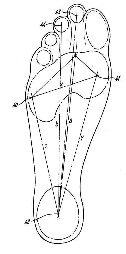
Fig. 4 is a xeLG~ a~hic rrint;
Fig. 5 is another rrint;
Fig. 6 shows a control arrang~m~nt.
Referring to Figs. l ana 2, ultrasound bone analyser
a~paratus 10 com~rises a hou~ing ll ~rovi~ing a flat
G.1310 FF
3 ~i 8~
surface 12 on which a patient' 8 foot 13 can rest with the
underface 14 of the foot 13 engaging the surface 12 and the
back of the heel 15 ~ust en~aging the front surface 16 of
an u~right ~ortion 17.
In assessing bone condition, it is desirable to take
several measurements over a period of time, and the
measurements may be se~arated by, for example, two months.
It i8 important for the usefulness of the assessments that
the measur~m~nts be taken of the same ~art of bone because
if there iB a difference in orientation of the body part
relative to the ultrasound means between measur~m~nts, this
can lead to-inconsistency reducing their usefulness in
assessing bone condition or change in bone condition.
In the ~resent case, before making a measurement, the
~osition of the body ~art is sensed and the ultrasound
trAn~ cers are ~ositioned in res~onse to the sensed
~osition of the body part.
~ ltrasound transducers 18, 19 are unted on op~osite
sides of the heel 15 and are connected to means 20 for
ad~usting the position of the res~ective transducers 18, 19
towards and away from the heel 15 (arrow A) and u~ and down
(arrow B).
The means 20 could for example be a gear 21 driven by
an electric tor 22, the gear engaging and moving a rack
23 connected to the transaucer (Fig. 3) and a second
electric tor 24 for driving a gear 21a engaging rack 23a
on sUvpo~t 25 for ving a su~port 25, 26 for the motor 22
and rac~ 23, the rack 23 sliding u~ and down on su~port 26.
Various means can be used for sensing the ~osition of
the body ~art.
In the arrangement of Figs. 1 and 2 the a~aratus
includes a xeLGy -~hic device 30 in housing 11 and the
G.1310 FF
~4~ 2 1 ~40~8
surface 12 18 a glass sheet forming ~art of the xerographic
device 30.
Fig. 4 shows an exa~ple of a xerographic print of the
underface 14 of a foot and the three points 40, 41, 42
5 indicate where the unaerface 14 of the foot produces the
highest pressure on surface 12.
The three aistances x, y, z between the points 40 and
41, 41 and 42, 42 and 40 are related to the height of a
~articular part of the heel above the surface 12, and also
10 to the position of the heel part horizontally in relation
to a vertical plane.
A large number of such relationshi~s are obtained by
accurately measuring a corresponding number of individuals
to provide a data store of such relationshi~s.
l S The particular ai~tances x, y, z for a given
measurement are then com~ared with those in the data store
and the height and lateral position of the body part (bone)
to be measured is obt~ine~3.
The position of one or both transducers 18, 19 is then
2 0 adjusted accoraingly. This can be done by manual control
of motors 22, 24, or the position data can be in~?ut to a
control 50 for the tors. The input can be manual or
electrical if the ~ata store is in a cos~uter 51.
The relative ~ositions of the bone ana the transaucers
2 5 can thus be reproaucea, ana the ~art of the bone being
measurea is thus essentially the same for each of the
various measurements.
The foot shoula not move auring the position sensing
and bone measurement.
3 0 In one example a xerographic print is not ~7roduced,
but the ~ositions of points 40, 41, 42 are notea by sensors
G.1310 FF
-s- 2 1 84~
52 in housing 12 res~onsive to electric charge which are
connected to com~uter 51 to in~ut the distances x, y, z to
a comparator 53 in the computer to ~roduce the tran~ducer
~osition control signals for a~pro~riately positioning the
tran8aucer8. The ~oints 40, 41, 42 corres~ond to highest
pressure and darkest ~oints if a ~rint were made.
In one arrangement a latent image of the foot on
surface 12 is stored in the com~uter and can be com~ared
with stored data; the image could be ~tored ana com~area
with a similar image at the next occasion of measurement to
bring the images into corres~on~ence prior to measurement.
If the measurement i8 largely done by hana, a normal
xerogra~hic ~rint can be obtained on ~a~er carrying grid
markings and the grid ~ositions x, y, z can be reaa off and
15 keyed into a control a~paratus for the transducers.
In another arrangement, the ~urface 12 is ~rovided by
or with a large number of ~ressure sensors which ~rovide
out~ut signals to the com~arator which res~onds to the
signals of highest ~ressure at points 40, 41, 42 to ~roduce
2 0 control signals for the positioning of the transducers.
It is ~ossible for another bone ~art of the body to be
measured, for exa~nple as in Fig. 5 a h~nd 54 may be ~ressed
against surface 12 ~roaucing lines 55 between maximum
pressure ~oints associated with the fingers and these can
25 be correlatea with corres~on~l~ng stored data. In this case
the position of the distal radius may be determined.
The transducers 18, 19 may be such that one tra~smits
and one receives or both may selectively transmit and
receive.
3 0 The transducers 18, 19 are controllea by unit 60 in
known manner to effect the measurement and obtain details
of bone condition; the ~rocedure is known to the skillea
~erson; examples are in BP-A-0576217 and GB-A-2257253.
G . 1 3 1 0 FF
-6- ~ 1 84 ~2 ~
The invention can also be a~lied in to the ~tuay of
bone condition in ~nl~
G.1310 FF
