Note: Descriptions are shown in the official language in which they were submitted.
CA 02184195 1999-09-23
t
1
A SCREENING METHOD FOR IDENTIFYING
LIGANDS FOR TARGET PROTEINS
Field of the invention
This invention pertains to novel methods for high-
throughput screening for pharmaceutical compounds, in
particular those that bind to proteins involved in
pathogenesis of disease or in regulation of a physiological
function.
10' Background of the Invention
Pharmaceuticals can be developed from lead
compounds that are identified through a random screening
process directed towards a target, such as a receptor.
Large scale screening approaches can be complicated by a
number of factors. First, many assays are laborious or
expensive to perform. Assays may involve expewimental
animals, cell lines, or tissue cultures that are dif-ficult
or expensive to acquire or maintain. They may require the
use of radioactive materials, and thus pose safety and
disposal problems. These considerations often place
20 practical limitations on the number of compounds that
reasonably can be screened. Thus, those employing random
screening methods are frequently forced to limit their
search to those compounds for which some prior knowledge
suggests that the compounds are likely to be effective.
This strategy limits the range of compounds tested, and many
useful drugs may be overlooked.
Furthermore, the specificity of many biochemical
assays may exclude a wide variety of useful chemical
v
2
compounds, because the interactions between the ligand and
the receptor protein are outside the scope of the assay.
For example, many proteins have multiple functions, whereas
most assays are capable of monitoring only one such activ-
ity. With such a specific assay, many potential
pharmaceuticals may not be detected.
Finally, in most existing biochemical screening
approaches to drug discovery, the activity of the target
protein must be defined. This requires that the system in
question be well-characterized before screening can begin.
Even when a protein sequence is known, as in e.g. a newly
cloned gene, the specific functions of the protein may not
be revealed simply by analysis of its sequence.
Consequently, biochemical screening for therapeutic drugs
directed against many target proteins must await detailed
biochemical characterization, a process that generally
requires extensive research.
Thus, there is a need in the art for a rapid,
cost-effective, high-throughput assay that enables the
screening of large numbers of compounds for their ability to
bind therapeutically or physiologically relevant proteins.
Furthermore, there is a need in the art for screening
methods that are independent of the biological activity of
the target proteins, and that will detect compounds that
bind regions of the target proteins other than~biologically
active domains.
Summary of the Invention
The present invention provides a method for
identifying a ligand that binds a target protein. The method
is carried out by:
(a) selecting as test ligands a plurality of
compounds not known to bind to the target protein;
(b) incubating each of the test ligands and
the target protein under conditions appropriate for the
target protein to unfold to a appropriate extent, thereby
producing a test combination;
21819
3
(c) incubating the target protein as in step
(b), but in the absence of a test ligand, to produce a
control combination;
(d) determining the extent to which the
target protein occurs in a folded state, an unfolded state,
or both, in the test combination and in the control
combination;
(e) comparing the determination made in step
(d) between the test and control combinations, wherein if
the target protein is present in the folded state to a
greater or lesser extent in the test combination than in the
control combination, the test ligand is a ligand that binds
to the target protein; and
(f) repeating steps (b)-(e) with a plurality
of said test ligands until at least one ligand that binds to
the target protein is identified.
In practicing the present invention, any method
may be used to determine the amount of target protein in
folded or unfolded states, including without limitation
proteolysis, antibody binding, surface binding, molecular
chaperone binding, differential binding to immobilized
ligand and differential formation of aggregated protein.
In one embodiment, the target protein is human
Hemoglobin S (HbS), and ligands are identified by their
ability to reduce the susceptibility of HbS to proteolysis.
Brief Description of the Drawings
Figure 1 shows an SDS-polyacrylamide gel profile
of carbonic anhydrase after proteolysis in the absence and
presence of increasing concentrations of acetazolamide.
Figure 2 shows an SDS-polyacrylamide gel profile
of carbonic anhydrase after proteolysis in the absence and
presence of l.OmM acetazolamide, in the absence and
presence of a fungal extract.
Figure 3 shows a graph representing a titration of
the binding of radiolabelled human neutrophil elastase to
nitrocellulose filters after proteolysis in the absence and
presence of increasing concentrations of elastatinal.
4
Figure 4 shows a graph representing a titration of
the ELISA detection of human neutrophil elastase after
proteolysis in the presence of increasing concentrations of
ICI 200,355.
Figure 5 shows a graph representing the
distribution of data for test ligands tested for binding to
human neutrophil elastase.
Figure 6 shows a graph representing the titration
of a ligand for human neutrophil elastase.
Figure 7 shows a graph representing the titration
of five ligands for their ability to inhibit the enzymatic
activity of human neutrophil elastase.
Figure 8 shows a graph representing a titration of
the ELISA detection of human hemoglobin after proteolysis in
the presence of increasing concentrations of 2,3-
diphosphoglycerate.
Figure 9 shows a graph representing a titration of
the binding of human hemoglobin to nitrocellulose filters
after proteolysis in the absence or presence of increasing
concentrations of 2,3-diphosphoglycerate.
Figure 10 shows a graph representing the
distribution of binding data for test ligands tested for
binding to human hemoglobin S.
Figure 11 shows a graph representing the titration
of a ligand for human hemoglobin.
Figures 12A to 12S show the structures of compounds
identified as ligands for human hemoglobin S (HbS) and their
activities in inhibiting HbS gelation relative to tryptophan
(Trp) .
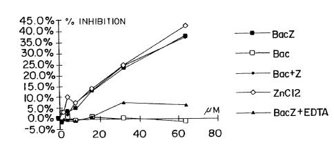
Figure 13 shows a graph representing the ligand-
binding activity for human hemoglobin of Zinc-bacitracin
(BacZ), zinc-free bactracin (Bac), zinc-free bacitracin to
which an equimolar concentration of ZnCl2 has been added
(Bac+Z), ZnCl2, and zinc-bacitracin to which a molar excess
of EDTA has been added (BacZ + EDTA).
CA 02184195 1999-09-23
t
Detailed Descri tion of the Invention
Definitions
As used herein, the term "ligand" refers to an
agent that binds a target protein. The agent may bind the
target protein when the target protein is in its native
conformation, or when it is partially or totally unfolded or
denatured. According to the present invention, a ligand is
not limited to an agent that binds a recognized functional
region of the target protein e.g. .the active site of an
enzyme, the antigen-combining site of an antibody, the
hormone-binding site of a receptor, a cofactor-binding site,
and the like. In practicing the present invention, a ligand
can also be an agent that binds any surface or internal
sequences or conformational domains of the target protein.
Therefore, the ligands of the present invention encompass
agents that in and of themselves may have no apparent
biological function, beyond their ability to bind to the
target protein in the manner described above.
As used herein, the irerm "test ligand" refers to
an agent, comprising a compound, molecule or complex, which
is being tested for its ability to bind to a target protein.
Test ligands can be virtually any agent, including without
limitation metals, peptides, proteins, lipids, poly-
saccharides, nucleic acids, small organic molecules, and
combinations thereof. Complex mixtures of substances such
as natural product extracts, which may include more than one
test ligand, can also be tested, and the component that
binds the target protein can be purified from the mixture in
a subsequent step.
As used herein, the term "target protein" refers
to a peptide, protein or protein complex for which identi-
fication of a ligand or binding partner is desired. Target
proteins include without limitation peptides or proteins_
21~~19~
6
known or believed to be involved in the etiology of a given
disease, condition or pathophysiological state, or in the
regulation of physiological function. Target proteins may
be derived from any living organism, such as a vertebrate,
particularly a mammal and even more particularly a human.
For use in the present invention, it is not necessary that
the protein's biochemical function be specifically
identified. Target proteins include without limitation
receptors, enzymes, oncogene products, tumor suppressor gene
products, viral proteins, and transcription factors, either
in purified form or as part of a complex mixture of proteins
and other compounds. Furthermore, target proteins may
comprise wild type proteins, or, alternatively, mutant or
variant proteins, including those with altered stability,
activity, or other variant properties, or hybrid proteins
to which foreign amino acid sequences e.g. sequences that
facilitate purification have been added.
As used herein, "test combination" refers to the
combination of one or more test ligands and a target
protein. "Control combination" refers to the target protein
in the absence of a test ligand.
As used herein, the "folded state" of a protein
refers to the native or undenatured form of the protein as
it is present in its natural environment, or after isolation
or purification, i.e. before exposure to denaturing
conditions. This includes native proteins that may be
detectably unfolded to differing extents in their natural
environment, and whose folding patterns may change during
their natural functioning. The "unfolded state" refers to a
situation in which the polypeptide has lost elements of its
secondary and/or tertiary structure that are present in its
"folded state." It will be recognized by those skilled in
the art that it is difficult to determine experimentally
when a polypeptide has become completely unfolded i.e. has
lost all elements of secondary and tertiary structure.
Thus, the term "unfolded state" as used herein encompasses
partial or total unfolding.
~1~~19~
As used herein, "detectable fraction" refers to a
quantity that is empirically determined and that will vary
depending upon the method used to distinguish folded from
unfolded protein. For example, when protease sensitivity is
used to monitor folding, conditions are chosen (e.g. by
adjusting temperature or adding denaturants) so that
approximately 800 of the target protein is digested within a
convenient incubation period. Alternatively, when
antibodies specific to the folded or unfolded state of a
target protein are used as the detection method, conditions
are chosen so that a sufficient amount of antibody is bound
to give a detectable signal.
The present invention encompasses high-throughput
screening methods for identifying a ligand that binds a
target protein. If the target protein to which the test
ligand binds is associated with or causative of a disease or
condition, the ligand may be useful for diagnosing,
preventing or treating the disease or condition. A ligand
identified by the present method can also be one that is
used in a purification or separation method, such as a
method that results in purification or separation of the
target protein from a mixture. The present invention also
relates to ligands identified by the present method and
their therapeutic uses (for diagnostic, preventive or
treatment purposes) and uses in purification ahd separation
methods.
According to the present invention, a ligand for a
target protein is identified by its ability to influence the
extent of folding or the rate of folding or unfolding of the
target protein. Experimental conditions are chosen so that
the target protein is subjected to unfolding, whether
reversible or irreversible. If the test ligand binds to the
target protein under these conditions, the relative amount
of folded:unfolded target protein or the rate of folding or
unfolding of the target protein in the presence of the test
ligand will be different, i.e. higher or lower, than that
observed in the absence of the test ligand. Thus, the
present method encompasses incubating the target protein in
8
the presence and absence of a test ligand, under conditions
in which (in the absence of ligand) the target protein would
partially or totally unfold. This is followed by analysis
of the absolute or relative amounts of folded vs. unfolded
target protein or of the rate of folding or unfolding of the
target protein.
An important feature of the present invention is
that it will detect any compound that binds to any sequence
or domain of the target protein, not only to sequences or
domains that are intimately involved in a biological
activity or function. The binding sequence, region, or
domain may be present on the surface of the target protein
when it is in its folded state, or may be buried in the
interior of the protein. Some binding sites may only
become accessible to ligand binding when the protein is
partially or totally unfolded.
In practicing the present invention, the test
ligand is combined with a target protein, and the mixture is
maintained under appropriate conditions and for a sufficient
time to allow binding of the test ligand to the target
protein. Experimental conditions are determined empirically
for each target protein. When testing test ligands,
incubation conditions are chosen so that most ligand:target
protein interactions would be expected to proceed to
completion. In general, the test ligand is present in molar
excess relative to the target protein. The target protein
can be in a soluble form, or, alternatively, can be bound
to a solid phase matrix. The matrix may comprise without
limitation beads, membrane filters, plastic surfaces, or
other suitable solid supports.
For each target protein, appropriate experimental
conditions, e.g. temperature, time, pH, salt concentration,
and additional components, are chosen so that a detectable
fraction of the protein is present in an unfolded form in
the absence of test ligand. For a target protein that
unfolds irreversibly, preferred experimental conditions
allow a detectable amount of the protein to unfold during a
convenient incubation period in the absence of test ligand.
2~8~~~
9
To adjust or optimize the ratio of folded: unfolded protein
or the rate of folding or unfolding, denaturing conditions
may be required, including the use of elevated temperatures,
the addition of chaotropes or denaturants such as urea or
guanidium salts such as guanidinium thiocyanate, detergents,
or combinations thereof. Furthermore, introduction of
stabilizing or destabilizing amino acid substitutions may be
used to manipulate the folded: unfolded ratio of target
proteins.
The time necessary for binding of target protein
to ligand will vary depending on the test ligand, target
protein and other conditions used. In some cases, binding
will occur instantaneously (e. g., essentially simultaneous
with combination of test ligand and target protein), while
in others, the test ligand-target protein combination is
maintained for a longer time e.g. up to 12-16 hours, before
binding is detected. When many test ligands are employed,
an incubation time is chosen that is sufficient for most
protein:ligand interactions.
Binding of a test ligand to the target protein is
assessed by comparing the absolute amount of folded or
unfolded target protein in the absence and presence of test
ligand, or, alternatively, by determining the ratio of
folded: unfolded target protein or the rate of target protein
folding or unfolding in the absence and presence of test
ligand. If a test ligand binds the target protein (i.e., if
the test ligand is a ligand for the target protein), there
may be significantly more folded, and less unfolded, target
protein (and, thus, a higher ratio of folded to unfolded
target protein) than is present in the absence of a test
ligand. Alternatively, binding of the test ligand may
result in significantly less folded, and more unfolded,
target protein than is present in the absence of a test
ligand. Similarly, binding of the test ligand may cause the
rate of target protein folding or unfolding to change
significantly.
In either case, determination of the absolute
amounts of folded and unfolded target protein, the
10
folded: unfolded ratio, or the rates of folding or unfolding,
may be carried out using one of the known methods as
described below. These methods include without limitation
proteolysis of the target protein, binding of the target
protein to appropriate surfaces, binding of specific
antibodies to the target protein, binding of the target
protein to molecular chaperones, binding of the target
protein to immobilized ligands, and measurement of
aggregation of the target protein. Other physico-chemical
techniques may also be used, either alone or in conjunction
with the above methods; these include without limitation
measurements of circular dichroism, ultraviolet and
fluorescence spectroscopy, and calorimetry. A preferred
embodiment involves measuring the relative proteolysis of a
target protein following incubation in the absence and
presence of a test ligand. However, it will be recognized
by those skilled in the art that each target protein may
have unique properties that make.a particular detection
method most suitable for the purposes of the present
invention.
For the purposes of high-throughput screening, the
experimental conditions described above are adjusted to
achieve a threshold proportion of test ligands identified as
"positive" compounds or ligands from among the total
compounds screened. This threshold is set according to two
criteria. First, the number of positive compounds should be
manageable in practical terms. Second, the number of
positive compounds should reflect ligands with an
appreciable affinity towards the target protein. A
preferred threshold is achieved when 0.1o to 1% of the total
test ligands are shown to be ligands of a given target
protein.
Binding to a given protein is a prerequisite for
pharmaceuticals intended to modify directly the action of
that protein. Thus, if a test ligand is shown, through use
of the present method, to bind a protein that reflects or
affects the etiology of a condition, it may indicate the
potential ability of the test ligand to alter protein
r
218~19~-
function and to be an effective pharmaceutical or lead
compound for the development of such a pharmaceutical.
Alternatively, the ligand may serve as the basis for the
construction of hybrid compounds containing an additional
component that has the potential to alter the protein's
function. In this case, binding of the ligand to the target
protein serves to anchor or orient the additional component
so as to effectuate its pharmaceutical effects. For
example, a known compound that inhibits the activity of a
family of related enzymes may be rendered specific to one
member of the family by conjugation of the known compound to
a ligand, identified by the methods of the present
invention, that binds specifically to that member at a
different site than that recognized by the known compound.
The fact that the present method is based on
physico-chemical properties common to most proteins gives it
widespread application. The present invention can be applied
to large-scale systematic high-throughput procedures that
allow a cost-effective screening of many thousands of test
ligands. Once a ligand has been identified by the methods
of the present invention, it can be further analyzed in more
detail using known methods specific to the particular target
protein used. For example, the ligand can be tested for
binding to the target protein directly e.g. by incubating
radiolabelled ligand with unlabelled target prbtein, and
then separating protein-bound and unbound ligand.
Furthermore, the ligand can be tested for its ability to
influence, either positively or negatively, a known
biological activity of the target protein.
In a preferred embodiment of the present
invention, binding of test ligand to target protein is
detected through the use of proteolysis. This assay is
based on the increased susceptibility of unfolded, denatured
polypeptides to protease digestion relative to that of
folded proteins. In this case, the test ligand-target
protein combination, and a control combination lacking the
test ligand, are treated with one or more proteases that act
preferentially upon unfolded target protein. After an
12
appropriate period of incubation, the level of intact i.e.
unproteolysed target protein is assessed using one of the
methods described below e.g. gel electrophoresis and/or
immunoassay.
There are two possible outcomes that indicate that
the test ligand has bound the target protein. Either a
significantly higher, or significantly lower, absolute
amount of intact or degraded protein may be observed in the
presence of ligand than in its absence.
Proteases useful in practicing the present
invention include without limitation trypsin, chymotrypsin,
V8 protease, elastase, carboxypeptidase, proteinase K,
thermolysin, papain and subtilisin (all of which can be
obtained from Sigma Chemical Co., St. Louis, MO). The most
important criterion in selecting a protease or proteases for
use in practicing the present invention is that the
protease(s) must be capable of digesting the particular
target protein under the chosen incubation conditions, and
that this activity be preferentially directed towards the
unfolded form of the protein. To avoid "false positive"
results caused by test ligands that directly inhibit the
protease, more than one protease, particularly proteases
with different enzymatic mechanisms of action, can be used
simultaneously or in parallel assays. In addition, co-
factors that are required for the activity of the
protease(s) are provided in excess, to avoid false positive
results due to test ligands that may sequester these
factors.
Typically, a purified target protein is first
taken up to a final concentration of 1-100 ~g/ml in a buffer
containing 50 mM Tris-HC1, pH 7.5, 10% DMSO, 50 mM NaCl, l00
glycerol, and 1.0 mM DTT. Proteases, such as, for example,
proteinase K or thermolysin (proteases with distinct
mechanisms of action), are then added individually to a
final concentration of 0.2-10.0 ~g/ml. Parallel incubations
are performed for different time periods ranging from 5
minutes to one hour, preferably 30 minutes, at 4°C, 15°C,
25°C, and 35°C. Reactions are terminated by addition of an
21819
13
appropriate protease inhibitor, such as, for example,
phenylmethylsulfonyl chloride (PMSF) to a final
concentration of 1mM (for serine proteases),
ethylenediaminotetraacetic acid (EDTA) to a final
concentration of 20 mM (for metalloproteases), or
iodoacetamide (for cysteine proteases). The amount of
intact protein remaining in the reaction mixture at the end
of the incubation period may then be assessed by any method,
including without limitation polyacrylamide gel
electrophoresis, ELISA, or binding to nitrocellulose
filters. It will be understood that additional experiments
employing a narrower range of temperatures can be performed
to establish appropriate conditions.
The above protocol allows the selection of
appropriate conditions (e.g., protease concentration and
digestion temperature) that result in digestion of
approximately 70% of the target protein within a 30 minute
incubation period, indicating that a significant degree of
unfolding has occurred. Preferably, conditions are chosen
so that proteolysis displays a temperature dependence
indicative of a cooperative protein unfolding transition.
To achieve this end, additional variables can be adjusted,
including, for example, the concentrations of glycerol,
salt, reducing agents, BSA or other "carrier proteins,"
target protein, denaturants and detergents. If a known
ligand for the target protein is available, the ligand is
included in the reaction mixture at a concentration above
the Kd for its binding to the target protein and at least
equal to the molar concentration of target protein, and the
digestion experiment is repeated. Typically, at least a two-
fold increase or decrease in the extent of digestion of the
target protein is observed, indicating that binding of a
known ligand changes the ratio of folded: unfolded target
protein and/or the rate of folding or unfolding.
Once conditions are established for high-
throughput screening as described above, the protocol is
repeated simultaneously with a large number of test ligands
at concentrations ranging from 20 to 200 ~M. Observation of
~~8~19~
14
at least a two-fold increase or decrease in the extent of
digestion of the target protein signifies a "hit" compound,
i.e., a ligand that binds the target protein. Preferred
conditions are those in which between 0.1% and 1% of test
ligands are identified as "hit" compounds using this
procedure.
In another embodiment, the relative amount of
folded and unfolded target protein in the presence and
absence of test ligand is assessed by measuring the relative
l0 amount of target protein that binds to an appropriate
surface. This method takes advantage of the increased
propensity of unfolded proteins to adhere to surfaces, which
is due to the increased surface area, and decrease in
masking of hydrophobic residues, that results from
unfolding. If a test ligand binds a target protein (i.e.,
is a ligand of the target protein), it may stabilize the
folded form of the target protein and decrease its binding
to a solid surface. Alternatively, a ligand may stabilize
the unfolded form of the protein and increase its binding to
20 a solid surface.
In this embodiment, the target protein, a test
ligand and a surface that preferentially binds unfolded
protein are combined and maintained under conditions
appropriate for binding of the target protein to a ligand
and binding of unfolded target protein to the surface.
Alternatively, the target protein and test ligand can be
pre-incubated in the absence of the surface to allow
binding. Surfaces suitable for this purpose include
without limitation microtiter plates constructed from a
30 variety of treated or untreated plastics, plates treated for
tissue culture or for high protein binding, nitrocellulose
filters and PVDF filters.
Determination of the amount of surface-bound
target protein or the amount of target protein remaining in
solution can be carried out using standard methods known in
the art e.g. determination of radioactivity or immunoassay.
If significantly more or less target protein is surface
bound in the presence of a test ligand than in the absence
218419
of the test ligand, the test ligand is a ligand of the
target protein. Similarly, the ratio of surface-
bound:soluble target protein will be significantly greater
or smaller in the presence of a test ligand than in its
absence, if a test ligand is a ligand for the target
protein.
In another embodiment, the extent to which folded
and unfolded target protein are present in the test
combination is assessed through the use of antibodies
10 specific for either the unfolded state or the folded state
of the protein i.e. denatured-specific ("DS"), or native-
specific ("NS") antibodies, respectively. (Breyer, 1989, J.
Biol. Chem., 264 (5):13348-13354).
Polyclonal and monoclonal DS and NS antibodies
specific for particular target proteins can be prepared by
methods that are well known in the art (E. Harlow & D. Lane,
Antibodies: A Laboratory Manual, Cold Spring Harbor
Laboratory, 1988; Zola, Monoclonal Antibodies: A Manual of
Techniques, CRC Press, Inc., Boca Raton, Florida,1987). For
DS antibodies, animals can be immunized with a peptide from
a region of the protein that is buried in the interior of
the protein when it is in the native state. If the three-
dimensional structure of the protein is unknown, antibodies
are prepared against several peptides. Alternatively, fully
denatured (i.e., unfolded) target protein is used as an
immunogen.
The resulting antibodies are screened for
preferential binding to the denatured state. For monoclonal
antibodies, culture supernatants derived from individual
cloned hybridomas are screened, and positive clones are used
directly as a source of individual DS antibodies. For
polyclonal antibodies, an unfractionated antiserum may
exhibit preferential binding to the denatured state.
Alternatively, DS antibodies may be purified from a
polyclonal antiserum by selective adsorption techniques
well-known in the art.
For NS antibodies, intact non-denatured protein,
or one or more peptides known to be on the surface of the
218419
16
native protein, may be used as an immunogen. The resulting
antibodies are screened as above for preferential binding to
the native protein and purified for use in the present
invention.
DS or NS antibodies can be utilized to detect a
ligand-induced change in the level of folded target protein,
unfolded target protein, the folded:unfolded ratio, or the
rate of folding or unfolding.
In one approach, a test combination containing the
DS antibody, the target protein, and the test ligand is
exposed to a solid support e.g. a microtiter plate coated
with the denatured target protein or a peptide fragment
thereof, under conditions appropriate for binding of the
target protein with its ligand and binding of the DS
antibody to unfolded target protein. A control combination,
which is the same as the test combination except that it
does not contain test ligand, is processed in the same
manner as the test solution. By comparing the amount of
antibody bound to the plate or the amount remaining in
solution in the test and control combinations, the
difference in target protein folding is detected. The
amount of antibody bound to the plate or remaining in
solution can be measured as described below.
In a second approach, a test combination
containing the DS antibody, the test ligand, and the target
protein is exposed to a solid support coated with a second
antibody, referred to as a solid phase antibody, which
cannot bind to the target protein simultaneously with the DS
antibody, and is specific for the target protein, but is
either specific for the folded state (NS antibody) or unable
to differentiate between the native and denatured states
("non-differentiating" or "ND" antibody). The resulting
test combination or solution is maintained under conditions
appropriate for binding of the target protein with a ligand
of the target protein and for binding of the antibodies to
the proteins they recognize. A control combination, which
is the same as the test solution except that it does not
contain test ligand, is processed in the same manner as the
17
test solution. In both combinations, denatured (unfolded)
target protein binds the DS antibody and is inhibited from
binding the solid phase antibody. The ability of the test
ligand to bind the target protein can be gauged by deter-
mining the amount of target protein that binds to the solid
phase antibody in the test solution and comparing it with
the extent to which target protein binds to the solid phase
antibody in the absence of test ligand, which in turn
reflects the amount of target protein in the folded state.
The amount of target protein bound to the plate via the
second antibody or remaining in solution can be detected by
the methods described below. This approach may be used in a
comparable manner with NS antibody as the soluble antibody
and DS or ND antibody on the solid phase.
In a third approach, a test solution containing
the target protein and the test ligand is exposed to a solid
support e.g. a microtiter plate that has been coated with a
DS or NS antibody and maintained under conditions
appropriate for binding of target protein to its ligand and
for binding of the antibody to target protein. Alter-
natively, the antibody can be present on the surfaces of
beads. The ability of the test ligand to bind the target
protein is gauged by determining the extent to which target
protein remains in solution (unbound to the antibody) or on
the solid surface (bound to the antibody), or the ratio of
the two, in the presence and in the absence of test ligand.
Alternatively, the antibody can be present in solution and
the target protein can be attached to a solid phase, such as
a plate surface or bead surface.
In another embodiment, molecular chaperones are
used to assess the relative levels of folded and unfolded
protein in a test combination. Chaperones encompass known
proteins that bind unfolded proteins as part of their normal
physiological function. They are generally involved in
assembling oligomeric proteins, in ensuring that certain
proteins fold correctly, in facilitating protein
localization, and in preventing the formation of
proteinaceous aggregates during physiological stress (Hardy,
18
1991, Science, 251:439-443). These proteins have the
ability to interact with many unfolded or partially
denatured proteins without specific recognition of defined
sequence motifs.
One molecular chaperone, found in E. coli, is a
protein known as SecB. SecB has a demonstrated involvement
in export of a subset of otherwise unrelated proteins.
Competition experiments have shown that SecB binds tightly
to all the unfolded proteins tested, including proteins
outside of its particular export subset, but does not appear
to interact with the folded protein. Other chaperones
suitable for use in the present invention include without
limitation heat shock protein 70s, heat shock protein 90s,
GroEI and GroES (Gething et al., Nature 355:33, 1992).
In this embodiment, a test combination containing
the test ligand and the target is exposed to a solid support
e.g. microtiter plate or other suitable surface coated with
a molecular chaperone, under conditions appropriate for
binding of target protein with its ligand and binding of the
molecular chaperone to unfolded target protein. The
unfolded target protein in the solution will have a greater
tendency to bind to the molecular chaperone-covered surface
relative to the ligand-stabilized folded target protein.
Thus, the ability of the test ligand to bind target protein
can be determined by determining the amount of 'target
protein remaining unbound, or the amount bound to the
chaperone-coated surface.
Alternatively, a competition assay for binding to
molecular chaperones can be utilized. A test combination
containing purified target protein, the test ligand, and a
molecular chaperone can be exposed to a solid support e.g. a
microtiter well coated with denatured (unfolded) target
protein, under conditions appropriate for binding target
protein with its ligand and binding of the molecular
chaperone to unfolded target protein. A control
combination, which is the same as the test combination
except that it does not contain test ligand, is processed in
the same manner. Denatured target protein in solution will
19
bind to the chaperone and thus inhibit its binding to the
denatured target protein bound to the support. Binding of a
test ligand to the target protein will result in a
difference in the amount of unfolded target protein, and,
thus, more or less chaperone will be available to bind to
the solid-phase denatured target protein than is the case in
the absence of binding of test ligand. Thus, binding of
test ligand can be determined by assessing chaperone bound
to the surface or in solution in the test combination and in
the control combination and comparing the results. In this
assay, the chaperones are generally not provided in excess,
so that competition for their binding can be measured.
Alternatively, a test combination containing the
target protein, the test ligand and a molecular chaperone
can be exposed to a solid support e.g. a microtiter well
that has been coated with antisera or a monoclonal antibody
specific for the folded target protein (NS antibody) and
unable to bind the target protein bound to the chaperone.
Unfolded target protein will bind chaperone in solution and
thus be inhibited from binding the solid phase antibody. By
detecting target protein in the solution or bound to the
well walls and comparing the extent of either or both in an
appropriate control (the same combination without the test
ligand), the ability of the test ligand to bind target
protein can be determined. If the test ligand~is a ligand
for the target protein, more or less target protein will be
bound to the antisera or monoclonal antibody bound to the
container surface in the test combination than in the
control combination, and correspondingly more or less target
protein will be present unbound (in solution) in the test
combination than in the control combination.
In another embodiment, a known ligand, cofactor,
substrate, or analogue thereof of the target protein is used
to assay for the presence of folded target protein. The
higher the fraction of protein in the folded form, the
greater the amount of protein that is available to bind to a
ligand that binds exclusively to the folded state.
Consequently, if a protein has a known ligand, it is
218419
possible to increase or decrease the binding of the protein
to the known ligand by adding a test ligand that binds
another site on the protein. For example, binding of
dihydrofolate reductase to methotrexate, a folic acid
analogue, can be used to assess the level of folding of this
enzyme.
In this approach, the ligand, cofactor, substrate,
or analogue thereof known to bind to the target protein is
immobilized on a solid substrate. A solution containing the
10 target protein and test ligand is then added. An increase
or decrease in the amount of target protein that binds to
the immobilized compound relative to an identical assay in
the absence of test ligand indicates that the test ligand
binds the target protein. The amount of target protein
bound to the solid substrate can be assessed by sampling the
solid substrate or by sampling the solution.
In another embodiment, the amount of unfolded
target protein in a test combination is assessed by
measuring protein aggregation. For proteins that unfold
20 irreversibly, unfolded protein often forms insoluble
aggregates. The extent of protein aggregation can be
measured by techniques known in the art, including without
limitation light scattering, centrifugation, and filtration.
In this approach, target protein and test ligand
are incubated and the amount of protein aggregation is
measured over time or after a fixed incubation time. The
extent of protein aggregation in the test mixture is
compared to the same measurement for a control assay in the
absence of test ligand. If a test ligand binds a target
protein, the rate of unfolding of target protein will be
lower or higher than in the absence of test ligand. For
measurements over time, the rate of appearance of aggregated
protein will be lower or higher if the test ligand is a
ligand for the target protein than if it is not. For
measurements at a fixed time, there will be more or less
unfolded protein and correspondingly more or less aggregated
protein if the test ligand is a ligand for the target
protein than if it is not. Thus, the ability of a test
z~s~~~~
21
ligand to bind a target protein can be determined by
assessing the extent of protein aggregation in the presence
and absence of test ligand.
It will be understood that the methods of the
present invention can be applied to fragments of target
proteins that constitute stable structural domains. As used
herein, "domain" refers to a fragment of a target protein
that retains a significant degree of native folded structure
after isolation. In some cases, a native protein will be
cleaved by a protease into one or more such domains when
proteolytic digestion of the native protein is performed at,
for example, a lower temperature than that at which complete
digestion of the protein occurs. In this case, it may be
advantageous to assay the binding of test ligands to each of
the domains independently. This may be achieved by either
or both of the following approaches. First, one or more
individual domains of a target protein can be prepared for
use as targets in the assays described above, either by
subjecting the intact protein to controlled proteolysis
followed by purification of domain-comprising fragments, or
by directing the synthesis of such fragments, either in
vitro or in vivo, from recombinant DNA molecules encoding
domain-comprising fragments of the protein. Second, domain-
specific detection may be used to quantify folding in a
reaction mixture in which the intact protein serves as the
target. Methods for domain-specific detection include
without limitation the use of domain-specific antibodies and
chemical or enzymatic methods which selectively label
particular domains. Domain-specific antibodies may be
prepared by any method known in the art. For example,
polyclonal domain-specific antibodies may be raised by using
as immunogens either the purified or recombinant domains
described above or domain-specific synthetic peptides.
Alternatively, a panel of monoclonal antibodies may be
prepared against the intact protein, and tested for reaction
with purified or recombinant domains.
The embodiments described above are summarized in
Table 1.
21~~~
22
TABLE 1
DETERMINING FOLDED AND UNFOLDED TARGET PROTEIN
Monitoring Method Used: ILesult Observed If Test Ligand
Binds Target Protein
Proteolysis
Protease that preferentially More or less intact target protein
hydrolyzes unfolded target in test combination than
protein is used in control combination
Surface Binding
Surface that preferentially More or less target protein unbound
binds unfolded target protein (in solution) to
is
used surface in test combination than
in control combination
Antibody Binding
DS antibody in solution/unfoldedMore or less DS antibody bound
target protein or peptide to unfolded target
fragment thereof on surface protein or peptide fragment thereof
on surface in test
combination than in control combination
DS antibody in solution/ antibodyMore or less target protein bound
that recognizes folded to antibody on surface
target protein on surface in test combination than in control
combination
NS antibody in solution/ antibodyMore or less target protein bound
that recognizes folded to antibody on surface
target protein on surface in test combination than in control
combination
DS antibody on surface More or less target protein bound
to DS antibody on
surface in test combination than
in control combination
NS antibody on surface More or less target protein bound
to NS antibody on
surface in test combination than
in control combination
2 Molecular Chaperones
0
Chaperone on surface More or less target protein bound
to chaperone on surface
in test combination than in control
combination
Competition Assay
Unfolded target protein on solidMore or less chaperone bound
phase, target protein in to unfolded target protein
solution on solid phase in test combination
than in control
combination
Chaperone in solution/antibody More or less target protein bound
that recognizes folded to surface-bound
target protein on surface antibody in test combination
than in control combination
Differential Binding to Immobilized
Ligand
Target protein in solution, More or less target protein bound
known ligand of target protein to surface bound ligand
attached to surface in test combination than in control
combination
3 Protein A~Qregation
0
Formation of aggregated proteinMore or less aggregated protein
by irreversible protein (higher or lower rate of
unfolding formation of aggregated protein)
in test combination than
in control
Protein Detection Methods
The embodiments described above require a final
step for detecting and/or quantifying the level of target
protein or digestion products thereof, or antibodies, in
~1841~~
23
order to quantify the relative amounts of folded and
unfolded target protein after exposure to test ligands. In
practicing the present invention, methods known in the art
are used to detect the presence or absence of protein, small
peptides or free amino acids. The method used will be
determined by the product (proteins, peptides, free amino
acids) to be detected. For example, techniques for
detecting protein size can be used to determine the extent
of proteolytic degradation of the target protein e.g. gel
electrophoresis, capillary electrophoresis, size exclusion
chromatography, high-performance liquid chromatography, and
the like. Measurement of radioactivity, fluorescence, dye
binding (Ciesiolka et al., Anal.Biochem. 168:280, 1988),
colloidal gold binding (Bradford, Anal.Biochem. 72:248,
1976), or enzymatic activity can detect the presence or
absence of products, either in solution or on a solid
support. Immunological methods including e.g. ELISA and
radioimmunoassay can detect the presence or absence of a
known target protein in solution or on a substrate. The
above methods are described in e.g. Harlow, E. and D. Lane,
Antibodies: A Laboratory Manual, Cold Spring Harbor
Laboratories, 1988; S.F.Y. Li, Capillary Electrophoresis,
Elsevier Press, 1993; Bidlingmeyer, Practical HPLC
Methodology and Applications, John Wiley and Sons, Inc.,
1992; and Cantor, C.R. and P.R. Schimmel, Biophysical
Chemistry, WH Freeman and Co., 1980.
In one embodiment, gel electrophoresis is used to
detect the presence or absence of protein, and can further
be used to detect the size of the protein. This latter
method is especially useful in conjunction with proteolysis,
as the presence of a greater or lesser amount of undigested
target protein in the test combination than in the control
combination indicates that the test ligand bound to the
target protein.
The following examples are intended to illustrate
the invention without limiting it thereof.
:~ ,
CA 02184195 1999-09-23
24
Example l: Methotrexate Binding Protects Dihydrofolate
Reductase (DHFR) From Proteolytic Digestion
by Proteinase R
The following were combined and incubated at 54°C
for 5 minutes: DHFR (100 ~g/ml), Proteinase K (80 ~g/ml),
0.1 M Tris-HC1 pH 7.5, and Methotrexate at 10-1° to 10~ M.
Samples were removed and undigested DHFR was
quantified by ELISA as follows:
(a) Protease incubations were diluted 50-fold
with Tris-buffered saline (TBS);
(b) 50 ~1 diluted samples were transferred to the
wells of an ELISA plate and incubated 60 minutes at room
temperature;
(c) the plate wells were thoroughly washed with
TBS plus 0.1% Tween-2f~(TBST);
(d) 50 ~l anti-DHFR rabbit serum diluted 250-fold
into TBST plus 5% nonfat dry milk was added to each
well and incubated 30 minutes at room temperature;
(e) plate wells were washed as in (c) above;
(f) 50 ul of goat anti-rabbit IgG alkaline
phosphatase conjugate diluted 500-fold in TBST plus 5% milk
was added to each well and incubated 30 minutes at room
temperature;
(g) plate wells were washed as in (c); and
(h) O.l ml of 1.0 mg/ml p-nitrophenylphosphate in
0.1% diethanolamine was added. Color development is
proportional to alkaline phosphatase antibody conjugate
bound.
The ELISA analysis showed that methotrexate
protects DHFR from digestion at concentrations of 10~M and
higher. By the same methods, nicotinamide adenine
dinucleotide phosphate (NADPH) and dihydrofolate at
concentrations of 10-SM and higher were shown to inhibit
proteolysis of DHFR in separate experiments.
(Trademark)
r
~P a
CA 02184195 1999-09-23
Example 2: Methotrexate, NADPH and Dihydrofolate Binding
Protects Dihydrofolate Reductase (DHFR) From
Proteolytic Digestion by Proteinase K in the
Presence of a Mixture of Aanino Acids
The following were combined and incubated at 54' C
for 5 minutes: DHFR (2.1 ~.g/ml), Proteinase K (80 ~.g/ml),
0.1M Tris-HC1 (pH 7.5), 10-SM of all 20 common amino acids
and either 0 or 10-SM ligand. The ligands used were the
10 inhibitor Methotrexate and the substrates dihydrofolate and
NADPH.
Samples were removed and undigested DHFR was
quantified by ELISA as follows:
(a) Protease incubations were diluted 50 fold with
Tris-buffered saline (TBS);
(b) 50 ~1 diluted samples were transferred to the
wells of an ELISA plate and incubated 60 minutes at room
temperature;
(c) the plate wells were thoroughly washed with
20 TBS plus 0.1% Tween-20*(TBST);
(d) 50 ~,1 anti-DHFR rabbit serum diluted 250 fold
into TBST plus 5% nonfat dry milk was added to each well and
incubated 30 minutes at room temperature;
(e) plate wells were washed as in (c) above;
(f) 50 ~,1 of goat anti-rabbit IgG alkaline
phosphatase conjugate diluted 500 fold in TBST plus 5% milk
was added to each well and incubated 30 minutes at room
temperature;
(g) plate wells were washed as in (c); and
(h) 0.1 ml of 1.0 mg/ml p-nitrophenylphosphate in
0.1% diethanolamine was added. Color development is
proportional to alkaline phosphatase antibody conjugate
bound.
The ELISA analysis showed that methotrexate and
the substrates protect DHFR from digestion relative to the
absence of ligands that bind to DHFR. Thus, specific
binding can be detected in the presence of a complex mixture
of compounds that do not bind to the target protein.
* (Trademark)
''
r
.,
CA 02184195 1999-09-23
26
Example 3: Methotrexate Binding Inhibits Binding of DHFR
to Microtiter Plates
The following were combined in a volume of 60 ~,1
and incubated in a Falcon 3072 "tissue-culture treated"
microtiter plate at 20 or 47°C: 100 mg DHFR, 50 MM Tris-C1
(pH 7 . 5 ) , and Methotrexate 10'1° to 10''~MM .
50 ~l of each sample was then transferred to the
wells of an ELISA plate, and the DHFR that remained in
solution was quantified by ELISA as follows:
(a) The 50 ~,1 samples were incubated for 60
minutes at room temperature;
(b) the plate wells were thoroughly washed with
TBS plus 0.1% Tween-20* (TBST) ;
(c) 50 ~.1 anti-DHFR rabbit serum diluted 250-fold
into TBST plus 5% nonfat dry milk was added to each well and
incubated 30 minutes at room temperature;
(d) plate wells were washed as in (b) above;
(e) 50 ~1 of goat anti-rabbit IgG alkaline
phosphatase conjugate diluted 500-fold in TBST plus 5% milk
was added to each well and incubated 30 minutes at room
temperature;
(f) plate wells were washed as in (b); and
(g) O.l ml of 1.0 mg/ml D-nitrophenylphosphate in
0.1% diethanolamine was added. Color development is
proportional to alkaline phosphatase antibody conjugate
bound.
The ELISA analysis revealed that methotrexate
inhibits DHFR binding to the Falcon 3072 plate at
concentrations of 10-'M and above.
Example 4: Inhibition or Enhancement of Unfolded-
Specific Antibody Binding
(1) ELISA plates are coated by incubation for 60
minutes with the following mixture: 4 ~g/ml irreversibly
denatured target protein or peptide fragments thereof in
Tris-buffered Saline (10 mM Tris-C1, pH 7.5, 0.2M NaCl;
TBS).
* (Trademarks)
, ;:
t
.
CA 02184195 1999-09-23
27
(2) The plates are washed 3 times with TBS plus
0.1% Tween-20* (TBST).
(3) The following mixture (total volume 50 ~1) is
incubated in the coated wells of the microliter plate for 60
minutes:
(a) Antibody specific for the unfolded state
of the target protein at a sufficient concentration to
give 50% of maximal binding (in the absence of competing
target protein).
(b) Target protein at a concentration
sufficient to achieve 90% inhibition of antibody binding to
the plate. The appropriate target protein concentration
differs for each target protein. The concentration depends,
in part, on the stability of the folded form of the target
protein. In some cases it may be desirable to reduce the
stability of the target protein by elevated temperature,
inclusion of chemical protein-denaturing agents, or
introduction of destabilizing amino acid substitutions in
the target protein.
(c) 10'9 to 10'SM test ligands
(d) 5% nonfat dry milk in TBST
(4) The plates are washed 3 times with TBST.
(5) 50 ~,l of goat anti-IgG alkaline phosphatase
conjugate at an appropriate dilution are added in TBST plus
5% nonfat dry milk and incubated for 30 minutes at room
temperature.
(6) Plates are washed 3 times with TBST.
(7) 0.1 ml of 1.0 mg/ml D-nitrophenylphosphate in
0.1% diethanolamine are added and the amount of color
development recorded by means of an ELISA plate reader.
ELISA analysis will reveal more or less antibody
bound to the plate when successful test ligand-target
protein binding has occurred than in the absence of such
binding.
* (Trademark)
28
Example 5: Inhibition or Enhancement of Chaperone
Binding
(1) ELISA plates are coated by incubation for
several hours with 4 ~.g/ml chaperone in TBS.
(2) The plates are washed 3 times with TBST.
(3) The following mixture (total volume 50 ~.1) is
then incubated in the coated. wells of the microtiter plate
for 60 minutes:
(a) Target protein at a concentration
10 sufficient to saturate about 50~ of the available binding
sites present on the chaperone proteins. Denaturing
conditions may be used in cases where the folded form of the
target protein is otherwise too stable to permit appre-
ciable binding to chaperones.
(b) 10'9 to 10~5M test ligands in TBST
(4) Aliquots of the well solutions are
transferred to wells of a new ELISA plate and incubated for
60 minutes at room temperature.
(5) The plate wells are washed 3 times with TBST.
(6) 50 ~.1 antibody specific for the target
protein at the appropriate dilution in TBST, plus 5% nonfat
dry milk, are added to each well and incubated 30 minutes at
room temperature.
(7) The plate wells are washed 3 times with TBST.
(8) 50 ~.l of goat anti-rabbit IgG alkaline
phosphatase conjugate at an appropriate dilution in TBST
plus 5°s nonfat dry milk are added to each well and incubated
minutes at room temperature.
(9) The plate wells are washed 3 times with TBST.
30 (10) 0.1 ml of 1.0 mg/ml p-nitrophenylphosphate
in 0.1% diethanolamine will be added. Color development
(proportional to alkaline phosphatase antibody conjugate
bound) is monitored with an ELISA plate reader.
ELISA analysis will reveal target protein in the
solution at higher or lower concentration when test ligand-
target protein binding has occurred than when it has not.
Example 6: Enhancement or Inhibition of Binding to a
Known Ligand
;.
CA 02184195 1999-09-23
29
(1) The following mixture (total volume 50 ~,1)
is incubated in the coated wells of the microtiter plate for
60 minutes:
(a) Ligand known to bind to the target
protein, covalently attached to solid beads such as
SephadeX This ligand can be a small molecule or a
macromolecule.
(b) Target protein at a concentration well
below saturation of the ligand and such that only 10% of the
protein binds to the ligand sites. The solution conditions
are such that most of the target protein is present in the
denatured state.
(c) 10-9 to 10-SM test ligands
(d) in TBST plus necessary denaturant, such
as urea.
(2) Aliquots of the well supernatant (free of
beads) are transferred to wells of a new ELISA plate and
incubated for 60 minutes at room temperature.
(3) The plate wells are washed 3 times with TBST.
(4) 50 ~,1 antibody specific for the target
protein at the appropriate dilution in TBST, plus 5% nonfat
dry milk, are added to each well and incubated 30 minutes at
room temperature.
(5) The plate wells are washed 3 times with TBST.
(6) 50 ~Cl of goat anti-rabbit IgG alkaline
phosphatase conjugate at an appropriate dilution in TBST
plus 5% milk are added to each well and incubated 30 minutes
at room temperature.
(7) The plate wells are washed 3 times with TBST.
(8) 0:1 ml of 1.0 mg/ml p-nitrophenylphosphate in
0.1% diethanolamine are added. Color development (propor-
tional to alkaline phosphatase antibody conjugate bound) is
monitored with an ELISA plate reader.
ELISA analysis will reveal a higher or lower
concentration of target protein in the solution when
successful test ligand-target protein binding has occurred.
* (Trademark)
l~ /~ ~'..~
Example 7: Low Throughput Assay for HIV Rev Protein
Reaction mixtures (0.03 ml total volume) contained
30 ~.g/ml HIV Rev protein that had been produced in E. coli,
0.05M Tris-HCl, pH 7.5, O.O1M calcium acetate, 2.5~.g/ml
proteinase K, loo DMSO, and varying amounts of tRNA as a
known ligand. The reactions were incubated on ice for 15
minutes. After addition of PMSF and EDTA as described in
Example 8 below, samples were prepared for gel
electrophoresis and analyzed as described in Example 8.
10 The results showed that in the absence of tRNA,
Rev protein is almost completely degraded by proteinase K
under these conditions. In the presence of tRNA, however, a
lower-molecular weight fragment of the protein is stabilized
against proteolysis. Thus, binding of a known ligand to HIV
Rev protein is detectable using the methods of the present
invention.
Example 8: Low throughput Assay for Carbonic Anhydrase
Ligande
20 Ligand binding to carbonic anhydrase I (Sigma) was
tested using proteolysis as a probe of target protein
folding, and denaturing gel electrophoresis was used as a
method for detection of intact protein remaining after
digestion with proteases.
To validate the assay, acetazolamide, a known
ligand of carbonic anhydrase, was tested. Though
acetazolamide is a known inhibitor of carbonic anhydrase
activity, these experiments make no use of that property,
and do not measure the enzymatic activity of the protein. In
30 addition, the sensitivity of the method to interference by a
natural product extract was examined.
Reaction mixtures contained 23.3 ~g/ml carbonic
anhydrase, 0.05 M Tris-HC1 pH 7.5, 0.01 M calcium acetate,
2.5 ~.g/ml proteinase K, 10% DMSO and acetazolamide (Sigma)
in concentrations ranging from 0.0 to 1.0 mM. The reactions
were incubated at 54°C for 15 minutes, and then chilled on
ice. Phenyl methyl sulfonyl fluoride (PMSF) was then added
from a 20 mM stock solution in ethanol to a final
F._': ~.,
w,
31
concentration of 1 mM, and EDTA was added from a 0.5M stock
solution to a final concentration of 20 mM. 0.01 ml of SDS
loading buffer (10% sodium dodecyl sulfate (SDS), 0.5 M
Dithiothreitol, 0.4 M Tris-HC1 buffer, pH 6.8, 50% Glycerol)
was added and samples were heated at 95°C for 3 minutes.
Samples were analyzed by SDS-polyacrylamide gel
electrophoresis using a 4-15% polyacrylamide (BioRad)
gradient gel, which was then stained with Coomassie Blue
dye.
As shown in Figure 1, binding of the known ligand
acetazolamide to carbonic anhydrase resulted in
stabilization of carbonic anhydrase against proteolysis by
proteinase K at 1 X 10-SM acetazolamide. The dissociation
constant for this interaction has been reported to be 2.6 X
10-6M (Matsumoto, K. et. al. (1989), Chem. Pharm. Bull,
37:1913-1915).
A fungal methanol extract was included in
reactions that were otherwise identical to that described
above such that the final concentration of an added small
molecule would be equal to its concentration in the source
culture. The presence of extract neither induced a false
signal nor diminished the response to 1.0 mM acetazolamide
(Figure 2.)
Example 9: High-Throughput Screening of Ligands for
Human Carbonic Anhydrase
A high throughput assay has been established for
carbonic anhydrase I. Each reaction mixture (in a final
volume of 0.05 ml) contains: 3.3 ~Cg carbonic anhydrase, 50
mM Tris-HC1, 50 mM NaCl, 1.0 mM Ca(OAc)Z, and 0.13 ~g
proteinase K, 10% DMSO, and the appropriate test compound at
a concentration of 20 Vim. Control reactions are identical,
except that the test compound is omitted. The mixtures are
incubated at 20°C for 10 minutes, followed by incubation at
54°C for 30 minutes, after which they are placed on ice.
Each mixture then receives 200 ~.l 50 mM sodium borate
. buffer, pH 8.5, containing 10 mM EDTA and 1.0 mM PMSF.
After 20 minutes incubation on ice, 7.5 ~l of the mixture
218~19~
32
are added to Dynatech Immulon-4 microplate wells containing
92.5 ~l per well of the borate buffer described above. The
plate is then incubated at 4°C overnight to permit carbonic
anhydrase binding. Bound carbonic anhydrase is quantified
by direct ELISA using HRP-conjugated anti-carbonic
anhydrase I antibody at a 1:2,000 dilution (Biodesign,
Kennebunk, ME); cat#K90046P) and Pierce (Rockford, IL) Turbo
TMB substrate.
Carbonic anhydrase I inhibitors (obtained from
Sigma Chemical Co., St. Louis, MO) were tested in the above
assay over a range of inhibitor concentrations. Table 2
shows the concentration that caused 50% inhibition of
proteolysis in the assay (ICso), the published Kd values for
the compounds (Matsumoto et al., Chem. Pharm. Bull., 37:
1913, (1989)), and the ratio of these values for each
compound. These data show a positive correlation between
the ICSO and Kd values for each inhibitor.
TABLE 2
Ligand: Kd ICSO Ratio
(IBM) (l~M) ICSn: K,,
Acetazolamide 2.6 13 5.0
Hydrochlorothi 23.4 246 10.5
azide
Sulfanilamide 36 350 9.7
Example 10: High-throughput Screening of Ligands for
Human Neutrophil Elastase
In practicing the present invention, the ability
to perform the binding assay on large numbers of compounds
is critical to its utility in discovering compounds with
potential pharmaceutical utility. Two different approaches
have been successfully implemented in a high-throughput
screening mode and each of these has been applied to two
target proteins: human neutrophil elastase (HNE) and human
hemoglobin, both hemoglobin A (HbA) and hemoglobin S (HbS)
.- (described in Example 11 below).
33
Notably, these target proteins differ from one
another in a number of important respects: HbS is an
intracellular, tetrameric protein that contains a prosthetic
group critical to its function. It is known to exist in two
conformations with different structural and functional
properties. In contrast, HNE is monomeric, lacks a
prosthetic group, and is secreted. HNE has an enzymatic
activity (proteolysis) and does not appear to undergo any
global conformational changes.
For high-throughput screening with both of these
target proteins, proteolysis is used as the probe of target
protein folding. The two high-throughput modes differ in
the methods used for detection of residual target protein
following proteolysis. The two detection methods are 1)
capture of radiolabelled protein on nitrocellulose filters
followed by quantitation of bound radioactivity and 2)
measurement of protein by enzyme linked immunosorbent assay
(ELISA.) Each of these methods was used successfully with
both hemoglobin and HNE.
A) Nitrocellulose binding of radiolabelled HNE:
0.1 mg HNE (Elastin Products) was labelled by reaction
with l2sl_Sodium Iodide (Amersham) in the presence of Iodogen
(Pierce) according to manufacturer's protocols (Pierce).
Reaction mixtures were prepared in a final volume of 0.05 ml
containing radiolabelled HNE (20,000 cpm, corresponding to
approximately 10 ~.g), 0.025 mg/ml Bovine Serum Albumin, 50
mM Tris-HC1, pH 7.5, 10 mM calcium acetate, 2.5 ~.g/ml
thermolysin (Boeringer Mannheim), 2.5 ug/ml proteinase K
(Merck), 10% DMSO, and the test compound at a concentration
of 200 ~M. Control mixtures were identical, except that the
test compound was omitted.
The mixtures were incubated at 20°C for 15
minutes, then at 65°C for 30 minutes, after which they were
placed on ice. 0.12 ml 50 mM sodium acetate buffer, pH 4.5,
was then added to each mixture. After an additional 15
minute incubation on ice, the samples were filtered through
nitrocellulose membrane sheets using the Schleicher and
t . ,,a
' a
CA 02184195 1999-09-23
34
Schuell Minifold. Each well of the apparatus was then
washed once with 0.2 ml 50 mM sodium acetate buffer, pH 4.5,
and twice with 0.5 m1 50 mM sodium phosphate, pH 5.5,
containing 2.0% SDS and 1.0% Triton X-100* After drying the
filter, bound radioactivity was determined by scintillation
counting using the Wallac MicroBeta apparatus.
To validate the assay, a known ligand for HNE,
elastatinal, was included in the assay at concentrations
ranging from 1-5 mM. As shown in Figure 3, inclusion of
elastatinal increased the retention of labelled HNE on the
nitrocellulose filters, indicating that it protected HNE
from proteolysis.
B ) ELISA Quan ti to ti on of HNE:
Reaction mixtures in final volume of 0.05 ml
contained 2 ~.g/ml HNE, 0.020 mg/ml Bovine Serum Albumin, 50
mM Tris-HC1, pH 7.5, 10 mM calcium acetate, 7.5 ~,g/ml
thermolysin (Boeringer Mannheim), 7.5 ~.g/ml proteinase K
(Merck), 10% DMSO, and the test compound at 20 or 200 ~M
concentration. Control mixtures were identical except that
the test compound was omitted. The mixtures were incubated
at 20°C for 15 minutes, then at 63°C, 30 minutes then placed
on ice.
0.1 ml of rabbit anti HNE antibody (Calbiochem) at
a dilution of 1:10,000 in TBST (10 mM Tris-HC1, pH 7.5, 0.15
M NaCl, 0.05% Tween-20'~ containing 5% nonfat dry milk
(Carnation' was then added to each reaction. After 10
minutes incubation at room temperature, the mixtures were
transferred to 96-well Immulon-4* plates (Dynatech) that had
been coated with HNE by overnight incubation with 0.1 ml per
well of 0.2 ~g/ml HNE in 50 mM Sodium Borate buffer, pH
8.5, and 3 mM sodium azide and then washed thoroughly with
TBST. The plates were then incubated at room temperature
for one hour, after which they were thoroughly washed with
TBST. 0.1 ml of alkaline phosphatase-conjugated goat anti-
rabbit IgG antibody (Calbiochem) diluted 1:1000 in TBST
containing 5% nonfat dry milk was added to each well, and
the plates were incubated at room temperature for 1-2 hours.
* (Trademarks)
t ,
,
CA 02184195 1999-09-23
The plates were then washed thoroughly with TBST and finally
with TBST lacking Tweeri 0.1 ml per ml of p-
nitrophenylphosphate (0.5 mg/ml) in 1X diethanolamine
substrate buffer (Pierce) was added to each well. Plates
were incubated at room temperature until color developed,
after which the absorbance of each well at 405 nm was
measured using a BioRad 3550-W microplate reader.
To validate the assay, a known ligand for HNE, ICI
200,355, was included in the assay at concentrations
10 ranging from 0.01-10 ACM. As shown in Figure 4, inclusion
of the ligand caused an inhibition of antibody binding to
the plate, indicating an increased level of immunoreactive
HNE in the reaction mixtures.
C) Results of High-Throughput Screening:
3,600 compounds have been screened for interaction
with HNE using proteolysis and ELISA detection as above
(Figure 5). Of these, 24 inhibited proteolysis of HNE by
proteinase K to an extent of 50% or more when assayed at a
20 concentration of 20 ~,M (positive hit compounds.) An
additional 6 compounds were found to increase the extent of
proteolysis at least two-fold when tested at 20 ~,M (negative
hit compounds.) The concentration dependence of the effects
of hit compounds was measured. Hit compounds showed half
maximal effects at concentrations as low as 8 ~,M; one
example is shown in Figure 6. Maximal inhibition was
usually, but not always, nearly 100%.
The hit compounds were assayed for their ability
to inhibit the enzymatic activity of HNE. Since compounds
30 identified in the binding assay may bind anywhere on the
protein surface, only a small fraction would be expected to
inhibit the enzymatic activity of HNE. The compounds were
tested as inhibitors of the proteolysis of Suc-(Ala)3-pNA
(Elastin Products Co., Owensville, MI), a chromogenic
synthetic substrate, according to the method of Bieth, J,
Spiess, B. and Wermuth, C. G. (1974, Biochemical Medicine,
11:350-357.) Two positive hit compounds and one negative
* (Trademark)
36
hit compound inhibit the proteolytic activity of HNE
significantly in these assays (Figure 7).
D) Comparison of Binding Activity and InhibiCory Activity
of Known HNE Inhibitors:
Three non-covalent inhibitors of human neutrophil
elastase catalytic activity were obtained from Marion
Merrell Dow, Inc. (Cincinnati, OH). Each of these compounds
was tested for HNE binding activity over a range of
concentrations using the methods of the present invention.
Binding activity (ICso) is expressed as the concentration
required to inhibit proteolysis by 500.
The compounds were also assayed for potency in
inhibiting the catalytic activity of HNE. For this purpose,
cleavage of the chromogenic substrate methoxysuccinyl-ala-
ala-pro-val-p-nitroanilide (Elastin Products Co.) was
monitored. In this assay, 100 ~.l reactions were prepared
containing the chromogenic substrate (1 mM), 100 nM
elastase, to DMSO, 100 mM Tris-HC1, pH 7.5, and 0.5 M NaCl.
Cleavage was monitored over time by measuring the increase
in absorbance of the reaction mixtures at 405 nm.
The ICso values reflecting binding to HNE, the ICso
values for inhibition of HNE catalytic activity, and the
ratio of these values are shown in Table 3.
37
TABLE 3
Compound Bindinaa Inhibition of Ratio of
(~.M) C a t a 1 v t i c Bindina/Inhibi
Activity ~ tion
( /.tM )
MDL 1.2 0.22 5.5
101,146
MDL 12 35 0.3
105,373
MDL 1.5 1.4 1.1
103,900
a expressed as ICso for inhibition of proteolysis of HNE
b expressed as ICso for inhibition of HNE activity
These data reveal a close correlation between
binding to HNE (as detected by the methods of the present
invention) and inhibition of HNE catalytic activity for each
of the inhibitors.
Example 11: High-throughput Screening of Ligands for
Human Hemoglobin
A) Nitrocellulose Binding of Radiolabelled Hemoglobin:
0.2 mg HbS or HbA (Sigma) was radiolabelled by
reaction with 1 mCi 'zsI_Bolton-Hunter reagent (Amersham) in
100 mM sodium borate buffer, pH 8.5, on ice for one hour.
Labelling was stopped by addition of borate buffer
containing 200 mM glycine. The mixture was then
fractionated by size on an execellulose GF-5 column (Pierce)
in 50 mM sodium phosphate buffer, pH 7.5, containing 0.250
gelatin.
For the binding assay, reaction mixtures in a
final volume of 0.05 ml contained radiolabelled hemoglobin
(20,000 CPM), 0.063 mg/ml unlabelled hemoglobin, 0.034 mg/ml
Bovine Serum Albumin, 50 mM Tris-HC1, pH 7.5, 10 mM calcium
acetate, 2.5 ~.g/ml thermolysin (Boeringer Mannheim), 2.5
~.g/ml proteinase K (Merck), l0a DMSO, and test compound.
Control mixtures were identical, except that the test
2~8~~9~
38
compound was omitted. The mixtures were incubated at 20°C
for 15 minutes, then 40°C for 30 minutes and then placed on
ice. 0.12 ml 50 mM sodium acetate buffer, pH 4.5, was then
added to each mixture. After an additional 15 minute
incubation on ice, the samples were filtered through
nitrocellulose membrane sheets using the Schleicher and
Schuell Manifold. Each well of the apparatus was then
washed once with 0.2 ml 50 mM sodium acetate buffer, pH 4.5,
twice with 0.5 ml of 50 mM sodium phosphate buffer, pH 5.5,
containing 2.Oo SDS and 1.0% Triton X-100. After drying the
filter, bound radioactivity was determined by scintillation
counting using the Wallac MicroBeta apparatus.
To validate the assay, a known ligand for
hemoglobin, 2,3-diphosphoglycerate, was included in the
reaction mixture at concentrations ranging from 10'5 to 10-1M.
As shown in Figure 8, 2,3-diphosphoglycerate significantly
increased the filter retention of hemoglobin.
B) ELISA Quantitation of Hemoglobin:
Reaction mixtures in a final volume of 0.05 ml
contained 0.063 mg/ml Hemoglobin, 0.034 mg/ml Bovine Serum
Albumin, 50 mM Tris-HC1, pH 7.5, 10 mM calcium acetate, 7.5
~g/ml thermolysin (Boeringer Mannheim), 7.5 ~g/ml proteinase
K (Merck), loo DMSO, and the test compound at 20 or 200 ~M
concentration. Control reactions were identical, except
that the test compound was omitted.
The mixtures were incubated at 20°C for 15
minutes, then at 44°C for 30 minutes, and then placed on
ice. To each mixture was then added 0.05 ml O.1M sodium
borate buffer containing 20mM EDTA and 1mM PMSF. After 10
minutes incubation on ice, the mixtures were transferred to
uncoated 96-well Immuulon-4 plates (Dynatech). The plates
were then incubated at 4°C overnight to allow binding of the
protein to the plate. The plates were washed thoroughly
with TBST, and 0.1 ml of rabbit anti-human hemoglobin
antibody (Calbiochem) dilute 1:500 was added to each well.
. The plates were incubated at room temperature for one hour,
then thoroughly washed with TBST. Next 0.1 ml of alkaline
~~~~9~
39
phosphatase conjugated goat anti rabbit IgG antibody
(Calbiochem) diluted 1:1000 in TBST plus 5o nonfat dry milk
was added to each well and the plates were incubated at room
temperature 1-2 hours. The plates were then washed
thoroughly with TBST and finally with TBST lacking Tween.
0.1 ml per ml of p-nitrophenylphosphate (0.5 mg/ml) in 1X
diethanolamine substrate buffer (Pierce) was added to each
well. Plates were incubated at room temperature until color
developed and the absorbance of each well at 405 nm was
measured using a BioRad 3550-W microplate reader.
To validate the assay, a known ligand for
hemoglobin, 2,3,-diphosphoglycerate, was included in the
reaction. As shown in Figure 9, this compound increased the
detection of immunoreactive hemoglobin.
C) Results of High-throughput Screening:
4,000 compounds have been screened for interaction
with HbS using proteolysis and ELISA detection as above
(Figure 10). Of these, 23 were found to inhibit proteolysis
to an extent of 20a or more when assayed at a concentration
of 20 ~M (positive hit compounds.)
The concentration dependence of the effects of hit
compounds was measured. Hit compounds showed half maximal
effects at concentrations ranging as low as 2.0 uM (For
example, see Figure 11). '
Example 12: High-Throughput Screening of Ligands for
Human Met-Hemoglobin S
The experiments described below were performed to
assay the binding of test ligands to Hemoblobin S. In these
experiments, Hemoglobin S is included in its oxidized form,
met-hemoglobin (met-HbS), wherein the heme iron is in the
ferric state.
Reaction mixtures (in a final volume of 50 ~1)
contained 0.32 mg/ml hemoglobin S, 50 mM Tris-HC1, pH 7.5,
50 mM NaCl, 0.4 mM potassium ferricyanide, 40 ~g/ml
proteinase K, 10% DMSO, and the test compound at a
concentration of 20 ~M. Control reactions were identical,
218~~~~
except that the test compound was omitted. Prior to the
addition of proteinase K (0.2 ~1) and DMSO (5 ~1) or DMSO
and test compound (5 ~1) to the above reaction mixtures, the
mixtures of hemoglobin S, Tris-HCl, NaCl, and potassium
ferricyanide (in a volume of 44.8 ~1) were incubated for 60
minutes on ice to allow the oxidation of the heme iron. The
complete mixtures were then incubated at 20°C for 10
minutes, at 28.3°C for 30 minutes, and then placed on ice.
Each mixture then received 150 ~1 of 50 mM sodium borate
10 buffer, pH 8.5, containing 10 mM EDTA and 1.0 mM PMSF.
After 10 minutes incubation on ice, 40 ~l of the mixture
were added to microplate wells containing 160 ~1 of BioRad~
protein assay reagent that had been diluted four-fold with
water. After mixing, the absorbance at 585 nm was measured
for each well.
Approximately 40,000 small molecules were screened
in this manner at a rate of 3,000 to 7,000 assays per day.
Of these, 108 compounds were found to inhibit proteolysis of
met-HbS to an extent of between 15 and 1000 when present at
20 a concentration of 20 ~M.
These "positive-hit" compounds were tested to
determine whether they also inhibit proteolysis of a
chromogenic peptide substrate, Succinyl-Ala-Ala-Ala-p-
nitroanilide. Of these compounds, 51 reduced the rate
chromogenic peptide hydrolysis at least two fold, indicating
that their effect on HbS is non-specific, i.e., via .
inhibition of the protease rather than by binding to HbS.
The remaining 57 compounds were individually
tested for their effects on the visible absorbance spectrum
30 of met-HbS. This was done to identify those compounds whose
apparent binding reflects contamination with cyanide or
azide ions. Both cyanide and azide test positive using the
assay of the present invention, which is presumably due to
the fact that each binds with high affinity to the ferric
iron atom of met-HbS and thereby increases met-HbS stability
substantially. Binding of these ions to HbS also causes
. characteristic changes in its visible absorbance spectrum.
The compounds were also tested for their ability to induce
~~v
CA 02184195 1999-09-23
41
aggregation of met-HbS under the assay conditions used
above; this was monitored by measuring changes in light
scattering of the HbS solution at 595 nm.
Three compounds caused substantial aggregation and
23 others caused characteristic spectral changes. Thirty-
one compounds caused neither aggregation nor spectral shifts
that might indicate cyanide or azide contamination. These
compounds represent ligands of HbS as defined by the methods
of the present invention.
Example 13: Activity of HbS Ligands in Inhibiting HbS
Polymerization
Compounds identified as ligands of hemoglobin S
(HbS) as described in Example 12 above were tested for their
ability to influence the in vitro polymerization of HbS,
using the CSAT method. In this method, the effect of
different agents on the equilibrium solubility (LSAT) of HbS
is assessed by measuring hemoglobin gelation.
A. METHODS
1) Purification of HbS: HbS was purified from the
blood of individuals homozygous for the sickle-cell trait.
Blood was collected in heparin or EDTA tubes to prevent
clotting. Erythrocytes were washed with cold phosphate-
buffered saline that had been equilibrated with CO and were
lysed with CO-equilibrated water. The lysate was desalted
on a Sephadex G-25* column (Pharmacia) in CO-equilibrated
0.05M phosphate buffer, pH 7.0, and the hemoglobin
concentration was adjusted to 15% (g/dl) tetramer.
Further purification of HbS using pH gradient
elution from DEAE-Sephadex* was used for samples from
heterozygotes or from individuals undergoing hydroxyurea
treatment (which results in the production of HbF).
The tetramer concentration was calculated using
the following formula: C% (g/dl) - 64500 x 0.25 x 0.00001 x
f x absorbance /e - 1.61 x 250 x absorbance / e, where
f = dilution factor (usually 250),
a - extinction coefficients:
E (HbCO at 570 nm) - 14.2; a (HbCO at 540 nm) - 14.3;
* (Trademarks)
f.~ ,.,
CA 02184195 1999-09-23
42
a (HbCO at 560 nm) - 12.1; a (HbCO at 630 nm) - 0.20;
E (MetHb at 570 nm) - 3.63; a (MetHb at 540 nm) - 5.96;
E (MetHb at 560 nm) - 3.72; a (MetHb at 630 nm) - 3.88.
Before use, 15% (g/dl) solutions of HbSCO are
oxygenated for 2 hours, after which the extent of
oxygenation was monitored spectroscopically (e (Hb02 at 577
nm) - 14.6; a (HbCO at 541 nm) - 13.8.) The oxygenated
solutions are then brought to a concentration of 35% (g/dl)
using Amicon* cone or CENTRICON*concentration (0°C, 3,500
rpm, 1.5 h).
2) Gelation Assav:
Reaction mixtures contained 250 ~1 HbS02 (35% g/dl)
and 80 ~,l of either buffer, control compounds (L-Phe or
Trp), or test ligands. The mixtures were incubated on ice
for 10 min, after which 10 ~,1 of a cold solution of 0.9M
sodium dithionite in nitrogen-purged phosphate buffer, pH
8.5, were added, and the mixtures incubated on ice for a
further 10 minutes to convert oxy- to deoxy-HbS. The
reaction mixtures were then incubated at 30°C to allow HbS
gelation.
The reaction mixtures were then subjected to
centrifugation at 35,000 rpm for 65 min at 30°C in a
Beckmanh SW55Ti rotor. The supernatant of each mixture was
then sampled in multiplicates and diluted 1:200 in Drabkins
solution (Sigma Chemical Co., St. Louis, MO), and the
absorbance of the diluted mixture at 540 nm was measured.
The HbS concentration in the supernatant was calculated
according to the following formula: HbS conc. - 1.61 x 200
x absorbance at 540 nm/ 11 = 29.32 x absorbance.
The solubility profiles were plotted using as the
x axis the concentration of test ligand (mM) and as the y
axis the CSaT (the concentration of HbS in the supernatant in
g/dl) . (The CSAT at 0 mM ligand should be about 16 thirough 18$. )
The slope of each plot (dy/dx g/dl M) was then obtained, as
well as the ratio of each slope to the control compounds (L=
Phe and Trp). The slopes are directly proportional to molar
effectiveness in inhibiting gelation.
* (Trademarks)
2184i9~
43
B. RESULTS:
Of the 23 compounds identified using the methods
of the present invention, 19 compounds showed detectable
activity in inhibiting HbS gelation. Figure 12 shows the
structures and commercial sources for these compounds and
indicates their activities relative to Trp. Thirteen of
these compounds exhibited anti-gelation activity at least
equal to that of Trp.
For example, zinc-bacitracin (designated ST56 in
Figure 12) is five times as effective as L-trp in inhibiting
HbS polymerization. To investigate the ligand-binding
properties of Zinc-bacitracin, Bacitracin and zinc Chloride
(ZnCl2) were tested individually in the hemoglobin S binding
assay described in Example 12 (Figure 13.) While zinc-free
bacitracin is inactive, ZnCl2 alone has an activity
equivalent to that of zinc bacitracin. Thus Zn++ is a
ligand of hemoglobin and zinc-free bacitracin is not. Zinc-
bacitracin complex itself may be a ligand of hemoglobin, or
its activity may stem solely from liberation of Zn++ from
the complex.
Without wishing to be bound by theory, it is
believed that the lack of activity in the remaining eight
compounds may be due at least in part to their limited
solubility at millimolar concentrations that are necessary
in the gelation assay.
In summary, the methods of the present invention
were successful in identifying a discrete number of HbS
ligands from among 40,000 test ligands.