Note: Descriptions are shown in the official language in which they were submitted.
wo 9sl2~237 ,, ~ ~ PCT~rS95/03076
21 84484
--1--
t~AT~T~TF~R ~:rU.~ ,Wlh'~ NIT~T RAT)IOppt~uE MARR~RR
RF~T.~TT"n AppTlT(~z~TIoN
This application is a rrlntlnll~tion-in-part of application
Serial No. 08/212,558, filed March ll, 1994.
FJ~T n OF T~T~ IN~ENTION
The invention relates to guidewires used to support and guide
dilatation catheters as they are advanced through body lumens
such as blood vessels.
R(.RO~lNn OF T~ INVENTI( 1N
A wide variety of guidewires are used for various medical
purposes in the treatment of the human body. Typically
guidewires are used to guide a catheter to a site within a
patient ' s blood vessel to perform the procedure for which the
catheter is adapted. For e~mple, guidewires, particularly
small diameter steerable guidewires, perform the important
function in percutaneous transluminal coronary a~gioplasty of
guiding a balloon catheter such that the balloor~ can be placed
at the s~te of the stenosis (obstruction) to be treated. The
balloon is then inflated to dilate the stenosis and
subsequently increase the blood f low through the artery .
SUBSTITUTE SHEET (RUI E 26)
W095/21237 2 } 8 4 4 8 4 r ~ 03Q76
--2--
Typical angioplasty steerabie guidewires include a
torsionally rigid, longitudinaliy flexible shaft and a
f lexible distal end that includes a coil, all or part of
which may be radiopaoue, so that a physician can monitor
fluoroscopically the position and advancement of the
guidewi re .
During procedures, such as coronary angioplasty, it is
often the practice to inject a radiopaque contrast liquid
into the artery so that the shape and path of the artery,
particularly in the region of the stenosis, may be visualized
1uoroscopically. The radiopacity of some guidewire coils
may be so dense as to visually obstruct the stenosed part of
the artery when the contrast liquid is injected. As a result
of the visual obstruction, the ability of the physician to
visualize and assess the nature of the stenosis is impaired.
U.S. Patent No. ~,144,959 (Gambale) describes a guidewire
which does not visually obstruct the desired part of the
artery when contrast liquid is injected. The distal region
of the Gambale guidewire includes a coil having a highly
radiopaque distal portion, a moderately radiopaque proximal
portion and a non-radiopague ; nt~ ate portion . The
guidewire may be advanced so that its distal portion advances
through and beyond the stenosis while the non-radiopaque
portion is disposed at the region of the stenosis to prevent
visual obstruction. The moderately radiopague proximal
portion provides an indication of the position and
configuration of the more proximally located portions of the
guidewire. Thorough assessment of the stenosis, however, is
difficult without ~he provision of a visual reference len-gth.
SU~SrIME SHEET (RULE 26~
~r, ~. ` y<~','
B0410/7227WO 8 4
-2A-
advanced 80 that its distal portion advances through and
beyond the stenosis while the non-radiopa~ue portion is
disposed at the region o~ the stenosis to prevent vi~ual
obstruction. The moderately radiopa~ue proximal portion
provides an indication of the position and configuration
of the more proximally located portions of the guidewire.
Thorough assessment of the stenosis, however, is
difficult without the provision of a visual reference
length .
AMENDED SHEET
IPEA/EP
~'0 '~ 3~ PCT~IS9~ 3()7~
3 2 1 ~3 4 4 8 4
The proYisiOn o~ a visible reference length would enable
the physician to make in vivo measurements of the lesion to
determine its length and shape and dimensions o~ the artery
ad~acent to the lesion. The assessment facilitates the
selection of an appropriately sized balloon catheter anl,
additionally, in the event that a stent is needed to prevent
the artery from collapsing in the area of the lesion, aids in
the selection of an appropriately sized stent. It is,
therefore, desirable for the distal region of non-obstructin~
guidewires to include a highly radiopaque distal section and
more proximal uniformly spaced radiopaque markers, which
provide such a reference length.
It is among the gene~al objects of the invention to
provide guidewires having the foregoing desirable
characteristics .
S~RY aF THE INVENTION
A guidewire, in accordance ~ith the invention, has an
elongate f lexible shaft . In a f irst ~ of the
invention, a distal radiopaque coil is ~ uL~ed about and is
att~rhod to a distal portion of the shaft. A prûximal
radlopaque coil is supported about and is attached to a
distal portion of the shaft, spaced proximally from the
distal coil. The distal coil may be more rA~l~oF~u~ than the
proximal coil which would make it appear darker than the
proximal coil under fluoroscopy. The two coils may have
identical radiopacity. A polymer sleeve encases the shaft
between the distal and proximal coils, its ends overlapping
the proximal end of the distal coil and the distal end of the
SU8STlTllTE SHEET (RUL 26)
wo 9sl2~37 PCTIUS95/03076
1 8 4 4 8 4 o
-4 -
proximal coil. The guidewire also includes at least one
radiopague marker band attached to the shaf t between the
distal and proximal coils and encased by the polymer sleeve.
Thus, the guidewire provides a distal region having a highly
radiopague distal portion, a non-radiopaque intermediate
portion (except for the radiopaque markers), and a moderately
or highly radiopaque proximal portion.
In a second ~ of the invention, a radiopaque
coil is supported about and is attached to the distal portion
of the distal region of the shaft. At least one radiopaque
marker band is attached to the proximal portion of the distal
region of the shaft. A polymer sleeve encases the marker
bands and the proximal portion of the distal region of the
shaft. In this embodiment, the guidewire provides a distal
region having a highly radiopas~ue distal portion and a
non-radiopaque proximal portion (except for the radiopaque
markers ) .
In a third embodiment of the invention, a radiopaque coil
is supported about and is attached to the distal region of
the shaft. The coil has varying pitch along its length and
includes highly radiopaque distal and proximal sections and a
moderately radiopague ;nter ~l;ate section. The guidewire
may also include an additional small radiopa~aue coil attached
to the distal end of the guidewire and located within the
distal section of the coil for increased radiopacity in the
distar section. The intermediate section may include one or
more tightly wound coil sections. Each coil section appears
as a dark marker under fluoroscopy, and is separated from the
proximal and distal sections by loosely wound coil sections,
which appear lighter under fluoroscopy. A polymer sleeve
SUBSTITUTE SHEET (RULE 26)
wo gsl2~237 ~ ~ ~ " ~ , 2 1 8 4 4 8 4 PC rlUsgs/03076
--5--
encases the coil along a majority of the length of the coil.
Thus, the guidewire provides a distal region having highly
radiopaque distal and proximal portions and a very lightly
radiopaque inte -~liate portion (except for the darker
radiopaoue markers ) .
In a fourth em~odiment of the invention, a radiopaoue
coil is supported about and is attached to the distal region
of the shaft. The coil has varying pitch along its leng~h
and includes highly radiopaque distal and proximal sections
and a lightly radiopaque intermediate window section. The
int~ -~iatp window section may include one or more smaller
tightly wound coil sections separated by loosely wound coil
sections. Each smaller tightly wound coil section appears as
a dark marker under f luoroscopy. A polymer sleeve encases
the int~ te window section of the coil. The sleeve is
attached to the guidewire only at the ends of the sleeve by
heat shrinking the ends of the sleeve to the tightly wound
coils immediately adjacent the int~rr~ iate window section.
A small gap exists between the coils of the intermediate
window section and the sleeve such that the contour of the
coils in the window section do not project through the
sleeve. Thus, the guidewire provides a distal region having
highly radiopaque distal and proximal portions and a lightly
radiopaque intermediate- portion (except for the darker
radiopaque markers ) .
The guidewire of the invention is int~n~lPd to be used
such that the ~on-radiopas;ue section ( if using the guidewire
of the first or second ~mhsrlim~nts), or lightly radiopaque
section ( if using the guidewire of the third or fourth
~mhcu1i tc), is placed within the stenosed region of the
SU8S1 1TUTE SHEET (RULE 26~
WO9~12.1237 ~ i- 2 ~ 84484 PCT~TS95103076
artery so that the radiopacity of the guidewire will not
interfere with the fluoroscopic imaging of the stenosis when
the artery is injected with radiopaque contrast liquid. The
radiopaque markers provide a reference for the physician to
measure the length of the stenosis, the dimensions of the
adjacent arterial area, and/or to mark the location of a
lesion or a stent. Thus, the markers aid in the selection of
an appropriate balloon and, if necessary, in the selection of
an appropriate stent. This invention allows the physician to
do this regardless of visualization angle or degree of
fluoroscopic magnification. The polymer sleeve insulates the
spaced coils or bands from the inner arterial wall and
provides a uniform outer diameter (in some embodiments) to
the distal region of the guidewire such that a catheter will
smoothly move over the guidewire.
DEST-RIPTION OF THE DRAWI~iTGS
The foregoing and other objects and advantages of the
invention will be appreciated more fully from the following
further description thereof, with reference to the
~-c ~ ying drawings wherein:
FIG. 1 is a longitudinal sectional frT~J ~od
illustration of f irst embodiment of the invention;
FI~. 2 is a longitudinal sectional fra~ d
illustration of a second embodiment of the invention;~
FIG. 3 is a longitudinal sectional fragmented
illustration of a third embodiment of the invention;
FIG. 4 is a longitudinal sectional fragmented
illustration of a variation to the third em~odiment of the
invent ion: and
SUBSTIME SHEET (RUL~
j ~ 2 1 84484
so410/7227WO
--7--
FIG. 5 is a longitudinal sectional fragmented illustration of
a fourth embodiment of the invention.
It is to be appreciated that the figures are not drawn to
scale and are highly diagrammatic to illustrate the concepts of
the invention.
DT'C~RTPTION OF T~T~ TT,T,USTR;~TIVE F~ lF3ODTl~FiNT
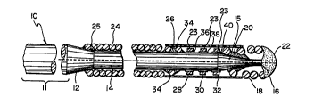
FIG. 1 shows a first embodiment of the invention. As shown,
the guidewire, when ;nt~n~Pd for percutaneous transluminal
coronary angioplasty may be approximately 175 cm to 300 cm in
total length and_includes an elongated rotationally rigid,
longitudinally flexible core wire 10, preferably made of
stainle3s steel or other material suitable for use as a guidewire
shaft. The majority of the length ~approximately 148 cm to 273
cm) is in the proximal segment ll of the core wire which ha~ a
substantially uniform diameter of approximately 0 . 25 to 0 . 457 mm
(0.010 to 0.018 inches). The proximal segment 11 merges into a
first tapered segment 12, about 3 cm long which, in turn, merges
into an intermediate barrel segment 14, approximately 22 . 5 cm
long and about 6 mils in diameter. Intermediate segment 14
merges into a second tapered segment 15, about 3 cm long which,
in turn, merges into a distal barrel segment 16, about 2.5 cm
long and approximately . 05 mm (2 mils) in diameter. The distal
segment 16 is more f lexible than the intermediate segment 14 .
Alternatively, the core wire (from taper 12 to distal segment 16)
may have a continuous taper along its length.
AMENDED SHEEr
I PEA/EP
~ 2 ~ ~4484
so410/7227Wo
--8 --
A distal coil 18, approximately 3 cm in length, is supported
about the distal segment of the core wire and preferably is
attached at its proximal end to the core wire by adhesive at
joint 20. Suitable adhesives include an ultraviolet curable
adhesive or a cyanoacrylate adhesive. The joint 20 alternatively
may be welded, soldered or brazed. The distal coil 18 is
attached at its distal end to the core wire by a distal
hemispherical tip weld 22. The distal coil may be formed from a
highly radiopaque material such as a gold/platinum or
platinum/tungsten alloy. In a guidewire intended for use in
percutaneous transluminal coronary angioplasty, the diameter of
the wire from which the distal coil 18 is wound preferably is
within the range of 0 . 025-0 .102mm ( . 001- . 004 inches) . The outer
diameter of the distal coil 18 preferably is within the range of
0.25 - 0.46 mm (.010-.018 inches).
A proximal coil 24 is 3upported about the intermediate
segment 14 of the core wire and is preferably attached at its
distal end to the core wire by adhesive at joint 26. The
proximal end of coil 24 extend3 to the proximal end of the
intermediate segment 14 of the core wire where it may be attached
to the core wire by adhesive at joint 25. Joints 25 and 26 may
alternatively be welded, soldered or brazed. The outer diameter
of the proximal coil is preferably the same as that of the distal
coil 18. The proximal coil preferably is 7-22 cm in length. The
proximal coil 24 may be less radiopaque than the distal coil 18
or of equivalent radiopacity. The proximal coil preferably is
formed from the same material a~ the distal coil but may be
formed from smaller diameter wire to achieve the desired reduced
radiopacity . AMENDED SHEFT
IPtAlEP
21 844~4
B0410t7227WO
g
The region of the guidewire between the proximal and distal
coils, inr]ll~;nJ a portion of the int~ te segment 14 and
tapered segment 15, is covered by a f lexible polymeric sleeve 34 .
The sleeve, preferably having an outer diameter er~ual to that of
the proximal and distal coils, provides a uniform outer ,1; i tPr
to the distal region of the guidewire such that the catheter will -
smoothly move over the guidewire during advi~nrpm~nt~ The sleeve
preferably is flexible, kink resistant and includes a lubricious
surface for aiding in guidewire maneuverability. The sleeve
preferably is formed from a polymer material (such as polyamide
or polyethrlene) which exhibits the above properties. A
hydrophilic or hydrophobic coating may be used to coat the outer
surface of the polymer sleeve for added lubricity.
The sleeve preferably is as thin as is practical, depending
on the polymer material from which the 81eeve iiY made, in order
that the region of the sleeve displays a desired degree of
flexibility. The wall thickness of the sleeve preferably falls
within the range of 0.005 - 0.05 mm (.0002-.002 inches). The
sleeve preferably covers the proximal end of the distal coil and
the distal end of the proximal coil and may be 5-20 cm in length.
As shown in FIG. 1, the distal end of the proximal coil 24 and
the proximal end of the distal coil 18 can be stretched and
tapered down to a smaller diameter in the region where they are
attached to the core wire and overlapped by the ends of the
polymer sleeve 34. The polymer sleeve preferably is adhesively
attached at both ends to the guidewire. If adhesively attached,
a small gap 23 exists between the inner surface of the sleeve and
outer surfaces of AMENDE~ SHEET
IPEA/EP
. _ , . , .. , .. , . . , _ . . .. .. .
2~8~484
so410/722~Wo
-10 -
/
the marker bands 28, 30 and 32 (discu3sed below). The size of
the gap is approximately equal to 0 . 025mm ( . 001 inches) but would
depend on the rh;~kn~qs of the sleeve wall and the outer diameter
of the marker bands. The gap prevents the outer contour of the
marker bands from projecting through the polymer sleeve.
Alternatively, the sleeve may be heat shrunk about the guidewire.
Radiopaque marker bands 28, 30 and 32 are attached to the
core wire between the proximal and distal coils. Marker bands
28, 30 and 32 preferably are attached by adhesive at joints 36,
38 and 40, respectively. Alternatively, ;oints 36, 38 and 40 may
be welded or brazed. The marker bands preferably are made from a
radiopaque material such as tantalum, platinum, gold or alloys
thereof. The marker bands will be spaced to provide optimum
usability for various l n vivo dlmension measurements by a
physician. Particularly, the spacing and dimensions of the
marker bands provide ref erence lengths such that the length and
shape of a lesion and adjacent arterial dimensions can be
determined fluoroscopically. Such a determination can aid a
physician in the selection of an appropriately sized balloon.
Additionally, if necessary, such a determilzation aids in the
selection of an appropriately sized stent The marker bands also
provide reference locations which can aid in placing a post-stent
balloon after stent placement. While the guidewire preferably
includes three marker bands, as shown and described, it is
envisioned that as few as one marker band could be used.
AMENDE~D SHEET
IPEAIEP
WO 95/2.1237 ~ t ~ PCTrUSs~rO3076
'
The guidewire of the first embodiment thus has a distal
tip section 16 that is highly radiopaque, an ;nt~rr^C~iate
section (between the coils) that is non-radiopaque (except
for the marker bands), and a proximal section that is
moderately or highly radiopaque. Such an arrangement may be
referred to as "grey/white/black" or "black/white/black"
~from the proximal to the distal ends), referring to its
relative appearance under fluoroscopy. The highly radiopaque
distal segment provides clear, Yisible, fluoroscopic
indication of the distal tip of the guidewire to indicate
clearly the guidewire position. Typically, the distal tip of
the guidewire is advanced through and beyond the stenosis to
be treated. The jn' -';ate, non--radiopague segment is
intf~n~led to be disposed at the region of the stenosis so that
the region will be ~ulob~L.Lcted by radiopague effects of the
guidewire. Thus, the full radiopaque effect of the
radiopa~ue contrast liquid injected into the artery can be
visualized on the fluoroscope, particularly in the critical
stenosed region of the artery. The moderately or highly
radiopaque proximal segment provides an indication of the
position and conf iguration of the more proximally located
portions of the guidewire and, therefore, the proximal
arterial anatomy.
The "grey/white/black" or "black/white/black"
configuration can be achieved with the appropriate relative
degrees of radiopacity by varying the thicknesses of the
wires from which the coils are wound, as described above.
Alternatively, as will be understood by those skilled in the
art, the coils may be plated with varying thicknesses of
radiopaque material to achieve the desired levels of
SU~SrIME SHEET (RULE 26)
wo ssl2~237 2 ~ 8 4 4 8 4 PCT/US9S/03076
, ~ " r t
--12-- ~
radiopacity. The plating process is described in U . S . Patent
No. 5,144,959 (Gambale), which is herein incorporated by
reference in its entirety.
FIG. 2 illustrates a second embodiment of the invention
which is fairly similar in construction to the first
i . Like elements in FIG. 2 are referred to by
identical reference characters ~to those in EIG. 1). In the
embodiment of FIG. 2, the construction of the core wire 10,
the distal coil 18, and the marker bands 28, 30 and 32 is
identical to that of the first ' ~i L ~shown in FIG. 1).
The guidewire of the second embodiment, however, omits the
proximal coil 24 of the first ~ . As in the first
: ~o~; ~, a polymeric sleeve 34, having the same properties
as those described above, encases the proximal end of the
distal coil and the marker bands. In the second ' ~
however, sleeve 34 extends proximally of the marker bands to
tapered segment 12 of core wire 10. The sleeve provides a
uniform outer diameter to the distal region of the guidewire
to aid in smooth advdllcG t of the catheter over the
guidewire. The sleeve preferably is adhesively attached at
both ends to the guidewire. Like the: ' of FIG. 1,
if adhesively attached, a small gap 39 exists between the
inner surface of the sleeve and the outer surface of the
marker bands 28, 30 and 32. The gap prevents the outer
contour of the marker bands from projecting through the
polymer sleeve. Alternatively, the sleeve may be heat shrunk
about the guidewire such that, proximally of the marker
bands, the sleeve 34 tapers down to f it tightly around the
int~r--~l; Ate segment 14 of core wire to create a smooth
transition .
SU35r1TUTE SHEET (RULE 26)
2 1 84484
so410,/7227wo
-13--
!
The guidewire of the second embodiment thus has a distal
region including a distal tip section 16 that i8 highly
radiopaque and a proximal section that is non-radiopaque. Such
an arrangement is referred to as "white/black" (from the proximal
to the distal ends), referring to its relative appearance under
f luoroscopy As with the embodiment of FIG . 1, the highly
radiopaque distal segment provides clear, visible fluoroscopic
indication of the distal tip of the guidewire to indicate clearly
the guidewire position and the proximal, non-radiopaque segment
provides for unobstructed visualization of the stenosis.
FIG. 3 shows a third embodiment of the invention in which a
radiopaque coil 50 is supported by and attached to a distal
region of the core wire 51 Core wire 51 is an alternate
embodiment core wire in which the distal barrel segment 16 of the
core wire 10 of the first embodiment is absent and is replaced by
a pair of round forming wires 53 and 55 which extend from tapered
segment 15 to hemispherical tip weld 22 The forming wires,
typically 3-7 cm in length, preferably are adhesively attached to
tapered segment 15 and extend approximately 2 cm beyond the
distal tip of core wire 51 ~he forming wires preferably are
formed from materials such as stainless steel, Sandvik lRK91,
PH455 or MP35~ Coil 50 can be used, however, with the core wire
10 of the first embodiment. Similarly, the guidewire
constructions of the first and second embodiments can be used
with the core wire 51
AMENDED SHEET
IPEA/EP
2 1 8~484
B0410/7227WO -14 -
As shown in FIG. 3, the distal end of the coil 50 is attached
to the hemispherical tip weld and the coil extends proximally to
the tapered section 12 of the core wire. The proximal end of the
coil 50 is attached to the core wire by adhesive at joint 52.
Alternatively, joint 52 may be soldered or brazed. Coil 50 may
also be attached to the core wire, preferably by adhesive, near
the distal end of the coil to add structural integrity to the
distal end of the guidewire such that unwinding of the coil
during adv~n~ ~mf~n~ of the guidewire through an artery i~
prevented. Spring 50 preferably is formed from a radiopaque
material such as a platinum/gold or other suitable alloy. The
diameter of the wire from which the coil 50 is wound preferably
is within the range of 0.025 - p.10 mm (0.001-0.004 inches). The
outer diameter of the coi1 preferably is within the range of o . 25
- 0.46 mm (.010-.018 inches). A typical coil length falls within
the range of 5-20 cm. If using a coil having a length of 5 cm,
the corresponding dimensions of the core wire would be less than
that disclosed above, as will be appreciated by those skilled in
the art.
Spring 50 has varying pitch along its length including
multiple tightly wound coil sections separated by loosely wound
coil sections. The tightly wound coil ~iections appear dark under
fluoroscopy and the loosely wound coil sections appear light.
Among the tightly wound coil sections are a distal section 54,
preferably within the range of 2-3 cm in length, and a proximal
section 55, within the range of 3-10 cm in length. Thus, the
proximal and distal sections are highly radiopaque.
AMENDED SHEET
~PEA/EP
wo 9sl2~237 , ~ PCTlUSsSI03076
~ 2 1 ~4484
--15--
An intermediate section 57 of the coil may include
multiple tightly wound coil sections 56; 58, 60, 62, and 64,
and loosely wound coil sections 65, 66, 68, 70, 72, and 7~.
The iLlL ~-'i ate section as a whole appears light under
fluoroscopy with the tightly wound sections appearing as dark
markers, providing reference lengths and location markers for
the physician. Markers 56, 58, 60 and 62 may be uniformly
spaced, preferably in 1 cm to 2 cm multiples. The number of
markers and the length and spacing thereof, however, can be
changed to suit a particular application. It is envisioned,
that as few as one marker band could be used.
The majority of the length of the spring, from a proximal
point of the distal section to the proximal end of the
spring, is covered by a f lexible polymeric sleeve 34 . Sleeve
34 exhibits the same ~ualities as the sleeve described above
in connection with the f irst and second embodiments . The
sleeve provides a uniform outer diameter to the distal region
of the guidewire. The sleeve, therefore, helps to ensure
smooth catheter ~ over the guidewire during
advAn,~- L. Sleeve 34 preferably is adhesively attached to
the guidewire at proximal 76 and distal 78 joints. If
adhesively attached, a small gap 80 exists between the outer
surface of spring 50 and the inner surface of sleeve 34. The
gap enables free bending ~l ,t of the coils of spring 50
beneath the sleeve 34 while the guidewire is maneuvered
through an artery. Additionally, the gap ~ v~ S the
co~tour of the outer surface of the coils from projecting
through the polymer sleeve 34. Alternatively, the ends of the
sleeve 34 maybe heat shrunk about the guidewire. It should
be unde~stood that the m-thod of attaching the sleeve to the
SuaST~ME SHEET (RULE 26)
~,~'0 95121237 2 1 8 4 ~ 8 4 PCTIUS95/03076
3 1,
-16-
guidewire only at its ends by adhesive or by heat shrirking
its ends can be used with any of the guidewire embodiments
disclosed herei~.
The guidewire of the third o-`~oAi ~ thus has a distal
tip section and a proximal section that are highly radiopaque
and an int: -~iate section that is lightly radiopaque
(except for the darker markers). Such an arrangement is
referred to as "black/grey/black" (from the proximal to the
distal ends), referring to its relative appearance under
fluoroscopy. As with the previously described ~ ts,
the highly radiopaque distal segment provides clear, visible
fluoroscopic indication of the distal tip of the guidewire to
indicate clearly the guidewire position and the int e~ii ate,
lightly radiopague segment provides for substantially
unobstructed vis~ a~; on of the stenosis .
As described above, the proximal and distal sections of
the coil preferably are equally highly radiopaque.
Alternatively, the distal section can be more radiopaque than
the proximal section. To achieve the desired relative
radiopacity, the distal section 54 of the coil may be wound
from a wire having a greater diameter than that of the
proximal sectio~. Alternatively, as will be understood by
those skilled in the art, the distal section 54 may include
another shorter radiopaque coil 90 which is supported by and
attached to the extreme distal part of the core wire and
which is located within the distal section of the coil, as
shown in FIG. 4.
Referring to FIG. 4, inner coil 90 is attached at its
distal end to the hemispherical tip weld 22 and may be
attached at its proximal end to the forming wires 53 and 55
SUBSTITUTE SHEET (RULE 26)
. O - 2i8'1~84
B0410/7227W0
-17-
and the tapered segment 15, preferably by adhesive.
Alternatively, the proximal end of the inner coil may not be
attached to any element. Be~ides inner coil 9o, the embodiment
of FIG. 4 is identical to that of FIG. 3. The inner coil 90
preferably has an outer diameter within the range of 0.15 - 0.30
mm(.006-.012 inches~, a length within the range of 1-4 cm, and is
made from a radiopaque material such as platinum, gold or a
platinum/gold alloy. The diameter of the wire from which the
inner coil is wound i5 preferably within the range of 0 . 025 -
0 . 076 mm ( . 001 to 003 inches) . The inner coil 90 is surrounded
by the distal section of the coil 50 such that the distal ~ection
will appear highly radiopaque.
FIG. 5 shows a fourth embodiment of the invention in which
the guidewire preferably falls within the range of 180-300 cm in
length and includes an elongated rotationally rigid,
longitudinally flexible core wire 10, preferably made of
stainle~s ~teel. The majority of the length (preferably 110-280
cm) of the core wire is in the proximal portion 11 which has a
substantially uniform diameter, typically within the range of
0.25 -~0.46 mm (10-18 mils) . The proximal portlon 11 merges into
a first tapered segment 12, preferably 2-10 cm in length, which,
in turn, merge~ into a first barrel segment 13, preferably 10-20
cm in length. The diameter of barrel segment 13 preferably is
within the range of 0.13 - 0.30 mm (5-12 mils), less than that of
proximal portion 11 First barrel segment 13 merges into a
second tapered segment 17, preferably 2-6 cm in length, which, in
turn, merges into a second barrel segment 19, preferably 5-15 cm
in length. The diameter of barrel segment 19 preferably is
within the range of 0.10 - 0.25 mm(4-10 mils), less than that of
AMENDEI~ EET
IPEA~'EP
~ - 2 1 84484
B0410/7227WO
-18-
f irst barrel segment 13 . Barrel segment 19 merges into a third
tapered segment 21, preferably 2-10 cm in length, which extends
to the distal end of the core wire.
One or more forming wires (only one of the forming wires 53
is shown) extend from tapered segment 21 to a rounded tip weld
22. The forming wires, typically one to five cm in length,
preferably are adhesively attached to tapered segment 21 at joint
78 and extend approximately 2 cm beyond the distal tip of core
wire 10. Alternatively, joint 78 can be soldered or brazed. The
forming wires preferably are made from a specially treated
precipitation hardenable alloy material. One such material is an
alloy of nickel, cobalt, molybdenum and chromium, colllmercially
available from Fort Wayne Metals of Fort Wayne, Indiana under the
trade designation MP35N. Another suitable material i8 a single
stage martensitic precipitation hardenable stainless ~teel having
modif ied proportions of chromium and nickel and with additional
elements of copper and titanium, commercially available f rom
Carpenter Steel Co. of Reading, Pennsylvania under the trade
designation 455PH. Still another suitable material i~ a
precipitation hardenable alloy that is commercially available
from Sandvik Steel under the trade designation Sandvik lRKgl.
While the distal region 27 of the core wire has been shown
and described herein as including two tapered segments, two
barrel segments and two forming wires, it should be understood
that other core wires can be used with this fourth embodiment
such as, for example, a core wire that has a distal region
including only a single barrel segment which merges into a
tapered segment that extends to the distal end
AMENDED SHEET
IPEA/EP
. ~ ` t~
- 2 ~ ~4484
sO410/7227WO
-19 -
of the core wire (with or without forming wires). Any of the
guidewire embodiments disclosed herein can be practiced with a
core wire having a distal region that either includes one or more
forming wires or, alternatively, a core wire that extends to the
distal tip of the guidewire.
A radiopaque coil 50 is supported by and attached to the
distal region 27 of the core wire 10. The distal end of the coil
50 is attached by the hemispherical tip weld. Just proximally of
the distal end of coil 50, the coil is attached to the core wire
at joint 78. Joint 78 also adds integrity to the structure of
the distal end of the guidewire to prevent the coil 50 from
separating from the core Mire if the forming wires break during
use. The coil extends proximally to the tapered sectipn 12 o
the core wire lO. The proximal end of the coil is attached to
the corc wire by adhesive at joint 52. Alternatively, joint 52
can be soldered or brazed. Spring 50 preferably is formed from a
radiopaque material such as platinum/gold or other suitable
alloy. The diameter of the wire from which the coil is wound
preferably is within the range of 0 . 038 - 0 . 076 mm ( . 0015 - . 003
inches). The outer diameter of the coil preferably is within the
range of 0 . 25 - 0 . 45 mm ( . 010 - . 018 inches) . A typical coil
length falls within the range of 15-40 cm. While the coil is
shown in FIG. S as having an outer diameter that is less than
that of the proximal portion 11 of the core wire, it is to be
appreciated that the outer diameter of the coil is preferably
equal to or approximately equal to the outer diameter of the
proximal portion 11 of the core wire.
Al~AENDED SHEET
IPEA/EP
wo g~/2~237 PCr~'S9~/03076
t -~
~ 21 ~4484
-20-
Spring 50 has varying pitch along its length including
distal and proximal tightly wound coil sections 54 and 55
separated by an intermediate loosely wound window coil
section 57. The tightly wound coil sections appear dark
under fluoroscopy and the loosely wound window coil section
appears light. Distal section 54 preferably falls within the
range of one to five cm in length, proximal section 55
preferably falls within the range o 10--25 cm in length, and
-~iiate window section 57 preferably falls within the
range of 5-20 cm in length.
Tntl -~iate window section 57 may include a number of
short tightly wound coil sections (not shown) separated by
loosely wound coil sections as in the embodiment of FIG. 3.
The tightly wound sections appear as dark marlcers under
fluoroscopy for providing reference lengths and location
marks for a physician. The number of markers and the length
and spacing thereof can be selected to suit a particular
application .
T L ~iate window section 57 of the coil 50 is covered
by a flexible polymeric sleeve 81. The sleeve 81 is kink
resistant and includes a lubricious surface for aiding in
guidewire maneuverability. The sleeve preferably is formed
from polyethylene terephthalate (PET) but may be formed from
other thermoplastic polymers such as polyethelene. A
hydrophilic or hydrophobic coating may be used to coat the
outer surface of the guidewire including the polymer sleeve
for added lubricity.
The sleeve preferably is as thin as is practical,
depending on the polymer material from which the sleeve is
made, in order that the region of the sleeve displays a
SUBSTITUTE SHEET (RUI.E 26~
~ - - 2 ~ 84~4
so410/7227Wo
-21-
desired degree of flexibility. The wall thickness of the sleeve
preferably falls within the range of 0.005 - 0.05 mm (.0002-.002
inches). The sleeve 81 is attached to the guidewire only at its
proximal and distal ends 82 and 84 leach preferably within the
range oi 0.5-2 cm in length) by heat shrinking the sleeve only at
those ends. Only the ends 82 and 84 of the sleeve 81 are
respectively heat shrunk about the tightly wound coils of the
proximal 55 and distal 54 regions immediately adjacent the window
section 57.
A small gap 80 exists between the outer surface of the
intermediate section 57 of coil 50 and the inner surface of
sleeve 81. The size of the gap is approximately equal to . 0001
inches but would depend on the thickness of the sleeve wall and
the outer diameter of the spring. The gap prevents the contour
of the outer surface of the coils from projecting through the
sleeve and contacting the inner arterial wall (which would occur
if the sleeve were heat shrunk about the entire length of the
coil) so that the guidewire can be navigated smoothly through the
artery during use. Additionally, the sleeve 81 insulates the
loosely wound coils of the intermediate section 57 from the inner
arterial wall to prevent them from significant m.,v t relative
to one another during advancement through an artery. Such
movement could result in altering the fluoroscopic image of the
guidewire and/or altering the structure and functionality of the
coils of the spring.
The distal region of the fourth embodiment (FIG 5) of the
guidewire thus has distal tip and proximal sections that are
highly radiopaque and an intermediate section that is lightly
radiopaque- Such an Alr\~Eln~Dem-eDn~ EErTeferred to as
IPEA/EP
wo 9512 1237 ~ PCTIUS95103076
' `i 21~4484
--22--
"black/grey/black" (from the proximal section to the distal
end), referring to its relative appearance under
fluoroscopy. The highly radiopaque distal segment provides
clear, visible fluoroscopic indication of the distal tip of
the guidewire to indicate clearly the guidewire position,
Typically, the distal tip of the guidewire is advanced
through and beyond the stenosis to be treated. The
;ate, lightly radiopaque segment is inr~n~ tl to be
disposed at the region of the stenosis so that the region
will not be materially obstructed by radiopaque effects of
the guidewire. Thus, the full radiopaque effect of the
radiopaque contrast liquid injected into the artery can be
visuali~ed on the fluoroscope, particularly in the critical
stenosed region of the artery. The radiopaque proximal
segment provides an irldication of the position and
configuration of the more proximally located portions of the
guidewire and, therefore, the proximal arterial anatomy, so
that a physician can observe the more proximally located
portions of the guidewire.
The proximal and distal sections of the coil preferably
are equally highly radiopaque. Alternatively, the distal
section can be more radiopaque than the proximal section. To
achieve the desired relative radiopacity, the distal section
54 of the coil may be plated with a radiopaque material.
The guidewire of the present inventio~ provides a number
of advantages. The guidewire facilitates better lesi~n
asse~ both by providing a non-obstructing region that
does not impair fluoroscopic evaluation of the shape of the
lesion and by providing a radiopaque proximal section that
allows visualization of the proximal portion of the wire. It
SUBSrlTUTE SHEET (RULE 26)
wo g~/2~237 ~ ;, PCT/US~/03076
; ~ 2 1 ~ 4 4 8 4
--23--
'
also provides radiopaque markers which offer a simple means
by which the physician can determine the length of the
lesion, adjacent arterial dimensions, and/or reference lesion
or stent location. Selection of an appropriately sized
balloon and, i necessary, selection of an appropriate stent
is, therefore, facilitated. Additionally, the sleeve
provides a uniform outer diameter to the distal region of the
guidewire to ensure smooth r,l~ v~ t of the catheter during
advancement. Also, as in the fourth embodiment, by heat
shrinking the sleeve only at its ends, the outer contour of
the coils is prevented from projecting through the sleeve,
and the sleeve insulates the loosely wound coils from the
inner arterial wall, preventing them from substantial
relative r ~ L.
Thus, we have described a guidewire having varied degrees
of radiopacity whereby, a distal section of the distal region
of the guidewire appears dark under fluoroscopy and an
jnt~ -aiate section o~ the distal region appears generally
white (or light in an alternate: ' -'; t). At least one
radiopaque marker may be located in the int, 'iate section
for providing rèference lengths and location marks for a
physician. A polymer sleeve encases at least part of the
distal region, providing a uniform outer diameter to the
region (in some: ' li tS), to aid in smooth catheter
advAn. ~. It should be understood, however, that the
foregoing description of the invention is ;nt~n~ ad merely to
be illustrative thereof and that other embo~ai and
modifications may be apparent to those skilled in the art
without departing from its objects, purposes and spirit.
Having thus described the invention, what we desire to
claim is:
SUBSrlTUTE SHt~T (RULE 26)