Note: Descriptions are shown in the official language in which they were submitted.
2185516
BEHRINGWERKE AKTIENGESELLSCHAFT 1995/B027 - Ma 1070
Dr. Bc/hg
Homogeneous gene probe test using a receptor directed
against the label
-------------------------------------------------------
The invention relates to a homogeneous gene probe test,
which is based on altering the signal of the
nonhybridized gene probe by means of a receptor
directed against the label.
Gene probe assays have already been described in the
literature in various embodiments. More frequently used
embodiments are the hybridization protection assay
(Clin. Chem. 35/8, 1989, 1588-1594), the kissing probes
technique (Nachr. Chem. Tech. Lab. 37/7, 1989, 698) and
the energy transfer principle.
A very general principle of a gene
probe assay according to the prior art is represented
in Fig. 1: in the first step, target sequence and
labeled gene probe are hybridized with one another,
wherefrom double-stranded constructs result if there is
sufficient homology of the two sequences. Moreover, as
a rule, however, nonhybridized single-stranded portions
of the gene probe also remain. In the second step, a
selective hydrolysis is carried out, which comprises,
on account of the conditions selected, essentially the
label of the single-stranded gene probe being attacked,
while the label of the double-stranded construct is
largely protected from hydrophilic attack. Thus in the
third step essentially the signal produced by the label
bound in the double-stranded construct is then
measured.
A disadvantage of the above method is that, despite the
treatment with the selection reagent (step 2), a
2185516.
2 -
remnant of single-stranded gene probe having an intact
= label remains, which distorts the measurement.
The present invention is therefore based on the object
of making available a method for the determination of a
nucleic acid sequence (a "gene probe assay") in which
the nonhybridized labeled gene probe contributes to a
smaller extent to undesired signal formation than in
the method according to the prior art. In particular,
the improvement aimed at should make possible an
improved homogeneous test procedure. The homogeneous
test procedure is fundamentally characterized by the
absence of a physical separation step between the
nucleic acid hybridization and the signal detection. In
such a method, according to the prior art, a
particularly severe interfering effect has to be taken
into account due to the nonhybridized labeled gene
probe.
The object was surprisingly achieved by employing in
the method according to the invention a receptor which
can bind to the label and as a result of the binding
detectably alters, for example attenuates ("quenches")
the signal to be attributed to the label. By means of
the method according to the invention described below,
it is therefore possible significantly to reduce the
interfering effect due to the nonhybridized labeled
gene probe and thereby to improve the sensitivity and
specificity of the test system decisively.
The present invention thus relates to a method for the
determination of a nucleic acid sequence (= target
sequence), in which a sample optionally containing the
target sequence is brought into contact with a gene
probe suitable for the determination of this target
sequence such that the target sequence and the gene
probe hybridize with one another, which comprises,
additionally
2185516
~'--' - 3 -
(a) adding a receptor which binds to the label of an
optionally excess fraction of the gene probe not
hybridized to the target sequence, whereby the
signal to be attributed to the label is
qualitatively and/or quantitatively altered and
(b) qualitatively or quantitatively detecting the
signal to be attributed to the label using a
method suitable for this purpose.
The present invention furthermore relates to a method
in which, in an additional step, the label of an
optionally excess, nonhybridized gene probe is partly
inactivated by incubation with a selection reagent.
A preferred embodiment of the present invention
comprises the incubation with the selection reagent
taking place first and then the addition of the
receptor.
A method is furthermore preferred in which the receptor
is a monoclonal or polyclonal antibody, an antibody
fragment, a chemically modified antibody- or a
chemically modified antibody fragment, if the antigen-
binding capacity after the chemical modification is
retained to an adequate extent.
Methods according to the invention which are
furthermore preferred comprise the label being a group
capable of fluorescence, phosphorescence, chemi-
luminescence, bioluminescence or electroluminescence.
In a particularly preferred embodiment of the present
invention, the label is an acridinium ester, an
acridinium acylsulfonamide, a luminol, an isoluminol or
a derivative thereof, a dioxetane, a luciferin, an
oxalic acid ester or an oxamide.
2185516
\' - 4 -
A method is also preferred in which the label is an
enzyme.
Label:
Suitable labels are all groups capable of fluorescence,
phosphorescence, chemiluminescence, bioluminescence or
electroluminescence, which on account of their chemical
structure can interact with a nucleic acid double
strand, for example by intercalating in the double
strand, - in such a way that the binding of a receptor
directed against this group is made difficult in
comparison with the binding to the corresponding single
strand-bound group. Particularly suitable are
acridinium ester and acridinium acylsulfonamide groups
which intercalate in a double-stranded nucleic acid.
Additionally suitable is a luminol, an isoluminol or a
derivative thereof, a dioxetane, a luciferin, an oxalic
acid ester or an oxamide.
Luminescent compounds already find various uses. They
are employed as indicators in enzyme immunoassays,
luminescence immunoassays (cf. W.P. Collins
"Alternative Immunoassays", Publishers John Wiley &
Sons Ltd.,- Chichester, 1985) and bioassays (tests which
are based not on antigen-antibody interactions, but on
binding affinities between molecules which are not
considered part of the immune system), but also in
nucleic acid hybridization assays (cf. J.A. Matthews et
al. "Analytical Biochemistry", 151, 205-209, 1985).
Additionally, chemiluminescence compounds are used in
flow injection analysis, in post-column detectors in
liquid chromatography, in flow research and for the
production of artificial light sources. Acridine
derivatives are furthermore suitable in test methods
for foodstuff and environmental analysis.
The use of acridinium labels in nucleic acid
hybridization assays is mentioned in EP-A-0 273 115 and
also in EP-A-0 212 951, EP-A-0 281 390,
2185516
-
EP-A-0 310 312, EP-A-0 313 219 and WO 89/02896.
EP-A-0 407 816 describes nucleotide derivatives with
the base uracil, which for its part is labeled with a
chemiluminescence compound via a spacer. EP-A 602 524
5 describes luminescent-labeled gene probes with
properties which are advantageous compared with the
prior art, and, inter alia, a homogeneous gene probe
assay according to the hybridization protection assay
principle, which is based on the advantageous
properties of the gene probes disclosed.
Anti-label antibodies:
Antibodies directed against the label can basically be
prepared in a conventional manner, e.g. by immunizing
an experimental animal with the label and subsequent
selection of suitable signal-affecting antibodies. Both
polyclonal and monoclonal antibodies are suitable,
monoclonal antibodies (MAbs) being preferred. Some
antibodies directed against luminogenic acridinium
labels have the property, by binding the label, of
reducing its signal strength (quench effect). Thus, for
example, in a single experimental batch under 10 mouse
MAbs directed against a luminogenic acridinium
acylsulfonamide label were found which, with respect to
possible signal-quenching properties, were not
preselected and one MAb was found which had the desired
signal-quenching properties.
An example of a highly suitable antibody is the
monoclonal mouse antibody secreted from the cell line
BW 90-614-8-04, which has been deposited in the German
Collection of Microorganisms and Cell Cultures GmbH,
Mascheroder Weg 1B, D-38124 Brunswick under the entry
number DSM ACC 2184. This MAb is directed against the
acridinium acylsulfonamide shown in Fig. 2.
Preparation of the gene probes:
2185516
' ~ - 6 -
The preparation of suitable gene probes can be
performed using methods known, in principle to the
person skilled in the art. Gene probes are discussed in
detail in a relatively large number of publications,
for example in: S. L. Beaucage and R. P. Iyer: "The
Functionalization of Oligonucleotides Via Phos-
phoramidite Derivatives", Tetrahedron 49, 1925-1963
(1993) and J. Goodchild: "Conjugates of Oligonucleo-
tides and Modified Oligonucleotides: A Review of Their
Synthesis and Properties", Bioconjugate Chemistry 1,
165-187 (1990).
The structure of a suitable gene probe is shown by way
of example in Fig. 3. Of course, the base sequence
shown can be replaced by any other suitable sequence.
Other labels known to any person skilled in the art can
also be linked to the nucleic acid to be used as a gene
probe by methods known to any person skilled in the
art.
The gene probe assay:
Generally, the method according to the invention can be
realized on the basis of all methods known in the prior
art. The gene probe technology according to the
invention can advantageously be employed in homogeneous
tests. On account of the strong signal quench by an
anti-label MAb, substantially more sensitive
homogeneous gene probe tests can be developed, with
comparatively good stability, than are known in the
prior art. Homogeneous gene probe assays according to
the invention are additionally distinguished by simple
handling and easy automatability.
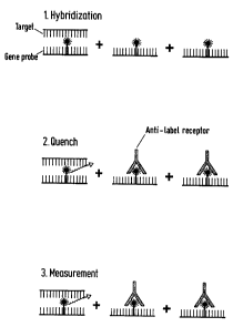
A preferred variant of the method according to the
invention is shown schematically in Fig. 4: in the
first step the target sequence and labeled gene probe
are hybridized with one another, wherefrom double-
stranded constructs result if there is sufficient
2185516
_ ~...- - 7 -
homology of the two sequences. Moreover, as a rule,
nonhybridized single-stranded portions of the gene
probe also remain. In the second step, a receptor, for
example an antibody, is employed which can bind to the
label of the single-stranded gene probe and, as a
result of the binding, detectably alters, for example
attenuates (quenches) the signal to be attributed to
the label. In the third step, the signal produced by
the label bound in the double-stranded construct is
then almost exclusively measured, as the label of the
hybridized gene probe cannot be bonded or can be bonded
less well by the receptor than the label of the
nonhybridized gene probe. Compared with known methods,
this method has the advantage that still less single-
stranded gene probe produces an (undesired)
contribution to the measured signal.
The above method can be further improved by performing,
after the first hybridization step, a selective
hydrolysis of the single-stranded gene probe by
treatment with a selection reagent and only
subsequently thereto employing a receptor directed
against the label, as shown above. The measurement
which then follows is virtually no longer distorted by
unbound label due to the prior double elimination of
the label of the single-stranded gene probe (see
Fig. 5 ) .
A further preferred working variant is based on the
hybridization protection assay (see above) and
comprises, according to the invention, detectably
altering, e.g. quenching, the label of the
nonhybridized single-stranded portions of the gene
probe in a further step by addition of a receptor which
can bind to the label of the single-stranded gene probe
such that these unbound gene probes can cause no
distortion of the measured signal.
2185516
~
- - 8 -
The receptor employed in the method according to the
invention is preferably a signal-quenching monoclonal
or polyclonal antibody directed against the label, as
already described further above.
The following examples are intended to illustrate the
present invention further, but not to restrict it in
any manner.
Example 1: Preparation of a monoclonal antibody against
a luminogenic acridinium acylsulfonamide label:
For the preparation of monoclonal antibodies, BALB-c
mice were injected subcutaneously or intraperitoneally
with 10 g of acridinium acylsulfonamide-BSA conjugate,
emulsified in complete Freund's adjuvant. The
acridinium acylsulfonamide-BSA conjugate can be
prepared by reaction of N-(4-methoxyphenyl)-
N-[4-(2-succinimidyloxycarbonylethyl)benzenesulfonyl]-
10-methylacridinium-9-carboximide fluorosulfonate or
trifluoroacetate (Fig. 2) with BSA by methods known to
the person skilled in the art. 4 to 5 additional
immunizations without adjuvant followed- every four
weeks. The last four -days before the fusion the mice
received intravenous booster injections (10 g per
day).
For the production of hybridomas, the immunized animals
were killed by means of cervical dislocation. The
spleen was asceptically removed and teased apart in
order to obtain an individual suspension of spleen
cells in serum-free Dulbecco's modified Eagle's medium
(DMEM). The cells were collected by means of
centrifugation (5 min.; 1800 rpm) and washed once in
DMEM. The total cell count was determined by
hemocytometer counting using the Trypan Blue exclusion
technique. The mouse myeloma cells (SP2/0) were washed
twice in serum-free DMEM, collected by means of
2185516
' ~ - 9 -
centrifugation (10 min., 1000 rpm) and counted as
described above.
Approximately 108 spleen cells were mixed with 2 x 10'
SP2/0 myeloma cells from the mouse. After
centrifugation at 1000 rpm for 10 minutes, the
supernatant was removed and 1 ml of polyethylene glycol
(PEG 4000, Merck, 50 %) was added to the vessel
containing the pellet. The pellet was then resuspended
with light tapping and incubated at 37 C for 1 minute._
10 ml of serum-free DMEM were added dropwise with light
tapping and the mixture was incubated for 2 to
4 minutes. The fused cells were then centrifuged at
1000 rpm for 10 minutes. The cell pellet obtained was
- suspended in DMEM containing 20 fetal calf serum
(FCS) and HAT (hypoxanthine 0.1 M; aminopterin 0.4 M;
thymidine 16 M) and plated out onto culture plates
(Nunc) with 24 wells using a concentration, by way of
approximation, of 5 x 10⁴ - 106 cells per well. After 2
to 3 weeks, individual cell colonies were removed from
the individual wells and cultured in wells of a new
culture plate.
The culture supernatants were examined for antigen-
specific antibodies by means of the EIA technique. Each
well of a microtiter plate coated with acridinium
acylsulfonamide-BSA (3 g/ml) was filled with 100 l of
the supernatant and incubated at room temperature for
1 hour. After washing, 100 l of a rabbit anti-mouse
peroxidase (POD) conjugate were added at room
temperature for a further hour. After incubation with
the substrate for 30 minutes, the color development at
492 nm was read off on a Behring-ELISA processor (BEP).
Hybridomas which produce antibodies having a suitable
antigen specificity were selected and cloned using an
individual cell manipulator. For the preparation of
large amounts of monoclonal antibodies, the clones were
replicated in mass culture. The subsequent purification
2185516
-
of the individual monoclonal antibodies was carried out
by means of protein A chromatography.
Example 2: Preparation of a gene probe (Fig. 3)
5
The synthesis of the oligonucleotide is described in
EP-A 0 602 524, p. 49, Example 14 b). Coupling with the
acridinium acylsulfonamide was carried out by known
methods, which are described, for example, in the
10 abovementioned European Patent Application.
Example 3: Homogenous gene probe test for detection on
E.coli without anti-label MAb
50 l of standard (from Flash Track test of Gen Probe,
Lot 11276/11278 for p.ositive/negative standard) are
pipetted into polystyrene tubes. 50 l of the gene
probe according to Fig. 3 (2.5 x 10⁶ RLU, 1 M tris
buffer, pH 7) are added and hybridized at 60 C for 15
minutes. 300 l of a selection reagent (0.2 M
tetraborate, pH 8) are then added, shaken for
2 x 3 seconds and again incubated at 60 C for
15 minutes. After this, the tubes are allowed to cool
for 5 minutes:
Measurement is carried out by addition of 300 l in
each case of analyzer reagent 1 (0. 1 M HNO3 , 0.5 % H202)
and analyzer reagent 2 (0.25 M NaOH) in a luminometer
(AutoCliniLumat from Berthold). The measurement time
is 1 sec/sample.
A clear signal differentiation between positive and
negative standard is determined (see table).
Example 4: Homogeneous gene probe test for the
detection of E.coli using anti-label MAb
50 l of standard (from Flash Track test of Gen Probe,
Lot 11276/11278 for positive/negative standard) are
pipetted into polystyrene tubes. 50 l of the gene
CA 02185516 2006-02-15
- 11 -
probe according to Fig. 3 (2.5 x 10⁶ RLU, 1 M tris
buffer, pH 7) are added and hybridized at 60 C for 15
minutes. 300 l of a selection reagent (0.2 M
tetraborate, pH 8) are then added, shaken for 2 x 3
seconds and again incubated at 60 C for 15 minutes.
After this, the tubes are allowed to cool for
5 minutes.
50 [Ll of anti-label MAb solution (10 g/ml of tris
buffer pH 7.4, 1 M, 0.1 m TritonTM X-100) are then
incubated at RT for 1 minute.
Measurement is carried out by addition of 300 l in
each case of analyzer reagent 1 (0.1 M HNO3, 0.5 's H202)
and analyzer reagent 2 (0.25 M NaOH) in a luminometer
(AutoCliniLumat from Berthold). The measurement time
is 1 sec/sample.
In comparison with the original hybridization
protection assay (Example 3), the signal
differentiation between positive and negative standard
is clearly improved by addition of the anti-label MAb
(see table). A signal differentiation is achieved which
otherwise is only possible in a heterogeneous
embodiment using magnetic particles as the solid phase
(see EP-A-0 602 524, pp. 50 to 51, Example 17).
Example 5: Homogeneous gene probe test for the
detection of E.coli using unspecific MAb (control
experiment)
50 l of standard (from Flash Track test of Gen Probe,
Lot 11276/11278 for positive/negative standard) are
pipetted into polystyrene tubes. 50 l of the gene
probe according to Fig. 3 (2.5 x 10⁶ RLU, 1 M tris
buffer, pH 7) are added and hybridized at 60 C for 15
minutes. 300 l of a selection reagent (0.2 M
tetraborate, pH 8) are then added, shaken for
2 x 3 seconds and again incubated at 60 C for
2185516
- 12 -
15 minutes. After this, the tubes are allowed to cool
for 5 minutes.
50 l of anti-TSH MAb solution (1 g of TSH MAb from
Medix, batch No.: SPHY052/ml of tris buffer pH 7.4,
1 M, 0.1 % Triton X-100) are then incubated at RT for
5 minutes.
Measurement is carried out by addition of 300 l in
each case of analyzer reagent 1(0.1 M HNO3, 0.5 % H202)
and analyzer reagent 2 (0.25 M NaOH) in a luminometer
(AutoCliniLumat from Berthold).
The measurement time is 1 sec/sample.
The signal differentiation between positive and
negative standard corresponds - as expected - to
Example 3 (see table).
Table:
Example 3 Example 4 Example 5
Positive standard 13449 14229 13872 -
[RLU] 14066 14041 14119
Negative standard 416 53 408
[RLU] 404 47 419
(RLU = relative light units)