Note: Descriptions are shown in the official language in which they were submitted.
218b~50
-1-
SOUND GENERATING APPARATUS AND METHOD, SOUND GENERATING SPACE
AND SOUND, EACH PROVIDED FOR SIGNIFICANTLY INCREASING THE
CEREBRAL BLOOD FLOW OF A PERSON
The present invention relates to a sound generating
apparatus, a sound generating space, a sound, and a method for
generating a sound. In particular the invention is directed
to a sound generating apparatus, a sound generating space, a
sound, a method for generating a sound, each provided for
significantly increasing the cerebral blood flow of a person.
It is generally accepted that audio frequencies above 20
Khz do not affect human sensory perception since they are
beyond the audible range. Due to this, the sampling frequency
for compact discs (Cbs) and mini discs (MDs) is set to 44.1
kHz, and the equipment for these discs are manufactured so
that audio signals up to about 22 kHz can be recorded and
reproduced. Additionally, the sampling frequency for digital
audio tape recorders (DATs) as well as digital compact
cassette recorders (DCCs) can be set to any one of the
frequencies, 48 kHz, 44.1 kHz, and 32 kHz, and the equipment
for these are manufactured so that audio signals up to about
24 kHz can be recorded and reproduced. As a result, human
beings can relax themselves by listening to favourite music
with the use of such equipment.
On the other hand, the strength of a-EEG
(a-electroencephalogram) or a waves generated from the human
brain is generally used as an index indicating that one has
less stress. Taking advantage of the fact that when an
ultra-low frequency signal of around 10 Hz is generated and
heard or listened to, one will generate a-EEG, there have been
developed and marketed apparatuses for generating such an
ultra-low frequency signal.
However, in the above-mentioned prior art, there are such
problems that, the degree to which stresses can be relieved
changes depending on the content of the music or the
frequencies of the ultra-low frequency signal, and moreover
the degree thereto is relatively small.
An essential object of the present invention is therefore
to provide a sound generating apparatus capable of increasing
2186050
-2-
cerebral blood flow of a person so as to improve and enhance
his or her state of mind and body, to relieve stresses,
thereby causing relaxation.
Another object of the present invention is to provide a
sound generating space capable of increasing cerebral blood
flow of a person so as to improve and enhance his or her state
of mind and body, to relieve stresses, thereby causing
relaxation.
A further object of the present invention is to provide a
sound capable of increasing cerebral blood flow of a person so
as to improve and enhance his or her state of mind and body,
to relieve stresses, thereby causing relaxation.
A still further object of the present invention is to
provide a method for generating a sound, capable of increasing
cerebral blood flow of a person so as to improve and enhance
his or her state of mind and body, to relieve stresses,
thereby causing relaxation.
In order to achieve the aforementioned objective,
according to one aspect of the present invention, there is
provided a sound generating apparatus comprising:
means for generating a sound which has a frequency within
a first frequency range beyond a predetermined audible
frequency range and up to a predetermined maximum frequency,
and which is non-stationary so as to change in a
micro-temporal area in a second frequency range beyond 10 kHz;
and
means for applying said sound to a person, thereby
increasing cerebral blood flow of the person.
According to another aspect of the present invention,
there is provided a sound generating apparatus comprising:
means for generating a sound which has a frequency within
a first frequency range beyond a predetermined audible
frequency range and up to a predetermined maximum frequency,
and which is non-stationary so as to change in a
micro-temporal area in a second frequency range beyond 10 kHz;
means for applying first sound components within the
audible frequency range out of said sound to an auditory
2186050
-3-
sensation of a person; and
means for applying second sound components having a
frequency range beyond the audible frequency range out of said
sound to the person, thereby increasing cerebral blood flow of
the person.
In the above-mentioned sound generating apparatus, the
maximum frequency is preferably 150 kHz.
According to a further aspect of the present invention,
there is provided a sound generating space comprising:
means for generating a sound which has a frequency within
a first frequency range beyond a predetermined audible
frequency range and up to a predetermined maximum frequency,
and which is non-stationary so as to change in a
micro-temporal area in a second frequency range beyond 10 kHz;
and
means for applying said sound to a person, thereby
increasing cerebral blood flow of the person.
According to a still further aspect of the present
invention, there is provided a sound generating space
comprising:
means for generating a sound which has a frequency within
a first frequency range beyond a predetermined audible
frequency range and up to a predetermined maximum frequency,
and which is non-stationary so as to change in a
micro-temporal area in a second frequency range beyond 10 kHz;
means for applying first sound components within the
audible frequency range out of said sound are applied to an
auditory sensation of a person; and
means for applying second sound components having a
frequency range beyond the audible frequency range out of said
sound are applied to the person, thereby increasing cerebral
blood flow of the person.
In the above-mentioned sound generating space, the
maximum frequency is preferably 150 kHz.
According to a still more further aspect of the present
invention, there is provided a sound which has a frequency
within a first frequency range beyond a predetermined audible
_ ~v s~o~o
-4-
frequency range and up to a predetermined maximum frequency,
and which is non-stationary so as to change in a
micro-temporal area in a second frequency range beyond 10 kHz,
said sound being applied to a person, thereby increasing
cerebral blood flow of the person.
In the above-mentioned sound, the maximum frequency is
preferably 150 kHz.
According to a more still further aspect of the present
invention, there is provided a method for generating a sound,
including the following steps of:
generating a sound which has a frequency within a first
frequency range beyond a predetermined audible frequency range
and up to a predetermined maximum frequency, and which is
non-stationary so as to change in a micro-temporal area in a
second frequency range beyond l0 kHz; and
applying said sound to a person, thereby increasing
cerebral blood flow of the person.
According to a more still more further aspect of the
present invention, there is provided a method for generating a
sound, including the following steps of:
generating a sound which has a frequency within a first
frequency range beyond a predetermined audible frequency range
and up to a predetermined maximum frequency, and which is
non-stationary so as to change in a micro-temporal area in a
second frequency range beyond 10 kHz;
applying first sound components within the audible
frequency range out of said sound to an auditory sensation of
a person; and
applying second sound components having a frequency range
beyond the audible frequency range out of said sound to the
person, thereby increasing cerebral blood flow of the person.
According to the present invention, therefore, when
applying the sound to the person as described above, the d-EEG
potential can be increased so that the person can be relaxed,
with stresses dissipated, and that the comfort of the mind as
well as the health of the body can be enhanced or maintained
successfully.
216050
-5-
These and other objects and features of the present
invention will become clear from the following description
taken in conjunction with the preferred embodiments thereof
with reference to the accompanying drawings throughout which
like parts are designated by like reference numerals, and in
which:
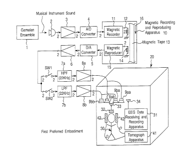
Fig. 1 is a block diagram of a signal sound generating
apparatus of a first preferred embodiment according to the
present invention, and a perspective view showing a room for
generating signals by the signal sound generating apparatus;
Fig. 2 is a partial block diagram of a signal sound
generating apparatus of a second preferred embodiment
according to the invention, and a perspective view showing a
room for generating signals by the signal sound generating
apparatus;
Fig. 3 is a graph showing frequency characteristics of
signals produced by the Gamelan ensemble, a cembalo, and a
piano used in the preferred embodiments;
Fig. 4 is a graph showing frequency characteristics of
environmental sounds in a forest in Tsukuba-shi, Ibaraki-ken,
Japan, in a tropical rain forest in Java Island, Indonesia,
and along a road in Tokyo-to, Japan;
Fig. 5 is a graph showing an MEM spectra array of the
Gamelan ensemble sound up to 100 kHz, which is used in the
preferred embodiments;
Fig. 6 is a graph showing an MEM spectra array of the
piano sound up to 100 kHz, which is a comparative example;
Fig. 7 is a graph showing an MEM spectra array of the
Gamelan ensemble sound up to 20 kHz, which is used in the
preferred embodiments;
Fig. 8 is a graph showing an MEM spectra array of the
piano sound up to 20 kHz, which is a comparative example;
Figs. 9A, 9B and 9C are projections showing parts of the
brain in which cerebral blood flow significantly increases in
the full range sound as compared with that in the high cut
sound alone in the first preferred embodiment, where Fig. 9A
is a sagittal projection which is a projection along the
2~8b050
-6-
sagittal suture of the human skull, Fig. 9B is a coronal
projection which is a projection along the coronal suture of
the skull, and Fig. 9C is a transversal projection of the
skull;
Figs. 10A, lOB and lOC are sectional views respectively
corresponding to Figs. 9A, 9B and 9C in the first preferred
embodiment, and showing a part of the brainstem in which the
cerebral blood flow significantly increases in the full range
sound as compared with that in the high cut sound alone, where
Fig. l0A is a longitudinal sectional view showing a sagittal
cross section along the sagittal suture of the human skull,
Fig. lOB is a longitudinal sectional view showing coronal
cross section along the coronal suture of the skull, and Fig.
lOC is a transversal cross sectional view of the skull;
Figs. 11A, 11B and 11C are sectional views respectively,
corresponding to Figs. 9A, 9B and 9C in the first preferred
embodiment, and showing parts of a left thalamus in which
cerebral blood flow significantly increase in the full range
sound as compared with that in the high cut sound alone, where
Fig. 11A is a longitudinal sectional view showing a sagittal
cross section along the sagittal suture of the human skull,
Fig. 11B is a longitudinal sectional view showing a coronal
cross section along the coronal suture of the skull, and Fig.
11C is a transversal cross sectional view of the skull;
Figs. 12A and 12B are graphs showing normalized r-CBF
values for the respective sounds in the first preferred
embodiment, where Fig. 12A is a graph showing the r-CBF values
at the brainstem, and Fig. 12B is a graph showing the r-CBF
values at the left thalamus;
Fig. 13 is a graph showing normalized a-EEG potentials
for the respective sounds in the first preferred embodiment;
Figs. 14A and 14B are graphs showing normalized r-CBF
values for the respective sounds in the first preferred
embodiment, where Fig. 14A is a graph showing the r-CBF values
at the brainstem, and Fig. 14B is a graph showing the r-CBF
values at the left thalamus;
- ~ ~ 86050
_7-
Fig. 15 is a graph showing normalized a-EEG potentials
for the respective sounds in the first preferred embodiment;
Figs. 16A, 16B and 16C are transversal cross sectional
views for different z's of the Talairach coordinates in the
first preferred embodiment, showing a part in which the a-EEG
potential significantly correlates with the r-CBF value, as
well as a part in which the cerebral blood flow significantly
increases, where Fig. 16A shows the cross section for z = -4
mm, Fig. 16B shows the cross section for z = 0 mm, and Fig.
16C shows the cross section for z = 4 mm;
Fig. 17 is a graph showing a correlation between r-CBF
values and normalized a-EEG potentials at the position of the
left thalamus in the first preferred embodiment;
Fig. 18 is a graph showing normalized a-EEG potentials
for the respective sounds in the second preferred embodiment;
Figs. 19A, 19B and 19C are projections showing parts in
which the r-CBF value more significantly increases when the
audible sound is applied from earphones while the low cut
sound is applied from speakers, than that when only the
audible sound is applied from earphones, in the second
preferred embodiment, where Fig. 19A is a sagittal projection
which is a projection along the sagittal suture of the human
skull, Fig. 19B is a coronal projection which is a projection
along the coronal suture of the skull, and Fig. 19C is a
transversal projection of the skull;
Fig. 20 is a graph showing normalized r-CBF values for
the respective sounds at the angular gyrus of the brain in the
second preferred embodiment;
Fig. 21 is a graph showing normalized r-CBF values for
the respective sounds at the posterior cingulate gyrus of the
brain in the second preferred embodiment;
Fig. 22 is a graph showing normalized r-CBF values for
the respective sounds at the boundary of the posterior
cingulate gyrus (precuneus) of the brain in the second
preferred embodiment;
Figs. 23A, 23B and 23C are sectional views respectively,
corresponding to Figs. 19A, 19B and 19C in the second
2 ~ s6~5o
_8_
preferred embodiment, and showing a part of the angular gyrus
of the brain in which the r-CBF value significantly increases,
where Fig. 23A is a longitudinal sectional view showing a
sagittal cross section along the sagittal suture of the human
skull, Fig. 23B is a longitudinal sectional view showing a
coronal cross section along the coronal suture of the skull,
and Fig. 23C is a transversal cross sectional view of the
skull;
Figs. 24A, 24B and 24C are sectional views respectively,
corresponding to Figs. 19A, 19B and 19C in the second
preferred embodiment, and showing a part of the posterior
cingulate gyrus of the brain in which the r-CBF value
significantly increases, where Fig. 24A is a longitudinal
sectional view showing a sagittal cross section along the
sagittal suture of the human skull, Fig. 24B is a longitudinal
sectional view showing a coronal cross section along the
coronal suture of the skull, and Fig. 24C is a transversal
cross sectional view of the skull;
Figs. 25A, 25B and 25C are sectional views respectively,
corresponding to Figs. 19A, 19B and 19C in the second
preferred embodiment, and showing a part of the boundary of
the posterior cingulate gyrus (precuneus) of the brain in
which the r-CBF value significantly increases, where Fig. 25A
is a longitudinal sectional view showing a sagittal cross
section along the sagittal suture of the human skull, Fig. 25B
is a longitudinal sectional view showing a coronal cross
section along the coronal suture of the skull, and Fig. 25C is
a transversal cross sectional view of the skull;
Figs. 26A, 26B and 26C are graphs showing frequency
characteristics of respective signals in the signal sound
generating apparatus shown in Fig. 1, where Fig. 26A is a
graph showing a frequency characteristic of a frequency
component outputted from a D/A converter shown in Fig. 1, Fig.
26B is a graph showing frequency characteristics of respective
frequency components outputted from the speakers shown in Fig.
1, and Fig. 26C is a graph showing a frequency characteristic
of the background noise in the room shown in Fig. 1; and
2$6050
-9-
Fig. 27 is a graph showing respective frequency
characteristics of (i) a high frequency component of a Gamelan
ensemble sound used in the signal sound generating apparatus
shown in Fig. 1, and (ii) a virtual stationary high frequency
component not having any fluctuation structure, changing in
the micro-temporal in the frequency range above 10 kHz, which
is obtained by filtering and waveform-shaping a stationary
white noise electrically generated so that the stationary
white noise approximates to dime-averaged spectral of the
temporal component of (i) the high frequency component.
Preferred embodiments according to the present invention
will be described below with reference to the attached
drawings.
First Preferred Embodiment
Fig. 1 is a block diagram of a signal sound generating
apparatus of a first preferred embodiment according to the
present invention, and a perspective view showing a room 20
which is a signal sound generating space for generating
signals by the signal sound generating apparatus. The signal
sound generating apparatus of the present preferred embodiment
is characterized in generating a signal which has a frequency
within a first frequency range beyond the audible frequency
range and up to a maximum frequency, and which is
non-stationary so as to change in a micro-temporal area in a
second frequency range beyond 10 kHz, and then applying the
above generated signal to a person, thereby increasing the
cerebral blood flow of the person.
It is noted that the first frequency range is a range
from about 20 Hz to about 150 kHz. In the first preferred
embodiment, as shown in Fig. 1, the same two line systems of
the sound recording and reproducing systems are prepared and
driven in the so-called stereophonic state.
As shown in Fig. 1, instrumental sounds obtained by
playing the Gamelan ensemble 1, which is bronze percussion
ensemble of Bali Island, Indonesia, are collected by a
microphone 2. The microphone 2 converts an input instrumental
sound into an analog electric signal, and the converted analog
2186050
-10-
electric signal is delivered to an A/D converter 4 via a
preamplifier 3. The A/D converter 4 converts the input analog
electric signal into a digital signal with a sampling
frequency of, for example, 1920 kHz, and then delivers the
analog-to-digital converted signal to a magnetic recorder 11.
A magnetic recording and reproducing apparatus 10 is a
so-called digital signal recorder which comprises the magnetic
recorder 11, a recording magnetic head 12, a reproducing
magnetic head 14, and a magnetic reproducer 15, and which
works to record digital signals onto a magnetic tape 13 or to
reproduce and output digital signals recorded on the magnetic
tape 13. The magnetic recording and reproducing apparatus 10
here used is a prior art DAT (Digital Audio Tape recorder)
invented by Dr. Yoshio YAMASAKI, having a uniform frequency
characteristic over a frequency range up to 200 kHz. The
magnetic recorder 11 modulates a carrier signal according to
the digital signal inputted from the A/D converter 4 by a
predetermined digital modulation method, and records the
modulated signal onto the magnetic tape 13 which is running
along a predetermined direction 16 indicated by an arrow of
Fig. 1, by using the recording magnetic head 12. On the other
hand, the magnetic reproducer 15 reproduces the modulated
signal recorded on the magnetic tape 13 by using the
reproducing magnetic head 14, and demodulates the reproduced
modulated signal by a digital demodulation method reverse to
the above-mentioned digital modulation method, so as to
extract and output the digital signal from the reproduced
modulated signal.
The demodulated digital signal is converted analog from
digital form into the original analog signal by a D/A
converter 5, and then output via a reproduction amplifier 6.
The analog signal outputted from the reproduction amplifier 6
is inputted via a switch SW1, a high-pass filter 7a having a
cut-off frequency of 22 kHz, and a power amplifier 8a, into a
right speaker 9aa and a left speaker gab, both of which can
generate signals within a frequency range from 20 kHz to 150
kHz. Moreover, the analog signal outputted from the
_ 2186054
-11-
reproduction amplifier 6 is inputted via a switch SW2, a
low-pass filter 7b having a cut-off frequency of 22 kHz, and a
power amplifier 8b, into a right speaker 9ba and a left
speaker 9bb, both of which can generate signals below 30 kHz.
In the present preferred embodiment, accordingly, the
crossover frequency of the two filters 7a and 7b is 22 kHz.
The speakers 9aa, gab, 9ba and 9bb are placed within the
room 20, which is an acoustically closed sound-shielded room.
The speakers 9aa, gab, 9ba and 9bb convert the input signals
into sounds, respectively, and apply them to a person 30, who
is a subject of measurement.
Detection electrodes are provided at, for example, 12
scalp sites of the person 30. An EEG detector and transmitter
32 connected to the detection electrodes converts an EEG
detected by each detection electrode into a radio signal and
transmits the resulting signal from an antenna 33 toward an
antenna 34. The radio signal of the EEG is received by the
antenna 34, and is then delivered to an EEG data receiving and
recording apparatus 31. In the EEG data receiving and
recording apparatus 31, the received radio signal of the EEG
is converted into an EEG signal, and is then recorded on a
magnetic recorder provided within the EEG data receiving and
recording apparatus 31. Further, the EEG signal is analyzed
by an analysis computer, while variations in the EEG are
recorded and outputted by using an output unit such as a CRT
display, a pen recorder or the like. On the other hand, the
head of the person 30 is placed so as to be sandwiched between
two detector elements of a detector 42 for the tomograph. A
detection signal derived from the detector 42 for the
tomograph is transmitted to a tomograph apparatus 41.
Subsequently, the tomograph apparatus 41 executes a
predetermined tomographical analysis process based on the
input detection signal, and displays a tomographical view of
the analysis result onto a built-in CRT display of the
tomograph apparatus 41.
Figs. 26A, 26B and 26C show frequency characteristics of
respective signals in the signal sound generating apparatus of
- 2186050
-12-
the first preferred embodiment shown in Fig. 1 as constructed
above, where Fig. 26A is a graph showing a frequency
characteristic of a frequency component outputted from a D/A
converter 5 shown in Fig. l, Fig. 26B is a graph showing
frequency characteristics of respective frequency components
outputted from the speakers 9aa, gab, 9ba and 9bb shown in
Fig. l, and Fig. 26C is a graph showing a frequency
characteristic of the background noise in the room 20 shown in
Fig. 1.
In the signal sound generating apparatus and the room 20
of the first preferred embodiment having the above-mentioned
construction, after the instrumental sounds produced by
playing the Gamelan ensemble 1 with both the switches SW1 and
SW2 turned on are recorded to the magnetic tape 13 of the
magnetic recording and reproducing apparatus 10, and
thereafter, when the sound signals are reproduced, the
reproduced sound signals substantially identical to the
instrumental sounds of the Gamelan ensemble 1 can be applied
to the person 30 by using the speakers 9aa, gab, 9ba and 9bb.
In this case, by turning on or off the switches SW1 and SW2,
the instrumental sound signals in various kinds of frequency
components can be generated by the speakers 9aa, gab, 9ba and
9bb. That is, with only the switch SW1 turned on, signals
having only high-frequency components above 22 kHz are applied
to the person 30, while with only the switch SW2 turned on,
signals having only low-frequency components below
22 kHz are applied to the person 30. In addition, with both
the switches SW1 and SW2 turned off, background noise
components of the baseline (hereinafter, referred to as
background noise components) including (i) aerial vibrations
generated by equipment provided in the room 20 and (ii)
negligible small noise components due to thermal noise
components of the power amplifiers 8a and 8b are applied to
the person 30.
The experimental results obtained using the signal sound
generating apparatus and the room 20 of the present preferred
embodiment are discussed below in detail.
21 X6050
-13-
Fig. 3 is a graph showing frequency characteristics of
signals generated by the Gamelan ensemble, the cembalo, and
the piano used in the preferred embodiments. The frequency
characteristic shown in Fig. 3 is an averaged power spectrum
of each instrumental sound with a duration of 30 seconds. As
is apparent from Fig. 3, the instrumental sound of the Gamelan
ensemble contains frequency components above 100 kHz, and
still, although not shown in Fig. 3, the instrumental sound of
the Gamelan ensemble instantaneously contains frequency
components up to about 150 kHz. Further, the instrumental
sound of the cembalo contains frequency components of low-
frequency components to about 50 kHz frequency components,
while the instrumental sound of the piano contains frequency
components up to about 10 kHz.
Fig. 4 is a graph showing frequency characteristics of
environmental sounds in a forest in Tsukuba-shi, Ibaraki-ken,
Japan, in a tropical rain forest in Java Island, Indonesia,
and along a road in Tokyo-to, Japan. As apparent from Fig. 4,
whereas the sound along the road in Tokyo-to has frequency
components up to as low as about 8 kHz, the sounds in the
forest in Tsukuba-shi and in the rain forest in Java Island
have high-frequency and low-frequency components up to about
50 kHz.
Next, the inventors performed digital signal processing
to analyze the instrumental sounds of the Gamelan ensemble and
the piano recorded with the magnetic recording and reproducing
apparatus 10, by using the Maximum Entropy Method (MEM) which
is publicly known to those skilled in the art. In this
analysis process, acoustic signal data of instrumental sounds
were sampled with a sampling frequency of 200 kHz, and 2000
pieces of data were obtained in every 20 msec. Then, the MEM
spectra of a maximum frequency of 100 kHz were calculated, by
which the MEM spectra of Figs. 5 to 8 were obtained in a time
series.
MEM spectra time-series arrays of the same part of the
composition "Gambang Kuta" played on both the Gamelan ensemble
of the present preferred embodiment and the piano of the
z t $050
-14-
comparative example are shown in Figs. 5 to 8. It should be
noted that the Gamelan ensemble music contained dynamic and
complex non-stationary structures over 50 kHz with the changes
between the frequency spectra as shown in Fig. 5. On the
other hand, frequency spectra over 10 kHz were hardly observed
in the same music played on the piano as shown in Fig. 6.
These results agreed with results from using an FFT analysis.
When the player pushed or hit the keys, we indicated as
"attack" in Figs. 5 to 8, where the pattern of MEM spectra
changed in both of the Gamelan ensemble and piano music. This
seemed to reflect a change of pitch. In the Gamelan ensemble
music, the change in the frequency spectra remained for a
while, and a fluctuation structure not caused by the change of
pitch in the micro-temporal area was observed. In the piano
music, the change in the spectra immediately stabilized after
the attack. As shown in Fig. 7, the tones of the Gamelan
ensemble sounds were observed stationary in the lower
frequency range under 10 kHz, however, in the higher frequency
range above 10 kHz there were obvious non-stationary
structures in the micro-temporal area. In the piano music, as
shown in Fig. 8, there was no such tendency of the tones.
As stated above, in the Gamelan ensemble music, a
fluctuation structure in the high-frequency range over 50 kHz
was observed, which was not caused by the key change. In the
piano music, one of the typical musical instruments of Western
classic music, no such fluctuation structure was found. In
addition, almost of the Gamelan ensembles are in pairs. In
the traditional way of tuning a Gamelan instrument, each
member of a pair is tuned to a slightly different pitch. It
is supposed that this "detuning" technique would be one of the
reasons for such a non-stationary structure. Thus, the
instrumental sounds of the Gamelan ensemble contain the
audible frequency range, for example, from about 20 Hz to
about 20 kHz as well as an extremely high frequency range
beyond the audible range and up to 150 kHz, and yet there are
fluctuations in the micro-temporal area of within 1 sec or
1/10 sec in the frequency components beyond 10 kHz. That is,
CA 02186050 2000-03-22
-15-
in the frequency components, there exists a non-stationary signal
sound that changes in the micro-temporal area.
Next discussed are the measurement of regional cerebral blood
flow value (hereinafter, referred to as an r-CBF value) and the
measurement of a-EEG. In the measurement of r-CBF value, scanning
was done with a multi slice PET scanner PCT3600W made by Hitachi
Medical, Tokyo, Japan, indicated by reference numeral 41, for 120
seconds at FWHM (Full Width at Half Maximum) of 9 mm in the trans-
axial direction and 6.5 mm in the axial direction, by which data of
15 slices with the center-to-center inter-slice distance of 7 mm
were obtained. Now, to the measurement-subject person 30, 150-
labelled water was injected with an intravenous syringe for 15
seconds by 30 mCi/6 ml, one minute after the playing was started.
Images resulting from the tomographical process were examined with
the ANALYZE~ system (BRU, Mayo Foundation, Rochester Minnesota,
U.S.A.), and a statistical analysis was performed with the
PROMATLAB~ system (Math Works, Natick, Massachusetts, U.S.A.) using
statistical parametric map (SPM, MRC Cyclotron Unit, United
Kingdom).
In Figs. 10 and 11, which will be discussed later, the
location of the maximum significant point for each activated area is
given by x, y and z referring to the stereotactic coordinates in the
three orthogonal dimensions of the atlas by Talairach and Tournoux
(referred to as a Talairach coordinates hereinafter).
In the measurement of the EEG, EEGs were measured from 12
scalp sites using linked earlobe electrodes as a reference and using
an EEG data receiving and recording apparatus 31 including the WEE-
6112~ telemetric system (Nippon Koden, Tokyo, Japan). The mean value
of each subject was taken as measurement basic data, and an output
value obtained by normalizing a-EEG potentials derived from the
posterior 2/3 sites of scalp based on the brain electric activity
map (BEAM) was taken as a measurement value. Throughout the
following figures, reference character P denotes the significant
threshold resulting after Fisher's PLSD post hoc test
2186050
- -16-
following ANOVA, meaning the possibility that the same results
as obtained here may occur absolutely by chance. Reference
character r denotes the correlation function, representing the
strength of the relation between increase or decrease of blood
flow and increase or decrease of a-EEG potential. The Z score
is a value that determines the significant threshold P,
representing a gap from the average value of observation
values obtained in the standardized whole data distribution.
Normalized r-CBF values and normalized d-EEG potentials
were measured in the following five divisions of frequency
components for comparison with one another:
(a) Full range sound: frequency components with both the
switches SW1 and SW2 turned on;
(b) Low cut sound or High-frequency components: frequency
components with only the switch SW1 turned on;
(c) High cut sound or Low-frequency components frequency
components with only the switch SW2 turned on;
(d) Only background noise: frequency components with
both switches SW1 and SW2 turned off; and
(e) Virtual full range sound (See Figs. 14 and 15):
frequency components including the high cut sound, and
virtual, stationary low cut sounds which are obtained by
filtering and waveform-shaping the electronically generated
stationary white noise by approximating them to the
time-average frequency spectrum of the low cut sounds and
which have no fluctuation structures that change in the
micro-temporal area in a frequency range beyond 10 kHz.
Fig. 27 shows respective frequency characteristics of (i)
a high frequency component of a Gamelan ensemble sound used in
the signal sound generating apparatus shown in Fig. 1, and
(ii) a virtual stationary high frequency component not having
any fluctuation structure, changing in the micro-temporal in
the frequency range above 10 kHz, which is obtained by
filtering and waveform-shaping a stationary white noise
electrically generated so that the stationary white noise
approximates to time-averaged spectral of the temporal
component of (i) the high frequency component.
218b050
_17_
Furthermore, in the correlation analysis between a-EEG
potentials and r-CBF values as shown in Figs. 16 and 17,
normalized a-EEG potentials and r-CBF values in the activated
objective sites were examined.
Figs. 9A, 9B and 9C are projections showing a part 100 of
the Talairach coordinates (x, y, z) - (4 mm, -26 mm, -8 mm)
corresponding to the brainstem and a part 200 of the Talairach
coordinates (x, y, z) - (-16 mm, -18 mm, 0 mm) corresponding
to the left thalamus, in which the cerebral blood flow
significantly increases in the full range sound as compared
with that in the high cut sound alone in the first preferred
embodiment, where Fig. 9A shows a sagittal projection which is
a projection along the sagittal suture of the human skull,
Fig. 9B is a coronal projection which is a projection along
the coronal suture of the skull, and Fig. 9C is a transversal
projection of the skull. As apparent from Figs. 9A, 9B and
9C, it can be seen that the cerebral blood flow significantly
increases in the brainstem and the left thalamus when the full
range sound is applied to the subject person 30, as compared
with that when only the high cut sound is applied.
Figs. 10A, lOB and lOC are sectional views respectively
corresponding to Figs. 9A, 9B and 9C in the first preferred
embodiment, and showing a part 100 of the brainstem in which
the blood flow significantly increases in the full range sound
as compared with that in the high cut sound alone, where Fig.
l0A is a longitudinal sectional view showing a sagittal cross
section along the sagittal suture of the human skull, Fig. lOB
is a longitudinal sectional view showing a coronal cross
section along the coronal suture of the skull, and Fig. lOC is
a transversal cross sectional view of the skull. Figs. 11A,
11B and 11C are sectional views respectively corresponding to
Figs. 9A, 9B and 9C in the first preferred embodiment, and
showing the part 200 of the left thalamus in which the
cerebral blood flow significantly increases in the full range
sound as compared with that in the high cut sound alone, where
Fig. 11A is a longitudinal sectional view showing a sagittal
cross section along the sagittal suture of the human skull,
_ 2~86~5a
-18-
Fig. 11B is a longitudinal sectional view showing a coronal
cross section along the coronal suture of the skull, and Fig.
11C is a transversal cross sectional view of the skull.
As is apparent from Figs. 10A, lOB and lOC and Figs. 11A,
11B and 11C, it can be seen that the cerebral blood flow
significantly increases in the brainstem and the left thalamus
when the full range sound is applied to the subject person 30,
as compared with that when only the high cut sound is applied.
Figs. 12A and 12B are graphs showing normalized r-CBF
values for the respective sounds in the first preferred
embodiment, where Fig. 12A is a graph showing the r-CBF values
at the brainstem, and Fig. 12B is a graph showing the r-CEF
values at the left thalamus.
As apparent from Fig. 12A, it can be seen that the r-CEF
value at the position of the brainstem increases and the
cerebral blood flow increases at the position of the brainstem
when the full range sound is applied to the subject person 30,
as compared with that when only the high cut sound, only the
low cut sound, or only the background noise is applied. As
apparent from Fig. 12B, it can also be seen that the r-CBF
value at the position of the left thalamus increases and the
cerebral blood flow increases at the position of the left
thalamus when the full range sound is applied to the subject
person 30, as compared with that when only the high cut sound,
or only the low cut sound, or only the background noise is
applied.
Fig. 13 is a graph showing normalized a-EEG potentials
for the respective sounds in the first preferred embodiment.
As apparent from Fig. 13, it can be seen that the cx-EEG
potential increases when the full range sound is applied to
the subject person 30, as compared with that when only the
high cut sound, only the low cut sound, or only the background
noise is applied.
Figs. 14A and 14B are graphs showing normalized r-CEF
values for the respective sounds in the first preferred
embodiment, where Fig. 14A is a graph showing the r-CBF values
at the brainstem, and Fig. 14B is a graph showing the r-CBF
21g605Q
-19-
values at the left thalamus. As apparent from Figs. 14A and
14B, it can be seen that the r-CBF values at the positions of
(a) the brainstem and (b) the left thalamus increase and the
cerebral blood flow increases at (a) the brainstem and (b) the
left thalamus when the full range sound is applied, as
compared with that when only the virtual full range sound
having no fluctuation structures that change in the
micro-temporal area in a frequency range beyond 10 kHz or only
the background noise is applied. In contrast to this, it can
be seen that when the virtual full range sound having no
fluctuation structures that change in the micro-temporal area
in a frequency range beyond 10 kHz is applied, the r-CBF
values decrease at (a) the brainstem and (b) the left thalamus
and the cerebral blood flow decreases at (a) the brainstem and
(b) the left thalamus, as compared with that when the full
range sound is applied and when the baseline background noise
is applied.
Fig. 15 is a graph showing normalized a-EEG potentials
for the respective sounds in the first preferred embodiment.
As apparent from Fig. 15, it can be seen that the a-EEG
potential increases when the full range sound is applied to
the subject person 30, as compared with that when only the
virtual full range sound having no fluctuation structures that
change in the micro-temporal area in a frequency range beyond
10 kHz, or only the background noise is applied. In contrast
to this, even if the virtual full range sound is applied, the
a-EEG potential does not increase, as compared with that when
the baseline background noise is applied.
Figs. 16A, 16B and 16C are transversal views for
different z's of the Talairach coordinates, showing a part in
which the a-EEG potential significantly correlates with the
r-CBF value, as well as a part in which cerebral blood flow
significantly increases, where Fig. 16A shows the cross
section for z = -4 mm, Fig. 16B shows the cross section for
z = 0 mm, and Fig. 16C shows the cross section for z = 4 mm.
As is apparent from Figs. 16A, 16B and 16C, the part 300
in which the a-EEG potential significantly correlates with the
2186050
-20-
r-CBF value, and the part 101 in which the cerebral blood flow
significantly increases are located so as to generally overlap
each other at the left thalamus, and this proves that the
a-EEG potential increases as the cerebral blood flow increases
at the position of the left thalamus.
Fig. 17 is a graph showing a correlation between r-CBF
values and normalized a-EEG potentials at the position of the
left thalamus in the first preferred embodiment. As apparent
from Fig. 17, as the a-EEG potential increases, the r-CBF
value increases, and this proves that there is a positive
correlation therebetween and their significant thresholds are
very close to each other. That is, it can be seen that the
a-EEG potential increases when the cerebral blood flow
increases at the left thalamus.
The cerebral thalamus is an aggregate of neuronal nuclei
located in the depth of the brain, playing an important role
as a basis for processing sensory input signals from the whole
body including the audio and visual sensations, and for
relaying them to the cerebral cortex. The thalamus plays
another important role as a key basis which receives and
integrates signals derived from the cerebral cortex or the
limbic system, and which administrates the control systems of
the whole body, such as the internal secretion system and the
autonomic nervous system, via the hypothalamus, thus having a
close relation to the control of relaxation and stresses that
allows one's strains to be alleviated. The thalamus has been
also received attention as one of the candidates for the
pacemaker of a-EEG that is widely known as an index of relaxed
state. Further, the thalamus, which forms the part of the
limbic system, is reported that its regional cerebral blood
flow value increases in conjunction with emotional variations.
According to recent studies, it is reported that in many
schizophrenia patients, regional abnormalities can commonly be
seen at the outer part of the thalamus, with an account that
various kinds of symptoms of schizophrenia take place as the
function of the thalamus is impaired. Thus, in order to
relieve one's strains, and then dissipate stresses, thereby
- 21$6050
-21-
relaxing him or her, so that the state of his or her mind and
body is enhanced or that these conditions are maintained
successfully, it is considerably effective to increase the
blood flow of the thalamus, thereby enhancing its activity.
The brainstem has a concentrated distribution of centers
of most important life functions having direct relations to
the support of life such as breathing, blood pressure, blood
sugar control or the like. The evaluation of the activity of
the brainstem is the decisive key to the decision of brain
death. Further, the brainstem also has the center of the
autonomic nervous system which controls the activities of the
internal organs of the entire body, the centers of the
fundamental actions for living things such as ingestion and
sexual actions, the centers of the circadian period such as
sleeping and awakening, and the like. As to the activity
level of the entire brain, it is considered that the reticular
activating system of the brainstem has a function of
controlling the activity level. Furthermore, important
neuronal pathways of monoaminergic systems which are
distributed to the entire brain, including the medial
forebrain bundle (MFB) where neural networks for pleasant
feelings and awakening, are derived from the neuronal nuclei
of the brainstem, and are thought to play an important role
for the emotional function. Thus, increasing the blood flow
of the brainstem to enhance its activity is considerably
effective to enhance the comfort of human mind as well as the
health of human body or to its maintenance.
By hearing or listening to the instrumental sounds of the
Gamelan ensemble, it is enabled to set a quasi-natural
comfortable environment. As shown in Fig. 4, by applying low
cut sounds beyond the audible frequency range that
significantly lack in the sound environments of today's
cities, it is enabled to increase the blood flow of the left
thalamus and/or the brainstem, to lead the human brain to a a-
EEG-dominant state free from stresses, and thus to obtain a
hyper sonic effect of a more comfortable auditory sensation.
As a result, the strains of the person 30 can be relieved so
_ 2186050
-22-
that he or she can be relaxed, stresses can be dissipated, the
comfort of the mind can be enhanced, and the physical health
can be maintained successful.
Second Preferred Embodiment
Fig. 2 is a partial block diagram of a signal sound
generating apparatus of a second preferred embodiment
according to the invention, and a perspective view showing a
room 20a for generating signals by the signal sound generating
apparatus. In Fig. 2, only the arrangements inside the room
20a different from the first preferred embodiment are shown.
Accordingly, the arrangement including and before the power
amplifiers 8a and 8b is the same as in the first preferred
embodiment.
In the second preferred embodiment, within the room 20a,
a right speaker 9aa and a left speaker gab are provided, while
a right earphone 9ca and a left earphone 9cb for applying
instrumental sounds to only the auditory sensation of the
person 30 are inserted and mounted into the right and left
ears of the person 30, respectively. The right earphone 9ca
comprises a low cut sound generator 9caa which is connected to
the right output terminal of the power amplifier 8a and which
generates low cut sounds above 22 kHz, and a high cut sound
generator 9cba which is connected to the right output terminal
of the power amplifier 8b and which generates high cut sounds
below 22 kHz. On the other hand, the left earphone 9cb
comprises a low cut sound generator Scab which is connected to
the left output terminal of the power amplifier 8a and which
generates low cut sounds above 22 kHz, and a high cut sound
generator 9cbb which is connected to the left output terminal
of the power amplifier 8b and which generates high cut sounds
below 22 kHz. This system is provided in two systems of the
same specifications, as that in the first preferred
embodiment, and is used in the so-called stereophonic state in
a manner similar to that of the first preferred embodiment. A
low cut sound output signal from the power amplifier 8a is
outputted to the speakers 9aa and gab via a switch SW3 while
it is outputted to the low cut sound generators 9caa and Scab
CA 02186050 2000-03-22
-23-
of the earphones 9ca and 9cb via the switch SW3. On the other hand,
a high cut sound output signal from the power amplifier Sb is
outputted to the high cut sound generators 9cba and 9cbb of the
earphones 9ca and 9cb. Accordingly, in the second preferred
embodiment, low cut sounds above 22 kHz and/or high cut sounds below
22 kHz can be applied to only the auditory sensation of both ears of
the person 30 while low cut sounds above 22 kHz can be applied to
the entire person 30.
In the measurement of r-CBF value of the second preferred
embodiment, scanning was done with a multi-slice PET scanner of
Advance type made by GE Yokogawa Medical, indicated by reference
numeral 41, for 90 seconds at FWHM (Full Width at Half Maximum) of
4.2 mm in the trans-axial direction and 4.2 mm in the axial
direction, by which data of 35 slices with the center-to-center
inter-slice distance of 4.25 mm were obtained. Now, to the
measurement-subject person 30, 150-labelled water was injected with
an automatic intravenous syringe for 40 seconds by 10 mCi/10 ml, at
the same time when the playing was started. Images resulting from
the tomographical process were examined with the ANALYZE system
(BRU, Mayo Foundation, Rochester Minnesota, U.S.A.), and a
statistical analysis was performed with the PROMATLAB~ system (Math
Works, Natick, Massachusetts, U.S.A.) using statistical parametric
map (SPM, MRC Cyclotron Unit, United Kingdom).
Fig.l8 is a graph showing normalized a-EEG potentials for the
respective sounds in the second preferred embodiment. In the second
preferred embodiment, signal sounds are applied to the subject
person 30 as follows:
(a) Only high cut sounds or low-frequency components are
applied via the high cut sound generators 9cba and 9cbb of the
earphones 9ca and 9cb (only the high cut sounds from the earphones
9ca and 9cb);
(b) With the switch SW3 turned to the earphones 9ca and 9cb,
low cut sounds or high frequency components are applied via the low
cut sound generators 9caa and Scab of the earphones 9ca and 9cb,
while high cut sounds or low-frequency components are applied
via the high cut sound generators 9cba
-22-
that he or she can be relaxed, s
2186p50
-24-
and 9cbb of the earphones 9ca and 9cb ((the low cut sounds
from the earphones 9ca and 9cb) + (the high cut sounds from
the earphones 9ca and 9cb));
(c) With the switch SW3 turned to the earphones 9ca and
9cb, only electronic background noise is applied via the low
cut sound generators 9caa and Scab and the high cut sound
generators 9cba and 9cbb of the earphones 9ca and 9cb (only
electronic background noise); and
(d) With the switch SW3 turned to the speakers 9aa and
gab, low cut sounds or high-frequency components are applied
via the speakers 9aa and gab, while high cut sounds or
low-frequency components are applied via the high cut sound
generators 9cba and 9cbb of the earphones 9ca and 9cb ((the
low cut sounds from the speakers 9aa and gab) + (the high cut
sounds from the earphones 9ca and 9cb)).
As apparent from Fig. 18, it can be seen that the a-EEG
potential increases in the case (d), as compared with those in
the cases (a), (b), and (c).
Figs. 19A, 19B and 19C are projections showing parts or
sites 400, 401 and 402 of the brain in which the r-CBF values
more significantly increase when the audible sound is applied
from the earphones 9ca and 9cb while the low cut sound is
applied from the speakers 9aa and gab, than that when only the
audible sound is applied from the earphones 9ca and 9cb, in
the second preferred embodiment, where Fig. 19A is a sagittal
projection which is a projection along the sagittal suture of
the human skull, Fig. 19B is a coronal projection which is a
projection along the coronal suture of the skull, and Fig. 19C
is a transversal projection of the skull.
As is apparent from Figs. 19A, 19B and 19C, it can be
seen that the cerebral blood flow statistically significantly
increases at the three sites including the site 400 belonging
to the angular gyrus located at the right brain, the site 401
belonging to the posterior cingulate gyrus, and the position
402 of the boundary of the posterior cingulate gyrus
(precuneus) .
Fig. 20 is a graph showing normalized r-CBF values for
_ ~1s6450
. -25-
the respective sounds at the angular gyrus of the brain in the
second preferred embodiment. Fig. 21 is a graph showing
normalized r-CBF values for the respective sounds at the
posterior cingulate gyrus of the brain in the second preferred
embodiment. Fig. 22 is a graph showing normalized r-CBF
values for the respective sounds at the boundary of the
posterior cingulate gyrus (precuneus) of the brain in the
second preferred embodiment.
As is apparent from Figs. 20 to 22, it can be seen that,
at the three sites of the brain including the angular gyrus of
the brain, the posterior cingulate gyrus thereof, and the
boundary of the posterior cingulate gyrus thereof, the
cerebral blood flow statistically significantly increases when
high cut sounds are applied from the earphones 9ca and 9cb and
low cut sounds are applied from the speakers 9aa and gab, as
compared with that when only background noise is applied, that
when only high cut sounds are applied from the earphones 9ca
and 9cb, and that when high cut sounds and low cut sounds are
applied from the earphones 9ca and 9cb.
Figs. 23A, 23B and 23C are sectional views respectively,
corresponding to Figs. 19A, 19B and 19C in the second
preferred embodiment, and showing a part 400 of the Talairach
coordinates (x, y, z) - (28 mm, -54 mm, 28 mm) corresponding
to the angular gyrus of the brain in which the r-CBF value
significantly increases, where Fig. 23A is a longitudinal
sectional view showing a sagittal cross section along the
sagittal suture of the human skull, Fig. 23B is a longitudinal
sectional view showing a coronal cross section along the
coronal suture of the skull, and Fig. 23C is a transversal
cross sectional view of the skull. Figs. 24A, 24B and 24C are
sectional views respectively, corresponding to Figs. 19A, 19B
and 19C in the second preferred embodiment, and showing a part
401 of the Talairach coordinates (x, y, z) - (14 mm, -34 mm,
32 mm) corresponding to the posterior cingulate gyrus of the
brain in which the r-CEF value significantly increases, where
Fig. 24A is a longitudinal sectional view showing a sagittal
cross section along the sagittal suture of the human skull,
2186050
-26-
Fig. 24B is a longitudinal sectional view showing a coronal
cross section along the coronal suture of the skull, and Fig.
24C is a transversal cross sectional view of the skull.
Further, Figs. 25A, 259 and 25C are sectional views
respectively, corresponding to Figs. 19A, 19B and 19C in the
second preferred embodiment, and showing a part 402 of the
Talairach coordinates (x, y, z) - (10 mm, -30 mm, 44 mm)
corresponding to the boundary of the posterior cingulate gyrus
(precuneus) of the brain in which the r-CBF value
significantly increases, where Fig. 25A is a longitudinal
sectional view showing a sagittal cross section along the
sagittal suture of the human skull, Fig. 25B is a longitudinal
sectional view showing a coronal cross section along the
coronal suture of the skull, and Fig. 25C is a transversal
cross sectional view of the skull.
As shown in Figs. 20 and Figs. 23A, 23B and 23C, the part
of the brain corresponding to the angular gyrus of the right
brain which showed the increase in the cerebral blood flow are
said to be the site having relations to the perception of
space, the perception of sites of the body, and the like.
Also, as shown in Fig. 21 and Figs. 24A, 24B and 24C, the part
of the brain belonging to the posterior cingulate gyrus which
showed the increase in the cerebral blood flow are said to
form a part of the limbic system and to serve for the
interface with emotions and actions. Further, as shown in
Fig. 22 and Figs. 25A, 25B and 25C, the part of the brain
belonging to the boundary of the posterior cingulate gyrus
(precuneus) which showed the increase in the cerebral blood
flow are said to be sites containing various kinds of
functions including the association function in the cerebrum.
As described above, in the second preferred embodiment,
it is apparent that the a-EEG potential increases and moreover
the cerebral blood flow increases at the three sites including
a site of the angular gyrus of the brain, a site within the
posterior cingulate gyrus, and a site of the boundary of the
posterior cingulate gyrus, more significantly when high cut
sounds are applied via the high cut sound generators 9cba and
218b050
_2~-
9cbb of the earphones 9ca and 9cb while low cut sounds are
applied via the speakers 9aa and gab ((the high cut sounds
from the earphones 9ca and 9cb) + (the low cut sounds from the
speakers 9aa and 9ab)). By applying the high cut sounds
directly to the auditory sensation and applying the low cut
sounds to not only the auditory sensation but also the entire
body of the subject person 30, the a-EEG potential can be
increased while the cerebral blood flow can be increased, so
that the person 30 can be relieved from strains thereby being
relaxed, and thus freed from stresses.
Modified Preferred Embodiments
In the above-mentioned preferred embodiments, the Gamelan
ensemble 1 is used to generate or to record and reproduce the
instrumental sounds of the Gamelan ensemble 1. However, the
present invention is not limited to this, and in the present
invention, an analog signal synthesizing process or a digital
signal synthesizing process used in synthesizers may be used
to generate a signal sound which includes the audible
frequency range from about 20 Hz up to 20 kHz as well as an
extremely high frequency range beyond the audible range and up
to 150 kHz, and yet which has fluctuations present in the
micro-temporal area within 1 second or 1/10 second in
frequency components above 10 kHz, that is, a signal sound in
which there exist non-stationary signal sounds changing in the
micro-temporal area in the frequency components. Also, the
frequency components of the instrumental sounds may have
frequencies of the audible frequency range from about 20 Hz to
20 kHz as well as frequencies beyond the audible range and up
to 100 kHz.
The above-mentioned preferred embodiments have been
described on the signal sound generating apparatuses, each of
which records instrumental sounds of the Gamelan ensemble and
thereafter reproduces them to generate the instrumental
sounds. However, the present invention is not limited to
this, and the signal sound generating apparatus may be one
which generates sound waves, or sounds, propagated by aerial
vibrations that are caused by vibrations of various objects,
218b050
-28-
such as:
(a) sounds produced or generated by musical instruments
including percussion instruments, stringed instruments, wind
instruments, and keyboard instruments, without being limited
to the Gamelan ensemble;
(b) sounds produced or generated by electronic
instrumental apparatuses that electronically produce or
generate instrumental sounds, including synthesizers;
(c) sounds produced or generated physically or
mechanically by vibrating an object;
(d) sounds produced or generated by animals or plants
including man or birds and beasts;
(e) sounds produced or generated by natural topographies
or other natural environments including, for example,
waterfalls and rivers; and
(f) sounds electrically produced or generated by signal
processing including analog signal processing or digital
signal processing.
In the above-mentioned preferred embodiments, the space
for generating sounds has been exemplified by the room 20 and
20a. However, the present invention is not limited to this,
and the space may be any of the spaces at which sounds will be
generated, including indoor spaces, vehicles such as trains,
automobiles, airplanes, ships, or the like, or outdoor spaces
such as gardens, parks, forests or the like.
In the above-mentioned preferred embodiments, the high
cut sounds have been frequency components below 22 kHz. The
high cut sounds may also be frequency components of, for
example, below 26 kHz to about 20 Hz, or frequency components
of below 22 kHz - 20 kHz, to about 20 Hz.
As described in detail hereinabove, according to the
sound generating apparatus of the preferred embodiments
according to the present invention, the sound generating
apparatus generates a sound which has a frequency within a
first frequency range beyond audible frequency range and up to
a predetermined maximum frequency, and which is non-stationary
so as to change in a micro-temporal area in a second frequency
2186050
-29-
range beyond 10 kHz, and then the sound is applied to a
person, thereby increasing the cerebral blood flow of the
person. Therefore, by applying such sounds to the person, the
a-EEG potential can be increased so that the person can be
relieved from any strains and thereby being relaxed, with
stresses dissipated, and that the comfort of the mind as well
as the health of the body can be enhanced or maintained
successful.
According to the sound generating apparatus of the
preferred embodiments according to the present invention, the
sound generating apparatus generates a sound which has a
frequency within a first frequency range beyond audible
frequency range and up to a predetermined maximum frequency,
and which is non-stationary so as to change in a
micro-temporal area in a second frequency range beyond 10 kHz,
and then, the first sound components within the audible
frequency range out of the sound are applied to an auditory
sensation of a person and, besides, the second sound
components having a frequency range beyond the audible
frequency range out of the sound are applied to the person,
thereby increasing the cerebral blood flow of the person.
Therefore, by applying such sounds to the person, the a-EEG
potential can be increased so that the person can be relaxed,
with stresses dissipated, and that the comfort of the mind as
well as the health of the body can be enhanced or maintained
successful.
According to the sound generating space of the preferred
embodiments according to the present invention, the sound
generating space comprises means for generating a sound which
has a frequency within a first frequency range beyond audible
frequency range and up to a predetermined maximum frequency,
and which is non-stationary so as to change in a
micro-temporal area in a second frequency range beyond 10 kHz,
wherein the sound is applied to a person, thereby increasing
the cerebral blood flow of the person. Therefore, by applying
such sounds to the person, the a-EEG potential can be
increased so that the person can be relaxed, with stresses
2186050
-30-
dissipated, and that the comfort of the mind as well as the
health of the body can be enhanced or maintained successful.
According to the sound generating space of the preferred
embodiments according to the present invention, the sound
generating space comprises means for generating a sound which
has a frequency within a first frequency range beyond audible
frequency range and up to a predetermined maximum frequency,
and which is non-stationary so as to change in a micro-
temporal area in a second frequency range beyond 10 kHz,
wherein the first sound components within the audible
frequency range out of the sound are applied to an auditory
sensation of a person while the second sound components having
a frequency range beyond the audible frequency range out of
the sound are applied to the person, thereby increasing the
cerebral blood flow of the person. Therefore, by applying such
sounds to the person as described above, the a-EEG potential
can be increased so that the person can be relaxed, with
stresses dissipated, and that the comfort of the mind as well
as the health of the body can be enhanced or maintained
successful.
According to the sound of the preferred embodiment
accords to the present invention, the sound is one which has a
frequency within a first frequency range beyond audible
frequency range and up to a predetermined maximum frequency,
and which is non-stationary so as to change in a
micro-temporal area in a second frequency range beyond 10 kHz,
wherein, when the sound is applied to a person, this causes
the cerebral blood flow of the person to be increased.
Therefore, by applying the sound to the person as described
above, the a-EEG potential can be increased so that the person
can be relaxed, with stresses dissipated, and that the comfort
of the mind as well as the health of the body can be enhanced
or maintained successful.
Although the present invention has been fully described
in connection with the preferred embodiments thereof with
reference to the accompanying drawings, it is to be noted that
various changes and modifications are apparent to those
2186050
-31-
skilled in the art. Such changes and modifications are to be
understood as included within the scope of the present
invention as defined by the appended claims unless they depart
therefrom.